Chondrocyte Metabolism and Matrix Nano-Electromechanics:
The Response to Cartilage Tissue Shear Deformation
by
Moonsoo Jin
S.M., Massachusetts Institute of Technology, 1999 S.B., Seoul National University, Seoul, Korea, 1997
Submitted to the Department of Mechanical Engineering in Partial Fulfillment of the Requirements for the Degree of Doctor of Science
at the
MASSACHUSETTS INSTITUTE OF TECHHOLOGY February 2002
© Massachusetts Institute of Technology 2002. All rights reserved.
Author... C
Departm nt of Mechanical Engineering February 2002
Certified by ...
lan. J. Grodzinsky Professor of Electrical, Mechanical and Bioengineering _,e----4iesis Supervisor
Accepted by...
Ain A. Sonin Chairman, Department Committee on Graduate Students
MASSACUSETTS INSTI UTE
OF TECHNOLOGY
L R
2002
Chondrocyte Metabolism and Matrix Nano-Electromechanics:
The Response to Cartilage Tissue Shear Deformation
by
Moonsoo Jin
Submitted to the Department of Mechanical Engineering in Partial Fulfillment of the Requirements for the Degree of Doctor of Science, February 2002
Abstract
Cartilage functions as a load bearing and friction reducing material in synovial joints and it is constantly exposed to in vivo loading which is coupled to electromechanical and physicochemical forces. The swelling pressure of cartilage originates from proteoglycans containing negatively charged carboxyl and sulfate groups within glycosaminoglycans. Proteoglycans are embedded within the network of collagen fibrils whose molecular structure (supercoiled helix of three alpha-chain subunits) provides resistance to tensile forces, and contributes to the overall poroviscoelastic behavior of the tissue. The dynamic balance between repulsive and tensile forces gives cartilage unique compressive and shear stiffness that varies with the rate of deformation. Chondrocytes synthesize and degrade matrix components influenced by the regulatory signals present in the extracellular matrix. The transduction mechanisms by which mechanical signals are converted to a biological response are not completely understood. Therefore, the knowledge of both biological and biophysical aspects of cartilage is important to understand the dynamic interaction between the cells and matrix.
In this study, the electromechanical properties of cartilage have been studied by measuring equilibrium and dynamic shear stiffness as a function of the ionic concentration of bath solution. Measured shear properties were dependent on ionic concentration; the shear modulus increased and the phase angle between stress and strain decreased with decreasing ionic concentration. Theoretical models were developed to interpret the experimental results: 1) the glycosaminoglycans (GAGs) were modeled as cylindrical rods (a unit cell model) with the geometry based on the experimental measurement; 2) GAGs were embedded within collagen network which supports the repulsive forces between GAGs; 3) macroscopic shear deformation was reflected on the randomly oriented unit cell; and 4) the Poisson-Boltzmann equation was used to calculate the change in the free energy and the shear modulus as a function of ionic concentration and shear deformation. The reasonable comparison between experimental results and theoretical calculations suggests that the microstructural rearrangement of GAGs during shear deformation is an important determinant in the shear stiffness of cartilage.
In vivo compression of cartilage influences chondrocyte biosynthesis through mechanical deformation, fluid flow, and concomitant electrical and physicochemical changes. In vitro systems utilizing one or a combination of biophysical forces which chondrocytes are exposed to during compressive deformation in vivo have shown the complexity of biophysical environment, which potentially could alter chondrocyte biosynthesis. In this study, we have hypothesized that 1) shear deformation on poroelastic tissue like cartilage does not induce pressure gradient and relative interstitial fluid motion and 2) cell-matrix deformation produced by tissue shear deformation, with little or no accompanying fluid flow, can regulate cartilage metabolism. For this purpose, we have developed an incubator-housed tissue loading apparatus that can mimic the shear deformation in vivo on cartilage explants ex vivo. The effects of tissue shear (0.5-6 % shear strain with frequencies between 0.01-1.0 Hz) on cartilage metabolism were evaluated across multiple pathways including phosphorylated ERKl/2 level, mRNA levels of aggrecan protein core and type II collagen, and matrix synthesis assessed by the proline and sulfate radiolabel incorporation and quantitative autoradiography. The synthesis of total protein (mostly collagen) and proteoglycan in response to shear deformation was significantly increased over static control by -50% and -25%, respectively. This increased matrix production was accompanied by the increases in mRNA levels of collagen and, less significantly, aggrecan core protein, which may be related, in part, to stimulated ERK1/2 pathways.
Thesis Supervisor: Alan J. Grodzinsky
Acknowledgments
This dissertation is the outcome of four and half years of dedicated work intermixed with joy and suffering at the Continuum Electromechanics Group. Throughout these years, 'Al' has been a great teacher as well as a friend, and with his guide I have been able to progress by asking myself what I'm after and what is important in research and life. I'd like to thank my thesis committee members, Dr. Deen and Dr. Kamm. Their input and comments added special flavors to this thesis.
During the stay in the Switzerland, the first in spring and the next in summer of year 2000, which had been solely focused on the autoradiography image analysis, I had really great time thanks to Ernst and his group members. His sense of humor and specialties on cell and tissue histology made our scientific discussion more special, which was mostly done at a beer table! I'm also obliged to thank Tom Quinn for his help on the autoradiography analysis; especially his precious comments on the unit cell model lead to the Appendix C. I also thank Elke Berger,
Veronique Gaschen, and Prasanna Perumbuli for their technical contributions.
All the work in this thesis has been performed using a tissue-loading machine, named 'Incudyne', of which design has been the focus of my master thesis. In this respect, Eliot Frank deserves a very special thanks and I'm sure he will continually do a pivotal role in any student's research. And thanks to Han-Wha. Not only her specialty on ordering and refurbishing lab supplies in a timely manner, but also she's been a good catering consultant ordering Chinese foods at Royal East. And here comes our secretary, Linda. Her organization skills have been invaluable in the lab's function and she's been a good tennis mate since I started learning tennis. Also I'd like to thank our talented group members. Parth has impressed me frequently with his general knowledge in history and literature, and often proofread my manuscripts. Kisiday set a high standard of lab work and we've worked together on a couple of projects such as investigating the effect of long-term stimulation on
hydrogel system, which appears in Appendix B. Greg set a new record on the number of plugs cored from one disk as well as the number of joints he processed for one year. I wish him the best luck on his medical studies. Paul, now Professor Fanning, has taught me the technique of Western blot and the underlying background. I also like to express my special thanks to a gang of Germans; Bodo, Jacob, and Thomas, who are warm-hearted and sincere guys. Especially, Thomas played a very important role when I started to set up RT-PCR. Also thank Aussie John and Yuichi Sakai who contributed a significant work on RT-PCR and Western blot experiments. Thanks Cindy for organizing a skip trip in Lake Tahoe before ORS meeting.
Of course, I want to thank other old and new group members, Alex, Joonil, Robin, John Szafranski, Laurel, Delphine, Nora, Jean-Noel, Christina, Emily, Michael, and they all contribute their specialties everyday in making our lab special. Thanks and congrats to Marc Levenston, who recently and finally got married. Special thanks to Emery Brown who hired me as a part time programmer and has taught me the beauty of statistical analysis.
Finally, I cannot express enough gratitude to my family and my wife, Miwan, for their love, confidence, and support. Thank you!
Moonsoo Jin December 10, 2001
Contents
Abstract 2 Acknowledgments 4 Contents 6 List of Figures 10 List of Tables 12 I. General Introduction 131.1 Electromechanics of Cartilage Matrix... 19
1.2 Signal Pathways & Matrix Synthesis... 22
1.3 T hesis O verview ... 25
II. Hardware Design 27 2.1 Tissue Loading A pparatus... 27
2.2 Tissue Loading Chambers... 30
III. Effect of Electrostatic Interactions of Glycosaminoglycans on The Shear 33 Stiffness of Cartilage 3.1 M aterials and M ethods... 37
3.1.1 E xperim ental Section... 37
3 .1.2 T heory ... .... 4 0 3 .2 R esu lts... ... . 4 9 3.2.1 E xperim ent... 49
3.2.2 FEM Implementation... 50
3.2.3 Calculation of Shear Modulus at Different Ionic Concentrations... 51
3.3 D iscussion ... . 57
IV. Tissue Shear Deformation Stimulates Proteoglycan and Protein 61 Biosynthesis in Bovine Cartilage Explants 4.1 M aterials and M ethods... 63
4.1.1 Isolation and Culture of Cartilage Disks... 63
4.1.3 B iochem ical A nalysis... 65
4.1.4 Core vs. Ring Comparison... 65
4.1.5 Quantitative Autoradiography... 65
4.1.6 Tissue Length Scale Analysis of Grain Densities... 66
4.1.7 Statistical A nalysis... 67
4 .2 R e su lts ... 6 7 4.2.1 Stimulatory Effect of Tissue Shear Loading... 67
4.2.2 Spatial Analysis of Newly Synthesized Protein and Proteoglycan by 72 A utoradiography ... 4 .3 D iscu ssion ... 7 5 V. Upregulation of Phosphorylated ERK1/2, mRNA Levels of Type II 79 Collagen and Aggrecan Protein Core, and Matrix Synthesis in Response to Tissue Shear Deformation 5.1 M aterials and M ethods... 81
5.1.1 Cartilage Explant and Loading... 81
5.1.2 Quantitative Cell-Length Scale Autoradiography... 82
5.1.3 Effect of Shear Strain Dose on Matrix Synthesis of Protein and PG... 82
5.1.4 Quantification of Phosphorylated ERK1/2 Level using Western Blot 84 5.1.5 Analysis of Type II Collagen and Aggrecan Protein Core mRNA 84 using R T -P C R ... 5.1.6 Statistical analysis... 85
5 .2 R esu lts... .... 8 6 5.2.1 Autoradiographic Analysis on Spatial Deposition of Extracellular 86 Protein and Proteoglycan... 5.2.2 Dependence of Phosphorylated ERK1/2 and Matrix Synthesis in 89 S hear S train-D ose... 5.2.3 Temporal Changes in the mRNA Level for Aggrecan Protein Core 91 and T ype II C ollagen ... 5.3 D iscussion ... ... . 95
VI. Combined Effects of Dynamic Tissue Shear Deformation and Insulin-Like 99 Growth Factor I on Chondrocyte Biosynthesis in Cartilage Explants 6.1 M aterials and M ethods... 99
6.1.2 Combined Mechanical and IGF-I Stimulation... 100
6.1.3 IGF-I Transport Studies... 100
6 .1.4 S tatistics... 10 1 6 .2 R e su lts ... 10 1 6.2.1 Dose-dependent Stimulatory Effect of Shear Strain Amplitude on 101 C artilage B iosynthesis... 6.2.2 Effects of IGF-I and Shear on Biosynthesis... 101
6.2.3 Effect of Shear on IGF-I Transport... 101
6 .3 D iscu ssio n ... 10 1 Appendix A. Effect of Injurious Compression on Biosynthesis and Material Properties 106 A .1 M aterials and M ethods... 107
A.1.1 Articular Cartilage Explants... 107
A .1.2 M echanical Injury... 108
A. 1.3 Biochemical and Biosynthesis Studies... 110
A. 1.4 Measurement of Mechanical Properties... 111
A .1.5 S tatistics... 1 12 A .2 R e su lts ... 1 12 A.2.1 Single Injurious Compression... 112
A.2.2 Biosynthetic Activity and Cell Viability after Injury... 115
A.2.3 Tissue Mechanical Properties after a Single Injurious Compression... 119
A .3 D iscu ssion ... 122
B. Effect of Dynamic Compressive Loading Duty Cycle on In vitro 127 Conditioning of Chondrocyte-Seeded Peptide and Agarose Scaffolds B .I M aterials and M ethods... 127
B.1.1 Hydrogel Scaffold Seeding and Culture... 127
B .1.2 C ulture C ham ber... 127
B .1.3 L oading Protocol... 128 B .1.4 A n alysis... 12 8 B .2 R e su lts ... 12 8 B .2 .1 A g aro se ... 12 8
B .2.2 P eptide G els... 129 B .3 D iscu ssio n ... 130
C. Detailed Description on the Unit Cell Model 135
C.1 Unit Cell and Deformation Models... 137 C.2 Mathematical Descriptions on the Deformation on Unit Cells... 141 C.3 Application to the torsional shear deformation... 145 C.4 Derivation of the Macroscopic Free Energy by Probabilistic Averaging at a 146 M icroscop ic L evel...
C.5 Calculation of the Shear Modulus... 147 C.6 Comparison of the Theoretical Calculation to the Experimental 148 M easu rem en ts...
D. Experimental Protocols... 150 D .1 W estern B lot... 150 D .2 R T -P C R ... 15 6
List of Figures
1.1 Interaction between chondrocytes and extracellular matrix. ... 14
1.2 Structure of collagen and fibrilogenesis. ... 17
1.3 Cartilage loading in vivo; compression and shear deformation. ... 18
1.4 Signal transduction to matrix synthesis; multiple regulatory pathways. 23 2.1 Tissue loading m achine. ... 29
2.2 V arious loading cham bers. ... 32
3.1 Structure of PG-aggregate, PG, and CS-GAG. ... 35
3.2 Schematic view of the loading apparatus. ... 38
3.3 Measurement of equilibrium and dynamic shear stress. ... 39
3.4 Unit cell model and effect of macroscopic shear on the unit cell. ... 42
3.5 The PB unit cell and boundary conditions. ... 44
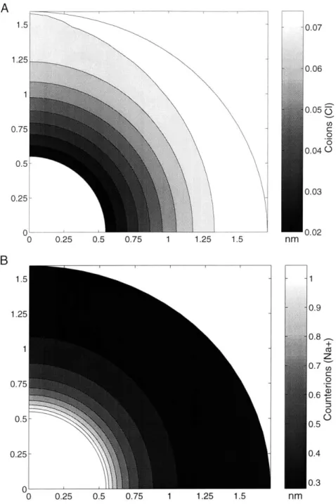
3.6 The equilibrium and dynamic shear modulus and phase angle with 53 varying ionic concentrations. ... 3.7 Mesh generation and the computed electrical potential. ... 54
3.8 Concentrations of co- and counter-ions... 55
3.9 Computed equilibrium shear stress, and shear modulus optimized with 56 CGAG and Gne as fitting param eters. ... 4.1 Schematic view of the shear loading device and waveforms of shear 64 stress and shear strain. ... 4.2 Tissue-level autoradiography and the definition of radial and vertical 68 v ariatio n s. ... 4.3 Frequency-dose response & Core vs. ring comparison... 70
4.4 Autoradiographic appearance of chondrocytes and proline and sulfate 71 g ra in s . ... 4.5 Radial and vertical variations of proline grains. ... 73
4.6 Radial and vertical variations of sulfate grains. ... 74
5.1 Autoradiographic histology of chondrocyte, proline, and sulfate 87 grains at the cell length scale. ... 5.2 Cell length scale analysis on proline and sulfate grain densities. ... 88
5.3 Shear strain-dose response on chondrocyte biosynthesis. ... 90
5.4 Phosphorylated ERK1/2 levels quantified for conditions free swelling, 92 0% control, and 1.5-4.5% shear strain. ... 5.5 RT-PCR products of aggrecan core, type II collagen, and G3PDH 93 m R N A s. ... 5.6 Quantified aggrecan core and type II collagen mRNA levels 94 norm alized by G 3PD H ... 6.1 Shear strain-dose response (0.5-6%) on chondrocyte biosynthesis... 103
6.2 Combined effect of shear and IGF-I on biosynthesis. ... 104
6.3 Effect of shear and static compression on IGF-I transport. ... 105
A. 1 Schematic timeline of the experimental design. ... 109
A.2 Representative waveforms of applied strain and resulting stress versus 113 tim e . ... A.3 Peak stress and stress after four minutes of stress-relaxation. ... 114
A.5 Effect of mechanical stimulation on chondrocyte biosynthesis after 117 injuriou s loading. ...
A.6 Percentage increase of cartilage explant wet weight after injurious 118 lo ad in g . ...
A.7 Changes in equilibrium and dynamic compressive- and shear-stiffness 121 of cartilage explants after injurious loading. ...
B.1 Loading chamber and machine... 131
B.2 Agarose-Repeated duty cycle for 8 and 4 days. ... 132
B.3 Agarose: Alternate day loading. ... 133
B.4 Peptide: Alternate day loading. ... 134
C.1 Unit cell geometry and notations. ... 138
C.2 The comparison of the deformed unit cell boundary ... 144
List of Tables
C.1 Calculation of equilibrium shear modulus using Model I and Model II 147 D. 1 Preparation of the albumin standards and the samples for BCA assay.. 152 D .2 R T R eaction M ix... 157 D .3 PC R R eaction M ix... 158
Chapter I
General Introduction
The synovial joints ensure load transmission within skeletal structures and allow relative movements between bones. The end of a synovial joint is covered with articular cartilage, a glossy and opaque tissue, which functions as a load absorbing and friction reducing material. These unique mechanical functions of articular cartilage are enabled by the interaction between extracellular matrix and synovial fluid; the major solid components (20-30% of wet weight) of extracellular matrix are proteoglycans (PGs) (30-35% of dry weight), hydrated collagen fibrils (50-60% of dry weight), and other non-collagenous proteins and glycoproteins (10-20% of dry weight) [Muir, 1995]. Negatively charged groups within proteoglycans induce repulsion and swelling pressure within collagen network and retard the relative movement of the interstitial fluid. The dynamic force balance between swelling pressure and a resistance of collagen fibrils to tensile forces gives rise to specialized mechanical properties by which an instantaneous large compression is prevented while a large compression is allowed to forces applied at a slow to moderate rate. The extracellular matrix, via its composition and structure, in turn, exerts a regulatory role in promoting or maintaining cellular differentiation and phenotypic expression [Yang, 1998; Bateman, 1996]. In response to the biosynthetic signals, chondrocytes synthesize and degrade matrix component, and the dynamic interaction between cell and matrix necessitates the understanding of biological and biophysical aspects of cartilage (Figure 1.1).
Glycosaminoglycans (GAGs) are the major molecules that give cartilage compressive stiffness due to their negatively charged sulfate and carboxyl groups. GAGs are attached to the serine residues of protein cores and this unit is called aggregating proteoglycan or aggrecan (-2,500 kDa). Proteoglycans are a family of ubiquitous proteins found on the cell surface, within intracellular vesicles, and extracellular space [Wight, 1991]. The multi-domain structure of the core protein of aggrecan includes a hyaluronic acid (HA)-binding region near the N-terminal (GI
domain) and a glycosaminoglycan-attachment domain containing a number of serine-glycine repeats in the middle portion. The C-terminal G3 domain comprises an alternatively spliced complement regulatory protein-like repeat at the extreme C-terminus, an adjacent repeat homologous with C-type animal lectins and an epidermal growth factor (EGF)-like domain that is also subject to alternative splicing [Wight, 1991]. The GI domain of aggrecan mediating specific interactions with hyaluronan (HA) is further stabilized by concomitant binding to the link protein, forming a supramolecular aggregate (refer to Figure 3.1).
In vivo loading:
electromechanical! physicochemical
forces
TISSUE:
Cell/ Matrix/ Fluid
4
0&Q 00
Turnover
Synthesis/ Degradation
MATRIX:
CHONDROCYTE
Proteoglycan!
Collagen
Influence biosynthesis
Figure 1.1: Interaction between cell and matrix. Cartilage, which is constantly exposed to the electromechanical and physicochemical forces in vivo, consists of cell, matrix, and interstitial fluid. Major components of articular cartilage matrix are proteoglycan and type II collagen fibrils whose dynamic interaction provides a macroscopic mechanical function. The physical structure of the matrix can itself impart regulatory information by altering cell morphology and phenotypic expression. In response to this regulatory signal, chondrocyte synthesize or degrade matrix leading to the remodeling of cartilage tissue.
Collagens constitute a superfamily of extracellular matrix proteins with a structural role as their primary function. All collagenous proteins have domains with a triple-helical conformation and each domain is formed by three subunits of X-chains. Each of the three polypeptide chains in the molecule forms an extended left-handed polyproline 11-type helix, which is stabilized by the high imino acid content. The three chains, staggered by one residue relative to each other, are supercoiled about a common axis in a right-handed manner to form the triple-helix. This helical structure has a requirement that every third amino acid is a glycine residue that allows the close packing along the central axis and hydrogen bonding between the three chains (Figure 1.2). Thus each domain possesses a characteristic tri-peptide motif (Gly-X-Y) and, frequently, X is proline and Y is hydroxylproline (Figure 1.2). The x-chains of the fibrillar collagens are synthesized as a large precursor pro oa-chains with N- and C-terminal globular extensions called propeptides. Following secretion, the N- and C-propeptide domains are proteolytically removed to produce the collagen monomers consisting of the triple helix and short non-helical telopeptide sequences at both the C- and N-termini. Following this extracellular processing, the processed collagen molecules self assemble into ordered fibrillar structures and this molecular arrangement is further stabilized by lysyl oxidase catalysed covalent cross-links. This process is called collagen fibrillogenesis.
Cartilage tissue is constantly exposed to external loadings that consist of a combination of compressive and shear deformation (Figure 1.3). Compressive deformation of cartilage induces a pressure gradient and the relative movement of interstitial fluid. Interstitial fluid contains net positive ions due to the negative charges of proteoglycan and the requirement of electroneutrality. The convective movement of fluid entraining net positive ions, again, generates electric currents and fields called electrokinetic coupling. Compressive deformation increases the fixed charge density, and this effect increases osmolarity and decreases pH of the intratissue fluid. One or the combination of these complicated electro-mechanical and physicochemical phenomena have been used to mimic aspects of in vivo
compression [Gray, 1988; Sah, 1989; Urban, 1993; Parkinnen, 1993; Giori, 1993; Sah, 1996; Bonassar, 2000]: cyclic hydrostatic pressure, fluid-induced shear, dynamic tissue deformation, changes in osmolarity and pH.
Newly synthesized proteoglycan molecules have been traced at the micron scale using quantitative autoradiography. The spatial profile and the estimated biophysical changes based on the poroelastic model of cartilage have been able to show that newly made proteoglycans were more localized in the tissue region where chondrocytes were exposed to a greater level of fluid flow under volumetric axial deformation of cartilage explants [Kim, 1994; Buschmann, 1999]. These studies suggest the importance of biophysical changes associated with the fluid flow on chondrocyte biosynthesis. Nonetheless, there have been little studies testing whether fluid flow-free deformation could stimulate chondrocyte matrix synthesis. In this study, we used tissue shear deformation to study the effect of cell and matrix deformation decoupled from the fluid flow since the shear deformation on the poroelastic tissue induces minimal volumetric deformation and pressure gradient.
In addition to the biological response of chondrocyte under shear deformation, tissue shear deformation, especially pure shear applied by torsional deformation, causes matrix deformation which is distinct from the changes due to the compressive deformation. The compressive stiffness of cartilage tissue originates from the electrostatic repulsion and swelling pressure due to GAG and other charge-independent macromolecules. In contrast, tensile stiffness of cartilage is considered to be from the collagen fibrils prestressed by the repulsive forces between PG [Zhu, 1993].
Band Spacing
D=67 nm
-4-FIBRIL
J
llTlhTll
Overlap Zone
Hole Zone (0.61)
(0.4D)
--MICROFIBRIL
kk.COLLAGEN
3000A
MOLECULE
Fqq 104AWPI
TRIPLE
HELIX
STRUCTURE
Glycinel
8.7A-
---
Glycine
IN
a-CHAIN
Y
X
Y
X
Figure 1.2: Structure of collagen. Three left-handed primary ct-chains are supercoiled about a common axis in a right-handed manner to form the triple-helix. After the secretion of procollagen into extracellular space, N- and C-termini are cleaved by specific endopeptidases and thus self-assemble into ordered fibrillar structures. The collagen fibrils are further stabilized by the formation of covalent-cross linking, which confers the mechanical properties of the collagen fibrils.
However, there were no previous systematic and theoretical studies on how PG contributes directly to shear stiffness and the interaction between PG and collagen network at the molecular level. Therefore, a theoretical model was developed to explain the direct role of PG in supporting the shear stiffness of cartilage. Glycosaminoglycans (GAGs) were modeled as a cylindrical rod (a unit cell model) with its geometry determined based on experimental measurements. Macroscopic shear deformation was imposed on the unit cell that is assumed to be randomly oriented. The Poisson-Boltzmann equation was used to calculate the change in the free energy and a shear modulus as a function of ionic concentration and shear deformation.
UNLOADED
COMPRESSION
SHEAR
Figure 1.3: Cartilage loading in vivo. Cartilage is constantly exposed to a combination of compressive and shear deformation. In response to compressive deformation, chondrocytes experience electromechanical and physicochemical forces which are known to affect chondrocyte metabolism. In contrast, in response to shear, the cell and matrix deformation is decoupled from the other biophysical changes.
1.1
Electromechanics of Cartilage Matrix'
The study of the electromechanical properties of cartilage matrix can be classified into macroscopic- or microscopic-scale approaches. Macroscopic approaches utilize native cartilage tissue and have studied the mechanisms by which cartilage provides in vivo functions. Testing configurations of compression, shear, and tension mode have been extensively used [Simon, 1990; Zhu, 1993; Basser, 1998]. Compressive, shear, and tensile stiffness reflect the content and integrity of proteoglycan and collagen fibrils, and also depend on their interaction with interstitial fluid. The tensile strength of the collagen network within native tissue has been studied using the force balance between an applied stress, proteoglycan (PG) swelling pressure, and the collagen tensile stiffness [Basser, 1998]. This study showed that the ability of collagen fibrils to limit the hydration of tissue and, thus, maintains a high PG concentration in normal cartilage was significantly compromised in osteoarthritis tissue.
Measurements of mechanical properties have often been accompanied by the enzymatic degradation of proteoglycan [Zhu, 1993], non-enzymatic glycation of collagen fibril [Kerin, 2001], and changes in ionic concentration and pH [Eisenberg, 1985; Frank, 1987; Jin, 2001]. These studies have revealed the electrostatic nature of proteoglycans in supporting compressive and shear stiffness, the contribution of collagen network to tensile and shear properties, and the complexity of the dynamic interaction among proteoglycan, collagen fibrils, and fluid phase. For example, injurious compression of cartilage explants can lead to collagen damage and GAG loss, and can diminish the ability of cartilage to withstand external deformation, which was evaluated by the measurements of compressive, and shear properties, and
Adapted from "Influence of Tissue Shear Deformation on Matrix Electromechanics and Metabolism in Cartilage Explants", In: The Many Faces of Osteoarthritis, [Jin; in press].
histology [Kurz, 2001; Chen, 1999]. Diagnostic tools using the electrokinetic coupling phenomenon within matrix have shown that the impedance and the current generated stress vary sensitively with the proteoglycan content and the hydration level of tissue [Evans, 2001].
Numerous theories have been used to describe the macroscopic behavior of cartilage tissue, focusing on the interaction between fluid and solid matrix, and the intrinsic properties of macromolecules. Poroelasticity and mixture theories have well predicted stress relaxation, creep, and electrokinetic coupling under volumetric deformation of cartilage tissue [Frank, 1987b; Mow, 1980]. Intrinsic viscoelastic properties of collagen network has been incorporated into the poroviscoelasticity theory, thus addressing coupled relaxation from the intrinsic properties of macromolecules and the interaction between solid and fluid phases [Setton, 1993]. Under the assumptions of linear, homogeneous, and isotropic conditions, these theories often provided the analytical solutions for simple geometry while the numerical methods have been used mostly for the more general cases.
The investigation of the molecular-level electromechanical properties of each component of matrix has enhanced our understanding of the origin of the macroscopic properties of cartilage. The osmotic swelling pressure of proteoglycan solution is associated with the surrounding mobile ions whose distributions are affected by the coupling of the electrostatic forces and the entropic effects. Direct measurement of the swelling pressure of proteoglycans has shown the dependence on the fixed charge density and the molar ratio of keratan sulfate and chondroitin sulfate groups [Comper, 1990; Venn, 1977]. Proteoglycans are attached to hyaluronate non-covalently and this bond is further stabilized by the globular link proteins within matrix. Loss of link protein is known to destabilize proteoglycan aggregate and lead to proteoglycan loss [Ratcliffe, 1986]. Viscosity and shear modulus of a link-stable proteoglycan aggregate in solution using rheometric device were found to be bigger than those of a link-free proteoglycan aggregate solution, emphasizing the stabilizing effect of a link protein in the aggregate [Zhu, 1991].
These rheological properties were also measured for the solutions containing collagen fibrils and the mixture of collagen and proteoglycan, and the theoretical predictions based on the statistical network model showed a reasonable fit to the experimental measurements [Zhu, 1996]. Diffusion properties of macromolecules such as aggrecan and aggregate have been measured using a confocal laser scanning microscope [Gribbon, 1998]; aggrecans were covalently labeled with fluorescein isothiocyanate and the radial distribution of the fluorescence intensity was analyzed after laser-induced photobleaching of the macromolecules. This measurement showed that the formation of aggregate caused a significant reduction in the diffusion coefficients. Nano scale structural visualization of aggrecan and collagen fibrils has been made by electron microscopy [Buckwalter, 1994; Holmes, 2000] and, recently, atomic force microscopy (AFM) has been used to visualize macromolecules in ambient air or in near physiological fluids [Chen, 2000; Sun, 2000].
Recently, AFM have been used to measure the intermolecular and intramolecular electrostatic repulsion between GAG chains, and showed the dependence of repulsive forces on the ionic strength and pH [Seog, 2001]. The Poisson-Boltzmann (PB) mean field theory has been used to model the physical phenomena of polyelectrolytes and colloidal systems containing charged macromolecules within electrolyte solutions [Katchalsky, 1971]. For example, the osmotic swelling pressure of proteoglycan solutions has been modeled quantitatively using PB equation [Buschmann, 1995]. GAG segment was modeled as a cylindrical rod with a surface charge, and the geometry of the unit cell was determined based on experimental measurements. Furthermore, the GAG unit cell model was extended to estimate the contribution of GAG electrostatic interactions to the compressive stiffness of cartilage [Buschmann, 1995]. The entropic portion of swelling pressure of macromolecules has been described using the lattice model based on the approach by Flory [Flory, 1953] and the thermodynamic consideration
1.2 Signaling Pathways & Matrix Synthesis
In vivo loading of cartilage has been shown to affect chondrocyte biosynthesis through multiple pathways including upstream signaling, transcriptional and translational regulation, post-translational modification, and facilitating vesicular transport (Figure 1.4).
A variety of extracellular signals trigger initial events upon association with their respective cell surface receptors and these signals are then transmitted to the interior of the cell, activating the appropriate cascades. Among numerous pathways that control transcriptional activities, the MAPKs (mitogen-activated protein kinases) have been shown to be the major signaling mechanism in eukaryotes [Seger, 1995]. MAP kinase was originally discovered as an insulin-activated protein-serine kinase and biochemical studies showed that the MAP kinase pathway consists of a cascade of three protein kinases, a MAPK kinase kinase (MAPKKK or MEKK), a MAPK kinase (MAPKK or MEK), and a MAPK [Waskiewicz, 1995]. The MEKK enzymes are Ser/Thr protein kinases that activate the MEK enzymes by phosphorylating two serine or threonine residues within a Ser-X-X-X-Ser/Thr motif. Once activated, the MEK enzymes, which are mixed function Ser/Thr/Try protein kinases, phosphorylate the MAPK enzymes on Thr and Try residues within the Thr-X-Tyr (TXY) motif. The central component of the MAPK is ERK1/2 (extracellular signal-regulated protein kinase) and these have dominated efforts to understand MAPK signaling. Other important kinases are JNK (c-jun amino terminal protein kinase) and p38, which are activated by stress, ultraviolet light, and inflammatory cytokines. The central amino acid differs for each MAPK superfamily member, corresponding to Glu for ERKl/2, Gly for p38, and Pro for JNK. A number of in vitro studies have been performed using chondrogenic cell lines (e.g., ATDC5 [Watanabe, 2001]) and isolated chondrocytes [Hung, 2000] to elucidate the involvement of MAPKs in controlling transcriptional activities of aggrecan mRNA.
In
vivo loading, soluble factors
Extracellular
Signaling
eC0Processing
ER
Gol
i
mRNA
TransciptiVesicular
Trancripio
t nsport
Translation
Post-translation
Figure 1.4: Multiple regulatory pathways. The pathway of aggrecan and collagen biosynthesis from gene transcription to secretion and aggregation of monomers into functional extracellular matrix is complex. Gene transcription and mRNA processing occurs within the nucleus and the mature mRNA is transported to the cytoplasm. Translation products are directed into the endoplasmic reticulum (ER) and, again, post-translationally modified in the Golgi complex. Final products are packaged into secretory vesicles and released into the extracellular space. In vivo loading and soluble factors are known to affect these multiple regulatory pathways influencing chondrocyte biosynthesis.The pathways of collagen biosynthesis and aggregation of collagen monomers into functional fibrils is complex. After following common processing to the vast majority of protein production involving mRNA processing inside nucleus and translation into the endoplasmic reticulum, extensive post-translation modifications of triple-helical molecules occurs inside the Golgi complex. These molecules are packaged into secretory vacuoles that release the procollagen into the extracellular space by exocytosis. Following secretion the N- and C-propeptides are
cleaved by specific endopeptidases, a procollagen N-proteinase and C-proteinase. Cleavage of the C-propeptide is a prerequisite for fibril formation and N-propeptide cleavage is essential for regular fibril morphology. Cleaved N- and C-propeptides may have a role in feedback regulation of procollagen biosynthesis. The processed collagen molecules assemble into fibrils that are stabilized by lysyl oxidase catalysed cross-links.
The precise biochemical mechanisms of collagen fibril assembly and growth are not well understood and our current understanding is based on in vitro experiments. Individual processed fibrillar collagen molecules (e.g., I, II, III, V)
spontaneously self-assemble into ordered fibrillar structures in vitro [Veis, 1988]. A critical feature of this in vitro fibrillogenesis is that collagens I, II, III, V (possibly type XI) all share the tendency to aggregate into ordered filamentous assemblages, which was shown by electron microscopy [Chapman, 1984] and X-ray diffraction [Brodsky, 1982]. This fibril-forming ability is encoded in the structure of the collagens, implying that precise interactions between collagen domains are involved in directing axial organization of the fibrillar aggregates. Both hydrophobic and electrostatic interactions between adjacent chains have been proposed as the mechanism [Veis, 1988].
The core protein of the aggrecan has a molecular weight of -200 kDa. About 80 chondroitin sulfate chains are attached to this core protein through covalent linkage to the hydroxyl groups of serine residues. In addition about 100 keratan sulfate chains and oligosaccharides are attached to serine or threonine residues of the core protein through glycosidic bonds. The presence in chondrocytes of an intracellular core protein precusor was first indicated in experiments using cell-free translation of isolated mRNA followed by immunoprecipitation with anti-proteoglycan antibodies [Upholt, 1979]. Subcellular fractionations have shown that at least 70% of the intracellular transit time for the core protein precusor is spent in the rough endoplasmic reticulum. The addition of the bulk of the glycosaminoglycan
chains and subsequent secretion occurs rapidly, occupying 30% of the intracellular dwell time of the precusor [Fellini, 1984].
Chondrocytes take up the building blocks for GAG synthesis, monosaccharides and sulfate, through specialized transporter complexes in the plasma membrane. Sugars and sulfate are then activated by nucleotide consumption in the cytosol to form UDP-sugars and 3'-phosphoadenosine 5'-phosphosulfate (PAPS), respectively. Specific transporters then translocate UDP-sugars and PAPS into the endoplasmic reticulum and Golgi lumens. Glycoproteins and glycolipids are also often sulfated. PAPS is the universal donor of sulfate to all sulfotransferases, both in the Golgi and the cytosol. GAG synthesis (except for in keratan sulfate (KS)) is initiated by sequential addition of four monosaccharides: xylose (Xyl), galactose (Gal), galactose, and glucuronic acid (GlcA). From this linker tetrasaccharide, the sugar chains are extended by addition of two alternating monosaccharides, an aminosugar and GlcA. In heparin and heparan sulfate (HS), the aminosugar is N-acetylglucosamine (GlcNAc) and in CS/DS it is N-acetylgalactosamine (GalNAc). The extent of epimerization of GlcA to iduronic acid and the sulfation pattern of the disaccharide units distinguish heparin from HS, and DS from CS. In KS, the GAGs are initiated as N-linked or 0-linked oligosaccharides and extended by addition of GlcNAc and Gal [Prydz and Dalen, 2000]. For the synthesis of chondroitin sulfate (CS), six different glycosyltransferases participate in the chain synthesis together with sulfotransferases 4/6: xylosyltransferase, galactosyltransferase I/II, glucuronosyl-transferase I, N-acetylgalactosaminylglucuronosyl-transferase, and glucuronosyltransferase II
[Lohmander, 1986].
1.3 Thesis Overview
This thesis describes the studies on the biological and biophysical aspects of cartilage in response to tissue shear loading. To simulate in vivo shear deformation
on cartilage explants, we have developed a shear loading apparatus and tissue culture chamber and they are described in Chapter II.
The effect of the electrostatic interaction between glycosaminoglycans in influencing the shear stiffness was studied experimentally and theoretically. On the experimental side, the shear modulus of cartilage was measured in a torsional configuration with varying ionic concentration. A molecular model of GAG electrostatic interaction was developed to propose the mechanism by which GAG contributes to shear modulus. From this model, the shear modulus was calculated by solving the Poisson-Boltzmann equation numerically using the finite element method (FEM) and it was compared with the experimental measurements. This biophysical aspect of cartilage is described in Chapter III.
Chapter IV-VI present the biological aspect of cartilage, especially focused on the effect of tissue shear (1) on chondrocyte matrix synthesis measured by radiolabel incorporation and tissue-level autoradiography (Chapter IV), (2) on the regulation of phosphorylated ERK1/2 and mRNA levels of aggrecan core and type II collagen in addition to the cell-level autoradiography (Chapter V), and (3) on the facilitated transport of Insulin-like Growth factor I (IGF-I) and the combined effects of tissue shear and IGF-I on chondrocyte biosynthesis (Chapter VI).
Appendix A presents the effect of injurious compression on the changes in the mechanical and biochemical properties, and the responsiveness of chondrocyte to dynamic compression. Recently, self-assembling peptides gel was developed as a potential scaffold for cartilage tissue engineering and a system for exploring the biosynthetic response of chondrocyte under mechanical stimulation. Appendix B presents the study on the chondrocyte-scaffold system based on agarose and self-assembling peptides, exploring the effects of long-term loading over 3-12 days using various duty cycles of dynamic compressive loading. Appendix C, at the end of this thesis, describes the detailed protocols for Western blot and RT-PCR technique.
Chapter II
Hardware Design
In vitro systems have been developed to mimic in vivo loading on cartilage ex vivo. Our aims were to develop a tissue loading apparatus capable of applying compressive and shear deformation to cartilage explants simulating physiological loading in its dynamic and static nature. In addition to the loading machine, various tissue loading-and-culture chambers were designed to adapt to different conditions of loading including dynamic- and static-shear/compression for stimulating cartilage explants, dynamic- and static-compression for stimulating chondrocyte-scaffold system, and the testing of material properties.
2.1 Tissue Loading Apparatus
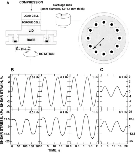
2We have developed an incubator housed, biaxial loading device capable of applying axial deformations as small as 1 gm and sinusoidal rotations as small as 0.001 degree with a resolution of 50 nm for applying sinewaves as low as 10 jim (or 1% based on a 1 mm thickness of sample) or up to greater than 100 Rm. The range of the ramping mode at axial and rotational deformation is up to 1mm. The machine is small enough (35.5 cm high x 25cm x 20cm) to be placed in a standard incubator for long-term tissue culture loading studies. A rigid frame was constructed consisting of two 0.95 cm thick stainless steel plates bolted to three 2.54 cm diameter stainless steel rods (Figure 2.1: Loading Machine). A third stainless steel plate was clamped to the support rods to allow repositioning for different experimental shear or compression chambers. A linear stepper motor (23A-6102A,
2 Adapted from "Regulation of Cartilage Metabolism by Dynamic Tissue Shear Strain and
the Mechanical Characterization of Cartilage", SM Thesis [Jin, 1999] and "A versatile shear and compression apparatus for mechanical stimulation of tissue culture explants", Journal of
American Precision, Buffalo, NY) mounted to the top plate has a threaded rotor that engages a threaded rod. The threaded rod in turn is attached to a carriage plate and pair of linear bearings that ride on two of the support rods.
The axial motor is capable of applying compressive ramps at rates up to 2 mm/s with an applied force up to 400 N. The rotational fixture consists of a rotary position table (6R180, Design Components, Franklin, MA) with a 180:1 gear reduction ratio driven by a conventional stepper motor (23D-6102, American Precision). A block placed at the outer edge of the rotating table contacts an LVDT to measure angular displacement. Each motor is driven by a micro-stepper drive (IM483, Intelligent Motion Systems, Marlborough, CT). The drives are optically isolated and each has their own power supply separate from the analog and digital electronics power supply. The micro-stepper drives, combined with the motors and gearing, provide a theoretical axial resolution of 50 nm and rotational resolution of 0.00010. Both axial and angular displacements are measured by linear variable differential transformers (LVDT, Model S5, Sensotec, Columbus, OH). Various load transducers (10 N, 100 N, and 500 N capacities, Model 31, Sensotec) can be attached to the carriage for various experimental conditions. A torque transducer (5 N-m capacity, Transducer Techniques, Temecula, CA, or 0.2 N-m capacity, QWLC-8M, Sensotec) in line with the load cell and attached to the chamber top is used to measure shear stress in the samples. Rotational control is integrated with axial control so that shear tests may be intermixed with compression tests in the same experimental procedure.
The control electronics, including the transducer signal conditioners for the LVDT's, load cells, and torque cells as well as the limit, feedback, and digital switching circuits were mounted on two prototype circuit boards with copper cladding acting as a ground plane. A digitally-controlled analog switch allows any one of the axial displacement, angular displacement, axial load, or torque signals to be used for closed-loop feedback control.
SU]LIEU I 1E I ilL I - -- --
-A
B
Figure 2.1: Tissue loading apparatus. A [earlier version]. Axial movement is actuated by the stepper motor which drives the carriage connecting load cell, torque cell, and the top chamber. Rotational motion is enabled by the combination of the stepper motor and the rotary table. Dynamic shear deformation (simple shear) can be applied to cartilage tissue by cyclically rotating the rotary table and, accordinaly, the bottom chamber with respect to the top. These axial and rotational deformations are measured by the transducers (LVDT). B [recent version]. The static control chamber is shown next to the machine.
A computer-based data acquisition system (National Instruments, Austin, TX) provides all control and monitoring of the apparatus. The data acquisition and control subsystem consisted of a multi-function I/O card (National Instruments AT-MIO-16DL-9), which provides high-speed analog-to-digital conversion (ADC) and digital I/O, and a 10-channel digital-to-analog converter (DAC) board (National Instruments AT-AO-10). The DAC channels provide signal offsets as well as ramp and sinewave control signals. In displacement feedback control, sinusoid waveform distortion is < 1% for a 10 gm amplitude sinewave, comparable to that of our Dynastat mechanical spectrometer (Dynastatics, Albany, NY) (-0.3 %), which we have used in our previous studies on cartilage metabolic response to dynamic compression [Sah, 1989].
2.2 Tissue Loading Chambers
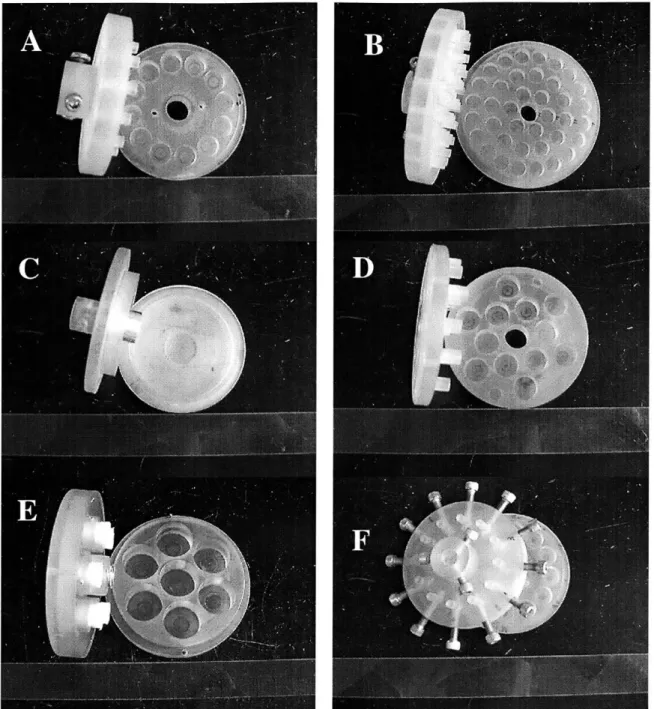
We have designed various chambers for experiments involving cartilage explant and chondrocyte-scaffold system. Most of chambers were made from autoclavable polysulfone except the material testing chamber that was plexiglas. Earlier version of shear chambers has 12 platens-and-wells that are located at equal distance (25.4 cm) from the center axis of rotation (Figure 2.2A). Each well can contain up to -650 gl of conditioned media and at the bottom of each well another small well (5 mm diameter by 0.2 mm thick) is made to hold cartilage specimens in place. To study shear strain-dose effect on chondrocyte biosynthesis, the recent version of shear chamber was made which can distribute cartilage specimens at three different distances from the center having a ratio of 1:2:3. The inner, middle, and outer wells of the chamber can accommodate 6, 12, and 20 specimens, respectively (Figure 2.2B).
For the measurements of shear modulus in the torsional configuration, a chamber was designed which can hold single cartilage disks at the center of the bottom half. An aluminum platen is attached to the top chamber and fine grit sand paper is glued to the end of the platen to prevent slipping between specimen and
chambers (Figure 2.2C). The new design of the static control chamber was adapted from the previous design and the new design allows up to 80% of axial compression on 1mm thick specimens (Figure 2.2D).
To study the effect of long-term stimulation on the chondrocyte-scaffold system including agarose and peptide gel, a chamber was specially designed whose upper half is attached with highly permeable (40% void, 120 gm pore size) polyethylene, with a coil spring inserted into the center hole (Figure 2.2E). The polyethylene material was selected to facilitate the transport of soluble factors to specimens during long-term stimulation. The center spring provides extra force to raise the top half since the agarose or the peptide gel may not be stiff enough to support the weight of the top chamber. The bottom chamber has 6 wells and each well can contain up to 12 mm diameter disks. To stimulate the chondrocyte-scaffold system with non-homogeneous thickness (e.g., collagen scaffold), the conventional shear chamber was modified so that each platen is located on to the surface of the each specimen by loosening the screw (Figure 2.2F).
Figure 2.2: Various chambers. Shown here are shear chamber (A), chamber for shear strain-dose experiment (B), material testing chamber (C), static control chamber (D), and chondrocyte-scaffold stimulating chambers (E & F).
Chapter III
Effect of Electrostatic Interactions between Glycosaminoglycans on
the Shear Stiffness of Cartilage
3Glycosaminoglycans (GAGs) are polymers of disaccharides that contain alternating sequences of glucuronic acid (GlcA) and either N-acetylglucosamine (GlcNAc) or N-acetylgalactosamine (GalNAc). The family of GAG molecules, including hyaluronic acid (HA), chondroitin sulfate (CS), keratan sulfate (KS), and heparan sulfate (HS) [Wight, 1991], plays an important role in the mechanical and transport properties of extracellular matrix (e.g., CS, HA) [Grodzinsky, 2000] and in cell surface ligand binding interactions (e.g., HS) [Lander, 2000]. For example, chondroitin sulfate GAG (CS-GAG) contains on the average one negatively charged carboxylate and sulfate group per disaccharide that is completely ionized under physiological pH conditions. Therefore the high negative charge density and associated electrical repulsion between CS-GAGs play an important role in electromechanical and physicochemical interaction within biological tissues such as cartilage.
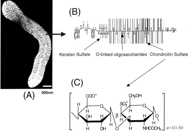
The role of electrical repulsive interactions is particularly critical in articular cartilage, a tissue that covers the ends of bones in synovial joints, providing compressive and shear stiffness during the relative motion of opposing joint surfaces. The compressive resistance of cartilage is mainly due to highly charged CS-GAGs (Figure 3.1C) that are attached to a core protein, forming the proteoglycan called aggrecan (Figure 3.1B). Aggrecan molecules, in turn, bind non-covalently to long hyaluronic acid (HA) chains forming supramolecular proteoglycan aggregates (Figure 3.lA) that are enmeshed within a resilient collagen network. At physiologic pH, the properties of the collagen fibrils do not change
3 Adapted from " Effect of electrostatic interactions between glycosaminoglycans on the shear stiffness of cartilage: a molecular model and experiments", Macromolecules [Jin, 2001].
significantly with ionic strength in the range of 0.01-1.0 M [Ripamonti, 1980; Bowes, 1948]. Therefore, the mechanical properties of cartilage can be modeled in terms of two distinct electrical and non-electrical contributions. Electrical
contributions to the mechanical properties of cartilage are mainly associated with the electrostatic repulsive forces between CS-GAGs. In contrast, non-electrical contributions to cartilage properties are associated with the resilience of the electrically neutral collagen fibrils as well as the elastic forces due to the steric and entropic effects induced by volumetric deformation of GAG and other matrix macromolecules.
Often the definition of free energy provides a way to derive macroscopic constitutive material properties such as compressive and shear moduli [Anand, 1996]. Intermolecular interactions and intramolecular conformational changes have been explained by using the free energy function [Honig, 1993]. In cartilage, the electrostatic free energy is mainly associated with the charged GAG constituents, and depends on the chemical environment (e.g., the pH and ionic concentration), temperature, and mechanical deformation, which can modulate the fixed charge density. However, the non-electrical free energy depends on mechanical deformation, volume change, and temperature, and not on the chemical environment such as ionic concentration and small pH changes around 7.0.
The Poisson-Boltzmann (PB) mean field theory has been used to model electrical properties of polyelectrolytes and colloidal systems containing charged macromolecules within electrolyte solutions. For example, the osmotic swelling pressure of proteoglycan solutions [Comper, 1978] has been modeled quantitatively using PB equation. The GAG constituent has been modeled as a cylindrical rod with a surface charge, and the geometry of the surrounding unit cell structure was based on experimental measurements [Buschmann, 1995]. Furthermore, the GAG unit cell model was extended to estimate the contribution of GAG electrostatic interactions to the compressive stiffness of cartilage [Buschmann, 1995].
Km
(B)
Keratan Sulfate 0-linked oligosaccharides Chondroitin Sulfate
(C)
(A)
5""m00- C0H H sc -H H OH H H 0 H \ H H . H OH 0 H NHCOC.. r--10-50Figure 3.1: Structure of PG-aggregate, PG, and GAG: The cartilage supramolecular
proteoglycan aggregate (A) consists of proteoglycan monomers called aggrecan (B)
which are non-covalently attached to hyaluronic acid (HA) molecules. Aggrecan, in turn, consists of glycosaminoglycan chains covalently attached to a core protein (B). The chondroitin sulfate GAG chains (C) of aggrecan are mainly responsible for the
compressive stiffness of cartilage through electrical repulsion interactions. On the average, CS-GAG disaccharides have one carboxylate and sulfate group which are negatively charged at physiologic pH. Figures A and B are adapted from literature
[Buckwalter, 1994; Hardingham, 1990].
In this study, we focused on the contribution of GAG electrostatic interaction in determining the shear stiffness of a material like cartilage. The role of GAGs in the shear properties of articular cartilage has been previously described as inflating the collagen network, causing a tensile prestress that enables the collagen-aggrecan matrix to resist shear deformation [Lai, 1991; Zhu, 1993; Basser, 1998]. Previous studies showed that cartilage shear modulus changed significantly after extraction of aggrecan [Zhu, 1993], and that increased ionic strength could decrease the shear
modulus inferred from the measurement of compressive modulus at confined compression tests [Bursac, 2000]. However, there has been little theoretical or experimental study of the possible mechanisms by which GAG electrostatic interactions may contribute directly to the tissue shear stiffness, and the importance of this contribution compared to that of other non-electrical interactions.
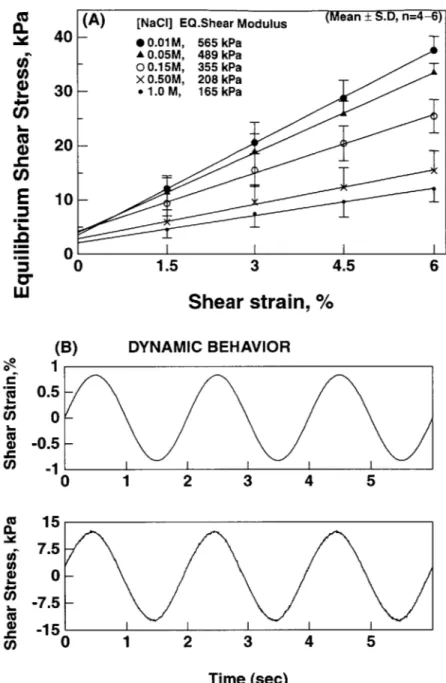
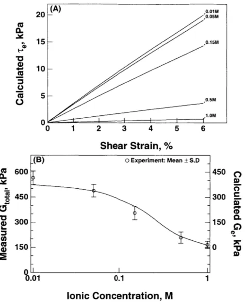
In this study, we first performed experiments to measure the equilibrium and dynamic shear stiffness of cartilage under torsional shear deformation at varying bath ionic concentrations. Experimental results showed a significant change in the measured shear modulus with changes in ionic strength between 0.01 and 1.0 M. We hypothesized that any dependence of the shear modulus with ionic strength comes from the GAG-associated electrical component of cartilage. However, using a macroscopic model of a polyelectrolyte system such as the Donnan model, the electrostatic free energy should be constant under torsional shear, since this deformation does not induce any volume change and therefore no change in fixed charge density. Therefore, in the present study, we hypothesized that (1) GAG molecules were embedded into the collagen, (2) the repulsive forces between GAGs were maintained by the collagen network, and (3) the rearrangement of GAG molecules at a microscopic scale up on the macroscopic deformation could provide increased resistance to shear deformation and increased shear stiffness with decreasing salt concentration. Using a PB unit cell model of GAG electrostatic interactions under shear deformation, we calculated the change in the electrical contribution to the equilibrium shear modulus at each ionic concentration. To account for the nonlinearity of the PB equation and the geometry of the model, we used the finite element method to solve the electrical potential and mobile ion distribution. From this calculation, the electrical contribution to shear stress was obtained by differentiating the electrostatic free energy with respect to the shear deformation. Finally, the electrical contribution to the equilibrium shear stiffness was calculated from the slope of stress vs. strain, and the resulting shear stiffness was compared to the experimental measurements.
3.1 Materials and Methods
3.1.1 Experimental SectionCartilage disks (9.65 mm diameter by 1 mm thick) were obtained under sterile condition from the femoropatellar groove of 1-2 week old bovine calves as previously described [Frank, 2000] and maintained in living organ culture for 1-3 days until testing (DMEM + 10 % fetal bovine serum). Prior to measurement, cartilage disks were equilibrated in 0.15 M NaCl solution up to 30 minutes. The disks were then placed in the base of a plexiglas chamber filled with a 0.15 M NaCl bathing solution. The base chamber was placed in the bottom grip of a specially designed biaxial rheometer capable of applying axial deformations as small as 1 gm and sinusoidal rotations as small as 0.0010 [Frank, 2000]. An upper aluminum platen fixed to a torque cell (Figure 3.2) was used to apply a 10% compressive offset strain to the specimen. After mechanical and chemical equilibration was achieved in this state (-15 minutes), torsional deformation was applied by rotating the bottom chamber with respect to the top platen, and the resulting torque was measured by the torque cell. From the measured torque and applied torsional deformation, the shear strain, y, and shear stress, r, were calculated as y = 0 r / h; r (r) = T r /I, where & is the angular deformation, r is the radial distance, and h is the height of the disk, T is the measured torque, and I is the polar moment of inertia (I =
ic R4 /2, R is the radius of the disk). Fine grit sand paper glued to the base and upper platen were used to prevent slippage between the specimen and these platens. For measurement of the equilibrium modulus, a ramp-and-hold shear strain of 1.5 % was applied, resulting in an initial increase and subsequent relaxation of the shear stress (Figure 3.3A). This sequence was repeated four times, and the slope of the relaxed equilibrium stress and strain was used to compute the equilibrium modulus. After returning the specimen to 0 % shear strain, a 0.8 % amplitude sinusoidal shear strain was applied at 0.5 Hz (Figure 3.3B). The magnitude of the dynamic shear modulus was calculated as G = 1z| / I1. The shear stress and strain signals were continuously recorded by a computer, and a discrete Fourier transform (DFT) was implemented to
![Figure 2.1: Tissue loading apparatus. A [earlier version]. Axial movement is actuated by the stepper motor which drives the carriage connecting load cell, torque cell, and the top chamber](https://thumb-eu.123doks.com/thumbv2/123doknet/14299251.493749/29.918.124.783.166.600/figure-tissue-loading-apparatus-movement-actuated-carriage-connecting.webp)