Current Concepts and Controversies
in Prion Immunopathology
Mathias Heikenwalder, Marco Prinz,
1Frank L. Heppner, and Adriano Aguzzi*
Institute of Neuropathology, University Hospital of Zürich CH-8091 Zürich, SwitzerlandReceived December 13, 2002; Accepted October 2, 2003
Abstract
Scrapie in sheep and new variant Creutzfeldt-Jakob disease in humans are typically initiated by extracere-bral exposure to prions. Both exhibit early prion accumulation in sites of the peripheral lymphoreticular system, such as splenic or lymph nodal germinal centers. In germinal centers, follicular dendritic cells (FDCs), whose development and maintenance depend on lymphotoxin and tumor necrosis factor signaling, are believed to be the main cell type for efficient prion replication in the periphery. Here, we discuss the molecular requirements for prion replication competence in stromal and lymphoid compartments of lymphoid organs. In addition, we examine the preconditions of transepithelial passage of prions in the mucosal-associated lymphoid system. Our results suggest that under specific conditions, efficient prion replication in mesenteric and inguinal lymph nodes is possible in the absence of mature FDCs. M cells are a plausible candidate for the mucosal portal of prion infection.
Index Entries:Immune system; prions; follicular dendritic cells (FDCs); lymphotoxin (LT); tumor necrosis factor (TNF).
Copyright © 2004 Humana Press Inc. All rights of any nature whatsoever reserved. ISSN0895-8696/04/23:3–11/$25.00
Introduction
Prion diseases are lethal, transmissible, neurode-generative conditions. The causative agent was pro-posed to be identical with prion protein (PrP)Sc, a
pathological conformer of the cellular protein PrPC
encoded by the cellular gene Prnp (Prusiner, 1982). PrPCis expressed in many sites, notably including
secondary lymphoid organs. Peripheral inoculation routes are likely to initiate most forms of spongiform encephalopathies such as sheep scrapie, bovine spongiform encephalopathy (BSE), iatrogenic Creutzfeldt-Jakob disease (iCJD) and variant CJD (vCJD). Also, intracerebral (ic) or peripheral admin-istration of prions to mice induces a rise of infectiv-ity in spleen and in other lymphoid organs long before the development of neurological symptoms and neuropathological changes. Intraperitoneal (ip)
inoculation has been used extensively to study the pathogenesis of transmissible spongiform encephalopathies because it causes rapid accumu-lation of infectivity in secondary lymphoid organs (Clarke and Haig, 1970; Hill et al., 1997; Hilton et al., 1998). The question of which compartments within lymphoreticular tissues support prion replication is of relevance to public health: Contamination with vCJD prions of germinal centers in lymph nodal and tonsillar follicles, for example, might call for pre-cautionary measures in the handling and steriliza-tion of surgical instruments. Also in sporadic CJD (sCJD), prions appear to be much more prevalent in extracerebral tissues than previously appreciated (Glatzel et al., 2003).
Tumor necrosis factor (TNF) and homotrimeric lymphotoxinα(LTα) signal through TNFR1, whereas membrane-bound LTα/β heterotrimers signal
R
EVIEWA
RTICLE*Author to whom all correspondence and reprint requests should be addressed. E-mail: [email protected]
through LTβR (Ware et al., 1995) (Fig. 1). TNFR1 and LTβR signaling is necessary for development and maintenance of secondary lymphoid organs (Rennert et al., 1996; Koni et al., 1997; Korner et al., 1997; Futterer et al., 1998) and their proper micro-architecture. LTβR signaling is also required for maturation and maintenance of follicular dendritic cells (FDCs), which are thought to be essential for prion replication and for accumulation of disease-associated PrPScwithin secondary lymphoid organs.
Inhibition of the LTβ signaling pathway with a sol-uble receptor, which depletes FDCs (Mackay and
In recent years we have studied peripheral prion pathogenesis in mice lacking TNFα, LTα/β, or their receptors. We found that ablation of LTβR signaling prevents peripheral pathogenesis (Montrasio et al., 2000; Prinz et al., 2002), whereas ablation of TNFR1 signaling prevents prion pathogenesis in spleen but not in lymph nodes, despite the absence of FDCs. Moreover we have investigated some of the pre-conditions of transepithelial passage of prions, iden-tifying M cells as a plausible candidate for the mucosal portal of prion infection.
Lymphoid Microarchitecture
and Efficient Peripheral Prion Replication
Even though FDCs are believed to be the key cell type for efficient prion replication, mice devoid of mature FDCs did show prion titers in secondary lym-phoid organs? The absence of TNFR1 and TNFα pre-served susceptibility to peripheral prion challenge, whereas deletion of LT signaling components confers high resistance to peripheral prion infection, as detected by Western blot analysis and transmis-sion experiments into tga20 indicator mice (Fig. 2).
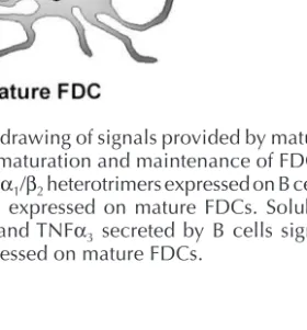
Fig. 1. Schematic drawing of signals provided by mature B cells essential for maturation and maintenance of FDCs. Membrane-bound LTα1/β2heterotrimers expressed on B cells signal through LTβR expressed on mature FDCs. Soluble homotrimeric LTα3and TNFα3 secreted by B cells signal through TNFR1 expressed on mature FDCs.
Fig. 2. (see opposite page) Western blot analysis of brains and spleens, as well as determination of prion infectivity titers in spleens and lymph nodes, of scrapie-challenged TNF- and LT-deficient mice. (A,B) Western blots of brain mater-ial electrophoresed natively (–), or after digestion with proteinase K (PK) (+). Large amounts of PK-resistant prion protein (PrPSc) were detected in the brain of all mice that had developed scrapie (terminal sick), independently of the genotype.
Clinical healthy mice deficient in LT signaling showed no PrPSc accumulation, excluding subclinical scrapie (A).
(C) Western blots of spleen homogenates electrophoresed natively (–) or after digestion with proteinase K (+). TNFR1–/–
and TNFα–/–mice accumulated lower amounts of PrPScas compared to TNFR2–/–and wild-type mice. No PrPScwas detected
in the spleens of clinical healthy LT-deficient mice. (D) Prion infectivity in lymphoid organs. Titers were determined in spleens (dark circles,
•
), inguinal (light circles,•
), mesenteric (crosses, X), and cervical (triangles) lymph nodes at the time points indicated below the graphs. Mice were inoculated ip with 6 logLD50or 4 logLD50of scrapie prions as indi-cated. Standard deviations within groups are drawn only when exceeding ±0.75 logLD50. (a,b) In each of two separate transmissions, 1 out of 4 tga20 mice died 24 h after inoculation, probably because of intracerebral bleeding after injec-tion. (j,k) Intercurrent death during incubation time. Symbols on the abscissa indicate prion titers below detection limit (none of the four indicator mice developed scrapie). If one or more indicator mice survived <180 dpi, or the mean incu-bation time was >120 d, titer was assumed to be close to the detection threshold of the bioassay. For these Fig. 2.(con-tinued) samples (labeled with lowercase letters) the numbers of animals succumbing to scrapie out of four inoculated tga20 mice and incubation time (in days) to terminal scrapie were as follows: (c) 1/4 (111), (d) 3/4 (93, 117, 132), (e) 2/4
(90, 95), (f) 1/4 (126), (g) 3/4 (112, 123, 128), (h) 4/4 (106, 115, 124, 136), (i) 3/4 (89, 102, 119), (l) 4/4 (115, 115, 126, 135), (m) 1/4 (98), (n) 2/4 (74, 78), (o) 2/4 (125, 151), (p) 2/4 (131, 193), (q) 3/4 (119, 125, 152), (r) 2/4 (109, 117), (s) 1/4 (108), (t) 1/4 (71) (u) 3/4 (80, 96, 98).( v ) 2/4 (98, 98).
transfer studies that complement the gene ablation experiments.
In Prnp0/0mice grafted with TNFR1–/–fetal liver
cells (FLCs), high infectivity loads (3.4–4.2 logLD50)
were detectable in lymph nodes but not spleens, strongly indicating a role of TNFR1-deficient hematopoietic cells in efficient prion propagation within lymph nodes. Conversely, lymph nodes of TNFR1–/–mice grafted with Prnp0/0FLCs contained
substantial amounts of infectivity. Therefore, stromal cells also contribute to the capability of TNFR1–/–-deficient lymph nodes to replicate prions.
Reciprocal reconstitution of wild-type and Prnp0/0
mice showed once again that efficient lymphoretic-ular prion propagation required PrPCexpression in
both stromal and hematopoietic compartments (Blättler et al., 1997; Kaeser et al., 2001).
These results imply that prion replication can take place in secondary lymphoid organs even in the absence of mature FDCs and that other cell types can maintain replication of prions to titers that are sim-ilar to those of wild-type mice. Moreover, prion pathogenesis in the lymphoreticular system can be topographically compartmentalized, and lymph nodes can represent an important reservoir of prion infectivity during disease.
This hypothesis is strengthened by the fact that even in wild-type lymph nodes, bright PrP signals outside FDC networks colocalized with a subset of ER-TR9- and MOMA-1-positive cells (Prinz et al., 2002).
TNFα signaling through TNFR1 is required for proper homing of macrophages to the splenic mar-ginal zone, and their absence can cause strong aber-rations in macrophage subsets. However, MOMA-1+
and ER-TR9+macrophages were normally
distrib-uted in the subcapsular area of TNFR1–/– and
TNFα–/–lymph nodes (Pasparakis et al., 2000) but
were strongly disturbed in mesenteric lymph nodes of LTβ–/–mice. It was suggested that FDC precursor
erly into the lymph node follicular areas where they would promote the formation of B-cell follicles and germinal centers (Pasparakis et al., 2000).
If the distribution of macrophages is important for peripheral prion pathogenesis, these histoarchi-tectural differences might account for the differences in splenic versus lymph nodal prion load of infected TNFR1–/–and TNFα–/–mice. Primary and secondary
follicles might be functionally different in spleen vs mesenteric lymph nodes of TNFR1–/–mice (Fu et al.,
1997). However, we did not identify morphological differences between splenic and mesenteric germi-nal centers, and the TNFR1–/–line used here (Rothe
et al., 1993) did not show abnormal germinal center responses after infection with vesicular stomatitis virus (Karrer et al., 2000).
As described repeatedly (Klein et al., 1997; Kaeser et al., 2001; Prinz et al., 2002), B-cell-deficient mice were entirely resistant to ip-administered prions, whereas TNF/LT-deficient mice were partially resis-tant. These findings differ from those reported by Mabbott and colleagues (2000) and suggest that the importance of B lymphocytes in prion pathogenesis might go beyond their role in FDC maintenance.
The unexpected finding of high prion titers in inguinal, mesenteric, and cervical lymph nodes of TNF-deficient—but not in mesenteric lymph nodes of LTβ-deficient—mice indicates that prion replica-tion within secondary lymphoid organs is LTβR dependent yet may occur in the absence of mature FDCs and functional germinal centers.
Therefore, cell types other than mature FDCs par-ticipate in the process of prion replication/accu-mulation in lymph nodes and, probably, in spleens. Because marginal zone macrophages might enter-tain close contacts to immature FDCs in the mar-ginal zone, whose presence was postulated for the TNFR1–/– mice (Pasparakis et al., 2000), and also
interact with marginal zone B cells, this cell type is certainly a candidate for supportive effects in the process of prion uptake and replication.
However, this interpretation has one caveat: Immunofluorescence detects PrP rather than infec-tivity and does not differentiate unequivocally between PrPCand PrPSc. Therefore, further studies
will need to focus on whether macrophage ablation, that is, using macrophage-specific suicide trans-genes, can suppress the infectibility of TNF-deficient lymph nodes.
Moreover, these findings are at striking variance with reports that LTβ–/–mice are fully susceptible to
infection with CJD prions (Rothe et al., 1993) and that TNFα–/–mice peripherally challenged with ME7
prions are largely protected (Mabbott et al., 2000). These and other discrepancies have been attributed to the use of different prion strains in these studies. This may well be the case, but our results indicate that resistance in each mouse strain is dose depen-dent and can always be overridden. Therefore, chal-lenge with one single size of inoculum, as done in other studies, might yield misleading results.
Invasion of lymphoid organs by prions occurs very rapidly after peripheral inoculation, and con-sistently high infectivity titers are detected until ter-minal disease. Lymphoinvasion most likely plays an important role in the pathogenesis of vCJD, as prion infectivity can be detected in the tonsils of virtually every vCJD patient (Bruce et al., 2001; Wadsworth et al., 2001). After lymphoinvasion, neuroinvasion occurs via autonomic nerves (Cole and Kimberlin, 1985; Race et al., 2000; Glatzel et al., 2001), but the nexus between germinal centers and nerves is still elusive. By virtue of their mobility, macrophages may represent a plausible candidate for transport of prion infectivity from germinal centers to sympa-thetic nerve terminals.
How could a possible prion amplification in macrophages be reconciled with their apparent pro-tective role, at least in the very early phase of prion pathogenesis (Beringue et al., 2000)? Maybe the action of macrophages is dose dependent: Small inoc-ula might be destroyed by phagocytosis, whereas larger inocula cannot be digested and will be trans-ported or amplified. Alternatively, the absence of TNFR1 might interfere directly with the interaction of macrophages and prions, as ablation of TNF sig-naling reduces the phagocytic ability of macrophages in several infectious models (Yap et al., 1998). Nev-ertheless, the fact that a cell type other than mature FDCs is involved in prion replication and accumu-lation within secondary lymphoid organs might help to develop postexposure prophylaxis strategies aimed at blocking prion neuroinvasion.
The Pathway of Orally
Administered Prions
Upon oral challenge, an early rise in prion infec-tivity can be observed in the distal ileum of infected organisms: This applies to several species but was investigated most extensively in sheep ( Wells et al., 1994; van Keulen et al., 1999). There, Peyer’s patches (PPs) acquire strong immunopositivity for the PrP. Immunohistochemical stains with antibodies to the PrP typically reveal a robust signal in primary B-cell follicles and germinal centers, which roughly colo-calizes with the complement receptor CD35 in a wide variety of secondary lymphoid organs, including appendix and tonsils (Hill et al., 1997). Although conventional light microscopy does not allow dif-ferentiation between PrPC and PrPSc, Western blot
analysis has not left any doubt about the fact that PPs do accumulate the disease-associated form of the PrP.
The latter is true also in the mouse model of scrapie, which is being used as a convenient exper-imental paradigm by many laboratories, including ours. Administration of mouse-adapted scrapie prions (Rocky Mountain Laboratory or RML strain, originally derived from the Chandler sheep scrapie isolate) induces a surge in intestinal prion infectivity as early as a few days after inoculation (Prinz et al., 2003b).
All of the above evidence conjures the suggestion that PPs might represent a portal of entry for orally administered prions on their journey from the lumi-nal aspect of the gastroenteric tube to the central ner-vous system. However, the question as to whether the same applies to BSE-affected cattle has been answered less definitely.
In a monumental study of BSE pathogenesis in cattle, carried out at the UK Veterinary Laboratory Agency, cows of various ages were fed with 100 g, 10 g, 1 g, or 100 mg of brain homogenate derived from BSE-sick cows (Bradley, 2000). A large variety of tissues were taken at various points in time, homog-enized, and transmitted ic to indicator to assess their prion content. This study was designed to be per-formed over a time frame of more than a decade and was still under way at the time of this writing: It has uncovered a transient surge in infectivity in the distal ileum of cows at approx 6 mo postinfection. Infectivity then subsides, but it appears to return to the terminal ileum at the end stages of disease, maybe by means of some sort of retrograde trans-port (Wells et al., 1998). Although this was not
major ports of entry for enteric pathogens in the gut via transepithelial transport (Neutra et al., 1996). Interestingly, maturation of M cells is dependent on signals transmitted by intraepithelial B cells. The group of Jean-Pierre Kraehenbuhl (Lausanne) has developed efficient in vitro systems, in which epithe-lial cells can be instructed to undergo differentia-tion to cells that resemble M cells by morphological and functional–physiological criteria (Kerneis et al., 1997). Therefore, we investigated whether M cells are a plausible site of prion entry in a coculture model (Kerneis et al., 1997) (Fig. 3). Colon carcinoma cells (line Caco-2) were seeded on the upper face of transwell filters and cultured until confluency was reached. Next, B-lymphoblastoid Raji cells were added onto the lower side of the filters. Lymphoid cells migrated through the pores of the filter and settled within the epithelial monolayer, inducing differentiation of some Caco-2 cells into M cells. Suc-cessful conversion was monitored by measuring transport of fluorescein-conjugated latex beads in cocultures that exhibited a high transepithelial resis-tance and were therefore tight. Active transepithe-lial transport of beads, but not passive leakage, was blocked at 4°C. Scrapie prions were administered to the apical compartment of cocultures that com-bined integrity and active transport of beads. After 24 h, infectivity was determined within the baso-lateral compartment by bioassay with tga20 mice, which overexpress a Prnp transgene and develop scrapie rapidly after infection (Fischer et al., 1996). Upon challenge with 5 logLD50 of scrapie prions,
we consistently recovered prions in the basolateral compartment of cocultures containing M cells, sug-gesting transepithelial prion transport. Even at low prion doses (3 logLD50), we found infectivity in at
least one M-cell-containing coculture. In contrast, there was hardly any prion transport in Caco-2 cul-tures without M cells (Heppner et al., 2001) (Fig. 3).
from the gastrointestinal tract (Aguzzi and Heppner, 2000; Mabbott et al., 2003). Therefore, prions might exploit M-cell-dependent transcytosis to gain access to the immune system.
Although these findings suggest that M cells are a plausible candidate for the mucosal portal of prion infection, it still remains to be established whether the pathway delineated above does indeed repre-sent the first portal of entry of orally administered prions into the body. This will necessitate in vivo exper-imentation, that is, by ablation of M cells through suicide transgenetic strategies, or by M-cell-specific expression of Prnp transgenes. Results of such experiments might help to define specific thera-peutic strategies to prevent prions from entering the immune system after oral uptake.
To this effect, it is interesting to note that a dimeric fusion protein composed of two immunoglobulin Fcγdomains fused to two PrPCmolecules (PrP-Fc
2
fusion protein) prolongs the latency period of prion infection upon expression in transgenic mice by com-peting with PrPCfor PrPSc(Meier et al., 2003). This
highly stable, soluble, dimeric fusion protein might serve as an effective antiprion therapeutic acting at the site of uptake, in the periphery, or even in the brains of infected individuals.
Acknowledgments
This work was supported by the Kanton of Zürich, and by grants from the Swiss Nationalfonds, the Bundesamt für Bildung und Wissenschaft, and the Coop foundations to A. A. M. H. was supported by the FAN Society for the Support of Young Academic Scientists and a generous education grant from the Catello family. M. P. was a fellow of the Deutsche Forschungsgemeinschaft. F. L. H. was supported by the Stammbach and by the Bonizzi-Theler foundations.
References
Aguzzi A. and Heppner F. L. (2000) Pathogenesis of prion diseases: a progress report. Cell Death Differ. 7, 889–902. Beringue V., Demoy M., Lasmezas C. I., Gouritin B., Weingarten C., Deslys J. P., et al. (2000) Role of spleen macrophages in the clearance of scrapie agent early in pathogenesis. J. Pathol. 190, 495–502.
Blättler T., Brandner S., Raeber A. J., Klein M. A., Voigtländer T., Weissmann C., and Aguzzi A. (1997a)
PrP-expressing tissue required for transfer of scrapie infectivity from spleen to brain. Nature 389, 69–73. Bradley R. (2000) Veterinary research at the Central
Veterinary Laboratory, Weybridge, with special reference to scrapie and bovine spongiform encephalopathy. Rev. Sci. Technol. 19, 819–830.
Bruce M. E., McConnell I., Will R. G., and Ironside J. W. (2001) Detection of variant Creutzfeldt-Jakob disease infectivity in extraneural tissues. Lancet 358, 208–209.
Fig. 3. Transepithelial transport of prions via M cells. Filter membranes with 3-µm pores were overlaid with epithe-lial cells (Caco-2). Human B-lymphoblastoid Raji cells were then cultured on the back side of the membrane. (A) Morphology of cocultures on filters, visualized by hematoxylin/eosin (HE) stains. (B) Immunohistochemical stain for cytokeratins (pan-CK) visualizing human Caco-2 epithelial cells, and (C) for the B-cell marker CD20, displaying B cells that have successfully migrated through the pores of the filter (arrow). (D) Flow cytometric analysis demon-strating active (i.e., temperature-dependent) transport of FITC-conjugated latex beads from the apical to the basolat-eral chamber compartment upon integration of B cells within pouches of the epithelial Caco-2 cell layer, and differentiation of M cells. Intactness of cocultures was assessed by measuring transepithelial resistance at 37°C, which was typically >200 Ω/cm2. The presence of additional peaks corresponds to aggregates of several latex beads.
(E) Recovery of prion infectivity upon challenge with different prion inocula (5 [dark] or 3 [light] logLD50input infec-tious units) was only visible in cocultures containing M cells (triangles, lanes 3,4). No prion transport was observed in Caco-2 cultures (circles, lanes 1,2) without M cells, except in one case in which traces of infectivity were present. Controls: mock inoculum (lane 1), Caco-2 culture (Ca) after slight mechanical manipulation resulting in a transep-ithelial resistance of <50 Ω/cm2(lane 2). Prion infectivity dilutions are as indicated before (positive control, lane 3,4)
and after incubating with either Caco-2 (lane 5) or M-cell-containing (M) cultures (lane 6). Each symbol represents the mean incubation time in days (ordinate) of four tga20 indicator mice until terminal disease.
Fu Y. X., Huang G., Matsumoto M., Molina H., and Chaplin D. D. (1997) Independent signals regulate development of primary and secondary follicle struc-ture in spleen and mesenteric lymph node. Proc. Natl.
Acad. Sci. USA 94, 5739–5743.
Futterer A., Mink K., Luz A., Kosco-Vilbois M. H., and Pfeffer K. (1998) The lymphotoxin beta receptor con-trols organogenesis and affinity maturation in periph-eral lymphoid tissues. Immunity 9, 59–70.
Glatzel M., Abela E., Maissen M., and Aguzzi A. (2003) Extraneural pathological prion protein in sporadic Creutzfeldt-Jakob disease. N. Engl. J. Med. 349, 1812–1820.
Glatzel M., Heppner F. L., Albers K. M., and Aguzzi A. (2001) Sympathetic innervation of lymphoreticular organs is rate limiting for prion neuroinvasion. Neuron 31,25–34.
Heppner F. L., Christ A. D., Klein M. A., Prinz M., Fried M., Kraehenbuhl J. P., and Aguzzi A. (2001) Transepi-thelial prion transport by M cells. Nat. Med. 7, 976–977. Hill A. F., Zeidler M., Ironside J., and Collinge J. (1997) Diagnosis of new variant Creutzfeldt-Jakob disease by tonsil biopsy. Lancet 349, 99.
Hilton D. A., Fathers E., Edwards P., Ironside J. W., and Zajicek J. (1998) Prion immunoreactivity in appendix before clinical onset of variant Creutzfeldt-Jakob disease [letter]. Lancet 352, 703–704.
Kaeser P. S., Klein M. A., Schwarz P., and Aguzzi A. (2001) Efficient lymphoreticular prion propagation requires prp(c) in stromal and hematopoietic cells. J. Virol. 75, 7097–7106.
Karrer U., Lopez-Macias C., Oxenius A., Odermatt B., Bachmann M. F., Kalinke U., et al. (2000) Antiviral B cell memory in the absence of mature follicular dendritic cell networks and classical germinal centers in TNFR1-/- mice. J. Immunol. 164, 768–778.
Kerneis S., Bogdanova A., Kraehenbuhl J. P., and Pringault E. (1997) Conversion by Peyer’s patch lymphocytes of human enterocytes into M cells that transport bacteria.
Science 277, 949–952.
Kitamura T., Sato N., Arai K., and Miyajima A. (1991) Expression cloning of the human IL-3 receptor cDNA reveals a shared beta subunit for the human IL-3 and GM-CSF receptors. Cell 66, 1165–1174.
photoxin-alpha and tumor necrosis factor in organo-genesis and spatial organization of lymphoid tissue.
Eur. J. Immunol. 27, 2600–2609.
Mabbott N. A., Williams A., Farquhar C. F., Pasparakis M., Kollias G., and Bruce M. E. (2000) Tumor necrosis factor alpha-deficient, but not interleukin-6-deficient, mice resist peripheral infection with scrapie. J. Virol. 74,3338–3344.
Mabbott N. A., Young J., McConnell I., and Bruce M. E. (2003) Follicular dendritic cell dedifferentiation by treatment with an inhibitor of the lymphotoxin path-way dramatically reduces scrapie susceptibility. J. Virol. 77,6845–6854.
Mackay F. and Browning J. L. (1998) Turning off follicu-lar dendritic cells. Nature 395, 26–27.
Meier P., Glenoud N., Prinz M., Maissen M., Rulicke T., Zubriggen A. et al. (2003) Soluble dimeric prion pro-tein binds PrPScin vivo and antagonizes prion disease.
Cell 113, 49–60.
Montrasio F., Cozzio A., Flechsig E., Rossi D., Klein M. A., Rulicke T., et al. (2001) B lymphocyte-restricted expres-sion of prion protein does not enable prion replication in prion protein knockout mice. Proc. Natl. Acad. Sci.
USA 98, 4034–4037.
Montrasio F., Frigg R., Glatzel M., Klein M. A. Mackay, F. Aguzzi A., and Weissmann C. (2000) Impaired prion replication in spleens of mice lacking functional fol-licular dendritic cells. Science 288, 1257–1259.
Neutra M. R., Frey A., and Kraehenbuhl J. P. (1996) Epithe-lial M cells: gateways for mucosal infection and immu-nization. Cell 86, 345–348.
Pasparakis M., Kousteni S., Peschon J., and Kollias G. (2000) Tumor necrosis factor and the p55TNF receptor are required for optimal development of the marginal sinus and for migration of follicular dendritic cell pre-cursors into splenic follicles. Cell. Immunol. 201, 33–41. Prinz M., Heikenwalder M. Junt T., Schwarz P., Glatzel M., Heppner F. L., et al. (2003a) Positioning of follicu-lar dendritic cells within the spleen controls prion neu-roinvasion. Nature 452, 957–962.
Prinz M., Huber G., Macpherson A. J., Heppner F. L., Glatzel M., Eugster H. P., et al. (2003b) Oral prion infec-tion requires normal numbers of peyer’s patches but not of enteric lymphocytes. Am. J. Pathol. 162, 1103–1111.
Prinz M., Montrasio F., Klein M. A., Schwarz P., Priller J., Odermatt B., et al. (2002) Lymph nodal prion replica-tion and neuroinvasion in mice devoid of follicular den-dritic cells. Proc. Natl. Acad. Sci. USA 99, 919–924. Prusiner S. B. (1982) Novel proteinaceous infectious
par-ticles cause scrapie. Science 216, 136–144.
Race R., Oldstone M., and Chesebro B. (2000) Entry versus blockade of brain infection following oral or intraperi-toneal scrapie administration: role of prion protein expression in peripheral nerves and spleen. J. Virol. 74, 828–833.
Rennert P. D., Browning J. L., Mebius R., Mackay F., and Hochman P. S. (1996) Surface lymphotoxin alpha/beta complex is required for the development of peripheral lymphoid organs. J. Exp. Med. 184, 1999–2006. Rothe J., Lesslauer W., Lotscher H., Lang Y., Koebel P.,
Kontgen F., et al. (1993) Mice lacking the tumour necro-sis factor receptor 1 are renecro-sistant to TNF-mediated tox-icity but highly susceptible to infection by Listeria monocytogenes. Nature 364, 798–802.
van Keulen L. J., Schreuder B. E., Vromans M. E., Langeveld J. P., and Smits M. A. (1999) Scrapie-associated prion
protein in the gastro-intestinal tract of sheep with natural scrapie. J. Comp. Pathol. 121, 55–63.
Wadsworth J. D. F., Joiner S., Hill A. F., Campbell T. A., Desbruslais M., Luthert P. J., and Collinge J. (2001) Tissue distribution of protease resistant prion protein in variant CJD using a highly sensitive immuno-blotting assay. Lancet 358, 171–180.
Ware C. F., VanArsdale T. L., Crowe P. D., and Browning J. L. (1995) The ligands and receptors of the lympho-toxin system. Curr. Top. Microbiol. Immunol. 198, 175–218. Wells G. A., Dawson M., Hawkins S. A., Green R. B., Dexter I., Francis, M. E., et al. (1994) Infectivity in the ileum of cattle challenged orally with bovine spongiform encephalopathy. Vet. Rec. 135, 40–41.
Wells G. A., Hawkins S. A., Green R. B., Austin A. R., Dexter I., Spencer Y. I., et al. (1998) Preliminary observations on the pathogenesis of experimental bovine spongiform encephalopathy (BSE): an update. Vet. Rec. 142, 103–106. Yap P. L., Leaver H. A., and Gillon J. (1998) Prions: prop-erties, occurrence, modes of transmission and relevance for blood transfusion and blood derivatives. Vox. Sang. 74,131–134.