Discovery of miRNA-regulated processes in mammalian
development
by
Amanda Garfinkel Young
B.A. Cornell University-Arts and Sciences Ithaca, NY 2003
SUBMITTED TO THE DEPARTMENT OF BIOLOGY
IN PARTIAL FULFILLMENT OF THE REQUIREMENTS FOR THE DEGREE OF DOCTORATE OF PHILOSOPHY
AT THE
MASSACHUSETTS INSTITUTE OF TECHNOLOGY
2010 Massachusetts Institute of Technology All rights reserved
March 2010 Signature of Author……….. Amanda G. Young Department of Biology Certified by……… Phillip A. Sharp Institute Professor of Biology Thesis Supervisor Accepted by………...
Stephen P. Bell Professor of Biology Chair, Biology Graduate Committee
Table of Contents
Abstract ... 6 Acknowledgements ... 7 CHAPTER 1: Introduction ... 9 Introduction ... 10miRNA biogenesis ... 10
pri-miRNA transcription and processing ... 10
Pre-miRNA processing ... 12
miRNA duplex unwinding and loading ... 13
RISC/miRNP ... 14
Modes of miRNA-mediated silencing... 16
Translational Repression ... 17
mRNA destabilization... 18
Translational Activation... 19
miRNA binding sites in 5’UTRs and coding sequences... 20
Interface of the miRNA pathway with other post-transcriptional regulatory pathways 21
Discovery of miRNA biological functions ... 21
Computational miRNA target prediction... 21
Biochemical miRNA target identification ... 25
miRNA loss-of-function ... 29
Introduction to miRNAs in murine Embryonic Stem cells... 32
ES cell biology ... 32
Dicer loss in ES cells ... 32
miRNA population in ES cells ... 33
CHAPTER 2: Identification of Lefty1 as an Ago2-associated miRNA target in mouse embryonic stem cells... 37
Abstract... 38
Introduction ... 39
Analysis of Ago2-associated mRNAs in mESCs ... 41
Leftys are targets of miR-290~295 cluster of miRNAs... 42
miR-290~295 controls endoderm differentiation of mESCs... 44
Discussion... 44
Methods ... 47
Cell culture ... 47
Immunoprecipitation... 47
Microarrays ... 48
Western Blots... 49
Transfections ... 50
Luciferase assays ... 51
mESC differentiation ... 51
Quantitative Real Time PCR... 52
Acknowledgements... 53
Figures ... 54
CHAPTER 3: Genome wide identification of Ago2 binding sites in mouse embryonic stem cells and their miRNA-deficient derivatives ... 65
Abstract... 66
Introduction ... 67
Results ... 68
CLIP-seq identified microRNA-dependent and -independent sequences cross-linked to Ago2... 68
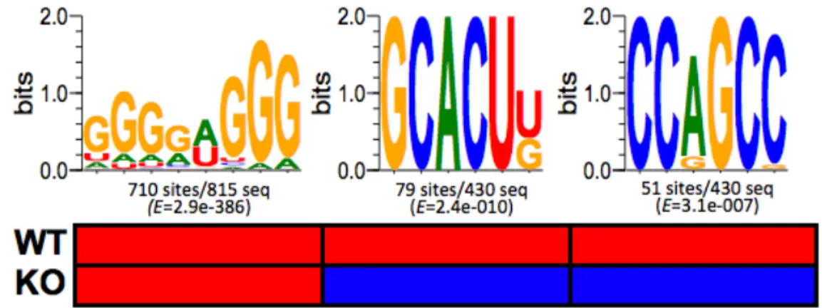
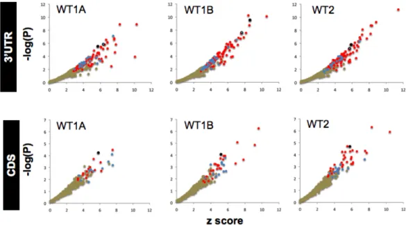
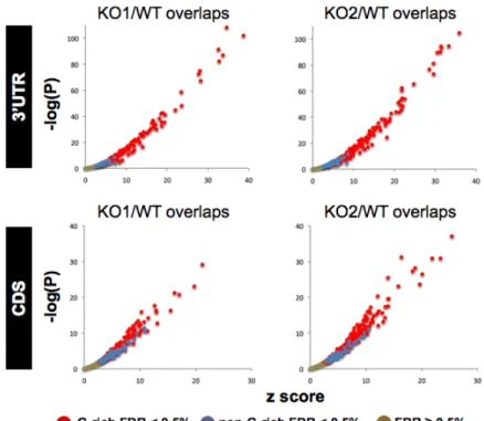
Identification of enriched motifs in Ago2-CLIP RNA tags... 69
Ago2- CLIP genes exhibit a miRNA dependent gene expression signature... 72
Identified GCACUU-seed match containing cluster is sufficient to confer miRNA-mediated repression ... 76
miRNA regulation can be modulated by G-rich motif that is associated with Ago2 .... 78
Discussion... 80
Methods ... 85
Cell culture ... 85
Derivation of Dicer wild-type and Dicer null mESCs ... 85
REAGENTS ... 85
EQUIPMENT... 86
SOLUTIONS ... 86
Ago2 CLIP protocol ... 87
Solexa-based small RNA cloning protocol ... 90
Short RNA cloning from Total RNA protocol: ... 94
Library Summary... 95
Data processing and bioinformatics... 95
Raw sequence processing ... 95
Data filtering ... 101
Motif enrichment analysis... 104
G-motif conservation analysis ... 108
Pathway enrichment analysis... 110
Western Blot analysis ... 111
Generation of luciferase constructs, mESCs transfection, and luciferase assays .... 111
mESC mRNA microarray analysis ... 114
TargetScan predictions and controls ... 114
Target statistics... 115
Statistical analyses ... 116
Acknowledgements... 117
Figures ... 118
CHAPTER 4: Targeted deletion reveals essential and overlapping functions of the miR-17~92 family of miRNA clusters ... 154
Abstract... 155
Introduction ... 156
Results ... 158
Expression pattern of miR-17~92 and its two paralogs ... 158
Generation and characterization of miR-106b~25 deficient mice ... 158
Generation and characterization of miR-106a~363-deficient mice ... 159
Generation of miR-17~92-deficient mice ... 159
Early postnatal lethality of miR-17~92-deficient mice ... 160
Lung hypoplasia and ventricular septal defect in miR-17~92-deficient mice ... 161
miR-17~92 is essential for fetal B cell development ... 161
miR-17~92 is essential for adult B cell development ... 162
miR-17~92 regulates B cell survival... 163
Functional interaction between miR-17~92 and miR-106b~25... 166
Discussion... 167
Role of miR-17~92 in heart and lung development... 167
Role of miR-17~92
in normal B cell development and lymphomagenesis... 168
Functional cooperation between miR-17~92 and miR-106b~25... 170
Bim and regulation of cell death by miR-17~92 and its paralogs... 171
Methods ... 172
ES cell manipulation, generation of chimeras and tetraploid blastocyst complementation... 172
Fetal liver reconstitution experiments ... 172
RNase protection assays and qPCR ... 173
Probes and Oligos used... 173
Bim 3’UTR reporter assays... 174
Antibodies, Immunohistochemistry and flow cytometry analysis ... 174
Histological analysis and serial sections... 175
Mouse husbandry ... 175
Acknowledgements... 176
Figures ... 177
CHAPTER 5: Conclusions and Future Directions... 206
Distribution of short RNA populations associated with the Ago2-miRNP ... 207
miRNA-targeted pathways in mESCs and developing mouse embryo... 209
Functional overlap between miRNAs with similar seeds ... 210
Understanding miRNA-targeting specificity ... 212
Roles for Ago2-directed regulation distinct from the miRNA pathway ... 214
CHAPTER 6: Bibliography ... 216
Discovery of miRNA-regulated processes in mammalian
development
by
Amanda Garfinkel Young
Abstract
The genomes of plants and animals encode hundreds of non-coding ~22nt RNAs termed “microRNAs” (miRNAs). These RNAs guide the sequence-specific inhibition of translation and destabilization of mRNA targets through short (6-7 nucleotide) stretches of complementarity between the 5’ end of the miRNA (seed region) and the 3’ untranslated region (UTR) of the targeted mRNA. Computational estimates based on prediction of conserved seed matches in 3’UTRs suggest that over half of genes in mammalian genomes are miRNA-regulated. Many miRNAs are also ubiquitously expressed in a variety of embryonic and adult tissues. Given these observations, we have sought to address the challenge of determining which cellular and
biological processes that miRNAs might regulate in different cell types and tissues. First, we have developed methods to isolate candidate miRNA-targeted mRNAs by immunoprecipitation of Ago2 in the miRNA effector complex (miRNP). In one purification method, the Ago2-bound mRNAs are identified by microarray. The second purification method adds crosslinking, RNase digestion, and cloning and deep-sequencing (CLIP-seq) to improve the specificity. These experiments were performed in mouse embryonic stem cells (mESCs) and a miRNA-deficient derivative cell line, lacking miRNA-processing enzyme, Dicer, to enrich for targets of miR-290~295, a miRNA cluster highly-expressed in mESCs. By these methods, we have identified a set of candidate mRNAs that bear a motif in their 3’UTRs that matches the seed of miR-290~295 and exhibit a miRNA-dependent gene expression signature. Gene ontology analysis of these candidate mRNAs shows they are involved in regulating the cell cycle, apoptosis, and the TGF-ß pathway. We further show that mESCs lacking miR-290~295 are unable to differentiate into endoderm, a TGF-ß regulated process.
We present additional evidence that RNAs crosslinked to Ago2 are significantly enriched in a G-rich motif in a miRNA-independent manner, as it is present in Ago2-CLIP libraries from Dicer wild-type and Dicer null mESCs. This G-rich motif exhibits higher conservation than the general 3’UTR background and appears to modulate the activity of miR-290~295 binding sites from the 3’UTRs of two genes. This G-rich motif may confer additional specificity to the Ago-miRNP and increase its affinity for miRNA-targeted mRNAs.
We have also investigated the function of a family of miRNA clusters in mouse embryonic development by targeted deletion of miR-17~92, and its paralogs, miR-106b~25 and
106a~363. 106b~25 and 106a~363 null mice are viable and fertile, however, miR-17~92-null mice die short after birth with lung hypoplasia and ventral septal defect. miR-17~92
null embryos also lack mature B cells, due to a block in the pro- to pre-B cell transition. Pre-B cells from the fetal livers of miR-17~92 null embryos have higher rate of apoptosis compared to wild-type embryos and upregulation of pro-apoptotic gene, Bim, a miR-17~92 target.
Our findings demonstrate roles for two different clusters of miRNAs in regulating
cellular processes of cell cycle, cell death, and cell signaling, that are important for differentiation of several different tissue types. Biochemical target identification and targeted deletion of
miRNA clusters in the mouse are both useful tools for uncovering miRNA functions. Thesis supervisor: Phillip Sharp
Acknowledgements
To Phil, thank you for the opportunity to receive excellent graduate training in your lab and your thoughtful and considerate mentorship in the last six years. Your ability to interpret data and extract the most salient and relevant points and then challenge all the weaknesses in a matter of minutes is inspirational. You always are looking at biology through a quantitative lens, and your scope of interest ranges from very broad to very deep. I will always carry your approach to science with me. You once told me when I wanted to join your lab that your goal for a graduate student is to conceive of a set of experiments to address a question, carry out those experiments, and then publish the results. This seemed so simple at the time, 6 years later I realized how devilishly
complicated it ended up being. You supported me the entire convoluted way, and I’d like to think that I learned more than if it had been as simple as I thought.
To my past and present thesis and defense committee members, Dave Bartel, Richard Hynes, Jianzhu Chen, Mike Hemann, Chris Burge, and Carl Novina, your advice and guidance over the years is much appreciated. You always tried to keep me going in the right direction, towards the finish line, now that I am about there, I have you to thank for it.
To the Sharpies, past and present, I started out needing a post-it with directions
indicating anterior and posterior, I would like to think that, thanks to you, I don’t need that anymore. Chris, John, Mauro, you taught me just about everything I know and had a lot of fun along the way. It will always be beer o’clock somewhere. Maggie, your interwebz skillz kept me entertained while in the lab. Lolcats 4eva! Grace, my running buddy and CDF guru, thanks for keeping me in shape and plotting my shifts. Anthony, many a long day spent with radioactivity in the cold room was less tedious because we did it together. I also appreciate you taking over so I didn’t have to irradiate my fetus. You are a
thorough and thoughtful experimentalist and your attention to detail is something I will always be trying to emulate. Sharp Lab Rules.
To my 5th floor colleagues and buddies and classmates, you made science easier and
fun. Andrea, thanks for letting me test my probes on your knock-outs. Turned out to be a pretty great collaboration for me, I miss having you as my coffee and golf buddy. Cheung and Keara, bottoms up. Hope we can reunite on the sunny coast. Irene, thanks for filling in as Nate’s drinking buddy while my pregnant butt was snoring on the couch. Michel, you helped introduce us to the glories of scotch, and thanks for helping us wind down from a long week with a glass (or 2). We will miss you and Amy Grace lots when we leave. DT and Heather, thanks for being awesome Dave fans and camping buddies. Thanks Dave especially for being such a good sport when we all made fun of you. To my former mentors, thank for all the opportunities to learn bench science and get my feet wet in the lab. I really appreciate your patience with me during the learning process and your encouragement to continue in science. Particular thanks to my undergraduate research advisor, Dr. Mariana Wolfer, Cornell University. I could not have asked for a more supportive, understanding, or intelligent teacher and scientist for the beginning of my research career. Your treatment of the undergrads in your lab as equal members, and your devotion to our projects and training, was key in cultivating my interest in biological problems as well as exploring the idea of research science as a career path.
Your lab was such a warm and friendly as well as intellectually stimulating environment for doing science, it will be my first model if I have a lab of my own. I also really
appreciate you allowing me to accompany you to the CSHL Germ cell meeting in 2002. This really cemented my decision to go to graduate school and was an amazing
opportunity to learn and think about exciting science as well as develop contacts that have continued here at MIT. Being your mentee and working in your lab is the singular most important opportunity offered to me at Cornell.
To my family, I may have been destined to be a scientist since I toddled around the halls of the Whitehead at a tender age and had my first acknowledgement in Cell 27 years before I was actually a published author there. You always supported and encouraged my love of learning, from “bus stuff”, to OM, to being a lab rat. Even when I broke down after my first Biology test at Cornell, you helped me pick up the pieces and keep going. Thanks especially to Emily for the note you wrote on my computer before I went to Cornell, knowing you believed in me and looked up to me helped me believe in myself. There have been a lot of successes and some amount of failures, but your love and support for me under both circumstances is a model that I will try my best to follow as I go down that road as a parent myself.
To Nathan Jr. (NJ), being 7 months old, it will probably be awhile before we pull
Mommy’s thesis off the shelf and look through it out of curiosity, but when we do it will be to remember about how you were just a little tiny baby, in our little tiny apartment in Somerville, when Mommy and Daddy were finishing their dissertations. Life was pretty stressful trying to get everything done and provide you with all the love and care you needed, but we made it. I would like to think having the flexibility of being a student and Phil’s generous support of my maternity leave let me experience a lot of your first moments and milestones. I thank you for very few sleepless nights after you were three months old, and loving us and your nannies. And even when times were tough and I was stressed about everything, a smile and laugh from you could make me forget all my worries.
To Nate Sr., I would never say that we couldn’t have done it without each other, because that would give each other too little credit. But, you have always been there for me with your love and support when I needed a friend, a colleague, a partner. We have come a long way since studying the catalytic triad on the second floor of building 68, and the challenges that we have faced in this period of our lives have left us much changed. But, there is no one I would rather have standing beside me to enter this next phase in California and beyond, as post-docs, as parents, as husband and wife. Love always.
Introduction
One of the fundamental processes of biology is how our genetic information is turned into functional molecules in the cell. Work over the last five decades has shown that this process begins with DNA being transcribed in the nucleus and spliced into messenger RNA (mRNA), which is then exported into the cytoplasm where it is
translated into protein, or stored, or degraded. Perhaps the most surprising discovery of the past decades is that RNA is not just a messenger, but can actually regulate gene expression post-transcriptional manner. This phenomenon is broadly defined as RNA interference (RNAi) and involves a short RNA reducing target gene expression in a sequence specific manner. One class of these RNAs are microRNAs (miRNAs), which are short ~22nt non-coding RNA genes. This gene class appears to have evolved independently in plants and animals and animal genomes encode hundreds of miRNA genes, many of which are conserved from C. elegans to humans. miRNAs control their target mRNAs through sequence-specific interactions that result in the destabilization and subsequent degradation and inhibition of translation of the target mRNAs. miRNAs play critical roles in diverse cellular processes such as apoptosis, proliferation,
differentiation: in organismal contexts such as embryonic development and cardiac electrophysiology; and in diseases such as cancer and Alzheimer's disease (reviewed in Singh et al. 2008). In Chapter 1, miRNA biogenesis, activity, and cellular functions are described, focusing mostly on mammalian miRNAs. Lastly, a brief description of the role of miRNAs in mouse embryonic stem cell biology is discussed, as it is the focus of research in this thesis.
miRNA biogenesis
pri-miRNA transcription and processing
RNA Polymerase II (Fig. 1), but a few are transcribed by Polymerase III (Cai et al. 2004; Lee et al. 2004; Monteys et al. 2010). Those generated by Pol II are capped and poly-adenylated and come from independent transcriptional units either in intergenic regions or in introns. By computational estimates, roughly 50% of miRNAs are found within other transcriptional units, either in the exon or intron of host non-coding RNAs or in the intron of host protein coding genes (Cai et al. 2004; Lee et al. 2004; Baskerville et al. 2005; Rodriguez et al. 2007; Saini et al. 2007; Griffiths-Jones et al. 2008). The miRNA resides in a hairpin structure, termed the pre-miRNA, with a ~33nt stem region that can be either mostly or completely paired, and a loop region. 36-47% of pre-miRNA hairpins in
vertebrates are also present in clusters of pre-miRNAs in the same primary transcript (Saini et al. 2007; Thatcher et al. 2008).
The pre-miRNA hairpin is processed from the primary transcript in the nucleus by the Microprocessor complex containing the RNase III-class enzyme Drosha, and its co-factor DGCR8 (Fig. 1)(Lee et al. 2003; Denli et al. 2004; Gregory et al. 2004; Han et al. 2004; Landthaler et al. 2004; Zeng et al. 2005b). The Microprocessor complex
recognizes the stem and the unpaired flanking regions of the hairpin. DGCR8 acts as a molecular ruler to guide Drosha's cleavage of the two strands at the base of the hairpin by its two RNase III domains acting as an intramolecular dimer (Zeng et al. 2003; Zeng
et al. 2005a). This leaves the stem with a 5' phosphate and a 3'OH on a 2-nt overhang.
The Microprocessor complex tracks with Pol II and its activity can precede splicing of the intron where the pre-miRNA resides (Kim et al. 2007b; Morlando et al. 2008). There also exists a class of miRNAs, termed mirtrons, that do not require Drosha for their
processing as the two ends of the pre-miRNA lie at the splice sites of introns (Okamura
Pre-miRNA processing
Following liberation from the primary transcript, the pre-miRNA is bound by Ran-GTP/Exportin-5 and exported to the cytoplasm (Fig. 1)(Yi et al. 2003). Once in the cytoplasm, the pre-miRNA is recognized by the RISC Loading complex (RLC). The RNAi silencing complex (RISC) is the effector complex of short RNA function that mediates targeting and regulation of the cognate mRNAs (Hammond et al. 2001; Hutvagner et al. 2002; Martinez et al. 2002; Martinez et al. 2004). This complex contains a member of the conserved Argonaute (Ago) protein family (Carmell et al. 2002; Caudy et al. 2002). The RLC contains Dicer, an RNase III-class enzyme, TRBP, Tar-RNA binding protein, PACT, protein activator of PKR, and Ago (Chendrimada et al. 2005; Gregory et al. 2005; Haase
et al. 2005; Maniataki et al. 2005; Lee et al. 2006). Dicer is a highly conserved protein
present in all species that have RNAi (Bernstein et al. 2001; Grishok et al. 2001; Hutvagner et al. 2001). Dicer's PAZ domain binds the 2nt overhang on the pre-miRNA (or long dsRNA) and its two RNase III domains, acting as an intramolecular dimer, cleave ("dice") the two strands of the stem 21-22nt away, removing the loop and leaving a 5' phosphate and 3'OH on a 2nt 3' overhang (Fig. 1)(Song et al. 2003; Yan et al. 2003; Lingel et al. 2004; Ma et al. 2004; Zhang et al. 2004; MacRae et al. 2007). TRBP and PACT, while not required for processing, appear to increase dicing efficiency and TRBP stabilizes Dicer protein (Chendrimada et al. 2005; Lee et al. 2006). Dicer, TRBP, and Ago2 make up a minimal complex that is sufficient for processing of the pre-miRNA and loading the mature miRNA into RISC (Gregory et al. 2005; Maniataki et al. 2005; Wang
et al. 2009b).
Several groups have shown that Drosha and Dicer processing of pri- and pre-miRNAs, respectively, can be regulated by other RNA binding proteins (Guil et al. 2007; Davis et al. 2008; Heo et al. 2008; Michlewski et al. 2008; Newman et al. 2008;
Piskounova et al. 2008; Rybak et al. 2008; Viswanathan et al. 2008; Trabucchi et al. 2009). Generally these proteins act by binding to pri-/pre-miRNA species and either facilitating or inhibiting cleavage by Drosha or Dicer. For example, TGF-ß and BMP activation of SMAD proteins stimulates their binding to pri-miR-21 along with p68 helicase, and consequently enhances its Drosha-mediated processing (Davis et al. 2008). Both Dicer and Drosha processing of the let-7 family of miRNAs can be inhibited by Lin-28, and this seems to be an important mechanism for regulating the proliferation of embryonic stem cells and their differentiated derivatives (Heo et al. 2008; Newman et
al. 2008; Piskounova et al. 2008; Rybak et al. 2008; Viswanathan et al. 2008; Melton et al. 2010).
miRNA duplex unwinding and loading
After Dicer processing, the remaining species is the miRNA duplex, which must be subsequently unwound (Fig. 1). One strand, the miRNA or guide strand, is loaded into the RISC-Ago complex and the other strand, the miR* or passenger strand, is discarded and degraded. Strand selection is, in part, based on asymmetry between the thermodynamic stabilities of the two ends of the duplex. The strand whose 5' end resides in the more unstable end of the duplex is more likely to be incorporated into RISC
(Khvorova et al. 2003; Schwarz et al. 2003). While protein "sensors" of this asymmetry, R2D2 and Loquacious, have been identified in Drosophila (Forstemann et al. 2005; Jiang et al. 2005; Saito et al. 2005); no such sensor has been found in mammalian systems and maybe the Ago protein itself (Tomari et al. 2004; Wang et al. 2009a; Yoda
et al. 2010).
Additionally, Ago2, an RNase-H-like endonuclease in RISC, can cleave the passenger strand of a perfectly complementary RNA duplex (siRNA) this rendering it incapable of being the guide strand in RISC (Matranga et al. 2005; Miyoshi et al. 2005;
Rand et al. 2005; Leuschner et al. 2006; Diederichs et al. 2007; Kim et al. 2007a). Ago2 can also cleave the miR* strand of the miRNA duplex and 3' arm of the pre-miRNA hairpin, however, this activity requires extensive base-pairing in the miRNA duplex or stem of the pre-miRNA, which is uncommon. In the absence of perfect base-pairing in the stem, Ago proteins in vitro can unwind miRNA duplexes and load the miRNA strand. The efficiency is increased if there are mismatches in the duplex between positions 2-10 and/or positions 12-15 (Wang et al. 2009a; Yoda et al. 2010). Ago proteins enhance the stability of overexpressed miRNAs in cultured cells (Diederichs et al. 2007). Ago2, in particular, is also required for stability of miRNAs in vivo, as loss of Ago2 in B cells results in the decrease of expression of some miRNAs. This appears to be independent of its endonuclease activity, as a cleavage-defective mutant of Ago2 can rescue the phenotype (O'Carroll et al. 2007).
RISC/miRNP
Minimal active RISC contains a single stranded RNA bound to an Ago protein. There are 8 members of the Ago super-family present in mammalian genomes
(reviewed in Hock et al. 2008). In the Ago subfamily, there are 4 genes in human,
hAgo1-4, and 5 genes in mouse, mAgo1-5; they are all expressed ubiquitously in many
tissues types and cell lines (Sasaki et al. 2003). In the Piwi sub-family, there is human
Hiwi, Hiwi2, Hiwi3 and Hili, and mouse Miwi, Miwi2, Mili; they are expressed specifically
in the germline and appear to regulate meiosis and spermatogenesis, at least in part, by the piRNA pathway. The Ago sub-family will be discussed here exclusively as it is most relevant to miRNA function, but for further discussion of piRNAs see Thomson and Lin (2009). The members of the Ago sub-family are highly related to one another both within and across species. Crystallographic studies of Ago proteins found in Archaea and corresponding biochemical analysis of metazoan Ago proteins have yielded important
insights into the structure and function of these proteins. Ago proteins consist of 3 domains: PAZ, Mid, and PIWI domains. The Ago PAZ domain, which is structurally similar to the Dicer PAZ domain, binds the 3'OH of the guide strand RNA (Song et al. 2003; Yan et al. 2003; Lingel et al. 2004; Ma et al. 2004). The Mid domain contains a basic pocket for binding the 5' phosphate of the short RNA, an important component the RNA binding specificity of Ago (Parker et al. 2004; Yuan et al. 2005). The PIWI domain is homologous to RNase-H catalytic domain, though only Ago2 possesses
endonucleolytic (slicer) activity. The catalytic triad for hAgo2 consists of Asp597, Asp669 and His807. Ago1 and Ago4 lack these residues, explaining why they lack slicer activity. However, Ago3 also has these residues, but has no slicing activity in vitro (Liu et al. 2004; Meister et al. 2004b; Parker et al. 2004; Song et al. 2004; Ma et al. 2005; Parker
et al. 2005; Rivas et al. 2005; Yuan et al. 2005; Wang et al. 2008c; Wang et al. 2008d;
Wang et al. 2009c). Surprisingly, Ago1 in vitro is able to cleave a perfectly
complementary RNA duplex with the sequence of miR-21, but is unable to catalyze cleavage of a perfectly complementary target RNA (Wang et al. 2009a). However, in
vivo, this activity does not seem to be robust enough to allow for appreciable recruitment
of mRNA targets to the Ago1-miRNP (Wang et al. 2009a).
Regulation of target mRNAs by miRNAs requires additional factors associated with RISC in a complex termed the miRNA ribonucleoprotein particle (miRNP)
(Mourelatos et al. 2002). Several studies of Ago-associated proteins have identified other components of the miRNP, including GEMIN3, GEMIN4, VIG, TSN, FMRP family members, MOV10, HSP90, HNRNP family members, RCK/p54, and others (Tahbaz et
al. 2001; Caudy et al. 2002; Mourelatos et al. 2002; Caudy et al. 2003; Tahbaz et al.
2004; Meister et al. 2005; Chu et al. 2006; Hock et al. 2007). Some of these associated co-factors can modulate the function of the miRNP. For example, FMRP is required in the mouse brain for regulation of NR2A 3'UTR by miR-125b complexed with Ago1 in the
miRNP (Edbauer et al. 2010). Another Ago-associated protein, Importin 8, in involved in regulating the subcellular localization of Ago proteins, as well as loading of miRNA target mRNAs into the miRNP complex (Weinmann et al. 2009).
The active RISC/miRNP identifies target mRNAs through base-pairing
interactions between the guide RNA and the target RNA. The base-pairing between the guide and target in an A-form helix is initiated at the 5' end of the guide RNA, although the first nucleotide is unpaired (Ma et al. 2005; Parker et al. 2005; Yuan et al. 2005). Pairing between the central region of the guide and the target aligns the duplex in the active site of Ago2 and results in cleavage of the target mRNA at the scissile phosphate group opposite the phosphate group between the tenth and eleventh nucleotides of the guide RNA strand, as measured from the 5' end of the guide strand (Elbashir et al. 2001a; Elbashir et al. 2001b; Liu et al. 2004; Ma et al. 2005; Parker et al. 2005; Yuan et
al. 2005; Wang et al. 2008c; Wang et al. 2008d; Parker et al. 2009; Wang et al. 2009b;
Wang et al. 2009c).
Few mammalian miRNAs have extensive pairing to their mRNA targets. Only mouse miR-196 has been shown to cause endonucleolytic cleavage of a target mRNA,
HoxB8 (Yekta et al. 2004) (Fig. 1). Instead, most mammalian miRNAs base pair with
their targets between nucleotides 2-8, as measured from the 5' end of the miRNA, have no pairing in the central region, and have variable amounts of pairing between nt 12-17 (Grimson et al. 2007). The region of pairing in the 5' end of the miRNA, termed the seed region, is usually critical for this recognition of the target mRNA and its regulation
(Doench et al. 2003; Lewis et al. 2003; Doench et al. 2004; Brennecke et al. 2005). The topic of miRNA targeting is discussed in more detail in a later section.
Modes of miRNA-mediated silencing
target mRNAs through two modes of regulation: inhibition of translation and destabilization and degradation of the transcript (Fig. 1).
Translational Repression
Initial work focused on the former mechanism as the first identified animal miRNA, C. elegans lin-4, caused a large decrease in the protein from the lin-14 transcript with little accompanying decrease in transcript levels (Lee et al. 1993; Wightman et al. 1993). Although, a later study indicates that lin-14 transcript is also destabilized by lin-4 (Bagga et al. 2005). Olsen et al. (1999) showed that lin-4 and lin-14 both associate with polyribosomes, and speculated that lin-4 causes an inhibition of translation after the initiation step. This mode of regulation was further supported by studies of both endogenous and artificial miRNA targets in mammalian cells that showed that target mRNAs associate with poly-ribosomes, miRNA repression leads to decreased readthrough at a stop-codon, and miRNAs repress internal ribosome entry site (IRES)-containing mRNAs whose initiation does not depend on canonical initiation factors (Maroney et al. 2006; Nottrott et al. 2006; Petersen et al. 2006).
However, several other groups showed that miRNAs inhibit translation of their target mRNAs by blocking initiation. These groups observed that miRNA target mRNAs are not associated with polyribosomes and fail to be repressed when they contain a cap-analog or IRES (Humphreys et al. 2005; Pillai et al. 2005). Additional experiments performed in intact cells and cell-free extracts also showed that an intact cap and a polyA tail are required for repression (Wang et al. 2006; Wakiyama et al. 2007; Beilharz
et al. 2009) and that miRNAs either inhibit 60S joining the loaded 40S complex (Wang et al. 2008a) or 80S initiation complex formation (Mathonnet et al. 2007). Addition of eIF4F
to the extract could relieve the repression, suggesting miRNA-mediated repression impinges on the recognition of the cap structure (Mathonnet et al. 2007). Indeed, Ago2
has been shown to bind the mRNA 7-methylguanosine cap, and may inhibit translation initiation by this activity (Kiriakidou et al. 2007; Djuranovic et al. 2010). It has also been shown that the miRNP can recruit eIF6 to the targeted mRNA and prevent association of the large ribosomal subunits (Chendrimada et al. 2007). Translational repression also requires RNA helicase RCK/p54 (Chu et al. 2006). At this point, it remains possible that miRNAs regulate the translation of their targets both at the step of initiation and at a step after initiation, potentially by enhancing ribosome drop-off.
mRNA destabilization
The importance of the second mode of miRNA-mediated regulation was later recognized with the observation that addition of exogenous miRNAs to human cells can cause downregulation of a large set of mRNAs (Lim et al. 2005). Additionally, several groups reported the localization of Ago2 to mRNA-processing bodies (P-bodies) (Liu et
al. 2005b; Pillai et al. 2005). These cytoplasmic foci are thought to be sites of mRNA
degradation and storage as they are enriched in many mRNA-degrading enzymes, including XRN1 and DCP1/2 (reviewed in Anderson et al. 2009). In human cells, about 10% of cytoplasmic Ago protein localizes to P-bodies, while the remaining protein is in the diffuse cytoplasm, and the protein shuttles rapidly between these two sites (Leung et
al. 2006). However, this localization is not required for miRNA target degradation (Chu et al. 2006; Eulalio et al. 2007).
Ago also interacts with the GW182 protein and this interaction is critical for destabilization of targeted mRNAs (Liu et al. 2005a; Rehwinkel et al. 2005; Ikeda et al. 2006; Eulalio et al. 2008). GW182 recruits CCR4-NOT deadenylase complex to the target mRNA; deadenylation is followed by decapping and 5' to 3' exonucleolytic degradation by XRN1 (Behm-Ansmant et al. 2006; Chen et al. 2009; Zekri et al. 2009). Ago2 interaction with GW182 is mediated through the C-terminal half on Ago2, and in
particular by two phenylalanines, F594 and F629 (Chen et al. 2009). These residues in Ago2 are required for repression of a target that is artificially tethered to Ago2 through the LambdaN-BoxB association (Eulalio et al. 2008). However, it has also been shown that these phenylalanines are required for Ago mRNA cap binding activity (Kiriakidou et
al. 2007), and thus may effect miRNA-mediated repression through inhibition of
translation initiation as well. mRNA deadenylation is not required for miRNA-mediated translational inhibition of the mRNA and some miRNA targets have been shown to have a decrease in protein output with little to no change in mRNA levels (Pillai et al. 2005; Wu et al. 2006; Baek et al. 2008; Eulalio et al. 2008; Selbach et al. 2008; Eulalio et al. 2009). Taken together, these observations suggest that the two modes of miRNA-mediated silencing occur independently and the contribution of each to the overall level of target repression depends on unknown factors or sequence contexts (Wu et al. 2006; Baek et al. 2008).
Translational Activation
miRNAs may also cause translational activation of their target mRNAs. This has been shown to occur for an endogenous mRNA, TNF-alpha, under conditions of cell cycle arrest (Vasudevan et al. 2007a). Vasudevan and colleagues (2007b) also
demonstrated that artificial miRNA targets are translationally repressed in cycling cells, but are translationally activated in G0/G1 quiescent cell state. Additionally, miR-10a upregulates the translation of TOP-motif (terminal oligopyrimidine tract) containing mRNAs by binding sites in the 5'UTR near the TOP motif, although these sites do not contain canonical seed matches to miR-10a (Orom et al. 2008). It is still unclear whether translational activation by miRNAs is a general phenomenon or a specialized activity of certain miRNA:mRNA target pairs under certain conditions.
miRNA binding sites in 5’UTRs and coding sequences
In addition to the report from Orom et al. (2008), several other groups have investigated miRNA binding sites in 5'UTRs. A proteomic analysis of neutrophils lacking miR-223 expression found no miRNA-dependent effects on protein abundance for mRNAs containing miR-223 seed matches in their 5'UTRs (Baek et al. 2008). However, an investigation of miRNA binding sites in the 5'UTR of luciferase reporter mRNAs found that they can confer miRNA-dependent translational inhibition (Lytle et al. 2007). In these experiments, initiation of translation of the reporter mRNAs was mediated by IRES elements, rather than the canonical mRNA cap, which could account of the differences observed.
miRNAs can also target sites in the coding sequence (CDS) of transcripts (Duursma et al. 2008; Tay et al. 2008). Tay et al. (2008) observed that several miRNAs in ES cells can post-transcriptionally regulate Oct4, Sox2, and Nanog, and thus affect the pluripotency of these cells. This phenomenon has also been observed on a genome-wide scale for miRNA-dependent expression changes (Grimson et al. 2007; Baek et al. 2008). The degree of mRNA change is significantly less for transcripts with miRNA sites in CDS as compared to those with sites in the 3'UTR. Consistent with this, translation of 3'UTRs sequences bearing miRNA binding sites fused to the CDS of a transgene abolishes their ability to confer miRNA-dependent downregulation (Gu et al. 2009). At this point, it remains unclear whether the regulatory mechanisms are the same for miRNA binding sites in the CDS and 3'UTRs of transcripts. Also, it is possible that the interaction of miRNAs with sites in CDS differs from the interaction with sites in the 3'UTR, as only 1 out of 5 of the sites identified by Tay et al. (2008) has a canonical miRNA seed match.
Interface of the miRNA pathway with other post-transcriptional regulatory
pathways
miRNA-regulated sites in 3'UTRs are only one kind of a larger category of 3'UTR sequence elements that are regulated by RNA-binding proteins (RBPs). Several groups have shown that miRNA-mediated effects can be abrogated by binding of other RBPs to sites near miRNA binding sites (Bhattacharyya et al. 2006b; Kedde et al. 2007;
Vasudevan et al. 2007a). For example, HuR binding to the CAT-1 transcript under stress conditions in liver cells relieves miR-122-mediated repression of this mRNA
(Bhattacharyya et al. 2006a). Given that miRNP complexes are quite stable in the cell (Lee et al. 2006; van Rooij et al. 2007; Gatfield et al. 2009), this is probably an important general mechanism for regulating miRNA-dependent effects on transcripts.
Discovery of miRNA biological functions
The discovery of miRNAs as an entirely new gene class led many groups to explore the biological function(s) of these RNAs on both the cellular and organismal levels. This question has been addressed in three main ways: 1) computational miRNA target prediction 2) biochemical miRNA target identification and 3) miRNA loss-of-function, by both use of specific miRNA inhibitors and classic reverse genetic approaches.
Computational miRNA target prediction
Seminal work on one of the first identified miRNAs (lin-4) in C. elegans
demonstrated that the phenotype of lin-4 null animals, reiteration of the L1 stage in later larval stages, is due to the upregulation of LIN-14 protein (Lee et al. 1993; Wightman et
al. 1993). They further showed that the lin-14 mRNA has complementary sites in its
3'UTR that can base-pair with the lin-4 miRNA, and that the region containing these sites is required for repression of LIN-14 protein synthesis. After both experimental and
computational methods identified many miRNAs present in the genomes of C. elegans,
Drosophila, mouse and human (Quintana et al. 2001; Lau et al. 2001;
Lagos-Quintana et al. 2002; Lim et al. 2003), it became clear, based on lin-4:lin-14 paradigm, that identification of miRNA-targeted mRNAs would provide many insights into the biological functions of miRNAs.
Given the large number of miRNAs, as well as mRNAs, present in animal genomes, computational approaches are necessary for prediction of miRNA-targeted mRNAs. Most target prediction algorithms were based on two features of the first validated miRNA:mRNA target pairs. First, there is at most 8-10 nt of continuous base-pairing between seed region in the 5' end of the miRNA and the target site in the 3'UTR of the mRNA, including G:U wobble base-pairs (Lee et al. 1993; Wightman et al. 1993; Brennecke et al. 2003).There was also variable amounts of complementarity between the 3' end of the miRNA and the mRNA, ranging from no complementarity to several nucleotides of complementarity. Second, the seed match region in the target mRNA is often conserved between related species. Employing these criteria, multiple groups used a thermodynamic approach that modeled the interaction between the miRNA seed and all the annotated 3'UTRs in the genome of interest. Some algorithms assessed further base-pairing interactions between the 3' end of the miRNA and mRNA or allowed G:U wobble pairs or mismatches in the seed. Furthermore, the conservation of these sites was determined between related genomes. Generally, these initial studies estimated that between 10-30% of human genes are targets of miRNAs (Lewis et al. 2003; John et al. 2004; Krek et al. 2005; Lewis et al. 2005; Miranda et al. 2006; Kertesz et al. 2007; Griffiths-Jones et al. 2008) and the overlap between predicted sets varied from 15%-40% (Kertesz et al. 2007). This could be due to the use of different 3'UTR annotations or genomic alignments, or the various permutations and weighting of the criteria.
of miRNAs into cells where they are not normally expressed could cause downregulation of hundreds of transcripts containing seed matches to the miRNA in their 3'UTRs (Lim et
al. 2005). When this set of targets identified by miRNA overexpression was compared to
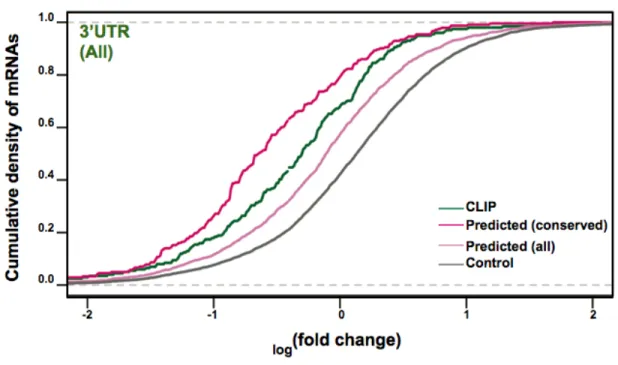
the set of computationally predicted targets, it became clear that those with longer seed matches (7 rather than 6 nt of base-pairing), an 'A' in the first position of the target site, and those sites that had a higher degree of conservation caused a greater
downregulation (Grimson et al. 2007; Nielsen et al. 2007). The preference for an ‘A’ in the first position of the target site, both at the level of conservation and degree of mRNA downregulation, could be due to the geometry of the miRNA:mRNA target duplex in the Ago protein (Lewis et al. 2005; Grimson et al. 2007). Crystallographic studies show that this nucleotide is unpaired, and since the first nucleotide of many miRNAs is ‘U’,
disrupting this pairing would be energetically more favorable (Lim et al. 2003; Lewis et
al. 2005; Ma et al. 2005; Parker et al. 2005; Yuan et al. 2005).
Additionally, this method allowed for the investigation of other criteria for miRNA targeting, including location of the miRNA binding site with respect to the stop codon and polyA tail, AU content around the site, proximity to other miRNA binding sites, and contribution of pairing between the 3'end of the miRNA and the target site (Doench et al. 2003; Grimson et al. 2007; Nielsen et al. 2007). miRNA binding sites appear to be more active if they are at least 14nt from the stop codon. This could be because the translation termination machinery inhibits miRNP from accessing the binding site. miRNA binding sites also appear to be more active if they are towards to ends of long 3'UTRs and in a region of high AU content. This likely is due to the miRNP having greater access to the miRNA binding site if it is not part of an RNA secondary structure. miRNA binding sites can also act cooperatively if they are within 40nt of each, potentially by increasing the likelihood the miRNP encounters the transcript. It has been experimentally determined that pairing between the 3'end of the miRNA and the target site confers additional
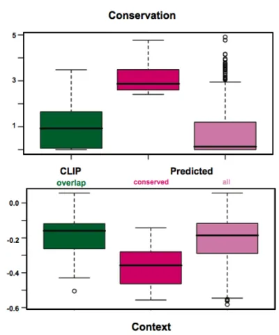
miRNA-dependent repression (Doench et al. 2004; Brennecke et al. 2005; Grimson et al. 2007). This appears to be particularly consequential for pairing of 4 nt starting at position 13 of the miRNA. Supporting this, there is also a detectable signal for conservation in the target for pairing to the 3' end of the miRNA (Grimson et al. 2007). These criteria have been incorporated into the newest version of the target prediction program, TargetScan 5.1, as a composite score (context score). Additionally, analysis of conservation of miRNA binding sites in 23 non-fish vertebrate genomes, predicts over 45,000 miRNA binding sites in ~50% of mammalian genes for hundreds of miRNAs (Friedman et al. 2009).
Several target prediction programs either do not use conservation as a criterion or separately allow the investigation of non-conserved sites (RNA22, PITA, TargetScan) by focusing on seed matching and site context (Miranda et al. 2006; Grimson et al. 2007; Kertesz et al. 2007; Friedman et al. 2009). Given that many non-conserved miRNA-binding sites have been shown to be functional, particularly if they have favorable sequence context, this is a useful aspect to target prediction programs (Krutzfeldt et al. 2005; Lim et al. 2005; Giraldez et al. 2006; Grimson et al. 2007; Rodriguez et al. 2007; Baek et al. 2008).
Given the potential for miRNAs to regulate sites in the 5'UTRs and CDS of transcripts, there have been efforts to also predict miRNA binding sites in these genic regions. This type of analysis relies on observing excess sequence conservation over the background sequence composition, since CDS sequences are already highly conserved, confidently predicting miRNA binding sites in this region is challenging (Lewis et al. 2005; Stark et al. 2007). Despite this, mRNAs bearing predicted seeds sites in the CDS do undergo miRNA-dependent changes in gene expression, although the degree of repression is less than those mRNAs bearing sites in the 3'UTR (Farh et al. 2005; Lim et al. 2005; Grimson et al. 2007; Baek et al. 2008). Searching for miRNA
binding sites in the 5' UTRs of mRNAs, Lee et al. (2009) found conserved sequences that were complementary to the 3' ends of miRNAs, rather than the seed region, similar to previous observations of miRNA sites in 5'UTRs (Orom et al. 2008). One 5'UTR sequence, when placed upstream of Luciferase coding sequence, was sufficient to confer downregulation on luciferase gene expression.
The sets of computationally predicted miRNA targets have provided the basis for validation of many miRNA:mRNA target interactions and investigation of the role of these interactions in a variety of cellular contexts. For example, identification of targeting of the oncogene E2F1 by miR-17-5p and miR-20 in human cells implicated these
miRNAs the control of the cell cycle and cell growth (O'Donnell et al. 2005). However, the utility of these predictions for determining the function of a given miRNA in a given cell type/tissue or a specific cellular process is limited by the fact that there are hundreds of targets predicted for each miRNA, that each prediction program may predict a unique set of targets, and that the likelihood that an mRNA is targeted by that miRNA depends on the expression level of the miRNA and mRNA in its native context. The degree of conservation of the miRNA binding site and the favorability of the sequence context around the site can be used as reliable indicators of the likelihood a miRNA binding site is functional (Grimson et al. 2007; Nielsen et al. 2007; Friedman et al. 2009). However, it is possible that many functional miRNA binding sites are overlooked because it is not feasible to analyze all predicted miRNA binding sites. These sets of computationally predictions of targets have been validated by complementary approaches to
biochemically identify miRNA targets.
Biochemical miRNA target identification
One such technique, as was previously mentioned, is to use microarray analysis to detect gene expression changes induced by miRNA overexpression or specific
miRNA loss, either by targeted deletion of the miRNA or miRNA inhibition by specific antisense oligo inhibitors (Lim et al. 2005; Rodriguez et al. 2007). Since miRNAs cause a decrease in the expression of their target mRNAs, the set of genes that possess miRNA seed-match sites and exhibit a change in expression are likely to be miRNA targets. This type of analysis has allowed for detection of a set of miRNA-regulated mRNAs under a give cellular context, and has verified many computational predicted targets. There are also several limitations this approach. First, the miRNA
overexpression approach is most effective under the conditions where the miRNA is introduced to a cell type where it is normally not expressed, such as expressing miR-1, a heart specific miRNA, in Hela cells, a cervical cancer cell line. However, miR-1 may regulate heart-specific transcripts that are not expressed in Hela cells, and thus would not be identified by this assay. Inhibition of miRNAs with specific inhibitors can get around this problem, although the signal tends to be weaker. Second, some miRNA target mRNAs exhibit translational repression with little to no miRNA-dependent change on the mRNA level, and thus would not be detected by this assay (Baek et al. 2008; Selbach et al. 2008).
A complementary approach to address this caveat is to analyze global miRNA-dependent protein changes by quantitative mass spectrometry using SILAC (stable isotope labeling with amino acids in cell culture) (Baek et al. 2008). This technique has been used to identify targets of miR-124, a brain specific miRNA, in Hela cells as well as targets of miR-223 in neutrophils, using cells that have a targeted deletion of miR-223. This yielded several important insights into miRNA-dependent regulation and the
performance of computational target prediction algorithms. First, some mRNAs exhibited a small change in protein expression without a change mRNA expression, however all genes that showed a greater than 30% repression had some mRNA destabilization component. This suggests that the most dramatic gene expression changes employ both
mechanisms of miRNA-dependent gene regulation.
Second, when comparing the set of observed targets in this assay with those predicted by several computational algorithms (miRANDA, miRbase, Pictar, PITA, TargetScan and RNA22), the two programs whose predicted targets had the greatest degree of expression change (Pictar and TargetScan) considered both exact 7mer and 8mer seed-match sites and conservation of those sites (John et al. 2004; Krek et al. 2005; Miranda et al. 2006; Kertesz et al. 2007; Griffiths-Jones et al. 2008; Friedman et
al. 2009). Those that allowed for mismatches or wobble base pairs in the seed region, or
did not consider conservation did no better than the expression change for all genes containing seed match sites. However, two thirds of the targets predicted by the
TargetScan and Pictar programs were not responsive to miRNA regulation in this assay, suggesting that there are additional unknown factors that govern miRNA-dependent regulation. One limitation of this approach is that quantitative mass spectrometry could only identify proteins whose abundance and solubility was such that they could be detected in this assay.
Expression based approaches have identified the seed match site as the only distinguishable common feature among genes that show miRNA-dependent mRNA and protein changes. There are some reports of miRNA-targeted mRNAs bearing seed matches with mismatches or pairing between positions 3-8, not the canonical 2-7 (Wu et
al. 2005; Didiano et al. 2006; Didiano et al. 2008; Tay et al. 2008; Friedman et al. 2009).
Furthermore, there are many genes whose expression levels have been observed to be responsive to miRNA effects but do not contain canonical seed matches. While some of these changes maybe secondary effects from miRNA overexpression or inhibition, other of these may associate with the miRNP through non-seed based interactions. These types of interactions between the miRNP and the mRNA target maybe common in a subset of targets, and may not have been observed by the expression-based
approaches.
Several groups have addressed this question by developing methods to
biochemically purify miRNA target mRNAs (Beitzinger et al. 2007; Karginov et al. 2007; Orom et al. 2007; Hendrickson et al. 2008; Landthaler et al. 2008; Orom et al. 2008; Chi
et al. 2009; Hong et al. 2009; Zisoulis et al. 2010). These methods employ
immunoprecipitation techniques to either isolate mRNAs bound to a biotin-tagged miRNA or mRNAs bound to the Ago protein in the miRNP and then detection of the mRNAs by microarray analysis or deep sequencing. Isolation of mRNAs bound to a biotinylated oligo has the advantage of identifying targets of a specific miRNA, however there could be other, spurious interactions of mRNAs with the labeled RNA. By this technique, an unusual class of RNA targets containing miRNA binding sites in their 5'UTRs was identified (Orom et al. 2008).
Immunoprecipitation of the Ago protein in cultured cells has also identified many potential target mRNAs, however it is a challenge to match up the mRNAs with the miRNAs that are targeting them to the miRNP (Beitzinger et al. 2007; Karginov et al. 2007; Hendrickson et al. 2008; Landthaler et al. 2008; Hong et al. 2009). Ultimately, bioinformatic analysis is necessary to look for seed matches in the 3'UTRs of the immunoprecipitated mRNAs. This approach is explored in Chapter 2 of this thesis, including an alternative experimental design to address this issue. A variation of the Ago immunoprecipitation technique termed CLIP uses cross-linking and RNase digestion to isolate short RNA tags that are in close physical association with the Ago protein. This has the advantage of improving the specificity of the isolation as well as identifying the sites in the mRNA were Ago is binding (Chi et al. 2009; Zisoulis et al. 2010). This method is described in Chapter 3 of this thesis to identify Ago-associated mRNAs in mouse embryonic stem cells.
candidate miRNA-regulated mRNAs that must be tested for direct miRNA-dependent regulation. The canonical approach for validation is to use a reporter-based system to determine if the prospective miRNA-regulated sequence can confer repression on a GFP or luciferase reporter mRNA (Zeng et al. 2002; Brennecke et al. 2003; Doench et
al. 2003). Typically, the sequence of interest is fused to the 3'UTR of a reporter
luciferase gene and this construct is introduced into cultured cells along with a different untargeted luciferase expression construct as an internal transfection control. The degree of repression conferred by the miRNA-regulated sequence is determined by comparing its expression in the presence and absence of the miRNA species. Additionally, miRNA-dependent repression can be attributed to that sequence by mutating the seed-match site where the miRNA putatively binds. These mutations should inhibit miRNA binding and thus abrogate repression of luciferase.
miRNA loss-of-function
The traditional approach to understanding a gene's function is to inhibit the activity of that gene, either by adding a small molecule that inhibits the activity of the gene product or by blocking the synthesis of the gene product. The former has been developed for miRNAs in the form of sequence-specific inhibitors, which are short nucleotide-analog oligos that are antisense to the miRNA sequence and can effectively act as competitive inhibitors. These anti-sense oligos (ASOs) stably bind the miRNA in the miRNP complex and block the complex from binding target mRNAs (Hutvagner et al. 2004; Meister et al. 2004a; Orom et al. 2006). ASOs have modifications on the ribose, such as 2' O-methyl groups or an extra bridge between the 2' oxygen and 4' carbon (locked nucleic acid-LNA) that increase their intracellular stability and inhibit them from being cleaved by RISC. Depending on the ASO modification used, the interaction between the miRNA and the ASO seems to enhance the turnover of the miRNA, and
thus decreases the expression of the miRNA (Krutzfeldt et al. 2005; Davis et al. 2006; Davis et al. 2009). These ASOs have been widely used to probe miRNA function in mammalian cells. For example, ASOs against miR-1 and miR-133 transfected into a muscle progenitor cell line, C2C12, inhibit myoblast proliferation and differentiation (Chen et al. 2006). These ASOs can be further modified to increase their stability in serum and tissue of whole animals and are called antagomirs. Antagomirs against miR-122 administered to mice caused an upregulation of miR-miR-122 target mRNAs, resulting in a decrease of plasma cholesterol levels (Krutzfeldt et al. 2005).
Another method to inhibit miRNA function is to express a high-copy reporter mRNA with multiple miRNA binding sites for a given miRNA, which outcompetes the endogenous miRNA targets for available miRNPs (Ebert et al. 2007). These RNAs, termed “miRNA sponges”, can be stably expressed from integrated transgenes and have been used to investigate the function of a variety of miRNAs, including demonstrating a role for miR-31 in breast cancer metastasis (Valastyan et al. 2009).
The advantages of miRNA sponges and ASOs is that they are relatively easy to make, are customizable to any miRNA sequence of interest, and work well in cell types that are easily transfected with oligos or infected with a virus expressing a sponge RNA. However, cell types that are not easily transfected or infected, such as primary neuron cultures or some immune cell types, are not amenable to this technology. Antagomirs are designed to work better in whole animal studies, but the current delivery methods only appreciably target the liver.
Classic reverse genetic approaches using gene knockout technology in mice have also been employed to study miRNA function. Analysis of targeted deletion of Dicer in the mouse revealed the effects of global miRNA loss on embryonic development. Dicer-null embryos fail to survive past embryonic day 8.5. Those that survive to this time point do not express markers of differentiation nor markers of pluripotent cell populations
(Bernstein et al. 2003). These observations showed that miRNAs are required for embryonic development. Ago2 is also required for embryonic development, although conflicting reports suggest that Ago2 loss arrests embryos either just after implantation or during gastrulation (Liu et al. 2004; Alisch et al. 2007; Morita et al. 2007). Since Ago2-null mouse embryonic fibroblasts and stem cells are still competent for
miRNA-dependent repression (Liu et al. 2004; Su et al. 2009), presumably through the other Ago family members, the mechanism underlying the Ago2 null embryonic phenotype remains unclear. It could be related to a decrease in miRNA expression, as has been observed for Ago2-deficient B-cells (O'Carroll et al. 2007).
Several groups have made targeted deletions in the mouse of specific miRNAs. Notably, Zhao and colleagues (2007) knocked out miR-1-2, a muscle-specific miRNA. They found that approximately 50% of miR-1-2 null mice die between E15.5 and
postnatally around the time of weaning. This was due to large ventricular septal defects in the heart, as well as embryonic pericardial edema. Those individuals that survived to adulthood had cardiac electrophysiological defects. The penetrance of these phenotypes was quite surprising given that the mouse genome encodes a paralogous miRNA,
miR-1-1, with identical sequence and pattern of expression as miR-1-2. Additionally, another
miRNA, miR-206, has a similar expression pattern as miR-1-2 and has the same seed sequence, and thus probably regulates the same targets. This highlights the difficulty in applying these approaches to studying miRNA function, as many miRNAs exist as part of a group of miRNAs that have the same seed and same expression pattern. In light of this, Ventura and colleagues (2008) made targeted deletions of three miRNA clusters present at three different genomic loci and that share the same seeds; 17~92,
Introduction to miRNAs in murine Embryonic Stem cells
ES cell biology
Chapter 2 and Chapter 3 of this thesis focus on the function of miRNAs in murine Embryonic Stem cells (mESCs). mESCs are a cultured cell type derived from the pre-implantation inner cell mass (ICM) of the developing mouse embryo. The ICM is made up of undifferentiated progenitor cells in an epigenetically plastic state that will give rise to the fully developed embryo (Niwa 2007). mESCs retain many properties of the ICM, including their pluripotency and ability to self-renew. These properties can be maintained indefinitely under defined culture conditions, but mESCs can also be instructed to
differentiate both in vitro and in vivo into a variety of cell types of all three germ layers (Beddington et al. 1989). These characteristics make ESCs a useful model for studying embryonic development in vitro, and are also a potential source for transplantable cells and tissues for therapeutic purposes (Keller 2005). Understanding the miRNA-regulated gene expression networks in ESCs will aid in these endeavors.
Dicer loss in ES cells
Intriguingly, mESCs are the only reported cultured cell type that can survive loss of mature miRNAs by removal of Dicer, the miRNA-processing enzyme. Dicer loss in mESCs results in a temporary cell cycle arrest, following recovery, these cells continue to express markers of pluripotency, including Oct4 and Nanog, and cycle continuously, albeit at a slightly slower rate than wild-type mESCs (Kanellopoulou et al. 2005;
Murchison et al. 2005). However, these cells fail to differentiate into any cell types from the 3 different germ layers, cannot form teratomas in nude mice, and cannot contribute to chimeras (Kanellopoulou et al. 2005; Murchison et al. 2005). Dgcr8-null mESCs, also lacking mature miRNA expression, have similar phenotypes (Wang et al. 2007; Wang et
results in apoptosis of these cells (Su et al. 2009). Taken together, these results suggest that miRNAs are necessary for the survival and pluripotency of mESCs. Additionally, Dicer null mESCs provide a useful miRNA negative background for investigating the effects of miRNA loss and add-back of single miRNA species.
miRNA population in ES cells
Multiple studies have characterized the mature miRNA population in mESCs (Houbaviy et al. 2003; Calabrese et al. 2007; Babiarz et al. 2008). Houbaviy et al (2003) first described the expression of the miR-290-295 miRNA cluster specifically in mESCs and early embryos and not in adult tissues. This miRNA cluster is the most highly expressed miRNA cluster in mESCs. miR-290-295 contains miRNAs with multiple distinct hexamer and 7mer seed sequences, however ~80% of the miRNA expression from this cluster has the seed “AAGUGC” (Fig. 2). Two related miRNA clusters in human, miR-302 cluster and miR-371 cluster have significant expression in human ESCs. Addition of members of the miR-290~295 cluster to Dgcr8-null mESCs rescues its reduced rate of proliferation compared to wild-type cells (Wang et al. 2008b). This
appears to be through regulation of the G1/S transition of the cell cycle by repression of p21. Additionally, these miRNAs regulate de novo DNA methylation in mESCs through repression of RBL2, a transcriptional repressor of the de novo demethylase DNMT3 (Benetti et al. 2008; Sinkkonen et al. 2008). Despite the prominence of the miR-290~295 cluster in the ES cell gene expression profile, there are still very few validated targets of this miRNA cluster. Thus the extent of miRNA-mediated regulation in mESCs remains relatively unexplored. For these reasons, much of the work in this thesis has focused on identification of bona fide miRNA targets in mESCs and their role in ESC biology.
Figure 1: microRNA pathway
The typical miRNA is transcribed in the nucleus by RNA Pol II. The mature miRNA resides in a hairpin structure in the primary transcript (pri-miRNA). The miRNA hairpin is excised from the pri-miRNA by the Microprocessor, composed of RNase III-like enzyme Drosha and co-factor DGCR8. The miRNA hairpin (pre-miRNA) is exported to the cytoplasm by Ran-GTP/Exportin-5. There the loop of the hairpin is excised by the RLC, composed of RNase III-like enzyme Dicer, and co-factors TRBP and PACT. The miRNA duplex is loaded into an Ago protein and
subsequently unwound, so the mature miRNA strand is the RNA guide in the complex. If the RNA guide has extensive complementarity to its target mRNA, the mRNA is endonucleolytically cleaved by RNase-H-like enzyme, Ago2, no other Ago family members possess this activity. Most miRNAs only have partial complementarity to their targets, in the 5’ region of the miRNA, termed the seed. This results in inhibition of translation of the mRNA, as well as deadenylation, decapping, and exonucleolytic degradation of the transcript from the 5’ and 3’ ends.
Mature miRNA sequence
mmu-miR-290-3p
A
AAGUGC
C
GCCUAGUUUUAAGCCC
mmu-miR-290-5p
A
CUCAAA
C
UAUGGGGGCACUUU
mmu-miR-291a-3p
A
AAGUGC
U
UCCACUUUGUGUGC
mmu-miR-291a-5p
C
AUCAAA
G
UGGAGGCCCUCUCU
mmu-miR-291b-3p
A
AAGUGC
A
UCCAUUUUGUUUGU
mmu-miR-291b-5p
G
AUCAAA
G
UGGAGGCCCUCUCC
mmu-miR-292-3p
A
AAGUGC
C
GCCAGGUUUUGAGUGU
mmu-miR-292-5p
A
CUCAAA
C
UGGGGGCUCUUUUG
mmu-miR-293
A
GUGCCG
C
AGAGUUUGUAGUGU
mmu-miR-293*
A
CUCAAA
C
UGUGUGACAUUUUG
mmu-miR-294
A
AAGUGC
U
UCCCUUUUGUGUGU
mmu-miR-294*
A
CUCAAA
A
UGGAGGCCCUAUCU
mmu-miR-295
A
AAGUGC
U
ACUACUUUUGAGUCU
mmu-miR-295*
A
CUCAAA
U
GUGGGGCACACUUC
Figure 2A: miR-290~295 miRNA sequencesmiR-290~295 miRNA sequences. Mirbase naming conventions are presented (Griffiths-Jones et al. 2008). 3p and 5p indicate the arm of the pre-miRNA hairpin from which the miRNA is
derived. * indicates the strand that is less likely to be incorporated into the miRNP. The hexamer seed is indicated in dark green. The additional nucleotide for the 7mer seed is indicated in light green.
B
C
Figure 2B, C: Seed distribution from miR-290~295
B) miR-290~295 gives rise to miRNAs with 4 different hexamer seeds. Shown is fraction of total
miRNA reads cloned from 290~295 that contain each hexamer seed (Chapter 3). C)
miR-290~295 gives rise to miRNAs with 8 different 7mer seeds (color-coded by hexamer). Shown is
fraction of total miRNA reads cloned from miR-290~295 that contain each 7mer seed (Chapter 3). 0 0.1 0.2 0.3 0.4 0.5 0.6 0.7 0.8 0.9
AAGUGC CUCAAA AUCAAA GUGCCG
Fraction miR290~295 reads 0 0.1 0.2 0.3 0.4 0.5 0.6 0.7 0.8 0.9 Fraction miR290~295 reads
CHAPTER 2: Identification of Lefty1 as an
Ago2-associated miRNA target in mouse embryonic stem cells
Experimental contributions:
All experiments conducted equally with Anthony Leung. Bioinformatic contributions from Cydney Nielsen, formerly of the laboratory of Christopher Burge, MIT Department of Biology.
Abstract
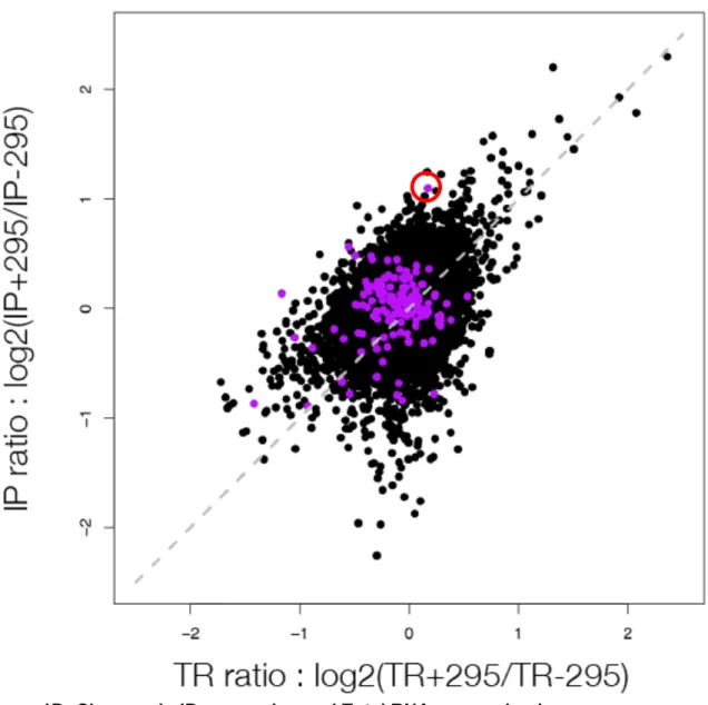
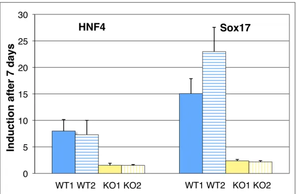
MicroRNAs (miRNAs) are a large family of genes encoding 19-22nt non-coding RNAs that post-transcriptionally regulate their mRNA targets through short regions of complementarity in their 5'ends (termed seed region). Computational predictions of miRNA targets based on conservation of seed matches suggest that more than half of all mRNAs are regulated by miRNAs, yet few have been experimentally identified. To directly identify potential miRNA target mRNAs, we performed microarray analysis of mRNAs in Ago2 immunoprecipitated and whole cell extracts from mouse embryonic stem cells (mESCs) in the absence of miRNAs and in the presence of a single miRNA species, 295. This method identified a set of mRNAs that had seed matches to miR-295 and were significantly down regulated in the whole cell extract in the presence of miR-295. Additionally, several mRNAs were found to be significantly enriched in the Ago2 immunoprecipitates, including Lefty1, a Nodal pathway inhibitor. mESCs that lack the miR-290~295 cluster have reduced induction of endodermal markers upon
differentiation, which may be due to upregulation of LEFTY protein, an inhibitor of differentiation down this lineage.