HAL Id: hal-01920858
https://hal.archives-ouvertes.fr/hal-01920858
Submitted on 19 Mar 2021
HAL is a multi-disciplinary open access
archive for the deposit and dissemination of sci-entific research documents, whether they are pub-lished or not. The documents may come from teaching and research institutions in France or abroad, or from public or private research centers.
L’archive ouverte pluridisciplinaire HAL, est destinée au dépôt et à la diffusion de documents scientifiques de niveau recherche, publiés ou non, émanant des établissements d’enseignement et de recherche français ou étrangers, des laboratoires publics ou privés.
by MLF and its partner DnaJ-1 during Drosophila
hematopoiesis
Marion Miller, Aichun Chen, Vanessa Gobert, Benoît Augé, Mathilde Beau,
Odile Burlet-Schiltz, Marc Haenlin, Lucas Waltzer
To cite this version:
Marion Miller, Aichun Chen, Vanessa Gobert, Benoît Augé, Mathilde Beau, et al.. Control of RUNX-induced repression of Notch signaling by MLF and its partner DnaJ-1 during Drosophila hematopoiesis. PLoS Genetics, Public Library of Science, 2017, 13 (7), pp.e1006932. �10.1371/journal.pgen.1006932�. �hal-01920858�
Control of RUNX-induced repression of Notch
signaling by MLF and its partner DnaJ-1 during
Drosophila hematopoiesis
Marion Miller1☯, Aichun Chen1☯, Vanessa Gobert1, Benoit Auge´1, Mathilde Beau2, Odile Burlet-Schiltz2, Marc Haenlin1*, Lucas Waltzer1*
1 Centre de Biologie du De´veloppement (CBD), Centre de Biologie Inte´grative (CBI), Universite´ de Toulouse, CNRS, UPS, Toulouse, France, 2 Institut de Pharmacologie et de Biologie Structurale, Universite´ de Toulouse, CNRS, UPS, Toulouse, France
☯These authors contributed equally to this work.
*lucas.waltzer@univ-tlse3.fr(LW);marc.haenlin@univ-tlse3.fr(MH)
Abstract
A tight regulation of transcription factor activity is critical for proper development. For instance, modifications of RUNX transcription factors dosage are associated with several diseases, including hematopoietic malignancies. In Drosophila, Myeloid Leukemia Factor (MLF) has been shown to control blood cell development by stabilizing the RUNX trans-cription factor Lozenge (Lz). However, the mechanism of action of this conserved family of proteins involved in leukemia remains largely unknown. Here we further characterized MLF’s mode of action in Drosophila blood cells using proteomic, transcriptomic and genetic approaches. Our results show that MLF and the Hsp40 co-chaperone family member DnaJ-1 interact through conserved domains and we demonstrate that both proteins bind and sta-bilize Lz in cell culture, suggesting that MLF and DnaJ-1 form a chaperone complex that directly regulates Lz activity. Importantly, dnaj-1 loss causes an increase in Lz+blood cell number and size similarly as in mlf mutant larvae. Moreover we find that dnaj-1 genetically interacts with mlf to control Lz level and Lz+blood cell development in vivo. In addition, we show that mlf and dnaj-1 loss alters Lz+cell differentiation and that the increase in Lz+blood cell number and size observed in these mutants is caused by an overactivation of the Notch signaling pathway. Finally, using different conditions to manipulate Lz activity, we show that high levels of Lz are required to repress Notch transcription and signaling. All together, our data indicate that the MLF/DnaJ-1-dependent increase in Lz level allows the repression of
Notch expression and signaling to prevent aberrant blood cell development. Thus our
find-ings establish a functional link between MLF and the co-chaperone DnaJ-1 to control RUNX transcription factor activity and Notch signaling during blood cell development in vivo.
Author summary
Tight regulation of proteins level is required for proper development. Notably, the aber-rant expression of key transcription factors or signaling pathway components controlling
a1111111111 a1111111111 a1111111111 a1111111111 a1111111111 OPEN ACCESS
Citation: Miller M, Chen A, Gobert V, Auge´ B, Beau
M, Burlet-Schiltz O, et al. (2017) Control of RUNX-induced repression of Notch signaling by MLF and its partner DnaJ-1 during Drosophila
hematopoiesis. PLoS Genet 13(7): e1006932.
https://doi.org/10.1371/journal.pgen.1006932
Editor: Claude Desplan, New York University,
UNITED STATES
Received: March 7, 2017 Accepted: July 18, 2017 Published: July 25, 2017
Copyright:© 2017 Miller et al. This is an open access article distributed under the terms of the
Creative Commons Attribution License, which permits unrestricted use, distribution, and reproduction in any medium, provided the original author and source are credited.
Data Availability Statement: The RNAseq data
were deposited on GEO under the accession number GSE93823.
Funding: This work was supported by grants from
the Agence Nationale de la Recherche (http://www. agence-nationale-recherche.fr), Fondation ARC (http://www.fondation-arc.org), Ligue Midi Pyre´ne´e contre le Cancer (http://www.liguecancer31.fr) and Fe´de´ration de Recherche en Biologie de Toulouse (https://www-frbt.biotoul.fr) to LW and in part by grants from the Re´gion Midi-Pyre´ne´es (http://
blood cell development contributes to hematological diseases such as leukemia. In this report, we useDrosophila as a model to study the function and mode of action of a family
of conserved but poorly characterized proteins implicated in leukemia called Myeloid Leukemia Factors (MLF). By combining proteomic, transcriptomic and genetic ap-proaches, we show thatDrosophila MLF acts in concert with an Hsp40 co-chaperone to
control the level and activity of a RUNX transcription factor and therefore RUNX+blood cell number and differentiation. Furthermore, we show that RUNX dosage directly im-pinges on the activity of the Notch signaling pathway, which is critical for RUNX+cell sur-vival and differentiation, by regulating the transcription of the Notch receptor. These findings shed light on a new mode of regulation of RUNX level and Notch activity to pre-vent abnormal blood cell accumulation, which could be involved in leukemogenesis.
Introduction
Proper blood cell development requires the finely tuned regulation of transcription factors and signaling pathways activity. Consequently mutations affecting key regulators of hemato-poiesis such as members of the RUNX transcription factor family or components of the Notch signaling pathway are associated with several blood cell disorders including leukemia [1,2]. Also, leukemic cells often present recurrent chromosomal rearrangements that participate in malignant transformation by altering the function of these factors [3]. The functional charac-terization of these genes is thus of importance not only to uncover the molecular basis of leu-kemogenesis but also to decipher the regulatory mechanisms controlling normal blood cell development.Myeloid Leukemia Factor 1 (MLF1) was identified as a target of the t(3;5)(q25.1;
q34) translocation associated with acute myeloid leukemia (AML) and myelodysplastic syn-drome (MDS) more than 20 years ago [4]. Further findings suggested thatMLF1 could act as
an oncogene [5–8] or a tumor suppressor [9] depending on the cell context and it was shown that MLF1 overexpression either impairs cell cycle exit and differentiation [10], promotes apo-ptosis [11,12], or inhibits proliferation [13,14] in different cultured cell lines. Yet, its function and mechanism of action remain largely unknown.
MLF1 is the founding member of a small evolutionarily conserved family of nucleo-cyto-plasmic proteins present in all metazoans but lacking recognizable domains that could help define their biochemical activity [15]. Whereas vertebrates have two closely related MLF para-logs,Drosophila has a single mlf gene encoding a protein that displays around 50% identity
with human MLF in the central conserved domain [16,17]. In the fly, MLF was identified as a partner of the transcription factor DREF (DNA replication-related element-binding factor) [16], for which it acts a co-activator to stimulate the JNK pathway and cell death in the wing disc [18]. MLF has been shown to bind chromatin [18–20], as does its mouse homolog [21], and it can either activate or repress gene expression by a still unknown mechanism [18,20]. MLF also interacts with Suppressor of Fused, a negative regulator of the Hedgehog signaling pathway [19], and, like its mammalian counterpart [13], with Csn3, a component of the COP9 signalosome [22], but the functional consequences of these interactions remain elusive. Inter-estingly the overexpression ofDrosophila MLF or that of its mammalian counterparts can
sup-press polyglutamine-induced cytotoxicity in fly and in cellular models of neurodegenerative diseases [17,23–25]. Moreover phenotypic defects associated with MLF loss inDrosophila can
be rescued by human MLF1 [17,26]. Thus MLF function seems conserved during evolution
andDrosophila appears to be a genuine model organism to characterize MLF proteins [15].
Along this line, we recently analyzed the role of MLF duringDrosophila hematopoiesis [26]. Indeed, a number of proteins regulating blood cell development in human, such as RUNX and
www.midipyrenees.fr/-Accueil-Enseignement-superieur-Recherche-), Fonds Europe´en de De´veloppement Re´gional (FEDER) (http://www. europe-en-france.gouv.fr/L-Europe-s-engage/ Fonds-europeens-2014-2020), Toulouse Me´tropole (http://www.toulouse-metropole.fr), and the French Ministry of Research for the
Programme Investissement d’Avenir Infrastructures Nationales en Biologie et Sante´ (PIA, ANR 10-INBS-08, French Proteomics Infrastructure, ProFI; http://www.agence-nationale-recherche.fr) to OBS. MM was supported by fellowships from Universite´ Paul Sabatier (http:// www.univ-tlse3.fr) and Fondation pour la Recherche Me´dicale (https://www.frm.org). AC was supported by a fellowship from the China Scholarship Council (CSC;http://www.csc.edu.cn/ laihua/noticeen.html). The funders had no role in study design, data collection and analysis, decision to publish, or preparation of the manuscript.
Competing interests: The authors have declared
Notch, also controlDrosophila blood cell development [27]. InDrosophila, the RUNX factor
Lozenge (Lz) is specifically expressed in crystal cells and it is absolutely required for the devel-opment of this blood cell lineage [28]. Crystal cells account for±4% of the circulating larval blood cells; they are implicated in melanization, a defense response related to clotting, and they release their enzymatic content in the hemolymph by bursting [27]. The Notch pathway also controls the development of this lineage: it is required for the induction of Lz expression and it contributes to Lz+cell differentiation as well as to their survival by preventing their rup-ture [28–31]. Interestingly, our previous analysis revealed a functional and conserved link between MLF and RUNX factors [26]. In particular, we showed that MLF controls Lz activity and prevents its degradation in cell culture and that the regulation of Lz level by MLF is critical to control crystal cell numberin vivo [26]. Intriguingly, although Lz is required for crystal cell development,mlf mutation causes a decrease in Lz expression but an increase in crystal cell
number. In human, the deregulation of RUNX protein level is associated with several patholo-gies. For instance haploinsufficient mutations inRUNX1 are linked to MDS/AML in the case
of somatic mutations, and to familial platelet disorders associated with myeloid malignancy for germline mutations [1]. In the opposite, RUNX1 overexpression can promote lymphoid leukemia [32,33]. Understanding how the level of RUNX protein is regulated and how this affects specific developmental processes is thus of particular importance.
To better characterize the function and mode of action of MLF inDrosophila blood cells,
we used proteomic, transcriptomic and genetic approaches. In line with recent findings [20], we found that MLF binds DnaJ-1, a HSP40 co-chaperone, as well as the HSP70 chaperone Hsc70-4, and that both of these proteins are required to stabilize Lz. We further show here that MLF and DnaJ-1 interact together but also with Lzvia conserved domains and that they
regu-late Lz-induced transactivation in a Hsc70-dependent manner in cell culture. In addition, using a null allele ofdnaj-1, we show that it controls Lz+blood cell number and differentiation as well as Lz activityin vivo in conjunction with mlf. Notably, we found that mlf or dnaj-1 loss
leads to an increase in Lz+cell number and size due to the over-activation of the Notch signal-ing pathway. Interestsignal-ingly, our results indicate that high levels of Lz are required to repress Notch expression and signaling. We thus propose a model whereby MLF and DnaJ-1 control Lz+blood cell growth and number by promoting Lz accumulation, which ultimately turn-downs Notch signaling. These findings thus establish a functional link between the MLF/Dna-J1 chaperone complex and the regulation of a RUNX-Notch axis required for blood cell homeostasisin vivo.
Results
MLF interacts with DnaJ-1 via conserved domains
To better characterize the molecular mode of action of MLF, we sought to identify its partners. Accordingly, we established aDrosophila Kc167 cell line expressing a V5-tagged version of
MLF close to endogenous levels in a copper-inducible manner (Fig 1A). After anti-V5 affinity purification from whole cell extracts of control or MLF-V5-expressing cells, isolated proteins were identified by mass spectrometry. Five proteins reproducibly co-purified with MLF and were either absent or at more than 4 fold lower levels in each control purification (Fig 1B): the Hsp40 co-chaperone DnaJ-1 (also known as DROJ1; [34]), the constitutively expressed Hsp70 chaperones Hsc70-4 and Hsc70-3, the RNA binding protein Squid (Sqd), and the retrotran-sposon-encoded protein Copia. Of note, as this manuscript was in preparation, Dyeret al. also
identified DnaJ-1 and Hsc70-4 as partners of MLF using a similar proteomic approach in the
this candidate and we further characterized its interaction with MLF as well as its function both in cell culture andin vivo.
First, we confirmed the interaction between MLF and DnaJ-1 by co-immunoprecipitation assays in Kc167 cells transfected with expression plasmids for tagged versions of these proteins using anti-tag antibodies (Fig 1C and 1D, andS1 Fig) or an anti-MLF antibody (S1C Fig). In addition, consistent with the hypothesis that these proteins interact in the cell, immunostain-ings showed that DnaJ-1 and MLF co-localize in the nuclei of Kc167 transfected cells (S1D Fig). Finally, we also observed a specific interaction between MLF and DnaJ-1 byin vitro GST
pull down assays (S1E Fig).
Fig 1. MLF and the co-chaperone DnaJ-1 interact via conserved domains. (A) Western blots showing MLF and MLF-V5 expression in Kc167 cells stably transfected with the copper-inducible pMT-MLF-V5 expression vector and treated or not with 50μm CuSO4for 24h. Tubulin (Tub) was used as an internal loading
control. (B) Proteins identified by mass spectrometry from CuSO4-induced Kc167-pMT-MLF-V5 cells using
anti-V5 antibody coupled to sepharose (IP1) or magnetic (IP2) beads for purification. The number of quantified peptides (#Qpep), sequence coverage and fold enrichment in comparison to control (parental Kc167 cells) are indicated for each experiment. Spe IP: not detected in control condition. (C, D) Schematic representation of DnaJ-1 (C) and MLF protein domains (D) and Western blots showing the results of immunoprecipitation experiments against GFP performed in Kc167 cells transfected with expression vectors for GFP-MLF and various HA-DnaJ-1 mutants (C) or GFP-DnaJ-1 and different HA-MLF mutants (D). Conserved domains are highlighted in grey. J: J-domain. G/F: glycine/phenylalanine-rich region. ter: C-terminal domain. MHD: MLF homology domain.
We then mapped the domains required for the interaction between DnaJ-1 and MLF. Hsp40/DnaJ co-chaperones play a crucial role in the regulation of protein folding and degra-dation; they chiefly act by delivering substrates to Hsp70/DnaK chaperones and stimulating their ATPase activity [35,36]. DnaJ-1 belongs to the DnaJB/class II subfamily of Hsp40/DnaJ proteins, which are characterized by an N-terminal J-domain required to stimulate Hsp70 ATPase activity (amino acids 4 to 57 in DnaJ-1), a central glycine/phenylalanine (G/F)-rich region (amino acids 64 to 144), and a conserved C-terminal region (amino acids 157 to 320) that contains the client proteins binding domain followed by a dimerization interface [36]. Immunoprecipitations of GFP-MLF expressed with different HA-tagged DnaJ-1 variants indi-cated that the DnaJ-1 C-terminal region mediates MLF binding (Fig 1C). In contrast, a point mutation (P32S) in the highly conserved HPD loop crucial for Hsc70 activation [36], deletion of the J-domain or deletion of the J and G/F domains did not affect the interaction between DnaJ-1 and GFP-MLF. MLF does not harbor characteristic domains apart from a central “MLF homology domain” (MHD, amino acids 96 to 202) conserved between MLF family members [15]. Using GFP-DnaJ-1 as bait and MLF deletion mutants as preys, we found that the MHD was sufficient for binding DnaJ-1, while MLF N- and C-terminal regions were dis-pensable (Fig 1D). Finally, consistent with the above results, the C-terminal region (amino acids 157 to 334) of DnaJ-1 bound to the MHD of MLF (S1F Fig). In sum, these data indicate that MLF and DnaJ-1 specifically bind each other through their conserved central and C-ter-minal region, respectively.
MLF and DnaJ-1 interact with Lz and control its activity
We have previously shown that MLF is required for Lz stability and transcriptional activity [26]. Interestingly, Dyeret al. reported that the knockdown of DnaJ-1 or of its chaperone
part-ner Hsc70-4 leads to a destabilization of exogenously expressed Lz in S2 cells [20]. However, the relationships between DnaJ-1, MLF and Lz were not further explored. We thus asked whether DnaJ-1 also controls Lz activity. As shown inFig 2A, transfection of a Lz expression plasmid in Kc167 cells induced a robust activation of the4xPPO2-Fluc reporter gene [37], which was significantly decreased when eithermlf or dnaj-1 expression was knocked down by
dsRNA treatment. Consistent with previous results [20,26], Western blot analyses showed
thatmlf and dnaj-1 knockdowns caused a drop in Lz protein level (Fig 2B). Moreover,
RT-qPCR experiments showed thatmlf and dnaj-1 knockdowns did not affect the expression of
each other or decreaselz transcript level, while they did cause a significant reduction in the
expression of Lz target geneppo2 (S2A–S2D Fig). Hence, like MLF, DnaJ-1 controls Lz protein stability and activity in Kc167 cells.
Next, we tested the effect of DnaJ-1 overexpression on Lz’s activity and protein level. Remi-niscent of MLF [26], we observed that DnaJ-1 over-expression was associated with an increase in Lz-induced transactivation and Lz level (Fig 2C and 2D). The overexpression of C-termi-nally-truncated DnaJ-1 proteins did not affect Lz-induced transcription or its expression. In contrast, the overexpression of Dna1 carrying the P32S point mutation or a deletion of its J-domain caused a decrease in Lz-induced transcription and a drop in Lz level (Fig 2C and 2D), suggesting that the activation of Hsc70 by DnaJ-1 is required for Lz’s stable expression and activity. In line with this hypothesis, knocking down Hsc70-4, which interacts with DnaJ-1 and MLF [20], caused a strong decrease in Lz-induced transactivation and a concomitant reduction in Lz protein level (S2E and S2F Fig). In sum, our results support the idea that MLF acts with DnaJ-1 in a Hsc70 chaperone complex to promote Lz stability and activity.
Given the impact of MLF and DnaJ-1 on Lz activity, we then asked whether these two pro-teins bind this RUNX transcription factor. Upon transfection of the corresponding expression
plasmids, both HA-DnaJ-1 and HA-MLF were co-immunoprecipitated by GFP-tagged Lz but not by GFP alone (Fig 2E and 2F). Furthermore,in vitro translated Lz bound to E. coli-purified
GST-MLF and GST-DnaJ-1 but not to GST alone in pull down assays (S2G Fig). Using differ-ent MLF variants in co-immunoprecipitation assays, we found that the N-terminal part of the MLF homology domain (amino acids 96 to 147) was crucial for the interaction with Lz (Fig 2G). Similarly the C-terminal domain of DnaJ-1 was required for binding Lz, while its J
Fig 2. MLF and DnaJ-1 bind Lz and control its stability and activity. (A) Luciferase assays in Kc167 cells treated with the indicated dsRNA and transfected with 4xPPO2-Fluc reporter plasmid in the presence or not (ctr) of the pAc-Lz-V5 expression plasmid. pAc-Rluc was used as an internal normalization control. (B) Western blots showing Lz-V5, MLF, Renilla luciferase (R luc) and Tubulin (Tub) expression in Kc167 cells treated with the indicated dsRNA and cotransfected with pAc-Lz-V5 and pAc-Rluc expression vectors. (A, B) dsDnaJ-1 (a) and (b) correspond to two distinct dsRNAs targeting dnaj-1. Of note, the multiple bands for Lz are only observed using C terminally (V5) tagged versions of Lz and not with N terminally (GFP) tagged Lz; they likely represent internal translation initiation events. The multiple bands observed using a MLF antibody could represent different MLF protein isoforms as described in [17]. (C, D) Luciferase assays (C) and Western blots (D) performed on Kc167 cells transfected with the 4xPPO2-Fluc reporter plasmid and pAc-based expression plasmids for Lz and for different DnaJ-1 variants as indicated. Rluc and Tubulin were used as internal controls. (E, F) Western blots showing the results of immunoprecipitation experiments against GFP performed in Kc167 cells transfected with expression vectors for HA-MLF (E) or HA-DnaJ-1 (F) and GFP or GFP-Lz as indicated in the upper part of the panels. (G, H) Western blots showing the results of immunoprecipitation experiments against GFP performed in Kc167 cells transfected with expression vectors for GFP-Lz and various HA-MLF (G) or HA-DnaJ-1 (H) mutants. (A, C) For luciferase assays means and standard deviations of results from biological triplicates are shown.***: p-value<0.001,**: p-value<0.01 (Students t-tests) as compared to Lz with dsGFP condition.
domain was dispensable (Fig 2H). Therefore it appears that MLF and DnaJ-1 interact with Lz through conserved domains and our results suggest that the MLF/DnaJ-1 complex regulates Lz stability and activity in Kc167 cells by binding it.
DnaJ-1 acts with MLF to control Lz
+blood cell number and size in vivo
Since DnaJ-1 interacts with MLF and controls Lz levelex vivo, we then sought to analyze
DnaJ-1 function in circulating larval crystal cells, whose proper development requires Lz stabi-lization by MLF [26]. Given that no mutant fordnaj-1 was available, we used a CRISPR/Cas9
strategy to generatednaj-1 null alleles (S3 Fig) [38]. In the following experiments, we used an allelic combination between two mutant lines obtained from independent founder flies (
dnaj-1Aanddnaj-1C), which harbor a complete deletion of thednaj-1 coding sequence (S3 Fig).
Around 65% of thednaj-1A/Cmutants reached the larval stage and 15% emerged as adult flies but they did not show obvious morphological defects. Reminiscent ofmlf phenotypes [26], bleeding of third instar larvae revealed thatdnaj-1 mutants exhibited a ±1.8-fold increase in
the number of circulating lz>GFP+blood cells as compared to wild-type (Fig 3A). In addition, as in themlf mutant, crystal cells from dnaj-1 mutant larvae still expressed the differentiation
marker PPO1 and were capable of melanization upon heat treatment (Fig 3C–3H). A closer examination also revealed the presence of unusually large lz>GFP+cells in thednaj-1 mutant
and quantitative analyses confirmed thatdnaj-1 loss caused a significant increase in lz>GFP+
cell size whereas lz>GFP-cells were unaffected (Fig 3B). Interestingly, a similar phenotype is observed inmlf mutant larvae (Fig 3B), suggesting that both genes not only control crystal cell number but also their differentiation (see below). Importantly, lz>GFP+cell number and size was restored to wild-type when DnaJ-1 was re-expressed in the crystal cell lineage ofdnaj-1A/C
mutant larvae using thelz-GAL4 driver (Fig 3A and 3B). This demonstrates not only that these phenotypes are specifically caused by thednaj-1 mutation, but also that DnaJ-1 acts cell
auton-omously and after the onset oflz expression in the crystal cell lineage. Of note, we did not
observe a rescue when we expressed a DnaJ-1 protein lacking its J-domain, suggesting that the interaction with Hsp70 chaperones is critical for the function of DnaJ-1 in the crystal cell line-age (S3C and S3D Fig). Furthermore, the increase in crystal cell number and size was also observed when we monitored crystal cell presence by immunostaining against PPO1 in larvae carrying adnaj-1Aordnaj-1Chomozygous mutation or over a deficiency uncovering the
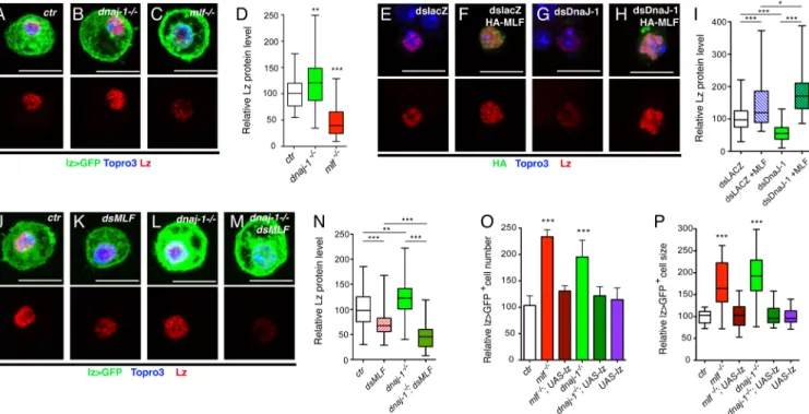
dnaj-Fig 3. dnaj-1 controls crystal cell development. (A, B) Quantification of circulating lz>GFP+cell number (A) and lz>GFP+or lz>GFP-cell size (B)
in lz-GAL4, UAS-mCD8-GFP/+ third instar larvae of the indicated genotypes. (C-E) Fluorescent immunostainings of the crystal cell differentiation marker PPO1 in third instar lz>GFP+hemocytes. The right panels show PPO1 immunostaining only. Nuclei were stained with Topro3. Scale bar: 10μm. (F-H) Bright field images of the posterior segments of third instar larvae heat-treated at 65˚C for 10 min to induce crystal cell melanization. (I, J) Relative lz>GFP+blood cell number (I) and size (C) in lz-GAL4, UAS-mCD8-GFP/+ third instar larvae of the indicated genotypes. (A, B, I, J)
*:p-value<0.05,**: p-value<0.01 and***: p-value<0.001 compared to control.
1 locus (S3E and S3F Fig). Overall, these results demonstrate that, likemlf, dnaj-1 controls
cir-culating larval lz>GFP+cell number and size.
Since MLF and DnaJ-1 bind to each other, we tested whether they genetically interacted to regulate crystal cell development. While heterozygous mutation in eithermlf or dnaj-1 did not
significantly alter circulating lz>GFP+cell number or size,mlfΔC1/+,dnaj-1A/+
transheterozy-gote larvae displayed a significant increase of both parameters (Fig 3I and 3J). We thus con-clude that DnaJ-1 and MLF act together to control crystal cell development. In sum, these results reveal a functional interaction between MLF and DnaJ-1in vivo.
High levels of MLF prevent Lz degradation in the absence of DnaJ-1
Next we assessed whether DnaJ-1 affects Lz stabilityin vivo as it does in cell culture.
Unexpect-edly, immunostaining against Lz did not reveal a decrease in Lz expression indnaj-1 mutant
crystal cells while the level of Lz was clearly lower in themlf mutant (Fig 4A–4C). Actually quantitative analyses revealed a slight (30%) but significant (p = 0.006) increase in Lz level in dnaj-1 mutant as compared to wild-type, whereas Lz level dropped by more than 2 folds in mlf
mutant (Fig 4D). Thus, unlikemlf, dnaj-1 loss is not sufficient to destabilize Lz in vivo. We
then tried to understand the reason for this discrepancy. One potentially important difference between Kc167 cells, in which DnaJ-1 is required to stabilize Lz, and crystal cells, in which it is not, is MLF expression. Indeed, in Kc167 cells, MLF is mainly detected in the cytoplasm and is present at low levels in the nucleus (S4A Fig). In contrast, MLF is present at high levels in the
Fig 4. High levels of MLF prevent Lz degradation in the absence of DnaJ-1. (A-C) Fluorescent immunostainings of Lz in circulating blood cells from lz-GAL4, UAS-mCD8-GFP/+ control (A), dnaj1-/-(B) and mlf-/-(C) third instar larvae. (D) Corresponding quantifications of Lz protein level.
(E-H) Immunostainings against Lz (red) and HA-MLF (green) in Kc167 cells treated with the indicated dsRNA and transfected with pAc-Lz-V5 alone (E, G) or in combination with pAc-3HA-MLF (F, H). (I) Corresponding quantification of Lz levels in Kc167 cells. (J-M) Immunostainings against Lz in circulating blood cells from lz-GAL4, UAS-mCD8-GFP/+ control (J), UAS-dsMLF (K), dnaj1-/-(L) and UAS-dsMLF; dnaj1-/-(M) third instar larvae. (N) Corresponding quantification of Lz protein levels in lz>GFP+larval blood cells. (A-C, E-H, J-M) Nuclei were stained with Topro3.
Lz staining only is shown in the lower panels. Scale bar: 10μm. (O, P) Relative lz>GFP+blood cell number (O) and size (P) in lz-GAL4,
UAS-mCD8-GFP/+ third instar larvae of the indicated genotypes. (D, I, N-P)*: p-value<0.05,**: p-value<0.01,***: p-value<0.001.
nucleus of larval crystal cells (S4B Fig). Moreover, MLF expression in this lineage is not affected bydnaj-1 loss (S4C and S4F Fig). We thus surmised that the presence of high levels of nuclear MLF might prevent Lz degradation in the absence of DnaJ-1.
To test this hypothesis, we designed two complementary experiments. On the one hand, we assessed whether MLF over-expression in Kc167 cells could protect Lz from degradation followingdnaj-1 knockdown. Lz level dropped when Kc167 cells were treated with a dsRNA
targetingdnaj-1 (Fig 4G) and increased upon over-expression of MLF (Fig 4F). Strikingly though, and in line with the observations indnaj-1 mutant crystal cells, the level of Lz was not
reduced but further increased whendnaj-1 was knocked down in MLF-overexpressing cells
(Fig 4H and 4I). On the other hand, we asked whether Lz would still be stable indnaj-1 mutant
crystal cells if MLF level is decreased. Accordingly, we expressed a dsRNA directed againstmlf
in lz>GFP+cells, which caused a significant and similar knock-down of MLF in wild-type and
dnaj-1 mutant larvae (S4D–S4F Fig). Remarkably, we found that the drop in Lz protein level
caused bymlf down-regulation was significantly enhanced in dnaj-1 deficient larvae, while the
dnaj-1 mutation alone increased Lz level (Fig 4J–4N). Hence it appears that in the absence of
DnaJ-1, high levels of MLF prevent Lz degradation.
Given that chaperones are important for proper protein folding [35,36], we postulated that Lz proteins accumulating in crystal cells in the absence of DnaJ-1 might be less active. Thus increasing Lz expression might be sufficient to rescue lz>GFP+cell number and size. In addi-tion, although re-expressing Lz is sufficient to restore lz>GFP+cell number inmlf mutant
lar-vae [26], it is not known whether this also rescues lz>GFP+cell size. Interestingly, lz>GFP+ cell count and cell size were restored to wild-type levels when we enforced Lz expression in this lineage either inmlf or dnaj-1 mutant larvae (Fig 4O and 4P). We thus conclude that DnaJ-1 and MLF act together to control crystal cell development by regulating Lz activityin vivo
MLF and DnaJ-1 control crystal cell differentiation
In parallel, to gain further insights into the function of MLF in the control of crystal cell devel-opment, we established the transcriptome of circulating lz>GFP+blood cells in wild-type and
mlf larvae. Heterozygous lz-GAL4,UAS-mCD8-GFP L3 larvae carrying or lacking a mlf null
mutation were bled, lz>GFP+cells were collected by FACS and their gene expression profile was determined by RNA sequencing (RNAseq) from biological triplicates. UsingDrosophila
reference genome dm3, we detected the expression of 7399 genes (47% of the total fly genes) in each of the 6 samples (Fig 5BandS1 Table). Consistent with the role of the crystal cells as the main source of phenoloxidases [39], the two most strongly expressed genes werePPO1 and PPO2. In addition, lz expression as well as that of several other crystal cell markers was readily
detected (see below). It was recently shown that larval circulating Lz+cells derive from plasma-tocytes, which express Hemolectin (Hml) and Nimrod C1 (NimC1), and transdifferentiate into crystal cells [40]. Accordingly, we detected the expression of these genes, as well as other “plasmatocytes” markers such asperoxidasin and croquemort (which were actually shown to be
also expressed in crystal cells [41,42]) in lz>GFP+cells.
Using DESeq2 to identify differentially expressed genes between wild-type andmlf mutant
lz>GFP+cells, we found 779 genes with significantly altered expression (adjusted p-value
<0.01): the transcript level of 469 genes was decreased and that of 310 genes was increased in
the absence of MLF (Fig 5A and 5B, andS2 Table). In line with our previousin situ
hybridiza-tion results [26], RNAseq analysis did not reveal a significant modification ofPPO1 or PPO2
expression in the absence ofmlf. However, the lz transcript level was reduced by ±2 fold
[43]. To assess whether other crystal cell markers were affected bymlf, we established a
compi-lation of genes expressed in (embryonic or larval) crystal cells based on Flybase data mining and re-examination of BerkeleyDrosophila Genome Project in situ hybridizations (http:// insitu.fruitfly.org/cgi-bin/ex/insitu.pl) (S3 Table). Among these 129 genes (i.e. excluding mlf
itself), 44 (34%) were differentially expressed in the absence ofmlf (19 repressed and 25
acti-vated) (Fig 5C), indicating a strong over-representation of deregulated gene in the “crystal
Fig 5. MLF and DnaJ-1 control crystal cell differentiation. (A) MA-plot of DESeq2 results for RNAseq data comparison between control and mlf -/-lz>GFP+blood cells sorted by FACS from third instar larvae. Genes that are significantly upregulated or downregulated in the mlf mutant (adjusted
p-value<0.01) are highlighted in red or blue, respectively. Red triangles: genes with log2fold change>5. (B) Pie chart showing the number of expressed
genes in lz>GFP+cells and the number of upregulated (red) or downregulated (blue) genes in the mlf mutant. (C) Heat map of “crystal cell”-associated genes differentially expressed (p-value<0.01) between control and mlf mutant lz>GFP+cells. Differential gene expression as per comparison to the mean of
the 6 samples (ctr 1, 2, 3 and mlf 1, 2, 3) is displayed as log2scale. Hierarchical clustering was performed using R-Bioconductor. (D-O) Immunostainings
against GFP and in situ hybridization against CG7860 (D-F), Oscillin (G-I), Jafrac1 (J-L) and CG6733 (M-O) in blood cells from lz-GAL4,UAS-mCD8-GFP/+ control (D, G, J, M), mlf-/-(E, H, K, N) or dnaj-1-/-(F,I, L,O) third instar larvae. RNA expression only is shown in the lower panels. Nuclei were stained with Topro3. Scale bar: 10μm.
cell” gene set as compared to all expressed genes (p-value = 2.6x10⁻¹³, hypergeometric test) and showing thatmlf plays a crucial role for proper crystal cell differentiation.
To substantiate these results, we analyzed byin situ hybridization the expression of 4 genes
that were either down-regulated (CG7860 and Oscillin) or up-regulated (CG6733 and Jafrac1)
in themlf mutant. CG7860 and Oscillin were specifically expressed in lz>GFP+but not in the surrounding lz>GFP-hemocytes in wild-type conditions (Fig 5D and 5G). Consistent with our RNAseq data, the expression ofCG7860 and Oscillin was strongly reduced in mlf mutant
larvae. AlthoughCG6733 is expressed in embryonic crystal cells [43], we did not detect its expression in circulating hemocytes of wild-type larvae, but it was expressed in the lz>GFP+ lineage inmlf larvae (Fig 5J and 5K). Finally,Jafrac1 expression increased in lz>GFP+
cells of
mlf mutant larvae as compared to wild-type, whereas its (lower) expression in lz>GFP-blood
cells seemed similar (Fig 5M and 5N). These data thus confirm the RNAseq results and dem-onstrate that MLF controls the expression of several crystal cell markers. Since the above results indicate that MLF functionally interacts with DnaJ-1 during crystal cell development, we also tested whether these four genes were deregulated in thednaj-1 mutant. As for mlf, we
observed that adnaj-1 mutation caused a down-regulation of CG7860 and Oscillin and an
up-regulation ofCG6733 and Jafrac1 expression in lz>GFP+blood cells (Fig 5F,5I,5L and 5O). In sum it appears that the loss ofmlf or dnaj-1 leads to a deregulation of the crystal cell gene
expression program characterized both by the overexpression and the downregulation of crys-tal cell markers. Thereforemlf and dnaj-1 are required for proper differentiation of the Lz+
blood cell lineage.
MLF and DnaJ-1 control Lz
+cell number and size by repressing Notch
signaling
Interestingly, the levels ofNotch receptor transcripts were significantly higher in the mlf
mutant (p = 1.3x10⁻⁶) (Fig 5C). Notch signaling plays a key role in crystal cell development [27]: Notch is first activated by its ligand Serrate to specify Lz+cells (crystal cell precursors) and its activation is subsequently maintained in Lz+cells in a ligand-independent manner to promote crystal cell growth and survival [29–31,40,44]. The rise in lz>GFP+cell number and size observed inmlf and dnaj-1 mutant could thus be due to increased ligand-independent
Notch signaling. However, the role of Notch signaling in crystal cell growth and survival has been mainly investigated in the larval lymph gland [30,31]. In agreement with these investiga-tions, inhibiting the Notch pathway in circulating Lz+cells, either by down-regulating the expression of Suppressor of Hairless [Su(H)], the core transcription factor in the Notch path-way, or by overexpressing Suppressor of Deltex [Su(dx)], a negative regulator of Notch [45], resulted in a decrease in lz>GFP+cell number and impaired their growth, whereas the overac-tivation of Notch signaling consecutive to the expression of a constitutively active Su(H)-VP16 fusion protein [46], caused a strong increase in lz>GFP+cell number and size (S5 Fig).
Then we further investigated the level of Notch expression and activation inmlf and dnaj-1
mutant blood cells. Immunostaining using an antibody against the Notch extracellular domain (NECD) showed that Notch accumulated at higher levels in lz>GFP+cells ofmlf and dnaj-1
mutant larvae than in wild-type conditions (Fig 6A–6C). Quantitative analyses confirmed that
mlf loss caused a significant increase in Notch level in lz>GFP+cell, whereas the (lower)
expression of Notch in lz>GFP-blood cells was not affected (Fig 6D). Similar results were obtained when we measured Notch protein levels using an antibody directed against its intra-cellular domain (NICD) (Fig 6EandS6 Fig). Thus Notch level is specifically increased in lz>GFP+cells ofmlf and dnaj-1 mutants. Next, we tested whether this resulted in increased
expressed in larval crystal cells: Klumpfuss-Cherry [31] and NRE-GFP [47]. Bothmlf and dnaj-1 loss were associated with a strong increase in the expression of these reporters (Fig 6F– 6J). Thusmlf and dnaj-1 are required to tune down Notch signaling in the crystal cell lineage.
Fig 6. The increase in lz>GFP+cell number and size in mlf and dnaj-1 mutant larvae is caused by overactivation of the Notch signaling pathway. (A-C) Immunostainings against Notch (NECD: Notch extracellular domain) in blood cells from lz-GAL4,UAS-mCD8-GFP/+ control (A), mlf-/-(B) and dnaj-1-/-(C) larvae. The immunostaining against Notch protein only is shown in the lower panels. Nuclei were stained with Topro3. (D) Quantification of NECD immunostainings in lz>GFP+and lz>GFP-blood cells from control, mlf-/-and
dnaj-1-/-larvae. (E) Quantification of NICD (Notch intracellular domain) immunostainings in lz>GFP+blood cells
from control, mlf-/-and dnaj-1-/-larvae. (F-H) Expression of the Notch pathway reporter Klu-Cherry in lz>GFP+ blood cells from control, mlf-/-or dnaj-1-/-larvae. Klu-Cherry expression only is shown in the lower panels. (I)
Corresponding quantification of Klu-Cherry level. (J) Quantification of the expression level of the Notch pathway reporter NRE-GFP in PPO1-expressing cells from control, mlf-/-or dnaj-1-/-larvae. (K, L) Relative lz>GFP+blood
cell number (K) and size (L) in third instar larvae of the indicated genotypes.
Finally, we asked whether the rise in lz>GFP+cell size and/or number observed inmlf and dnaj-1 mutants depends on Notch. Strikingly, when we reduced Notch dosage by introducing
one copy of theN55e11null allele in these mutants, both parameters were restored to control levels, whileN55e11heterozygote mutation had no effectper se (Fig 6K and 6L). Collectively, these data strongly support the hypothesis that the increase in Notch level underlies lz>GFP+ cell expansion inmlf and dnaj-1 mutants.
MLF and DnaJ-1 are required to turn-down Notch expression during
crystal cell maturation
It was shown that crystal cells tend to increase their size as they mature in response to Notch signaling [31,40], which is consistent with the results we obtained by manipulating Notch sig-naling activity in Lz+cells (S5 Fig). To better characterize the defects associated withmlf or
dnaj-1 loss, we analyzed the distribution of lz>GFP+cells as well as Notch level according to
lz>GFP+cell size categories. Whereas cells more than 1.3-fold larger than the mean wild-type cell size represented a small fraction (±10%) of the lz>GFP+population in wild-type larvae, they constituted the prevalent population inmlf or dnaj-1 mutant (respectively 49.6 and 37%)
(Fig 7A). Interestingly, Notch protein level was maximum in the population of lz>GFP+cells of mean cell size but lower in larger cells of wild-type larvae (Fig 7B), whereas it continued to increase in the larger cell populations ofmlf or dnaj-1 larvae (Fig 7B–7D). Actually we ob-served a similar trend when we monitoredNotch expression by in situ hybridization. In
wild-type larvae,Notch transcripts were readily seen in small/medium lz>GFP+cells but barely
Fig 7. MLF and DnaJ-1 are required to turn down Notch expression in large crystal cells. (A) Quantificationd of the proportion of lz>GFP+cells according to their size in control, mlf-/-or dnaj-1-/-larvae. Cells were grouped into 5 categories as compared to the mean size of lz>GFP+cells in the control condition.
(B-D) Quantification of NICD immunostaining (relative to control) in each of the five lz>GFP+cell size
categories in control (B), mlf-/-(C) and dnaj-1-/-(D) larvae.
*:p-value<0.05,**: p-value<0.01,*** p-value<0.001, n.s.: not significant. (E-J) Fluorescent immunostainings of GFP and in situ hybridizations of Notch in circulating blood cells from lz-GAL4,UAS-mCD8-GFP/+ third instar larvae of the indicated genotypes. Representative images of Notch expression in small/medium (E, G, I) versus large (F, H, J) lz>GFP+cells. Scale bar: 10μm. Nuclei were stained with Topro3. The lower panels show Notch expression
only.
detectable in large lz>GFP+cells (Fig 7E and 7F). In contrast,Notch transcripts continued to
accumulate in large lz>GFP+cells frommlf or dnaj-1 mutant larvae (Fig 7H and 7J). Hence, MLF/DnaJ-1 loss is associated with the accumulation of large crystal cells exhibiting aberrant maintenance ofNotch expression. Since the Notch pathway is activated in a
ligand-indepen-dent manner in Lz+cells [30], a tight regulation of the level of Notch is particularly critical to control crystal cell growth and number. All together, our data suggest that inmlf or dnaj-1
mutant larvae,Notch expression fails to be turned down when lz>GFP+cells reach a critical size, leading to the maintenance of a high level of Notch signaling and thus to increased crystal cell growth and survival.
High levels of Lz prevent accumulation of lz
>
GFP
+cells and repress
Notch expression/signaling
We showed above that forcing the expression of Lz rescues the increase in crystal cell number and size caused bymlf or dnaj-1 loss. It is thus plausible that this RUNX transcription factor
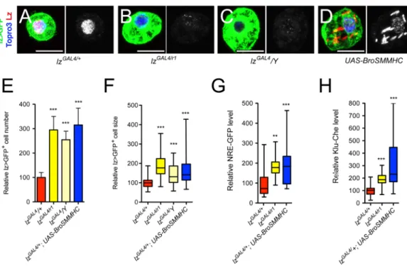
directly participates in down-regulation of Notch signaling. To explore this hypothesis, we asked whether a reduction inlz activity might cause an expansion of the Lz+cell lineage associ-ated with an over-activation of the Notch pathway. Accordingly, we introduced thelzr1null allele into thelzGAL4context. This hypomorphic allelic combination caused a decrease in Lz expression (Fig 8B) and resulted in an increase in lz>GFP+cell number and size (Fig 8E and 8F). Interestingly,lzGAL4/Y hemizygous larvae displayed similar phenotypes (Fig 8C,8E and 8F), indicating that this P{GAL4} insertion inlz alters its expression in the crystal cell lineage.
As an alternate strategy, we interfered with Lz activity by expressing a fusion protein between
Fig 8. High levels of Lz prevent lz>GFP+cell accumulation and Notch signaling overactivation. (A-D) Fluorescent immunostainings of Lz in circulating blood cells from lz-GAL4, UAS-mCD8-GFP/+ (A, control), lz-GAL4, UAS-mCD8-GFP/lzr1(B), lz-GAL4, UAS-mCD8-GFP/Y (C) and lz-GAL4, UAS-mCD8-GFP/+; UAS-BroSMMHC (D) third instar larvae. Nuclei were stained with Topro3. Scale bar: 10μm. Lz immunostaining only is shown in the right panels. (E-H) Quantifications of lz>GFP+cell number (E) and size (F) as well as NRE-GFP (G) and Klu-Cherry (H)
expression levels in third instar larvae of the indicated genotypes.**: p-value<0.01,***p-value<0.001.
Lz partner Brother (Drosophila CBFß homolog) and the non-muscular myosin heavy chain
SMMHC [48]. This chimera mimics the CBFß-MYH11 fusion protein generated by the Inv (16) translocation in human AML and can sequester RUNX factors in the cytoplasm [1,49]. Bro-SMMHC expression in lz>GFP+cells titrated Lz from the nucleus and also caused an increase in lz>GFP+cell number and size (Fig 8D–8F). Importantly, the expression of the Notch pathway reporters NRE-GFP and Klu-Cherry was strongly increased inlzGAL4/lzR1
mutant or upon Bro-SMHCC expression in the Lz+blood cell lineage (Fig 8G and 8H). More-over, knocking down Su(H) or over-expressing the Notch protein inhibitor Su(dx) was suffi-cient to prevent the rise in lz>GFP+cell number and size oflzGAL4/Y hemyzigotes (S5 Fig). Thus, a reduction inlz activity causes similar defects as the mlf or dnaj-1 mutations and likely
involves the overactivation of the Notch pathway.
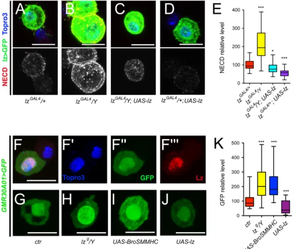
Then we analyzed the relathionship between Lz and Notch levels. In Lz+cells of increasing size, Lz levels continuously increased while Notch became less abundant (S7A Fig). This suggested that Lz level rises as crystal cells grow/mature and, in view of the above results, we surmised that this increase might participate in the down-regulation of the Notch receptor. Indeed, we found that the Notch receptor level was significantly augmented in lz>GFP+cells of hypomorphiclzGAL4/Y hemyzigote mutant larvae, whereas it was reduced when Lz was
over-expressed (Fig 9A–9E). In addition, the increase in Notch expression observed inlzGAL4/ Y larvae was suppressed by forcing Lz expression. Moreover, in situ hybridization experiments
revealed that, unlike in control larvae,Notch expression was not repressed in large lz>GFP+
cells inlzGAL4/Y larvae (S7 Fig). ThereforeNotch might be a direct transcriptional target of
Lz. By analyzing the expression of different GAL4 lines that cover potentialNotch regulatory
regions [50], we identified two lines that drive expression in circulating Lz+blood cells (Fig 9F
andS7 Fig). The regulatory elements carried by these two lines (GMR30A01 and GMR30C06) overlap on a 668bp DNA segment that contains two consensus binding sites for RUNX tran-scription factors conserved in otherDrosophila species (S7A Fig), suggesting that Lz might directly regulateNotch transcription by targeting this region. We thus tested the effect of Lz
dosage manipulation on the activity of this enhancer-GAL4 line. Strikingly, a hypomorphic
lozenge mutation (lzg/Y) [51] or the expression of Bro-SMMHC caused an increase in the
expression of this enhancer, whereas the over-expression of Lz resulted in its down-regulation (Fig 9G–9K). These findings strongly argue that Lz directly repressesNotch expression.
All together, these results demonstrate that high levels of Lz are required to prevent the accumulation of over-grown lz>GFP+cells as well as over-activation of the Notch pathway, and we propose that Lz-mediated repression ofNotch transcription is critical during this
process.
Discussion
Members of the RUNX and MLF families have been implicated in the control of blood cell development in mammals andDrosophila and deregulation of their expression is associated
with human hemopathies including leukemia [1,9,15,52]. Our results establish the first link between the MLF/DnaJ-1 complex and the regulation of a RUNX transcription factorin vivo.
In addition, our data show that the stabilization of Lz by the MLF/DnaJ-1 complex is critical to control Notch expression and signaling and thereby blood cell growth and survival. These findings pinpoint the specific function of the Hsp40 chaperone DnaJ-1 in hematopoiesis, re-veal a potentially conserved mechanism of regulation of RUNX activity and highlight a new layer of control of Notch signaling at the transcriptional level.
In line with results published as this manuscript was in preparation [20], we found that MLF binds DnaJ-1 and Hsc70-4 and that these two proteins, like MLF, are required for Lz
stable expression in Kc167 cells. In addition, our data show that MLF and DnaJ-1 bind to each othervia evolutionarily conserved domains and also interact with Lz, suggesting that Lz is a
direct target of a chaperone complex formed by MLF, DnaJ-1 and Hsc70-4. Of note, a system-atic characterization of Hsp70 chaperone complexes in human cells identified MLF1 and MLF2 as potential partners of DnaJ-1 homologs, DNAJB1, B4 and B6 [53], a finding corrobo-rated by Dyeret al. [20]. Therefore, the MLF/DnaJ-1/Hsc70 complex could play a conserved role in mammals, notably in the regulation of the stability of RUNX transcription factors. How MLF acts within this chaperone complex remains to be determined.In vivo, we demonstrate
thatdnaj-1 mutations lead to defects in crystal cell development strikingly similar to those
observed inmlf mutant larvae and we show that these two genes act together to control Lz+
cells development by impinging on Lz activity. Our data suggest that in the absence of DnaJ-1, high levels of MLF lead to the accumulation of defective Lz protein whereas lower levels of MLF allow its degradation. We thus propose that MLF stabilizes Lz and, together with DnaJ-1, promotes its proper folding/conformation. In humans, DnaJB4 stabilizes wild-type E-cadherin
Fig 9. Lz represses Notch expression. (A-D) Immunostainings against NECD (Notch extracellular domain) in blood cells from lz-GAL4, UAS-mCD8-GFP/+ (A), lz-GAL4, UAS-mCD8-GFP/Y (B), lz-GAL4, UAS-mCD8-GFP/Y; UAS-lz (C) and lz-GAL4, UAS-mCD8-GFP/+; UAS-lz (D) third instar larvae. NECD immunostaining only is shown in the lower panels. Nuclei were stained with Topro3. (E) Corresponding quantifications of NECD in lz>GFP+blood
cells. (F-F”‘) Immunostaining against Lz in circulating blood cells from NotchGMR30A01-GAL4, UAS-GFPnls third instar larvae. Nuclei were stained with Topro3. (F’-F”‘): single channel images. (G-J) NotchGMR30A01-GAL4-driven expression of GFP in circulating blood cells from larvae of the indicated genotypes. (K) Corresponding
quantifications of the level of GFP. (A-D, F-J) Scale bar: 10μm. (E, K)*: p-value<0.05,***p-value<0.001.
but induces the degradation of mutant E-cadherin variants associated with hereditary diffuse gastric cancer [54]. Thus the fate of DnaJ client proteins is controlled at different levels and MLF might be an important regulator in this process.
In this work, we present the first null mutant for a gene of theDnaJB family in metazoans
and our results demonstrate that a DnaJ protein is requiredin vivo to control hematopoiesis.
There are 16 DnaJB and in total 49 DnaJ encoding genes in mammals and the expansion of this family has likely played an important role in the diversification of their functions [55,56]. DnaJB9 overexpression was found to increase hematopoietic stem cell repopulation capacity [57] and Hsp70 inhibitors have anti-leukemic activity [58], but the participation of other DnaJ proteins in hematopoiesis or leukemia has not been explored. Actually DnaJ’s molecular mecha-nism of action has been fairly well studied but we have limited insights as to their rolein vivo.
Interestingly though, both DnaJ-1 and MLF suppress polyglutamine protein aggregation and cytotoxicity inDrosophila models of neurodegenerative diseases [17,23,24,59–63,64], and this function is conserved in mammals [24,25,65,66]. It is tempting to speculate that MLF and DnaJB proteins act together in this process as well as in leukemogenesis. Thus a better character-ization of their mechanism of action may help develop new therapeutic approaches for these diseases.
As shown here,mlf or dnaj-1 mutant larvae harbor more crystal cells than wild-type larvae.
This rise in Lz+cell number is not due to an increased induction of crystal cell fate as we could rescue this defect by re-expressing DnaJ-1 or Lz with thelz-GAL4 driver, which turns on after
crystal cell induction, and it was also observed inlz hypomorph mutants, which again suggests
a post-lz / cell fate choice process. Moreover mlf or dnaj-1 mutant larvae display a higher
frac-tion of the largest lz>GFP+cell population, which could correspond to the more mature crys-tal cells [31,40]. It is thus tempting to speculate thatmlf or dnaj-1 loss promotes the survival of
fully differentiated crystal cells. Our RNAseq data demonstrate thatmlf is critical for
expres-sion of crystal cell associated genes, but we observed both up-regulation and down-regulation of crystal cell differentiation markers inmlf or dnaj-1 mutant Lz+cells. Also these changes did not appear to correlate with crystal cell maturation status since we found alterations in gene expression in the mutants both in small and large Lz+cells. In addition our transcriptome did not reveal a particular bias toward decreased expression for “plasmatocyte” markers in Lz+ cells frommlf-mutant larvae. Thus, it appears that MLF and DnaJ-1 loss leads to the accumula-tion of mis-differentiated crystal cells.
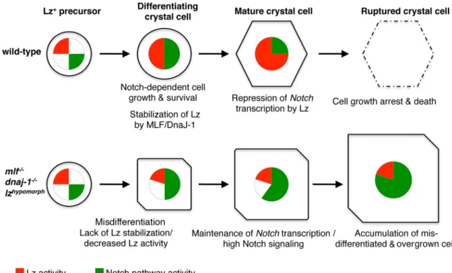
Our data support a model whereby MLF and DnaJ-1 act together to promote Lz accumula-tion, which in turn represses Notch transcription and signaling pathway to control crystal cell size and number (Fig 10). Indeed, we observe an abnormal maintenance ofNotch expression
in the larger Lz+cells as well as an over-activation of the Notch pathway in the crystal cell line-age ofmlf and dnaj-1 mutants or when we interfere with Lz activity. Moreover our data as well
as previously published experiments show that Notch activation promotes crystal cell growth and survival [30,31,40]. Importantly too the increase in Lz+cell number and size observed in
mlf or dnaJ-1 mutant is suppressed when Notch dosage is decreased. Yet, some of the
mis-dif-ferentiation phenotypes in themlf or dnaj-1 mutants might be independent of Notch since
changes in crystal cell markers expression seem to appear before alterations in Notch are apparent. At the molecular level, our results suggest that Lz directly repressesNotch
transcrip-tion as we identified a Lz-responsiveNotch cis-regulatory element that contains conserved
RUNX binding sites. The activation of the Notch pathway in circulating Lz+cells is ligand-independent and mediated through stabilization of the Notch receptor in endocytic vesicles [30,45]. Hence a tight control ofNotch expression is of particular importance to keep in check
the Notch pathway and prevent the abnormal development of the Lz+blood cell lineage.
auto-activation loop might rapidly go awry in a context in which Notch pathway activation is independent of ligand binding. By promoting the accumulation of Lz during crystal cell matu-ration, MLF and DnaJ-1 thus provide an effective cell-autonomous mechanism to inhibit Notch signaling. Further experiments will now be required to establish how Lz repressesNotch
transcription. RUNX factors can act as transcriptional repressors by recruiting co-repressor such as members of the Groucho family [68]. Whether MLF and DnaJ-1 directly contribute to Lz-induced-repression in addition to regulating its stability is an open question. MLF and DnaJ-1 were recently found to bind and regulate a common set of genes in cell culture [20]. They may thus provide a favorable chromatin environment for Lz binding or be recruited with Lz and/or favor a conformation change in Lz that allows its interaction with co-repressors. The scarcity of lz>GFP+cells precludes a biochemical characterization of Lz, MLF and DnaJ-1 mode of action notably at the chromatin level but further genetic studies should help decipher their mode of action. While the post-translational control of Notch has been extensively stud-ied, its transcriptional regulation seems largely overlooked [69]. Our findings indicate that this is nonetheless an alternative entry point to control the activity of this pathway. Given the importance of RUNX transcription factor and Notch signaling in hematopoiesis and blood cell malignancies [1,2], it will be of particular interest to further study whether RUNX factors can regulate Notch expression and signaling during these processes in mammals.
In conclusion, our study shows that MLF and DnaJ-1 act together to regulate RUNX tran-scription factor activity, which in turn controls Notch signaling during hematopoiesisin vivo.
Fig 10. A model for the control of crystal cell development by MLF/DnaJ-1, Lz, and Notch. In wild-type conditions, lz expression is induced in crystal cell precursors and Lz protein gradually accumulates thanks to its interaction with MLF/DnaJ-1. At the same time, ligand-independent Notch signaling promotes crystal cell growth and survival. Once it reaches a sufficient level, Lz represses Notch transcription. This leads to a down-regulation of Notch signaling, thereby limiting crystal cell growth and promoting the death (rupture) of mature crystal cells. In conditions where Lz activity is impaired (decreased expression or lack of stabilization by MLF/DnaJ-1), crystal cells do not differentiate properly and Notch activity is maintained at high levels, which causes the accumulation of a higher number of Lz+cells and their overgrowth.
We anticipate that the extraordinary genetic toolbox available inDrosophila will help shed
fur-ther light on the mechanism of action of these evolutionarily conserved proteins and will bring valuable insights into the control of protein homeostasis by MLF and DnaJ-1 during normal or pathological situations.
Materials and methods
Fly strains
The followingDrosophila melanogaster lines were used: mlfΔC1,UAS-mlf [17],UAS-ds-mlf
(National Institute of Genetics),UAS-lz, lzGAL4,UAS-mCD8-GFP, lzg,lzr1,N55e11,UAS-dsSu(H),
P{EPgy2}DnaJ-1EY04359, UAS-dnaj-1, Def(3L)BSC884, vas-Cas9, UAS-GFPnls, NRE-GFP,
GMR30C06, GMR30A01, UAS-dsSu(H) (Bloomington Drosophila Stock Center), Bc-GFP [70],
Klu-mCherry [31]UAS-Bro-SMMHC [48],UAS-DnaJ-1ΔJ [61],UAS-dsSu(H),
UAS-Su(H)-VP16 [46],UAS-Su(dx) [71]. To generatednaj-1 deficient flies, we designed two guide RNA
targetingdnaj-1 locus (S4 Fig) and the corresponding DNA oligonucleotides (g2: GTCGAC CACAACGCGCCGGATCAA; g3: GTCGCATCACAGTCACGCTTTCCT) were cloned in pCFD3 (Addgene).vas-cas9 females were crossed to P{EPgy2}DnaJ-1EY04359 males and the
resulting embryos were injected using standard procedures with both g2 and pCFD3-g3 plasmids (500ng/ul). Deletion of the P{EPgy2}EY04359transposon, as revealed by loss of the
w+marker, was screened for at the F2 generation, and deletion ofdnaj-1 locus was assessed by
PCR and sequencing.
All crosses were conducted at 25˚C on standard food medium as described in [72].
Immunostainings and in situ hybridizations
For each sample, four third instar larvae were bled (or 5.103Kc167 cells were dispensed) in 1ml of PBS in 24-well-plate containing a glass coverslip. Unless mentioned otherwise, only female larvae were used. The hemocytes were centrifuged for 2 min at 900g, fixed for 20 min with 4% paraformaldehyde in PBS and washed twice in PBS. For immunostainings: cells were permeabilized in PBS-0.3% Triton (PBST) and blocked in PBST- 1% Bovine Serum Albumin (BSA). The cells were incubated with primary antibodies at 4˚C over night in PBST-BSA, washed in PBST, incubated for 2h at room temperature with corresponding Alexa Fluor-la-beled secondary antibodies (Molecular Probes), washed in PBST and mounted in Vectashield medium (Eurobio-Vector) following incubation with Topro3 (ThermoFisher). The following antibodies were used: anti-Lz, anti-Notch intracellular domain, anti-Notch extracellular domain (Developmental Studies Hybridoma Bank, DSHB), anti-MLF [73], anti-PPO1 [74], anti-GFP (Fisher Scientific), anti-HA (Sigma).
Forin situ hybridizations: after fixation, the cells were washed and permeabilized in
PBS-0.1% Tween 20 (PBSTw), pre-incubated for 1h at 65˚C in HB buffer (50% formamide, 2x SSC, 1 mg/ml Torula RNA, 0.05 mg/ml Heparin, 2% Roche blocking reagent, 0.1% CHAPS, 5 mM EDTA, 0.1% Tween 20) and incubated over-night with anti-sense DIG-labeled RNA probes (againstCG6733, CG7860, Jafrac, Notch and Oscillin) diluted in HB. The samples were washed
in HB for 1h at 65˚C, in 50% HB- 50% PBSTw for 30 min at 65˚C and three times in PBSTw for 20 min at room temperature. Then the cells were incubated for 30 min in PBSTw- 1% BSA before being incubated with anti-DIG antibody conjugated to alkaline phosphatase (Roche, 1/2000 in PBSTw) for 3h. After 4 washes in PBSTw,in situ hybridization signal was revealed
with FastRed (Roche). The cells were then processed for immunostaining against GFP as described above, incubated in Topro3, washed in PBS and mounted in Vectashield medium for analysis.
Experiments were performed using at least biological triplicates. Samples were imaged with laser scanning confocal microscopes (Leica) and images were analyzed with ImageJ. Cell size and protein expression levels were measured on maximal intensity projections of Z-sections through the whole cell on a minimum of 25 cells per genotype.
Plasmids
The following previously described plasmids were used: pAc-Lz-V5, 4xPPO2-Firefly luciferase (originally named 4xPO45-Fluc, [37]), pAc-MLF [17]. We generated the followingDrosophila
expression plasmids for C-terminally tagged or N-terminally tagged proteins using standard cloning techniques: pAc-Lz-EGFP, pAc-MLF-EGFP, pMT-MLF-V5-His, pAc-DnaJ-J1-EGFP, pAc-Hsc70-4-EGFP, pAc-3xHA-DnaJ-1 (2–334), pAc-3xHA-DnaJ-1 (P32S), pAc-3xHA-D-naJ-1 (58–334), pAc-3xHA-DpAc-3xHA-D-naJ-1 (2–156), pAc-3xHA-DpAc-3xHA-D-naJ-1 (2–191), pAc-3xHA-DpAc-3xHA-D-naJ-1 (2–269), pAc-3xHA-DnaJ-1 (157–334), pAc-3xHA-MLF (2–309), pAc-3xHA-MLF (2–147), 3xHA-MLF (2–202), 3xHA-MLF (202–309), 3xHA-MLF (148–309), pAc-3xHA-MLF (96–309), pAc-pAc-3xHA-MLF (96–202). DnaJ-1 and MLF cDNA were also cloned into pBlueScript II to generate pBS-DnaJ-1 and pBS-MLF and in pGEX-2T to generate pGEX-DnaJ-1 and pGEX-MLF. All constructs were verified by sequencing.
Cell culture, dsRNA treatments and transfections
Drosophila Kc167 cells were grown at 25˚C in Schneider medium (Invitrogen) supplemented
with 10% fetal bovine serum (FBS) and 50μg/ml of penicillin/streptomycin (Invitrogen). For RNAi experiments, double stranded RNA duplexes (dsRNA) corresponding to 400-600bp exonic regions were produced using T7 promoter-containing primers and MEGAscript T7 transcription kit (Ambion). After an annealing step, dsRNA probes were purified using the RNeasy cleanup protocol (Qiagen). Independent dsRNA targeting different regions ofdnaj-1
were produced. The sequences of the T7-containing primers used to generate the dsRNA are available on request. Cells were seeded at 2x10⁶/ml on dsRNA (16μg/well for 6-well-plate, 8 μg for 12-well-plate and 1μg for 96-well-plate) and incubated in Schneider medium without FBS for 40 min before being transferred to 5% FBS containing medium. 24h later, cells were trans-fected with the plasmids of interest using Effectene (Qiagen) and they were collected 72h later for subsequent analyses.
Luciferase reporter assays
For luciferase assays, 50 ng of4xPPO2-Firefly luciferase reporter plasmid, were contransfected
with 20 ng of pAc-Renilla luciferase plasmid, 10 ng of pAc-Lz-V5 and/or 10 ng of pAc
expres-sion plasmid for the protein of interest in 96 well-plate.Firefly and Renilla luciferases activities
were measured 72h after transfection using Promega Dual luciferase reporter assay. Three bio-logical replicates were performed for each transfection assay.
Real-time quantitative PCR
For RT-qPCR, RNAs were prepared from Kc167 cells using RNeasy kit (Qiagen) with an addi-tional on-column DNAse treatment step. 1μg of total RNA was used for reverse transcription using Superscript II and random primers (Invitrogen). 10μl of a 1/300 dilution of cDNA was used as template for real time PCR using HOT Pol Evagreen qPCR mix (Bio-rad). The sequences of the primers used to assess the expression ofdnaj-1, mlf, lz, PPO2, Renilla luciferase
andrp49 are available upon request. All experiments were performed using biological
In vitro pull down assays
pET-3c-Lz, pBS-MLF and pBS-DnaJ-1 plasmids were used as template to produce35 S-methio-nine-labeled proteinsin vitro using Rabbit Reticulocyte Lysate coupled
transcription-transla-tion system (Promega). pGEX-2T, pGEX-MLF and pGEX-DnaJ-1 were used to produce GST, GST-MLF and GST-DnaJ-1 inEscherichia coli BL21. Equivalent amounts of GST purified
pro-teins immobilized on Gluthation-Sepharose beads were used to pull down Lz, MLF or DnaJ-1. Proteins were incubated for 2h at 4˚C in buffer A (20 mM Tris–HCl, pH 8.0, 150 mM NaCl, 10 mM KCl, 1 mM EDTA, 0.1mg/ml BSA, 1 mM DTT, 0.05% NP40). After extensive washing in buffer buffer B (20 mM Tris-HCl, pH 8.0, 150 mM NaCl, 1 mM EDTA, 1mM DTT, 0.05% NP40), bound proteins were eluted in SDS-loading buffer, separated by SDS–PAGE and visu-alized by autoradiography.
Protein extraction, immunoprecipitations and western blots
Kc167 cells were collected, washed in PBS and incubated for 30 min in IP buffer (150 mM NaCl, 0.5% NP40, 50 mM Tris-HCl, pH8.0, 1mM EGTA) supplemented with protease inhibi-tor cocktail (Roche). The extracts were cleared by centrifugation at 13.000g for 15 min at 4˚C and subjected to SDS-PAGE (50μg of proteins par lane) or immunoprecipitation (1 mg per point). For immunoprecipitation, proteins were preadsorbed with 100μl of sepharose beads slurry for 1h at 4˚C before being incubated with 20μl of anti-GFP (Chromotek), anti-V5 (Sigma-Aldrich) or anti-HA (Covance) antibody coupled to sepharose beads, or with 10μl of rabbit anti-MLF [19] or rabbit IgG (SantaCruz) in the presence of 20μl of protein A sepharose beads (Sigma), for 4h at 4˚C. The beads were spun down and washed in IP buffer and immu-noprecipitated proteins were processed for SDS-PAGE and Western Blot analyses. Western blots were performed using standard techniques and the blots were developed by photolumi-nescence procedure using Lumi-LightPLUSWestern Blotting Substrate (Roche) and Amersham HyperfilmTMECL (GE Healthcare) or Chemidoc Touch Imaging System (BioRad). The fol-lowing antibodies were used for Western blots: anti-V5 (Invitrogen), anti-HA (BioLegend), anti-GFP, anti-tubulin (Sigma-Aldrich), anti-Renilla luciferase (MBL), and anti-MLF [19].
Affinity purification and mass spectrometry analysis
Stable Kc167 cells carrying an inducible expression vector for MLF were obtained by cotrans-fecting pMT-MLF-V5-His and pCoBlast (Thermo Fisher Scientific) expression plasmids and selecting individual clones with 25μg/ml blasticidin. For affinity purification, MLF-inducible or parental Kc167 cells were seeded at 106/ml and cultivated for 24h in the presence of 50 mM CuSO4 to induce MLF expression. 20 mg of proteins extracted in IP buffer were then incu-bated on 200μl of anti-V5 coupled sepharose beads (Sigma-Aldrich) or 400 μl of anti-V5 cou-pled magnetic beads (MBL). After several washes in IP buffer, affinity purified proteins were eluted in Laemmli buffer, reduced in 30 mM DTT and alkylated with 90 mM Iodoacetamide before being loaded on 12% SDS-PAGE. The single band of proteins was cut and digested overnight at 37˚C with 1μg of Trypsin (Promega) in 50 mM NH4CO3. Digested peptides were
extracted from the gel by incubating 15 min at 37˚C in 50 mM NH4CO3and twice for 15 min
at 37˚C in 5% formic acid/acetonitrile (1:1). The dried peptide extracts were dissolved in 17μl of 2% acetonitrile, 0.05% trifluoroacetic acid and the peptide mixtures were analyzed by nanoLC-MS/MS using an Ultimate3000-RS system (Dionex) coupled to an LTQ-Orbitrap Velos mass spectrometer (Thermo Fisher Scientific). 5μl of each peptide extract were loaded on a 300μm ID x 5 mm PepMap C18 precolumn (LC Packings, Dionex,) at 20 μl/min in 5% acetonitrile, 0.05% trifluoroacetic acid. After 5 minutes desalting, peptides were online sepa-rated on a 75μm ID x 50 cm C18 Reprosil C18 column. The flow rate was set at 300 nl/min.