Journal of Antimicrobial Chemotherapy (1992) 29, 563-573
Development of resistance during ceftazidime and cefepime therapy in a
marine peritonitis model
Jean-Claude Pechere and Ion Rusan Vladoiana
Department de Ginetique et Microbiologie, Centre Midical Universitaire, 9 ay. de Champel CH-1211 Geneva, Switzerland
Resistance emerging after ceftazidime or cefepime therapy was investigated in a peritonitis model. Mice were given a peritoneal challenge (10* cfu plus talcum) and treated by either antibiotic (50 mg/kg/dose, which produced similar antibiotic concentrations in peritoneal fluid in both cases). After one or three doses, resistance never developed in Serratia marcescens or Citrobacter freiouiii infections. After Enterobacter cloacae and Pseudomonas aeruginosa challenge, ceftazidime selected more resistance (21/36 cases) than did cefepime (1/36 cases). In mice challenged with resistant strains selected by ceftazidime therapy, cefepime (six doses) successfully treated 7/18 E. cloacae infections but 0/18 P. aeruginosa infections; ceftazidime was never effective. Neither cefepime nor ceftazidime cured mice infected with the resistant strain selected by cefepime. MICs were poor predictors of further emer-gence of resistance in mice inoculated with strains classified as susceptible, but antibiotic-containing agar gradients plated with a high inoculum (10* cfu) allowed better prediction. In selected clinical situations, cefepime may be preferable because it may be associated with less frequent emergence of resistance.
Introduction
Development of bacterial resistance during a therapy with third generation cephalo sporins such as cefotaxime and ceftazidime has been repeatedly documented (Pechere, 1989) and is of concern in hospital practice. It is a particular problem in infections caused by Enterobacter cloacae or Pseudomonas aeruginosa (King et al., 1983; Quinn, Di Vicenzo & Foster, 1987). Recently, other cephalosporins have been developed, aimed at limiting this problem. Cefepime is one of these recent /Mactam drugs, showing poor affinities for /Mactamases combined with stability to enzymatic hydrolysis (Phelps et al., 1986; Hiraoka et al., 1988; Bellido, Pechere & Hancock, 1991) and maintaining in-vitro activity against ceftazidime and cefotaxime resistant strains belonging to the genera Enterobacter, Pseudomonas and Citrobacter (Vuye & Pick, 1985; Fung-Tome et al., 1989).
In order to evaluate the risk of resistance arising during antibiotic therapy, a murine model has been developed (Pechere et al., 1986; Marehou et al., 1987*). In this model, emergence of resistance depended on the bacterial species inoculated in the animal and on the compound administered. Resistance was seen more often with E. cloacae followed by P. aeruginosa and Serratia marcescens. A greater risk of resistance was associated with therapy by third generation cephalosporins (cefotaxime, ceftriaxone,
563
564 J.-C. Pecbtre and L R. VUdoianu
ceftazidime) followed by monobactams (carumonam, aztreonam), while resistance during imipcnem therapy was uncommon and limited to P. aeruginosa. With a similar model, we have examined the phenomenon of resistance acquired during therapy with cefepime in comparison with ceftazidime, as well as the relevant virulence capabilities of resistant strains obtained in the model.
Methods Bacteria
The bacterial strains used in this study were clinical isolates. E. cloacae 218S and 219S were isolated from cerebrospinal fluid in patients with purulent meningitis (Eng et al.,
1987). S. marcescens 239 and 240, C.freunaHi 151 and 158 were strains isolated in blood cultures from patients not treated with /Mactam agents before the blood sampling. P. aeruginosa 302S and 305S were isolated from infected wounds (Michea-Hamzeh-pour et al., 1987).
Antibiotics
Cefepime, (sulphate salt), was provided by Bristol-Myers-Squibb, Syracuse, NY, USA; ceftazidime was obtained from Glaxo AG, Bern, Switzerland. Working solutions were freshly prepared from powders of known potency according to the recommendations of the manufacturer.
Growth-inhibitory activity
MICs were determined by two methods. The first was an agar dilution method (National Committee for Clinical Laboratory Standards, 1985). The inoculum contained c. 104 cfu per spot. The second method used antibiotic gradient containing plates, as described previously (Marchou et al., 1987a). The inoculum was c. 10* per dish. This method allowed the definition of two levels of antibiotic activity. The first level was read as the boundary concentration, the boundary being the relatively sharp limit separating the confluent growth at lower antibiotic concentrations from the zone of higher antibiotic concentrations, where only single colonies grew. The second level of antibiotic activity, called no-growth concentration, was the minimal concentration inhibiting all visible growth.
Antibiotic resistance acquired in vivo
Swiss ICR female mice, weighing 20-30 g, were conditioned for one week after receipt from the breeder and kept in conventional cages with free access to water and antibiotic-free chow. Inoculum was prepared from an overnight broth culture and diluted with 0-9% NaCl. One mL of the diluted culture, containing approximately 10* cfu, and 125 mg of sterile talcum (magnesium hydropolysilicate), was injected intraperitoneaUy to establish peritonitis. Talcum was used as a foreign body in order to ascertain lethal sepsis in all cases. Two hours after bacterial challenge the antibiotic therapy started, with the administration of subcutaneous doses of 50 mg/kg of cefe-pime or ceftazidime. Three therapeutic schedules were used: single dose, three doses (dosing intervals: 2 h) or six doses (dosing intervals: 2 h). Analysis of the peritoneal
Rcsistaoce after cefepfane or ceftazfafiroe 565 bacterial population was performed by sampling the peritoneal fluid at various time intervals with a syringe and needle. Mice were killed by cervical dislocation 24 h after inoculation. The peritoneal fluid was plated after appropriate dilutions on free agar for colony counts, whereas the rest of the sample was placed into antibiotic-free broth to allow overnight growth for further susceptibility testing. Data were compared from treated and untreated control animals. At autopsy, heart blood and spleen pulp were sampled for viable counts after careful local heat-searing of the organ surface, followed by collection with syringe and needle. A significant shift to resistance was defined as an increase of the boundary concentration by at least four-fold. Antibiotic assays
Mice were injected intraperitoneally with 1 mL of 0-9% NaCl, containing 250 mg of sterile talcum. Two hours later, antibiotics were administered subcutaneously, 50 mg/kg. Mice were killed by cervical dislocation (three or four animals per time interval), peritoneal exudate was sampled and deposited on 6-5 mm paper discs. Assays were performed by using a disc-diffusion method, with Mueller-Hinton agar and Staphylococcus aureus as the test organism.
Virulence assay
Using groups of five mice over an inoculum range of approximately 10' to 109 cfu (i.e. 25-30 animals for each strain studied), the virulence of initial and post-therapy strains injected intraperitoneally was estimated after determination of the inoculum able to kill 50% of the animals inoculated (LD^).
Results Susceptibility testing before therapy
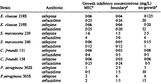
Before therapeutic exposure, the eight strains used in the study were susceptible to both cefepime and ceftazidime (Table I). As judged by MICs in agar dilution, and on a weight basis, cefepime was more potent than ceftazidime against E. cloacae, S. marcescens and Citrobacter freundii, but ceftazidime was more active against P. aeruginosa. MICs, as measured by agar dilution, were very similar to the boundary concentrations as determined on antibiotic-containing agar. On gradient plates, no-growth concentrations were in the same range (a difference of four-fold or less) as the corresponding boundary concentration for both cefepime and ceftazidime in S. marcescens and C. freundii, and for cefepime only in E. cloacae and P. aeruginosa. In the two latter species, no-growth concentrations were higher than boundary concen-trations by ten- to 150-fold when ceftazidime was tested, and overall, no-growth concentrations were higher for ceftazidime than for cefepime.
Antibiotic assays
Ceftazidime and cefepime displayed similar pharmacokinetic profiles in the peritoneal fluid, with antibiotic concentrations far above the MICs of initial strains 60 and
566 J.-C. Pechdre and L R. Vkdoiann
Table L Growth inhibitory activity of cefepime and ceftazidime before therapeutic exposure Strain E. cloacae 218S E. cloacae 219S 5. marcescens 239 S. marcescens 240 C. freundii 151 C. freundii 158 P. aeruginosa 3O2S P.aeruginosa 3O5S Antibiotic cefepime ceftazidime cefepime ceftazidime cefepime ceftazidime cefepime ceftazidime cefepime ceftazidime cefepime ceftazidime cefepime ceftazidime cefepime ceftazidime Growth M I C 006 0-25 006 0-25 1-6 4 0O6 0-12 006 04 O06 025 1 05 2 2 inhibitory concentrations (mg/L) boundary* 004 024 004 020 1-5 30 005 012 O03 03 003 024 2-5 1-5 6 1-5 no-growth 0125 20 020 32 2-5 6 015 1 008 05 006 05 5 30 6 15
"As measured by agar dilution (inoculum: c 104 cfu per spot); * as measured on antibiotic gradient
containing agar (inoculum: c. 10* cfu per dish).
Emergence of resistance after a first therapeutic exposure
Thirty-two untreated mice, serving as controls (four per strain) were killed and autopsied 24 h after inoculation challenge. Severe peritonitis was observed in all cases, and the peritoneal fluid contained from 91 x 10' to 7 x 10¹⁰ cfu/mL (all bacterial strains being considered), when the inoculum used for challenging the animals was 1-6 ±3-5 x 10* cfu/mL. Autopsy of control mice also showed enlarged spleens which yielded growth of the pathogen inoculated in all cases. Blood cultures were positive in 90% of the cases. One hundred and forty-four mice were treated by cefepime or ceftazidime, one or three doses of SO mg/kg each (Table III). Resistance did not develop in any mouse infected with S. marcescens or C. freundii. In mice infected with E. cloacae or P. aeruginosa, resistance commonly occurred after ceftazidime therapy with either the one or three dose regimens; with cefepime resistance developed in only a
Table II. Antibiotic concentration in peritoneal fluid after subcutaneous administration of cefepime or ceftazidime in mice with aseptic peritonitis
Antibiotic administered (dosing) Ceftazidime (50 mg/kg) Cefepime (50 mg/kg) Time (min) after dosing 30 60 120 180 60 120 180 Antibiotic concentration (mgL) in peritoneal fluid' 64-6 57-8 13-0 <2-5 38-2 12-9 3-3
Beahtanre after cefepime or ceftazfcttme 567 Table IIL Resistance emerging after therapy with cefepime or ceftazidime, one or three doses
(50mg/kg)
Strain Therapy
Number of mice with acquired resistance (fold increase of boundary concentration) after one dose* After three doses* E. cloacae 218 E. cloacae 219 S. marcescens 239 S. marcescens 240 C.freundU 151 C.frewtdii 158 P. aeruginosa 302 P. aeruginosa 305 cefepime ceftazidime cefepime ceftazidime cefepime ceftazidime cefepime ceftazidime cefepime ceftazidime cefepime ceftazidime cefepime ceftazidime cefepime ceftazidime
"Out of six mice inoculated. *Out of three mice inoculated.
0 3 (125-200) 0 5(250-640) 0 0 0 0 0 0 0 0 0 2(8) 0 2(8) 0 3(80) 1(50) 3 ( > 320) 0 0 0 0 0 0 0 0 0 2(20) 0 1(17
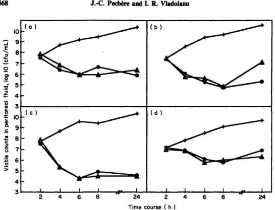
single mouse infected with E. cloacae 219S and dosed three times (Table 111). Chi-square statistics, after Yates* correction, indicated that in E. cloacae infected mice receiving a single antibiotic dose, resistance was significantly more frequent after ceftazidime than after cefepime therapy (P < 001). When resistance emerged, MIC increases were impressive (at least 80-fold) in E. cloacae infections, and more limited (eight-fold) in P. aeruginosa infections. As to the peritoneal viable counts, an antibiotic effect was observed in all cases, i.e. a decrease of at least two logs compared to parallel counts in the corresponding untreated mice. Twenty-four hours after challenge, treated animals had 2-3 to over 6 logs lower counts than did untreated animals, even when resistance occurred. For four of the strains under study, peritoneal viable counts were determined over 24 h in animals receiving three antibiotic doses or no antibiotic (controls) (Figure 1). Within the first 8 h following bacterial challenge, these counts were indistinguishable whether cefepime or ceftazidime was administered; after 24 h, viable counts remained similar in three experiments, while cefepime therapy was associated with a lower peritoneal cfu count in E. cloacae 219S (Figure 1) where resistance emerged after ceftazidime therapy.
Prediction of emergence of resistance in vitro
The eight strains exposed to cefepime or ceftazidime therapy were initially classified as susceptible to these antibiotics according to standard MIC determination, but resist-ance emerged in some of the animals treated. So we tried to find other in-vitro parameters which would be able to ensure a better prediction of the therapeutic outcome in the model. Higher no-growth concentration on antibiotic-containing gradient agar was associated with greater risk of resistance in vivo, except in the
568 J.-C. Ped&e and L R. Vladoianii 10 3 2. o § •a ftu l
s
S In pe ii
o o 9 8 7 6 5 4 3 10 9 8 7 6 ( o ) ( b ) (d ) 24 2 4 Time course ( h ) 24Figure 1. Viable counts of peritoneal bacteria in treated and untreated control mice after peritoneal challenge with 10* cfii. (a) E. cloacae 218S; (b) E. cloacae 219S; (c) C.freunOi 151; (d) P. aeruginosa 302S.
+ , No treatment; A , ceftazidime; • , cefephne.
cefepime-treated mouse infected with E. cloacae 219S where resistance emerged (Table II) despite low no-growth concentration (Table I). A greater correlation was found with the ratio of no-growth concentration to the boundary concentration versus the number of mice in which resistance developed (Figure 2).
256
3 4 5 6 7 8 Number of mice with resittance
Flgare 2. Correlation of emergence of resistance in mice with resistance as judged by cefepime- or ceftaTklimr-gradient containing agtr.
Resistance after cefepime or ryff Ttrftww 569 Table IV. Growth inhibitory activity of cefepime and ceftazidime against
resistant isolates selected in vivo Strain 218 R (CTZ) 219 R (CTZ) 219 R (CPM) 302 R (CTZ) 305 R(CTZ) Antibiotic ccfcpiroc ceftazidime cefepime ceftazidime cefepime ceftazidime cefepime ceftazidime cefepime ceftazidime Growth inhibitory MIC 0-5 32 4 > 128 2 128 8 32 32 128 concentrations (mg/L) boundary* 4 50 256 256 128 128 128 128 128 128
'As measured by agar dilution (inoculum: c. 104 cfu per spot).
*As measured by antibiotic gradient containing agar (inoculum: c. 10* cfu per dish).
Second therapeutic exposure
From each group of mice in which resistance emerged during therapy, one or two resistant isolates were chosen randomly for further studies. Five strains originated from ceftazidime-treated mice (respectively 218 R (CTZ) I and 2, 219 R (CTZ), 302 R (CTZ) and 305 R (CTZ)). A sixth strain came from the only cefepime-treated mouse showing bacterial resistance (219 R (CPM)). Growth inhibitory activity of cefepime and ceftazidime in vitro against these strains is summarized in Table IV. According to the agar dilution method with a 104 cfu inoculum, MICs of cefepime always remained lower than those of ceftazidime, by four-fold against P. aeruginosa and by at least 64-fold against E. cloacae. With the antibiotic containing gradient agar however, using a 10" cfu inoculum, boundary concentrations increased markedly, becoming similar for the two antibiotics.
In order to evaluate the therapeutic impact of these diminished in-vitro activities, the six resistant isolates were inoculated into naive mice, according the same protocol. Ten untreated mice were used as controls for these experiments, and killed 24 h after challenge. The macroscopic appearance of peritoneal cavities, and viable counts in the peritoneal fluid were similar to those found in untreated mice inoculated with cephalo-sporin-susceptible parent strains. This was a first indication that the resistant isolates had kept at least part of their initial virulence. Forty-eight mice were inoculated with one of the resistant isolates and treated with six antibiotic doses. The peritoneal cavity was sampled one day after challenge to confirm establishment of infection. All peritoneal fluids yielded growth of the species inoculated. Nine ceftazidime-treated mice had a peritoneal bacterial population which was more resistant to ceftazidime by two- to eight-fold, compared to the organism inoculated, (strains 218 R (CTZ)1 and 302 R (CTZ)). All ceftazidime-treated animals were dead by day 3, and most of them by day 2 after challenge (Table V). After 30 days of observation, cefepime ensured survival for the six mice infected with 219 R (CTZ), and one out of 12 mice infected with 218 R (CTZ), but no protection against infections produced by 219 R (CPM) or the pseudomonas isolates. For comparison purposes, 24 mice were inoculated with E. cloacae 218S and 219S (susceptible to cephalosporins) and treated as above
570 J.-C. Pecnere and L R. Yladoianu
Table V. Therapeutic effects on survival of mice challenged with resistant isolates and treated by cefepime or ceftazidime (six doses, 50 mg/lcg each, 2 h
apart) Strain 218S 219S 218 R (CTZ) 219 R (CTZ) 219 R (CPM) 302 R (CTZ) 305 R (CTZ) Therapy cefepime ceftazidime cefepime ceftazidime cefepime ceftazidime cefepime ceftazidime cefepime ceftazidime cefepime ceftazidime cefepime ceftazidime total inoculated 6 6 6 6 12 6 6 6 3 3 3 3 3 3 Number of mice surviving 2 6 5 6 4 3 0 6 5 0 0 0 0 0 0 3 6 3 6 0 1 0 6 0 0 0 0 0 0 0 at day: 30 6 3 6 0 1 0 6 0 0 0 0 0 0 0
(Table V). One month after challenge, the 12 cefepime-treated animals survived, compared to three out of the 12 mice which were given ceftazidime (P < 005).
Overall, the 22 mice still alive one month after challenge (E. cloacae in all cases) were apparently healthy. However, at autopsy we found small peritoneal abcesses and adhesions in seven of them (one ceftazidime-treated, six cefepime treated). Culture of the pus yielded E. cloacae, with the same pattern of antibiotic activities as the strain inoculated.
Virulence studies
The LDM of each pair of pre-and post therapy (resistant) strains were less than five-fold different in all cases, except for the 302 pair, which was approximately one log
Table VL Virulence of parent strains and cephalosporin-rcsistant mutants of E. cloacae
and P. aeruginosa Strain JO (cfu) E. cloacae 21SS 9-5x10* E. cloacae 219S 1-8x10* E. cloacae 218 R (CTZ)1 2-0 x 10* E. cloacae 218 R (CTZ)2 4-9 x 10* E. cloacae 219 R (CTZ) 2-9 x 10* E. cloacae 219 R (CPM) 1-0 x 10* P. aeruginosa 302S 2-7 x 10⁴ P. aeruginosa 3O5S 1-0 x 103 P. aeruginosa 302 R (CTZ) 1-9 x 10* P. aeruginosa 305 R (CTZ) 2-5 x 10³
Reafaanre after cefeptme or ceftazidime 571 different No consistent trend towards an increase or decrease in virulence was noted after occurrence of resistance (Table VI).
Discussion
After therapy of P. aeruginosa and E. cloacae infections, cefepime produced signifi-cantly less emergence of resistance than did ceftazidimc, and cefepime therapy occasionally remained efficient in treating mice infected with resistant E. cloacae selected by ceftazidime. Several factors could account for this 'dissociated resistance' in
vivo. Host factors were not investigated, and antibiotic concentrations in peritoneal
fluid were similar for the two drugs under study. However differences exist at the bacterial level. Resistance emerging during 0-lactam therapy of infections caused by aerobic non-fastidious Gram-negative bacilli is thought to result from the selection of low frequency mutants characterized by high ^-lactamase production, decreased outer membrane permeability or both (Marchou ex al., 1987a; Sawai, Yamaguchi & Hiruma, 1988). On antibiotic-gradient containing agars these mutants grew as single colonies well over the boundary concentration on ceftazidime, but not on cefepime gradients, in accordance with previous observations demonstrating the cefepime activity against ceftazidime-resistant bacteria (Vuye & Pick, 1985; Fung-Tome et al., 1989). Recent studies conducted with E. cloacae 218 (Bellido et al., 1991) showed that the V,,,,,, reflecting the stability of /Mactamase-substrate complex, was in the same range for cefepime and 'conventional' third generation cephalosporins; by contrast, the K,,, value for cefepime was remarkably high, reflecting the previously described low affinity for the /Mactamase (Hiraoka et al., 1988). In addition, and again in comparison with older cephalosporins, cefepime was shown to cross the outer membrane more rapidly and this was so even in a porin F-deficient mutant of E. cloacae 218 (Bellido et al., 1991). Altogether, in a given time, more cefepime molecules penetrate into the periplasmk space, avoid the /Mactamase attack and get access to the target molecules, for which cefepime does not show improved affinity (Bellido et al., 1991).
We have attempted to address the difficult question of the significance of the resistance emerging during therapy. Clinically, this resistance is associated with thera-peutic failure in about half of the cases (Milatovic & Braveny, 1987). The time course of viable counts in peritoneal fluid from animals treated by either antibiotic showed that, whether or not resistance occurred, the bacterial populations remained similar, at least for the first 8 h. A rather dramatic antibacterial effect was seen in all cases, which may correspond to the elimination of the susceptible population. The resistant mutants, which form a small minority within this population, grew at the same rate as their susceptible counterparts in broth medium (data not shown). So we assume that during the first hours following initiation of therapy, the lack of visible resistant cells in viable counts was due to their small number at the time of challenge. As an example, if the frequency of resistant cells was one in a million, a challenge inoculum of 10s cfu would have provided only 100 resistant cells. So resistance is not necessarily synonymous with therapeutic failure probably because, in some cases, the host defence can eliminate such small number of bacteria. However, despite the important structural and functional changes associated with the mutation to resistance (derepressed /Mactamase produc-tion, which represents a significant expense of energy, and altered permeability with possible consequences on bacterial uptake of various substances) the resistant mutants kept their initial virulence as determined here by the LDg, assays. They also resisted
572 J.-C. Pechere and L R. VUdoUm
treatment with their selective agents in vivo, leading to lethal therapeutic failure. These observations contrast with other examples where antibiotic resistance is associated with lower virulence such as gentamicin resistance in S. aureus (Musher et at., 1977; Pelletier, Richardson & Feist, 1979) or P. aeruginosa (Keys & Washington, 1977; Khakoo & Kluge, 1978), isonazid resistance in Mycobacterium tuberculosis (Middle-brook, 1957) or antibiotic resistance in Neisseria gonorrhoeae (Stollerman, 1978).
Therapeutic failure occurring when the pathogen is classified as susceptible to the antibiotic administered in vitro represents a very major error of susceptibility testing which has been seen with recent broad spectrum /Mactam compounds used for treating non-fastidious Gram-negative aerobic bacteria like E. cloacae and P. aeruginosa (Sanders, 1984). In this setting, both animal and clinical studies (Michea-Hamzehpour
et at., 1989) have shown that MICs (or their equivalent in disc diffusion techniques)
were poor predictors of emergence of resistance, at least when the strains were 'so-called' susceptible. This is likely to be due to the bacterial inoculum used in the testing procedures (typically 10* or 105 cfu, and less in microtechniques) compared with the scarce number of mutants associated with resistance (often less than one in 105 cells). Using a higher inoculum (10* cfu), the antibiotic gradient method described here allowed detection of these mutants. Resistance occurred in the mice only when the difference in /Mactam inhibitory concentrations between the wild type cells and the mutants was at least five-fold, as determined by the ratio no-growth concentration/ boundary concentration. No resistance was observed when the two subpopulations were relatively similar (less than four-fold difference). By contrast, as shown previously (Michea-Hamzehpour & Pechere, 1989), the presence and frequency of resistant mutants, and even the level of their resistance in vitro to the /Mactam given to the animal, did not predict faithfully further development of resistance in vivo. The links which are likely to exist between these observations and pharmacokinetics obviously deserve further studies.
Acknowledgements
We thank R. E. Kcssler and L. Lamb, from Bristol-Myers Squibb Pharmaceutical Research Institute USA for the determination of LDjoS, Mehri Michea-Hamzehpour and Lasta Kocjancic for antibiotic assays, and Lasta Kocjancic for technical assistance in animal experiments. This work has been supported by Fonds National Suisse de la Recherche Scientifique (Grant 31-28007-89).
References
BclMdo, F., Pechere, J. C. & Hancock, R. E. W. (1991). Revaluation of the factors involved in the efficacy of new /Mactams against Enterobacter cloacae. Antimicrobial Agents and
Chemotherapy 35, 73-8.
Eng, R. H. K., Cherubin, C. E., Pechere, J. C. & Beam, T. R. (1987). Treatment failures of cefotaxhne and latamoxef in meningitis caused by Enterobacter and Serratia spp. Journal of
Antimicrobial Chemotherapy 20, 903-11,
Fung-Tome, J., Dougherty, T. J., De Orio, F. J., Simich-Jacobson, V. & Kessler, R. E. (1989). Activity of cefepime against ceftaridime- and cefotaxime-resistant Gram-negative bacteria and its relationship to /f-lactamase levels. Antimicrobial Agents and Chemotherapy 33, 498-502.
Resistance after cefepime or cefbuttbne 573
Hiraoka, M., Masuyoshi, S., Mitsuhashi, S., Tomatsu, K. & Inoue, M. (1988). Cephalosporinase interactions and antimicrobial activity of BMY 28142, ceftazidime and cefotaxime. Journal of Antibiotics 41, 86-93.
Keys, T. F. & Washington, J. A. (1977). II. Gentamicin-resistant Pseudomonas aentginosa: Mayo Clinic experience, 1970-1976. Mayo Clinic Proceedings 52, 797-801.
Khakoo, R. A. & Kluge, R. M. (1978). Decreased virulence of genetamicin-resistant strains of Pseudomonas aeruginosa in a rat model. Journal of Laboratory and Clinical Medicine 91, 96-103.
King, A., Shannon, K., Eykyn, S. & Phillips, I. (1983). Reduced sensitivity to 0-lactam antibiotics arising during ceftazidime treatment of Pseudomonas aeruginosa infections. Journal of Antimicrobial Chemotherapy 12, 363-70.
Marchou, B., Bellido, F., Charnas, R., Lucain, C. & Pechere, J. C. (1987a). Contribution of p-lactamase hydrolysis and outer membrane permeability to ceftriaxone resistance in Enterobacter cloacae. Antimicrobial Agents and Chemotherapy 31, 1589-95.
Marchou, B., Michea-Hamzehpour, M., Lucain, C. & Pechere, J. C. (19876). Development of /*-lactam-resistant Enterobacter cloacae in mice. Journal of Infectious Diseases 156, 369-73. Michea-Hamzehpour, M., Auckenthaler, R., Regamey, P. & Pechere, J. C. (1987). Resistance
occurring after fluoroquinolone therapy of experimental Pseudomonas aeruginosa peritonitis. Antimicrobial Agents and Chemotherapy 31, 1803-8.
Michea-Hamzehpour, M. & Pechere, J. C. (1989). How predictable is development of resistance after /J-lactam therapy in Enterobacter cloacae infection? Journal of Antimicrobial Chemotherapy 24, 387-95.
Middlebroolc, G. (1957). Diagnosis and biological problems of isoniazid-resistant tubercle bacilli. In Proceedings of the 14th International Tuberculosis Conference, New Delhi, pp. 71-97. Tuberculosis Association of India.
Milatovic, D. & Braveny, I. (1987). Development of resistance during antibiotic therapy. European Journal of Clinical Microbiology 6, 234 44.
Musher, D. M., Baughn, R. E., Templcton, G. B. & Minuth, J. N. (1977). Emergence of variant forms of Staphylococcus aureus after exposure to gentamicin infectivity of the variants in experimental animals Journal of Infectious Diseases 136, 360-9.
National Committee for Clinical Laboratory Standards. (1985). Methods for Dilution Antimicrobial Susceptibility Tests for Bacteria that Grow Aerobically; Approved Standard
M7-A. NCCLS, Villanova, PA.
Pechere, J. C. (1989). Emergence of resistance in Gram-negative bacilli during beta-lactam therapy: a challenge for the future. European Journal of Cancer and Clinical Oncology 25, Suppl. 2, S17-23.
Pechere, J. C , Marchou, B., Michea-Hamzehpour, M. & Auckenthaler, R. (1986). Emergence of resistance after therapy with antibiotics used alone or combined in a murine model. Journal of Antimicrobial Chemotherapy 17, Suppl. A, 11—8.
Pelletier, L. L., Richardson, M. & Feist, M. (1979). Virulent gentamicin-induced small colony variants of Staphylococcus aureus. Journal of Laboratory and Clinical Medicine, 94, 324-34. Phelps, D. J., Carlton, D. D., Farrell, C. A. & Kessler, R. E. (1986). Affinity of cephalosporins for ^-lactamases as a factor in antibacterial efficacy. Antimicrobial Agents and Chemotherapy 29,845-8.
Quinn, J. P., DiVincenzo, C. A. & Foster, J. (1987). Emergence of resistance to ceftazidime during therapy for Enterobacter cloacae infections. Journal of Infectious Diseases 155,942-7. Sanders, C. C. (1984). Failure to detect resistance in antimicrobial susceptibility tests. A "very
major" error of increasing concern. Antimicrobic Newsletter 1, 27-31.
Sawai, T., Yamaguchi, A. & Hiruma, R. (1988). Effect of interaction between outer membrane permeability and /J-lactamase production on resistance to /Mactam agents in Gram-negative bacteria. Reviews of Infectious Diseases 10, 761-4.
Stollerman, G. H. (1978). Trends in bacterial virulence and antibiotic susceptibility: streptococci, pneumococci, and gonococci. Annals of Internal Medicine 89, 5 Pt 2 Suppl., 746-8. Vuye, A. & Pijck, J. (1985). In vitro antibacterial activity of BMY-28142, a new
extended-spectrum cephalosporin. Antimicrobial Agents and Chemotherapy Tl, 574-7. (Received 14 October 1991; revised version accepted 22 January 1992)