Combined hepatocellular-cholangiocarcinomas exhibit progenitor features and
activation of Wnt and TGFβ signaling pathways
Cédric Coulouarn
1, Catherine Cavard
2,3,4, Laura
Rubbia-Brandt
5, Anne Audebourg
6, Florent Dumont
2,3,4, Sébastien
Jacques
2,3,4, Pierre-Alexandre Just
2,3,4, Bruno Clément
1,
Hélène Gilgenkrantz
2,3,4, Christine Perret
2,3,4and Benoît
Terris
2,3,4,6,*
1INSERM, UMR991, Université de Rennes 1, Liver Metabolisms and Cancer, Rennes, France, 2INSERM, U1016, Institut Cochin, France, 3CNRS UMR8104, France, 4Université Paris Descartes, Paris, France, 5Service de Pathologie Clinique, Hôpitaux Universitaires de Genève, 1211 Geneva, Switzerland and 6Service d’Anatomie et Cytologie Pathologiques, AP-HP, Hôpital Cochin, Université Paris Descartes, Paris, France
*To whom correspondence should be addressed. Tel: +33 1 58 41 14 79; Fax: +33 1 58 41 14 80; Email: [email protected]
Intrahepatic malignant tumours include hepatocellular carci-nomas (HCC), cholangiocarcicarci-nomas (CC) and combined hepa-tocholangiocarcinomas (cHCC-CC), a group of rare and poorly characterized tumours that exhibit both biliary and hepatocytic differentiation. The aim of the study was to characterize the molecular pathways specifically associated with cHCC-CC patho-genesis. We performed a genome-wide transcriptional analysis of 20 histologically defined cHCC-CC and compared them with a series of typical HCC and of CC. Data were analysed by gene set enrichment and integrative genomics and results were further validated in situ by tissue microarray using an independent series of 152 tumours. We report that cHCC-CC exhibit stem/progeni-tor features, a down-regulation of the hepatocyte differentiation program and a commitment to the biliary lineage. TGFβ and Wnt/ β-catenin were identified as the two major signalling pathways activated in cHCC-CC. A β-catenin signature distinct from that observed in well-differentiated HCC with mutant β-catenin was found in cHCC-CC. This signature was associated with microen-vironment remodelling and TGFβ activation. Furthermore, inte-grative genomics revealed that cHCC-CC share characteristics of poorly differentiated HCC with stem cell traits and poor progno-sis. The common traits displayed by CC, cHCC-CC and some HCC suggest that these tumours could originate from stem/progenitor cell(s) and raised the hypothesis of a potential continuum between intrahepatic CC, cHCC-CC and poorly differentiated HCC.
Introduction
Hepatocellular carcinoma (HCC) and cholangiocarcinoma (CC) are the two major types of primary liver cancers. More than 80% of primary liver cancers are HCC usually occurring in fibrotic and cir-rhotic livers related to chronic viral infections, metabolic disorders, high alcohol intake or hemochromatosis (1). CC, which account for about 15% of primary liver cancers, derive either from small intrahe-patic bile ducts or ductules (intraheintrahe-patic cholangiocarcinoma, ICC), or from large hilar or extrahepatic bile ducts (extrahepatic cholan-giocarcinoma, ECC (1)). Another rare and aggressive subtype of pri-mary liver cancer has been defined according to the World Health Organization criteria, referred to as combined hepatocholangiocarci-noma (cHCC-CC), sharing clinicopathological features of both HCC and ICC, and can arise in both non-cirrhotic and cirrhotic livers (2–5). Besides the classical cHCC-CC, which contain areas of typical HCC and CC, three subtypes with stem cell features were defined, one of them called cholangiolocellular carcinoma (3). The tumoural cells of
these particular subtypes show intermediate features between hepato-cytes and cholangiohepato-cytes. Today, the diagnosis of cHCC-CC and their different subtypes relies on histopathological analyses and unified cri-teria, including molecular markers, are still pending.
Genomic approaches have been successful in identifying genes and pathways involved in the pathogenesis of CC and HCC. Recently, by gene expression profiling of a large cohort of HCC and a small ser-ies of CC and cHCC-CC, Woo et al. identified a subtype of HCC with CC-like traits associated with a poor prognosis (6), thus suggest-ing a phenotypic overlap between HCC, CC and cHCC-CC. In the present work, we profiled a series of 20 histologically well-defined cHCC-CC and a series of typical HCC and ICC. Functional ana-lysis, cross-comparison with published datasets and in situ validation using an independent set of tumours showed that cHCC-CC displayed stem/progenitor features, a down-regulation of an HNF4-driven hep-atocyte differentiation program and a commitment to the biliary lin-eage. Wnt/β-catenin and TGFβ were the main signaling pathways activated in cHCC-CC. Importantly, the Wnt/β-catenin signature in cHCC-CC differed dramatically from that in well-differentiated HCC with mutant β-catenin. Finally, integrative genomics revealed that cHCC-CC recapitulated a subset of HCC, which exhibits progenitor features and poor prognosis.
Materials and methods
Patients and histopathological variables
Two different series of surgically resected primary liver tumours (Cochin Hospital, Paris, France) were used in this study. Series #1 included 20 cHCC-CC, 3 IcHCC-CC, 7 typical HCC (Table I), which were profiled by microarray. Since we showed that HCC with mutant β-catenin were well-differentiated tumours with specific morphologic and metabolic features (7), we discarded them and selected Edmondson-Steiner grade 3 HCC (8) with macrotrabecular morphology. Epithelial cell adhesion molecule (EpCAM) being a marker of HCC with stem/progenitor features (9), we selected half of them with posi-tivity for EpCAM. The β-catenin status of the 20 cHCC-CC and ICC were all of WT genotype. Series #2 included an independent validation set of 48 cHCC-CC, 54 ICC and 152 HCC (among them 60 with mutant β-catenin (7)). The use of clinical and pathologic records was in agreement with French laws and ethical guidelines related to the protection of the patient. Hematoxylin and eosin-stained sections were histologically assessed by two of the authors (B.T. and P-A. J.). Hepatic fibrosis in non-cancerous livers was graded accord-ing to the METAVIR classification (10).
Tissue microarray
Core tissue biopsies were taken from individual paraffin-embedded tumours and arranged in recipient tissue array-blocks using a microarrayer (Beecher Instruments, Woodland, CA, USA). Three cores of 0.8 mm in diameter were sampled from each tumour.
Immunohistochemistry
Paraffin sections were processed as described previously (7). Immunohistochemistry was done on standard (series 1) and tissue microarray slides (series 2) using antibodies against β-catenin, EpCAM, GLUL, KRT19, LEF1 and SOX9 (supplementary materials and methods).
In situ hybridisation
The PCR template coding for almost the full-length human albumin (ALB) was generated from the MGC clone containing the complete cDNA for the human protein (IMAGE ID: 4734617) purchased from Invitrogen (Invitrogen, Carlsbad, USA). Detailed protocol is given as supplementary materials and methods.
RNA extraction and gene expression profiling
Total RNA was isolated with TRIzol (Invitrogen, Carlsbad, USA) and quality controlled on Agilent’s 2100 Bioanalyzer (RNA integrity number was above 8). Detailed protocol for GeneChip® human Gene (Affymetrix) hybridization is given as supplementary methods. MIAME compliant microarray data are available in Gene Expression Omnibus (GEO) database (GSE35306).
Abbreviations: CC, cholangiocarcinoma; cHCC-CC, combined
Statistical analysis and data mining
Briefly, differentially expressed genes were identified by a univariate two-sample t-test with a random variance model. Data mining was based on gene ontology, gene set enrichment analysis (GSEA) and ingenuity pathway analysis (Mountain view, CA, USA) as described previously (11,12). Details are provided in supplementary methods. Integrative genomics was performed using publicly available gene expression datasets downloaded from GEO. Results
Histopathological features of profiled tumours
The 20 cHCC-CC displayed the typical histopathological features described by N.D. Theise (4). All these tumours, recognized on hematoxylin-eosin staining, were classified in the cholangiolocellular carcinoma stem cell subtype (Figure 1A). Tumoural cells arranged in a tubular, cord-like, anastomosing pattern and were frequently embedded in a fibrous stroma. At the tumour periphery, the tumoural cords were continuous with the non-tumoural liver-cell cords in a replacing growth pattern. None of the tumours showed mucin forma-tion. All cHCC-CC were positive for KRT19 and EpCAM, 75% of them being positive for ALB (Table I and Figure 1A–C). All three ICC corresponded to moderately differentiated adenocarcinomas and were strongly positive for KRT19 and EpCAM (Table I and Figure 1D–F). All seven HCC expressed ALB mRNA (Table I and Figure 1I).
cHCC-CC and CC exhibit overlapping gene expression profiles
Microarray experiments were performed to establish and to compare gene expression profiles of the 30 tumours described above and two normal liver samples harvested at the periphery of hepatic tumourec-tomy specimens (untreated metastatic colon carcinoma). Based on the
expression of the most variant genes, two main clusters were identified both by hierarchical clustering and multidimensional scaling analysis (Supplementary Figure 1). Cluster 1 included all normal liver samples and HCC, and cluster 2 included all CC and cHCC-CC. Thus, unsuper-vised genome-wide expression profiling demonstrated that cHCC-CC tumours exhibited more similarity with CC than with HCC. Next, by using a supervised approach, which combined class comparison and class prediction algorithms, we identified 1244 non-redundant genes dif-ferentially expressed (P < 0.001, 2-fold change, FDR < 0.5%, 96–100% correct prediction) between HCC and cHCC-CC (Supplementary Figure 1B and Supplementary Table I). By hierarchical clustering analysis (Supplementary Figure 2), differentially expressed genes were further divided into a cHCC-CC gene set (i.e. 588 genes up-regulated in cHCC-CC) and a HCC gene set (i.e. 656 genes up-regulated in HCC). Validating the gene selection, HCC gene set included well-known HCC markers such as α-fetoprotein, glypican 3 or the non-coding H19 gene (Supplementary Figures 1B and 3D).
Down-regulated genes in cHCC-CC are linked to differentiated hepatocyte phenotype
Functional analysis of HCC and cHCC-CC gene sets was performed by using an unsupervised GSEA. This approach clearly demonstrated that gene signatures associated with hepatic metabolism were significantly (P < 0.05) enriched in the gene expression profiles of HCC compared with those of cHCC-CC (Figure 2A and Supplementary Figure 4A). Notably, numerous genes encoding plasma proteins (e.g. ALB) or those involved in glucose and lipid metabolism (e.g. G6PC), and detoxica-tion (e.g. CYP2A6) were repressed in cHCC-CC (Supplementary Figure 3A). These results were further confirmed by gene ontology analysis (Supplementary Table II). Interestingly, down-regulated genes
Table I. Clinicopathological features and in situ expression data of profiled tumours
Case Age Gender Etiologyb Fibrosisc Size(cm) β-catenin mutation EpCAM IHCd KRT19 IHC ALB ISHd
1 70 male GH 2 5 wt 1 1 2 2 62 male AC 3 4 wt 2 1 1 3 62 male AC 4 8 wt 1 1 2 4 50 male GH 4 2 wt 1 1 0 5 61 male AC 3 5 wt 2 1 2 6 69 male AC 3 7 wt 2 2 2 7 73 female None 0 3.5 wt 2 2 2 8 60 male GH 4 4 wt 2 1 1 9 35 female None 0 10 wt 2 1 0 cHCC-CCa 10 37 male None 2 11 wt 2 1 1 11 62 male None 0 14 wt 2 2 2 12 69 male AC 3 9 wt 2 1 2 13 57 female AC 4 8 wt 2 2 1 14 68 male AC 4 4 wt 2 1 0 15 58 male AC 4 5 wt 1 1 2 16 80 male AC 1 8 wt 1 1 1 17 59 female None 1 17 wt 1 1 0 18 54 male HCV 2 5 wt 2 1 2 19 56 female None 0 3 wt 2 1 1 20 69 female None 0 5 wt 2 1 0 1 73 male None 1 7 wt 2 1 0 CCa 2 53 male HBV 2 5 wt 2 2 0 3 61 female None 1 20 wt 1 1 2 1 67 male AC 3 10 wt 0 0 2 2 55 male AC 3 15 wt 0 0 1 3 39 male HBV 1 10 wt 0 0 2 HCCa 4 69 male AC 3 9 wt 0 0 2 5 74 male AC 4 7 wt 2 0 1 6 68 male HBV 4 6 wt 2 0 1 7 50 male AC 1 16 wt 2 0 2
acHCC-CC, combined hepatocholangiocarcinomas; CC, intrahepatic cholangiocarcinomas; HCC, hepatocellular carcinomas. bGH, genetic hemochromatosis; AC, alcohol consumption; HBV, hepatitis B virus; HCV, hepatitis C virus.
cFibrosis was graded according to the METAVIR score.
dEpCAM positivity was defined as membranous expression in ≥10% of tumour cells with moderate (1) or strong intensity (2), as well ALB ISH cytosolic positivity.
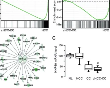
in cHCC-CC were specifically associated with hepatocyte-enriched transcription factor HNF4A, as judged by GSEA (Figure 2A), inge-nuity network analysis (Figure 2B) and promoter regulatory elements analysis (Supplementary Tables II and III). Accordingly, HNF4A itself was significantly repressed in cHCC-CC (Figure 2C). Altogether, these results demonstrated that genes related to mature hepatocyte differen-tiation were down-regulated in cHCC-CC.
Up-regulated genes in cHCC-CC are associated with biliary com-mitment and extracellular matrix remodelling
In agreement with our first observation that cHCC-CC and CC exhib-ited similar expression profiles, analysing the cHCC-CC gene set highlighted signalling pathways typical of CC (i.e. biliary differen-tiation and fibrosis). Notably, numerous fibrosis-associated genes, including potent pro-fibrotic factors (e.g. TGFβ, IL8), extracellular matrix (ECM) genes (e.g. COL1A1) and ECM-remodelling genes (e.g. MMP2, TIMP1) were up-regulated in cHCC-CC (Supplementary Figure 3E). These features were further validated by ingenuity way analysis as illustrated by the analysis of enriched canonical path-ways and gene networks (Figure 3A and 3B, respectively).
Unsupervised GSEA revealed that gene signatures of early liver development and stem cells were enriched in cHCC-CC (Supplementary Figure 4B) indicating that cHCC-CC exhib-ited progenitor features. Interestingly, early biomarkers of biliary
commitment (e.g. SOX9), as well as master genes of signalling pathways, which regulate the differentiation of hepatoblasts to chol-angiocytes (e.g. TGFβ, Wnt, Notch), were induced in cHCC-CC (Supplementary Figure 3B). Enrichment of Wnt/β-catenin and TGFβ pathways in cHCC-CC was further validated by unsupervised GSEA (Supplementary Figure 4C). Corroborating this data, LEF1 transcrip-tion factor was significantly increased in cHCC-CC (Figure 3C) and LEF1 binding sites occurred frequently in the promoter of cHCC-CC genes (Supplementary Table III). In addition, cHCC-CC exhibited a gene signature characteristic of the activation of the Wnt/β-catenin pathway in mouse hepatoblasts, which was associated with biliary ductal morphogenesis (13) (Supplementary Figure 4D). These obser-vations suggested that a Wnt-mediated transcriptional program linked to cholangiocyte differentiation was present in cHCC-CC.
cHCC-CC signature in HCC with progenitor features and poor prognosis
Next, we compared our data with publicly available HCC datasets (11,14). By integrative genomics, we showed that cHCC-CC clustered with a specific HCC subset, which exhibited progenitor features, activa-tion of TGFβ and poor prognosis (Cluster 1 in Figure 4A). Accordingly, TGFβ (11) and hepatoblast (14) signatures were significantly enriched in cHCC-CC (Figure 4B). Integration with another cohort of 98 HCC (15) further demonstrated that cHCC-CC recapitulated the previously
Fig. 1. Microscopical pattern, EpCAM immunohistochemical staining and ALB mRNA expression in profiled tumours. (A–C) HES staining, expression of
EpCAM by IHC and expression of ALB mRNA by in situ hybridization in cHCC-CC with features of cholangiolocellular carcinoma. (D–F) HES staining, expression of EpCAM by immunohistochemistry and expression of ALB mRNA by in situ hybridization in CC. (G–I) HES staining, expression of EpCAM by immunohistochemistry and expression of ALB mRNA by in situ hybridization in typical macrotrabecular HCC.
described S1 HCC subtype, which was characterized by an aberrant Wnt and TGFβ activation along with a poor prognosis (Figure 4C–E). Further reinforcing the notion that cHCC-CC are associated with a bad prognosis, unsupervised GSEA demonstrated a specific enrich-ment of genes linked to poor survival and recurrence in liver cancer (Supplementary Figure 4D). In addition, several markers of epithelial-mesenchymal transition (EMT) and metastasis (e.g. SNAI2, VIM) were up-regulated in cHCC-CC as well as several genes that have been also proposed as cancer stem cell markers (e.g. EPCAM, CD133/
PROM1, CD44, THY1) (Supplementary Figure 3B,F).
Tissue microarray validation of hepatic progenitor features and Wnt/β-catenin activation in cHCC-CC
To validate our results, we analysed the in situ expression of markers of β-catenin activation together with markers of hepatocyte and
cholangiocyte differentiation using tissue microarray, which included an independent set of 48 cHCC-CC, 54 CC and 152 HCC among them 60 (39%) displaying β-catenin mutation (Table II). As expected, HCC with mutant β-catenin displayed β-catenin nuclear positivity, expressed the hepatocyte β-catenin target GLUL (16) and were negative for EpCAM, in agreement with their well-differentiated phenotype (7). In contrast, cHCC-CC did not show β-catenin nuclear staining (30% displayed a faint cytosolic β-catenin staining) and
Fig. 2. Absence of HNF4A-linked hepatocytic gene signature in cHCC-CC.
(A) Unsupervised GSEA using cHCC-CC and HCC gene expression profiles (respectively on the left and right sides of each enrichment plot). Displayed are the enrichment plots for two gene signatures representative of liver-specific genes and HNF4A-regulated genes. Hits (bar code) represent genes included in each signature. Enrichment scores are shown on the y-axis. The two signatures were shown to be significantly enriched in HCC and absent in cHCC-CC (P < 0.05). (B) Ingenuity analysis of HCC gene set (i.e. genes specifically down-regulated in cHCC-CC) highlighted a gene network centered on the liver-enriched HNF4A transcription factor. (C) HNF4A mRNA levels in normal livers (NL), hepatocellular carcinomas (HCC), cholangiocarcinomas (CC) and combined HCC-CC (cHCC-CC). HNF4A was significantly expressed at a lower level in CC and cHCC-CC. Statistical significance was evaluated using a two-sample unpaired Student’s t-test between HCC and cHCC-CC. *P < 0.001.
Table II. In situ expression of selected markers by IHC and ISH on TMA containing independent cHCC-CC, CC and HCC
β-catenin+ GLUL+ LEF1+ EpCAM+ SOX9+ KRT19+ ALB+
c-HCC-CC (n = 48) 0a (0%) 0 (0%) 17 (35%) 47 (98%) 20 (47%) 48 (100%) 34 (77%) CC (n = 54) 0a (0%) 0 (0%) 18 (32%) 54 (100%) 25 (46%) 54 (100%) 18 (33%) HCC - with mutant β-catenin (n = 60) - without mutant β-catenin (n = 92) 38 (63%) 2 (2%) 54 (90%) 2 (2%) 7 (12%) 12 (13%) 4 (7%) 17 (18%) 0 (0%) 4 (6%) 4 (7%) 26 (28%) 60 (100%) 92 (100%)
Negative (0) β-catenin expression indicates that no nuclear staining was observed. EpCAM positivity was defined as membranous expression in ≥10% of tumour cells with moderate or strong intensity, as well ALB ISH positivity. For LEF1 and SOX9, tumours were considered as positive when the intensity of nuclear expression was greater than those observed in normal biliary cells.
aFaint positive cytosolic positivity alone in 30% of the cases.
Fig. 3. Signaling pathways associated with CC and biliary differentiation
in cHCC-CC. (A) Ingenuity analysis of canonical pathways significantly enriched in cHCC-CC gene set (i.e. genes specifically up-regulated in cHCC-CC). Top-ranked pathways were associated with hepatic fibrosis, TGFβ and Wnt/β-catenin signaling. (B) Gene network analysis using cHCC-CC gene set. A specific network associated with TGFβ and
extracellular matrix remodelling was identified. (C) Wnt/β-catenin associated LEF1 transcription factor was significantly expressed at a higher level in cHCC-CC and CC. Shown are LEF1 mRNA levels in normal livers (NL), hepatocellular carcinomas (HCC), cholangiocarcinomas (CC) and combined HCC-CC (cHCC-CC). Statistical significance of LEF1 mRNA abundance between HCC and cHCC-CC was evaluated using a two-sample unpaired Student’s t-test. *P < 0.001. (D) cHCC-CC exhibited a gene signature characteristic of the activation of the Wnt/β-catenin signaling in hepatoblast, which is associated with biliary cell fate. GSEA was performed using a gene set that signs APC invalidation in hepatoblast as described (13). Up- and down-regulated gene sets were found to be specifically enriched in cHCC-CC and HCC gene expression profiles, respectively (P < 0.05).
showed a completely different spectrum of β-catenin target genes (all GLUL (-) and EpCAM (+) (17)) (Table II). As expected, expression of EpCAM and KRT19 hepatic progenitor markers was observed in >98% cHCC-CC. In agreement with the biliary differentiation trait, expression of LEF1 and SOX9 was similar in cHCC-CC and CC. ALB mRNA were detected in >75% cHCC-CC and >30% CC. Altogether, these observations corroborated the overlapping features between cHCC-CC and CC and supported the hypothesis that these tumours may arise from hepatic progenitor(s).
Discussion
cHCC-CC are aggressive and heterogeneous tumours, which exhibit both biliary and hepatocytic differentiation features. Their diagno-sis can be difficult and relies on histopathological observations. The closed morphological pattern frequently observed between cHCC-CC and CC probably explains both the poor reproducibility of diagnosis
among experts and the numerous terminologies employed for their characterization (18). Very few genomic studies (1) were designed to depict the molecular pathogenesis of these tumours. Here, we per-formed a transcriptional analysis, which relies on a well-characterized series of cHCC-CC with stem cell features versus CC and typical macrotrabecular HCC. By unsupervised genome-wide expression profiling, we demonstrated that cHCC-CC clustered with CC rather than with HCC. This was confirmed by an altered hepatocytic dif-ferentiation program, as underlined by the down-regulation of HNF4 and its target genes.
By comparing our data with previously published HCC gene sig-natures, we found that cHCC-CC recapitulated (i) HCC with the hepatoblast signature published by Lee et al. (14); (ii) the S1 HCC subgroup described by Hoshida et al. (15); (iii) the G1-3 HCC groups from Boyault et al. (19) (Supplementary Figure 5); and (iv) the EpCAM-positive HCC described by Yamashita et al. (9) (data not shown). Strikingly, all these subgroups included poorly
Fig. 4. cHCC-CC recapitulated HCC with a bad prognosis and hepatoblast features along with an aberrant activation of Wnt and TGFβ pathways. (A) Through an integrative genomic approach, cHCC-CC, HCC and CC gene expression profiles were merged with those derived from an independent cohort of 139 cases of human HCC (GSE1898 dataset from the GEO database). Integration was based on the expression of genes differentially expressed between HCC and cHCC-CC. Shown is the dendrogram overview of the integrated dataset where the identity of samples is displayed at the beginning of each row. Top, samples from the present study: HCC, hepatocellular carcinoma; CC, cholangiocarcinoma; cHCC-CC, combined HCC-CC. Bottom, HCC from the independent cohort that were previously assigned to specific subgroups with respect to cell origin (14) (HB, hepatoblast; HC, hepatocyte), prognosis (23) (A, bad prognosis; B, good, prognosis) and TGFβ signaling (11) (E, early; L, late TGFβ signature). Clustering analysis identified two major clusters. Cluster 1 included cHCC-CC and CC from the present study as well as HCC characterized by a poor prognosis and by the hepatoblast and late TGFβ signatures (subgroups A, HB and L, respectively) from the independent cohort. (B) GSEA validation of specific enrichment of TGBβ and liver cancer hepatoblast signatures in the gene expression profiles of cHCC-CC (P < 0.05). (C) Integration of cHCC-CC, HCC and CC gene profiles (the present study) with those derived from 98 human HCC (GSE10186 dataset from the GEO database). Those HCC were previously assigned to three robust subclasses characterized by an aberrant activation of the Wnt and TGFβ signaling pathways (S1), proliferation as well as MYC and AKT activation (S2), and hepatocyte differentiation (S3) (15). Clustering analysis of the integrated dataset identified two main clusters. Cluster 1 included 90–100% of cHCC-CC and CC from the present study as well as most of S1-type HCC from the independent cohort. Cluster 2 included all HCC from the present study and S3-type HCC. (D) Distribution of specific HCC subtypes from Hoshida’s classification (S1–3) between clusters C1 and C2 (P < 0.001). (E) Unsupervised gene set enrichment analysis confirmed the close association between cHCC-CC and HCC from subclass S1 defined in Hoshida’s study (P < 0.05) (15).
differentiated tumours and were associated with a greater incidence of recurrence after surgical treatment and with stem cell/progenitor traits. Accordingly, it has recently been reported that scirrhous HCC, a rare variant of HCC, harbours cholangiocarcinoma traits with expres-sion of EMT-related genes as in our cHCC-CC series, which also dis-play an abundant stroma (20) supporting the idea of a continuous liver cancer spectrum between these tumours.
We also report that an activation of both TGFβ and Wnt/β-catenin cell signalling pathways is a prominent feature of cHCC-CC. TGFβ pathway is involved in biliary differentiation and in EMT. Accordingly, unsupervised GSEA demonstrated the up-regulation of EMT-related markers in cHCC-CC. Wnt/β-catenin pathway is also critical for bil-iary/hepatocyte cellular commitment (13,21). We previously reported the important role of Wnt/β-catenin pathway in controlling the fate of hepatoblasts, preventing them from differentiating toward the hepato-cyte lineage and guiding them to biliary ductal morphogenesis (13). Interestingly, we evidenced in cHCC-CC a particular Wnt/β-catenin signature that was not associated with β-catenin mutation. Although the mechanism of β-catenin activation remained to be identified in cHCC-CC, we hypothesize that a crosstalk with the TGFβ pathway may be involved based on previous observations by Hoshida et al. (15) who reported a TGFβ-driven activation of β-catenin signalling in the S1 HCC subset, which clustered with our cHCC-CC series.
In an attempt to establish a discriminative fingerprint between cHCC-CC and CC, we observed in CC a higher expression of genes involved in the regulation of Wnt signalling (data not shown). This is consistent with the fine-tuning of Wnt activation necessary for the biliary commitment and further differentiation (22). Expression of such markers associated to ALB expression, which is more frequently observed in cHCC-CC than in CC, might distinguish these two enti-ties. It remains an opened question that has to be strengthened by the analysis of larger series.
Whether hepatic primitive tumours arise from progenitor cells and/or from the dedifferentiation of neoplastically transformed mature hepatocytes is still a matter of debate. By demonstrating that cHCC-CC clustered both with CC and with a specific subset of HCC with progenitor features, our data support the former concept. Altogether, our data raise the hypothesis that ICC might derive from cHCC-CC originating from progenitor cells, with an ultimate induc-tion of the CC phenotype.
Supplementary data
The following supporting information may be found in the online ver-sion of the article.
Supplementary materials and methods.
Supplementary Fig. 1. Genome-wide transcriptional profiling of
cHCC-CC; clustering analysis.
Supplementary Fig. 2. Hierarchical clustering analysis based on
genes differentially expressed between HCC and cHCC-CC.
Supplementary Fig. 3. Examples of genes differentially expressed
between HCC and cHCC-CC.
Supplementary Fig. 4. Examples of relevant gene sets specifically
enriched in cHCC-CC and HCC gene expression profiles.
Supplementary Fig. 5. Integrative genomics analysis of HCC and
combined HCC-CC using independent gene expression profiles.
Supplementary Table I. List of genes differentially expressed
between HCC and combined HCC-CC tumours.
Supplementary Table II. Gene Ontology (GO) analysis of HCC
and cHCC-CC gene sets.
Supplementary Table III. Top ranked regulatory elements in the
promoter of HCC and cHCC-CC gene sets.
Funding
This work was supported by INSERM, CNRS, the Ligue Nationale contre le Cancer (équipe labellisée 2011–2013), the Institut National du Cancer, Association pour la Recherche sur le Cancer, the Agence
Nationale de la Recherche and the Charles Debray Association. P.A Just is a recipient of a fellowship from CNRS-CEA/AP-HP.
Acknowledgements
We thank Brigitte Radenen and Nathalie Lin Mark for their technical assis-tance. We are grateful to the members of C. Perret’s group and we thank all the members from the Genomic and Sequencing Facility of the Cochin Institute and from the Biobank of Cochin Hospital.
Conflict of Interest Statement: None declared.
References
1. Cazals-Hatem,D. et al. (2004) Clinical and molecular analysis of combined hepatocellular-cholangiocarcinomas. J. Hepatol., 41, 292–298.
2. Wakasa,T. et al. (2007) A histopathological study on combined hepa-tocellular and cholangiocarcinoma: cholangiocarcinoma component is originated from hepatocellular carcinoma. Hepatogastroenterology, 54, 508–513.
3. Komuta,M. et al. (2008) Clinicopathological study on cholangiolocellu-lar carcinoma suggesting hepatic progenitor cell origin. Hepatology, 47, 1544–1556.
4. Theise,N.D. et al. (2010) Combined hepatocellular-cholangiocarcinoma. IARC, Lyon.
5. Roskams,T. et al. (2010) Hepatic progenitor cells: an update. Clin. Liver Dis., 14, 705–718.
6. Woo,H.G. et al. (2010) Identification of a cholangiocarcinoma-like gene expression trait in hepatocellular carcinoma. Cancer Res., 70, 3034–3041. 7. Audard,V. et al. (2007) Cholestasis is a marker for hepatocellular
carcino-mas displaying beta-catenin mutations. J. Pathol., 212, 345–352.
8. Edmondson,H.A. et al. (1954) Primary carcinoma of the liver: a study of 100 cases among 48,900 necropsies. Cancer, 7, 462–503.
9. Yamashita,T. et al. (2009) EpCAM-positive hepatocellular carci-noma cells are tumor-initiating cells with stem/progenitor cell features. Gastroenterology, 136, 1012–1024.
10. Bedossa,P. et al. (1996) An algorithm for the grading of activity in chronic hepatitis C. The METAVIR Cooperative Study Group. Hepatology, 24, 289–293.
11. Coulouarn,C. et al. (2008) Transforming growth factor-beta gene expres-sion signature in mouse hepatocytes predicts clinical outcome in human cancer. Hepatology, 47, 2059–2067.
12. Coulouarn,C. et al. (2009) Loss of miR-122 expression in liver cancer cor-relates with suppression of the hepatic phenotype and gain of metastatic properties. Oncogene, 28, 3526–3536.
13. Decaens,T. et al. (2008) Stabilization of beta-catenin affects mouse embry-onic liver growth and hepatoblast fate. Hepatology, 47, 247–258. 14. Lee,J.S. et al. (2006) A novel prognostic subtype of human hepatocellular
carcinoma derived from hepatic progenitor cells. Nat. Med., 12, 410–416. 15. Hoshida,Y. et al. (2009) Integrative transcriptome analysis reveals common
molecular subclasses of human hepatocellular carcinoma. Cancer Res., 69, 7385–7392.
16. Cadoret,A. et al. (2002) New targets of beta-catenin signaling in the liver are involved in the glutamine metabolism. Oncogene, 21, 8293–8301. 17. Yamashita,T. et al. (2007) Activation of hepatic stem cell marker EpCAM
by Wnt-beta-catenin signaling in hepatocellular carcinoma. Cancer Res.,
67, 10831–10839.
18. Malouf,G. et al. (2009) Is histological diagnosis of primary liver carcino-mas with fibrous stroma reproducible among experts? J. Clin. Pathol., 62, 519–524.
19. Boyault,S. et al. (2007) Transcriptome classification of HCC is related to gene alterations and to new therapeutic targets. Hepatology, 45, 42–52. 20. Seok,J.Y. et al. (2012) A fibrous stromal component in hepatocellular
carcinoma reveals a cholangiocarcinoma-like gene expression trait and epithelial-mesenchymal transition. Hepatology, 55, 1776–1786.
21. Tan,X. et al. (2008) Beta-catenin deletion in hepatoblasts disrupts hepatic morphogenesis and survival during mouse development. Hepatology, 47, 1667–1679.
22. Lemaigre,F.P. (2010) Molecular mechanisms of biliary development. Prog. Mol. Biol. Transl. Sci., 97, 103–126.
23. Lee,J.S. et al. (2004) Classification and prediction of survival in hepatocel-lular carcinoma by gene expression profiling. Hepatology, 40, 667–676.
Received March 12, 2012; revised June 04, 2012; accepted June 09, 2012