Mutagenicity of 2-amino-3-methylimidazo[4,5-f]quinoline in human lymphoblastoid cells
6
0
0
Texte intégral
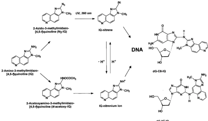
(2) P.M.Leong-Morgenthaler et al.. Fig. 1. Metabolic activation of IQ by cytochrome P450 enzymes and the N,O-acetyltransferase and an alternate activation pathway through the photoactivation of N3-IQ leading to the formation of the dgN2–IQ and dG-C8–IQ adducts.. lymphoblastoid cell line TK6 to rat liver S9-activated IQ and to photoactivated N3-IQ. We have determined the adducts formed and their removal, and also characterized some of the mutations induced following treatment with photoactivated azido-IQ in these cells. Materials and methods Chemicals RPMI1640, glutamine, gentamycin and horse serum were obtained from Gibco, Life Technologies (Basel, Switzerland). IQ was purchased from Toronto Research Chemicals (North York, Ontario) and Moltox arochlor1254-induced rat liver S9 was from Molecular Toxicology (Boone, MD). N3-IQ was synthesized as previously reported (18). Micrococcal nuclease, nuclease P1, and spleen phosphodiesterase (type I) were purchased from Sigma (St Louis, MO). Cloned T4 polynucleotide kinase was obtained from New England Biolabs (Beverly, MA). [32P]ATP (7000 Ci/mmol) was obtained from ICN Chemicals (Irvine, CA). PEI-cellulose thin layer plates were from MacheryNagel (Du¨ren, Germany). Isoelute C18 end-capped cartridges (100 mg) were purchased from ICT AG (Basel, Switzerland). Qiagen tip-2500 columns, QIA amp genomic kit and the QIAquick Gel extraction kits were obtained from Qiagen AG (Basel, Switzerland). The f-mol cycle sequencing system was purchased from Promega (Zurich, Switzerland). Mutation assays Mutation assays using the TK6 human lymphoblastoid cells were performed as described in Skopek et al. (19) and Liber and Thilly (20). The TK6 cells were maintained in RPMI1640 supplemented with 2 mM glutamine and 10% horse serum. The cells were cultured in sterile flasks in an incubator maintained at 37°C with 5% CO2. The media used for experimental cultures were also supplemented with 10% gentamycin. Prior to the mutation assays, the TK6 cells were treated with CHAT medium to reduce the spontaneous mutants. To determine the mutagenicity of IQ, cultures of TK6 cells were treated with different concentrations of the compound in the presence of an exogenous metabolizing system (1.5% rat liver S9) for 4 h. An alternate route to generate the reactive intermediates of IQ by the photoactivation of N3-IQ is shown in Figure 1. Treatment with N3-IQ was performed by the addition of different concentrations of N3-IQ to cells suspended at a concentration of 5–83105 cells/ml in phosphate-buffered saline (PBS) followed by the activation of the N3-IQ by irradiation of the cell suspension at 366 nm. Irradiation was done from a distance of 6 cm for 10 min using a Spectroline ENF-260 UV lamp.. 1750. Following treatment, the cells were maintained as stationary cultures in T-flasks with daily dilution to 53105 cells/ml for 6 days before the determination of the mutant fraction in the hypoxanthine guanine phosphoribosyltranferase (hprt) locus. Calculations to determine the mutant fractions were done as described in Furth et al. (21). Cells treated for adducts determination were maintained in stationary cultures in T-flasks for a period of 0, 4, 11, 24 or 51 h. After this time, the cells were spun and stored at –70°C until preparation of their genomic DNA. Determination of adducts by 32P-post-labelling TK6 cells were treated with N3-IQ as described above and genomic DNA was isolated by Qiagen chromatography. DNA digestion, adduct enrichment and 32P-post-labelling were done as described (18). Briefly, 30 µg of DNA were digested with 3.0/0.38 U of micrococcal nuclease/spleen phosphodiesterase at 37°C for 8 h. DNA adducts were then enriched by solid phase extraction using a Bond-Elut C18 cartridge prior to post-labelling. Adducts were resolved with the following TLC solvents: D1, 1.0 M NaH2PO4, pH 5.8; D3, 3.6 M lithium formate, 8.5 M urea, pH 3.5; D4, 1.0 M LiCl, 0.5 M Tris–HCl, 8.5 M urea, pH 8.0; D5, 1.7 M NaH2PO4, pH 6.0. Adduct visualization and quantification were done with a Packard Instant Image Analyzer. Analysis of mutations induced by IQ Mutation analysis were done on PCR amplified fragments of exon III from genomic DNA or RT–PCR fragments from total RNA. Genomic DNA and total RNA were isolated from the mutants using the QIA amp genomic kit and Qiagen RNeasy Mini kit. Exon III was amplified using the pair of primers 59-TTTGCAGGCATGGGGTCTCACTATATT-39 and 59-AATAAGTATATATCCTCCAAGGTGACTAG-39 and sequenced with the primer 59GAGGCCATCACATTG-39. The cDNA of the hprt gene were amplified and sequenced using the primers described in Leong-Morgenthaler and Holzha¨user (22). Following amplification, the bands of interest were isolated following migration in 1.5% agarose gel and purified using the QIAquick Gel Extraction Kit. The DNA sequence of the isolated fragments were determined using the f-mol cycle sequencing system as described by the suppliers.. Results Responses of TK6 cells to IQ in the presence of S9 The responses of TK6 cells to IQ activated by an exogenous metabolizing system (1.5% S9 mix) are shown in Figure 2. Under these conditions, the mutant fractions observed in the.
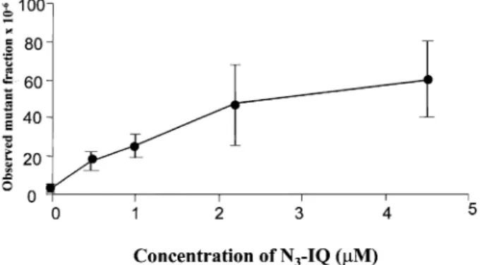
(3) Mutagenicity of IQ in lymphoblastoid cells. Fig. 2. Responses of TK6 cells at the hprt locus following treatment with IQ. Cells were treated in the presence of different concentrations of IQ and 1.5% S9 mix as described in the text. The error bars show the SD of the mutant fractions observed from two independent experiments. The observed mutant fractions in the cultures treated with 25 µM and greater IQ were different (α , 0.05) from those observed in the non-treated cultures.. Fig. 4. DNA adducts found following treatment of TK6 cells with photoactivated N3-IQ. (A) 500 fmol dG-C8–IQ and dGN2-IQ standards. (B) DNA from TK6 cells treated with 0.45 µM N3-IQ. (C) DNA from untreated cells. Fig. 3. Responses of TK6 cells at the hprt locus following treatment with photoactivated N3-IQ as described in the text. N3-IQ was activated by irradiation of the cells in the presence of different concentrations of N3-IQ with ultraviolet light (366 nm). The error bars show the SD of the mutant fractions observed from two independent experiments. The observed mutant fractions in cultures treated with 0.5 µM and greater N3-IQ were different (α , 0.05) from the non-treated cultures.. non-treated cultures were 3310–5 whereas treatments with 25 µM IQ resulted in mutant fractions of 14310–5. Treatment with 8 µM IQ did not result in a significant (α , 0.05) increase in mutant fraction. The cultures treated with 25 µM IQ or higher showed induced mutant fractions that were at least 4-fold higher and different (α , 0.05) than those in nontreated cultures. The response was similar to that observed following treatment of the cells with PhIP (22). Under the same conditions, the two other heterocyclic amines, 4-MeIQ and 4-MeIQx, which have been found to be more active in the Ames test, were not mutagenic and treatment of TK6 cells with up to 200 µM of these compounds did not induce any genotoxic effects (P.M.Leong-Morgenthaler and J.Horlbeck, unpublished data). Responses of TK6 cells to N3-IQ To circumvent the inefficient activation of IQ to its reactive N-acetoxy intermediate, possibly because of the low expression of N,O-acetyltransferase within the cells, we treated the TK6 cells with the photoactivated N3-IQ (18). Azides of arylamines and heterocylic arylamines have been previously reported to chemically modify DNA after photoactivation and result in mutagenesis in S.typhimurium (25,26). Figure 3 shows the response of TK6 cells to N3-IQ following irradiation at 366 nm. Treatment of cells with 0.45 µM N3-IQ activated by irradiation at 366 nm resulted in induced mutant. fractions of 14.4310–5. These induced mutant fractions were 3.5 times higher and significantly different (α , 0.05) than those observed in the control cultures. To obtain similar mutagenic responses, 25 µM of IQ activated by S9 was required, showing that the photoactivated N3-IQ was at least 50-fold more potent than IQ activated by rat liver homogenate (S9). To ensure that TK6 cells were not sensitized to the mutagenicity of N3-IQ by irradiation, cells were also irradiated at 366 nm for 10 min, prior to treatment with IQ and S9 mix. Pre-irradiation of the cells did not increase the mutagenic response of TK6 to IQ activated by S9 mix (results not shown). DNA adduct formation and removal in TK6 cells following treatment with N3-IQ The DNA adducts formed following treatment of TK6 cells with photoactivated N3-IQ were evaluated by 32P-post-labelling (Figure 4). The major lesion was the dG-C8–IQ adduct and it accounted for ~60–80% of the observed post-labelled material. The minor adduct, dG-N2–IQ, was also present, and it accounted for 15–20% of the total radioactivity. Several other adducts were detected, which may be the products of incompletely digested oligomers of dG-C8–IQ (12,18). The ratio of the dG-C8–IQ and the dG-N2–IQ adducts did not change significantly in the cells as a result of the different dose treatments with N3-IQ (Table I). A dose-dependent increase in adducts was observed as a function of N3-IQ treatment, which closely followed the induction of observed mutations (Figure 3). The removal of the adducts was determined after treatment of TK6 cells with 2.2 µM of photoactivated N3-IQ where 50% survival of the cells was observed. Samples were taken at 0, 4, 11, 24 and 51 h post-treatment. There was no significant difference in the rate of the removal of the two adducts, 1751.
(4) P.M.Leong-Morgenthaler et al.. Table I. Following treatment with the different concentrations of N3-IQ activated by irradiation at 366 nM UV, DNA were prepared from the cells and the adduct levels present measured by 32P-post-labelling Concentration of N3-IQ (µM). dG-N2–IQ adduct (310–9) basesa (SD). dG-C8 adduct (310–9) basesa (SD). Total adducts (310–9) basesa (SD). 0.45 2.2 4.5 22. 100 700 1560 5109. 170 1581 4058 21231. 367 3353 8544 35860. a 32P-post-labelling. was performed in triplicate.. (5) (60) (369) (971). (45) (224) (1005) (3901). (68) (281) (2643) (5466). Fig. 5. Removal of adducts following treatment of TK6 cells with 2.2 µM photoactivated N3-IQ. TK6 cells were grown for 0, 4, 11, 24 or 51 h following treatment. DNA was prepared from the cells and the adduct levels measured by 32P-post-labelling as described in the text. The top panel shows the quantity of the two adducts present at the different times. The lower panel shows the ratios of the two adducts at the different times.. showing that there was no preferential removal of either adduct (Figure 5). The adduct removal appeared to be biphasic with an initial higher rate of removal, which led to a loss of 50% of the adducts within 9 h. A slower decrease, which resulted in a loss of ~90% of the adducts after 24 h, was observed following the initial phase. Mutations induced by photoactivated N3-IQ in TK6 cells To gain further insight into the mutagenicity of IQ, we have characterized 44 hprt mutants that were isolated following treatment of TK6 cells with photoactivated N3-IQ. Because more than a third of the mutations following treatment with the heterocyclic aromatic amine, PhIP were found in the exon III of the gene (22), we initially focused on identifying mutations here. Subsequently, nucleotide sequence analysis was performed on most of the cDNA of the hprt gene following amplification of the region by RT–PCR of total RNA from the mutants. Of these 44 mutants, 15 mutations were identified and 10 others were found to be missing an exon (Table II). Further characterizations of these mutants are necessary to determine if they contain splice site mutations or genomic deletions. The remaining 19 mutants that are resistant to 6TG have not been characterized further. They could have attained resistance because of a mutation in their trans membrane protein, which resulted in their inability to take up 6TG or 1752. could have altered expression of the hprt. Southern blot analysis of the DNA of the mutants has not been done and, therefore, we cannot exclude the absence of deletions within that gene. Because RT–PCR with primers within the hprt coding sequence of the total RNA of these mutants produced a fragment of the expected size (data not shown), it is unlikely that they contain large deletions within the coding sequence of the gene. Of the mutations identified, six were located in the exon III. All of these mutations were single base deletions in the runs of six guanine present in the exon. This run of six guanines has been found to be a ‘hotspot’ in the mutations induced by the frameshift mutagen ICR-191, but unlike the mutations observed in the IQ treated populations, those mutations induced by ICR-191 were single base (guanine) additions resulting in 11 frameshifts (23,24). At this position, single base deletions were also induced by another heterocyclic aromatic amine, PhIP (22). Apart from this one mutation, no other hotspots were found in this collection of mutations. Among the mutations observed, several different base substitutions were found. They were mainly transversions, including two GC→TA mutations at positions 568 and 582, two GC→CG mutations at positions 538 and 574 and an AT→TA mutation at position 591. Of the GC→TA mutations observed, the one mutation observed in position 568 was also represented (2/39) in the PhIP-treated mutants. One GC→AT transition at position 599 was found. In the HPRT database, there are 10 entries for this mutation, of which four are derived from spontaneous cultures. With the data available, we cannot conclude whether this mutation is of spontaneous origin or induced by IQ. Among this limited collection of mutants, we found three mutations that were not represented in the hprt database (HPRT Analysis Program 6.0; Mutabase Software, Durham, NC). They were a GC→TA transversion at position 582, which resulted in the amino acid change of an aspartate to glutamate, a AT→TA transversion at position 591, which resulted in the change of a glutamate residue to an aspartate, and a single guanine deletion at position 403, which resulted in a –1 frameshift. Two multiple mutations that included a single base deletion and a deletion of five bases were also observed. Whether all these new mutations represent true IQ-induced mutations, or are of spontaneous origins, needs to be confirmed. Discussion IQ was mutagenic to TK6 cells at concentrations .25 µM following activation with an exogenous metabolizing (S9) mix. The response of these cells to IQ is similar to those observed following treatment with PhIP. Two other heterocyclic amines, 4-MeIQ and 4-MeIQx were not found to be mutagenic under the conditions used. The mutagenic activities of HAAs in these cells do not reflect their potencies as observed in the Ames test, but are consistent with the responses seen in other mammalian cells, including Chinese hamster ovary cells (5). Several studies have shown that the weaker potency observed in mammalian mutation assays is the result of the lack of N,O-acetyltransferases in these systems (7). We observe that treatment of TK6 cells with photoactivated N3-IQ was at least 50-fold more active than treatment with the parent compound following activation by rat liver S9. The increase in mutagenicity is not caused by the sensitization of the cells as a result of irradiation. Because photoactivated N3-IQ does not require metabolizing enzymes for its ultimate activation to its.
(5) Mutagenicity of IQ in lymphoblastoid cells. Table II. Types of mutation observed in photoactivated N3-IQ-treated populations Class of mutations Transition Transversion. GC→AT AT→GC GC→TA GC→CG. Frameshift Multiple mutationsc Putative splice site mutationsd Total. AT→CG AT→TA –1 guanine –1 thymidine and deletion of 5 bp Not identified. Positiona. Sequence contextb. Strand with affected guanine. 599. ACTTCAGGGATTT. NT. 582 568 574 538. CCTTGACTATAAT GTGTTAGGATATG GGATATGCCCTTG TTTGTTGGATTTG. T NT NT NT. 591 207 403 627 and 631. TAATGAATACTTC CTCAAGGGGGG GTGGAAGATATAA TTGGATTTGA and GAAATTCCAGAC. NT NT. No. observed 1 0 1 1 1 1 0 1 6 1 2 10 25. aThe. position refers to the base pair on the coding sequence of the hprt gene. A mutation not present in the HPRT Mutation database (HPRT Analysis program 6.0; Mutabase Softbase) is denoted in bold. bThe mutated base is shown in bold. In the case of a single deletion of a guanine in a string of guanines, all six bases are in bold as it is not possible to distinguish the actual base deleted. cBoth of these mutants were from independent cultures. They both contained a deletion of a T at position 627 and deletion of five base pairs at positions 631–636. dOf these mutants, two were found to be missing exon V, three were missing exon VII and five were missing exon VIII. The actual mutations responsible have not been identified.. reactive metabolite, we conclude that the weak mutagenicity of IQ in TK6 cells is the result of the inefficient formation of the reactive metabolite of IQ within the cell and is probably caused by the lack of N,O-acetyltransferase activity in these cells. Following treatment of the TK6 cells with photoactivated N3IQ, two DNA adducts, the dG-C8–IQ adduct and another one present in lesser quantity (dG-N2–IQ), were found. The DNA adduct profile was similar to that observed following treatment of calf thymus DNA with photoactivated N3-IQ or N-acetoxy– IQ (18). There was a dose-dependent increase in both adducts, which correlated with an increase in mutant fraction; however, the ratio of the two adducts did not change with increasing N3IQ concentration. Under treatment conditions that resulted in 50% survival, ~90% of the adducts were removed after 24 h and there was no preferential removal of either adduct. The lack of preferential adduct removal is in contrast to the slowly dividing tissues of rodents, such as liver and kidney, where the dGC8–IQ adduct was removed more rapidly than the dG-N2–IQ lesion (11). The 15 mutations that were identified in the IQ-treated cultures consisted of single base deletions (7/15) and transversions (5/ 15). Most of the mutations (12/15) identified were at guanines. The single base deletion in the run of six guanines at position 207 was represented six times in the 15 identified mutations. From this observation, we propose that such a sequence is a hotspot for IQ-induced deletions. The same hotspot was also found in PhIP-induced mutants (22). Whether this hotspot mutation is a signature for IQ and PhIP awaits confirmation by the analysis of more mutants using the mutational spectrometry approach (27). Nearest neighbour analysis of the remaining mutations at the guanine residues showed a preference for an adenine (4/6) 59 to the mutated base. However, unlike the PhIPinduced mutations, no preference for a guanine 59 to the mutated base was observed. Because of the small number of mutations analysed, we cannot conclude if this represents a difference in the mutational specificity of the two compounds. In addition to the mutations observed at guanine residues, one. AT→TA transversion and two multiple mutations that resulted in deletion of a thymidine and a 5-adenine-thymidine residue were found. Whether these are IQ-induced or spontaneous mutations are not known. Because the mutants were isolated in a population that had a mutant fraction that was 4fold greater than the background, we could expect ~25% of the characterized mutants to be of spontaneous origins. This would translate to four mutants in our collection. Because most of the characterized mutations (12/15) and the IQ–DNA adduct were found at guanines, the dG–IQ adducts are candidates as the pre-mutagenic lesions. Further studies are needed to determine whether the dG-C8–IQ adduct or the dG-N2–IQ adduct, or both adducts are indeed responsible for the deletions and transversions observed. Acknowledgements We thank Professor W.G.Thilly, MIT, USA for the TK6 cells and the reviewers for their helpful comments. This work was supported by a grant from Nestec SA to P.L.-M. and C.O. and by the Swiss National Science Foundation (grant no. 31-50743.97) to P.L.-M.. References 1. Wakabayashi,K., Nagao,M., Esumi,H. and Sugimura,T. (1992) Food derived mutagens and carcinogens. Cancer Res., 52 (suppl.), 2092s–2098s. 2. Yamashita,M., Wakabayashi,K., Nagao,M., Sato,S., Yamaizumi,Z., Takahashi,M., Kinae,N., Tomita,I. and Sugimura,T. (1986) Detection of 2amino-3-methylimidazo[4,5-f]quinoline in cigarette smoke condensate. Jpn. J. Cancer Res., 77, 419–422. 3. Ohgaki,H., Hasegawa,H., Kato,T., Suenaga,M., Ubukata,M., Sato,S., Takayama,S. and Sugimura,T. (1986) Carcinogenicity in mice and rats of heterocyclic amines in cooked foods. Environ. Health Perspect., 67, 129– 134. 4. Thorgeirsson,U.P., Snyderwine,E.G., Gomez,D.E. and Adamson,R.H. (1996) Dietary heterocyclic amines as potential human carcinogens: experimental data from non-human primates. In vivo, 10, 145–152. 5. Thompson,L.H, Carrano,A.V., Salazar,E., Felton,J.S. and Hatch,F.T. (1983) Comparative genotoxic effects of the cooked-food related mutagens Trp-P2 and IQ in bacteria and cultured mammalian cells. Mutat. Res., 117, 243–257. 6. Thompson,L.H., Wu,R.W. and Felton,J.S. (1991) Introduction of cytochrome P450IA2 metabolic capability into cell lines genetically matched for DNA repair proficiency/deficiency. Proc. Natl Acad. Sci. USA, 88, 3827–3831.. 1753.
(6) P.M.Leong-Morgenthaler et al. 7. Yanagawa,Y., Sawada,M., Deguchi,T., Gonzalez,F.J. and Kamataki,T. (1994) Stable expression of human CYP1A2 and N-acetyltransferases in Chinese hamster CHL cells: mutagenic activation of 2-amino-3-methylimidazo[4,5f]quinoline and 2-amino-3,8-dimethylimidazo[4,5-f]quinoxaline. Cancer Res., 54, 3422–3427. 8. Thompson,L.H., Wu,R.W. and Felton,J.S. (1995) Genetically modified Chinese hamster ovary (CHO) cells for studying the genotoxicity of heterocyclic amines from cooked foods. Toxicol. Lett., 82–83, 883–889. 9. Snyderwine,E.G., Roller,P.P., Adamson,R.H., Sato,S. and Thorgeirsson,S.S. (1988) Reaction of N-hydroxylamine and N-acetoxy derivatives of 2-amino3-methylimidazolo[4,5-f]quinoline with DNA. Synthesis and identification of N-(deoxyguanosin-8-yl)–IQ. Carcinogenesis, 9, 1061–1065. 10. Turesky,R.J., Rossi,S.C., Welti,D.H., Lay,J.O. Jr and Kadlubar,F.F. (1992) Characterization of DNA adducts formed in vitro by reaction of N-hydroxy2-amino-3-methylimidazo[4,5-f]quinoline and N-hydroxy-2-amino-3,8dimethylimidazo[4,5-f]quinoxaline at the C-8 and N2 atoms of guanine. Chem. Res. Toxicol., 5, 479–490. 11. Turesky,R.J., Markovic,J. and Aeschlimann,J.M. (1996) Formation and differential removal of C-8 and N2-guanine adducts of the food carcinogen 2-amino-3-methylimidazo[4,5-f]quinoline in the liver, kidney and colorectum of the rat. Chem. Res. Toxicol., 9, 397–402. 12. Turesky,R.J., Gremaud,E., Markovic,J. and Snyderwine,E.G. (1996) DNA adduct formation of the food-derived mutagen 2-amino-3methylimidazo[4,5-f]quinoline in non-human primates undergoing carcinogen bioassay. Chem. Res. Toxicol., 9, 403–408. 13. Endo,H., Schut,H.A. and Snyderwine,E.G. (1995) Distribution of the DNA adducts of 2-amino-3-methylimidazo[4,5-f]quinoline and 2-amino-1methyl-6-phenylimidazo[4,5-b]pyridine in the supF gene as determined by polymerase arrest assay. Mol. Carcinogen., 14, 198–204. 14. Fruscoe,J.C., Wu,R., Shen,N.H., Healy,S.K. and Felton,J.S. (1988) Basechange analysis of revertants of the hisD3052 allele in Salmonella typhimurium. Mutat. Res., 201, 241–251. 15. Marwood,T.M., Meyer,D. and Josephy,P.D. (1995) Escherichia coli lacZ strains engineered for detection of frameshift mutations induced by aromatic amines and nitroaromatic compounds. Carcinogenesis, 16, 2037–2043. 16. Endo,H., Schut,H.A. and Snyderwine,E.G. (1994) Mutagenic specificity of 2-amino-3-methylimidazo[4,5-f]quinoline and 2-amino-1-methyl-6phenylimidazo[4,5-b]pyridine in the supF shuttle vector system. Cancer Res., 54, 3745–3751. 17. Lee,H. and Shih,M.K. (1995) Mutational specificity of 2-amino-3methylimidazo-[4,5-f]quinoline in the hprt locus of CHO-K1 cells. Mol. Carcinogen., 13, 122–127. 18. Turesky,R.J. and Markovic,J. (1994) DNA adduct formation of the food carcinogen 2-amino-3-methylimidazo[4,5-f]quinoline at the C8 and N2 atoms of guanine. Chem. Res. Toxicol., 7, 752–761. 19. Skopek,T.R., Liber,H.L., Penman,B.W. and Thilly,W.G. (1978) Isolation of a human lymphoblastoid line heterozygous at the thymidine kinase locus: possibility for a rapid human cell mutation assay. Biochem. Biophys. Res. Commun., 84, 411–416. 20. Liber,H.L. and Thilly,W.G. (1982) Mutation assay at the thymidine kinase locus in diploid human lymphoblasts. Mutat. Res., 94, 467–485. 21. Furth,E.E., Thilly,W.G., Penman,B.W., Liber,H.L. and Rand,W.M. (1981) Quantitative assay for mutation in diploid human lymphoblasts using microtiter plates. Anal. Biochem., 110, 1–8. 22. Leong-Morgenthaler,P.M. and Holzha¨user,D. (1995) Analysis of mutations induced by 2-amino-1-methyl-6-phenylimidazo[4,5-b]pyridine (PhIP) in human lymphoblastoid cells. Carcinogenesis, 16, 713–718. 23. Cariello,N.F., Keohavong,P., Kat,A.G. and Thilly,W.G. (1990) Molecular analysis of complex human cell populations: mutational spectra of MNNG and ICR-191. Mutat. Res., 231, 165–176. 24. Taft,S.A., Liber,H.L. and Skopek,T.R. (1994) Mutational spectrum of ICR191 at the hprt locus in human lymphoblastoid cells. Environ. Mol. Mutagen., 23, 96–100. 25. Wild,D., Dirr,A., Fasshauer,I. and Henschler,D. (1989) Photolysis of arylazides and generation of highly eletrophilic DNA-binding and mutagenic intermediates. Carcinogenesis, 10, 335–334. 26. Wild,D., Watkins,B.E. and Vanderlann,M. (1991) Azido- and nitro-PhIP, relatives of the heterocyclic arylamine and food mutagen PhIP-mechanism of their mutagenicity in Salmonella. Carcinogenesis, 12, 1091–1096. 27. Khrapko,K., Andre,P., Cha,R., Hu,G. and Thilly,W.G. (1994) Mutational spectrometry: means and ends. Progr. Nucl. Acid Res. Mol. Biol., 49, 285– 312. Received on November 4, 1997; revised on June 22, 1998; accepted on June 29, 1998. 1754.
(7)
Figure


Documents relatifs
(Bruxelles) Bruxelles Sous la pluie Bruxelles Infinie Tu décolles à jamais Tu décolles à regret Zaventem Terminal des envies aériennes Zaventem Terminal des envies aériennes Je
While the total time of employees absent (e.g., sick leave) as a percentage of the total working time stayed constant for 2004 and 2009, fluctuation (employee turnover on site
Calculez le perim` etre et l’aire de chaque triangle.. Perim`etre et Aire d’un Triangle
Aparece assim a necessidade de uma educação ambiental de caracter integral que promova o conhecimento dos problemas do meio ambiente natural e social no seu
Biofeedback-Enhanced Interactive Computer Play to Improve Hand Function in Youth with Cerebral Palsy
occupational therapist: Box and Blocks, Assisting Hand Assessment and COPM Goals. As in single-case design, all participants allocated to same intervention.. All other control
Original Code (Binary) Original Printed Code Print & Scan Decoding Print & Scan Copied Code (Binary) Authentication Test Opponent Copied Printed Code Receiver X N Y N ˆ X N
The observed cumulative irradiations -UVB, UVA and total solar irradiation (TSI)- over the course of measurement for three stations -a northern (Ostende), central (Uccle) and
