HAL Id: tel-01674230
https://tel.archives-ouvertes.fr/tel-01674230v2
Submitted on 8 Mar 2018HAL is a multi-disciplinary open access
archive for the deposit and dissemination of sci-entific research documents, whether they are pub-lished or not. The documents may come from teaching and research institutions in France or abroad, or from public or private research centers.
L’archive ouverte pluridisciplinaire HAL, est destinée au dépôt et à la diffusion de documents scientifiques de niveau recherche, publiés ou non, émanant des établissements d’enseignement et de recherche français ou étrangers, des laboratoires publics ou privés.
The sleeping brain at work : perceptual processing and
learning in human sleep Thomas Andrillon
Thomas Andrillon
To cite this version:
Thomas Andrillon. The sleeping brain at work : perceptual processing and learning in human sleep Thomas Andrillon. Cognitive Sciences. Université Paris sciences et lettres, 2016. English. �NNT : 2016PSLEE004�. �tel-01674230v2�
Paris Sciences et Lettres!–
PSL Research University
préparée à
l’École normale supérieure
L
E CERVEAU DORMANT AU TRAVAIL:
traitement et apprentissage
perceptifs durant le sommeil
chez l’Homme
par Thomas Andrillon
Ecole doctorale n°158
Spécialité : Neurosciences
Soutenue le 14 Avril 2016
Composition du Jury : M. Shihab SHAMMA
Ecole Normale Supérieure Président du jury M. Jan BORN Université de Tübingen Rapporteur M. Marcello MASSIMINI Université de Milan Rapporteur
Mme. Hélène BASTUJI
Université de Lyon Membre du jury M. Karim BENCHENANE ESPCI Membre du jury M. Sid KOUIDER
Ecole Normale Supérieure Directeur de thèse
École Normale Supérieure
École Doctorale Cerveau Cognition Comportement
Laboratoire de Sciences Cognitives et Psycholinguistique Équipe Cerveau et Conscience
Le cerveau dormant au travail :
traitement et apprentissage perceptifs
durant le sommeil chez l’Homme
Thomas Andrillon
Thèse de doctorat en Neurosciences
Sous la direction de Sid Kouider
Présentée et soutenue publiquement le 14 Avril 2016
Devant un jury composé de :
Marcello MASSIMINI
Université de Milan
Rapporteur
Jan BORN
Université de Tübingen
Rapporteur
Hélène BASTUJI
Université de Lyon
Examinatrice
Karim BENCHENANE
ESPCI
Examinateur
Shihab SHAMMA
ENS
Examinateur
Sid KOUIDER
ENS
Directeur
École Normale Supérieure
École Doctorale Cerveau Cognition Comportement
Laboratory of Cognitive Sciences and Psycholinguistics Brain and Consciousness Team
The sleeping brain at work:
perceptual processing and learning
in human sleep
Thomas Andrillon
Dissertation for the degree of Doctor in Philosophy
Under the supervision of Sid Kouider
Publicly defended on April 14
th, 2016
Before the Doctoral Committee composed of:
Marcello MASSIMINI
University of Milan
Referee
Jan BORN
University of Tubingen
Referee
Hélène BASTUJI
University of Lyon
Examiner
Karim BENCHENANE
ESPCI
Examiner
Shihab SHAMMA
ENS
Examiner
Sid KOUIDER
ENS
Supervisor
! !
!
! À!Sophie,!à!Julien,! à!ces!êtres!aimés! dont!les!voix!se!sont!tues.!
! !
« Je ne cesserai jamais de m’émerveiller que cette chair soutenue par ses
vertèbres, ce tronc joint à la tête par l’isthme du coup et disposant autour de
lui symétriquement ses membres, contiennent, et peut-être produisent un esprit
qui tire parti de mes yeux pour voir et de mes mouvements pour palper… J’en
sais les limites, et que le temps lui manquera pour aller plus loin, et la force, si
par hasard lui était accordé le temps. Mais il est, et, en ce moment, il est Celui
qui Est. Je sais qu’il se trompe, erre, interprète souvent à tort les leçons que lui
dispense le monde, mais je sais aussi qu’il a en lui de quoi connaître et parfois
rectifier ses propres erreurs. J’ai parcouru au moins une partie de cette boule
où nous sommes ; j’ai étudié le point de fusion des métaux et la génération des
plantes ; j’ai observé les astres et examiné l’intérieur des corps. Je suis
capable d’extraire de ce tison que je soulève la notion de poids et de ces
flammes la notion de chaleur. Je sais que je ne sais pas ce que je ne sais pas ;
j’envie ceux qui sauront d’avantage, mais je sais qu’ils auront tout comme moi
à mesurer, peser, déduire et se méfier des déductions produites, faire dans le
faux la part du vrai et tenir compte dans le vrai de l’éternelle admixtion du
faux. Je ne me suis jamais entêté à une idée par crainte du désarroi où je
tomberais sans elle. Je n’ai jamais assaisonnée un fait vrai à la sauce du
mensonge, pour m’en rendre à moi-même la digestion plus facile. Je n’ai
jamais déformé les vues de l’adversaire pour en avoir plus aisément raison
(…). Ou plutôt si : je me suis surpris à le faire, et me suis chaque fois
réprimandé (…). J’ai rêvé mes songes ; je ne les tiens pas pour autre chose que
des songes. Je me suis gardé de faire de la vérité une idole, préférant lui
laisser son nom plus humble d’exactitude. Mes triomphes et mes dangers ne
sont pas ceux qu’on pense ; il y a d’autres gloires que la gloire et d’autres
bûchers que le bûcher. J’ai presque réussi à me défier des mots.
Je mourrai un peu moins sot que je ne suis né. »
RÉSUMÉ...I! SUMMARY...III! ACKNOWLEDGEMENTS...X! GENERAL&INTRODUCTION...1! WHAT!IS!SLEEP?! 2! Sleep!is!ubiquitous... 2! Sleep!as!a!state... 3! Sleep!as!a!process... 4! The!function(s)!of!sleep... 5! THE!PHYSIOLOGY!OF!SLEEP! 8! NREM!and!REM!sleep ... 8! The!hypnogram!and!the!structure!of!sleep...10! Macro!and!micro!dynamics!of!NREM!sleep...10! NREM!sleep!spatial!dynamics ...12! The!REM!sleep!paradox ...14! THE!SLEEPING!BRAIN!IS!NOT!DORMANT! 15! Sleep!and!phenomenology ...15! Sleep!and!its!reversibility ...16! Are!sleepers!isolated!from!their!environment? ...17! Can!sleepers!learn!while!they!sleep? ...20! SLEEP!AS!A!TOOL! 22! The!study!of!consciousness!and!the!contrastive!approach...22! Studying!a!brain!in!a!vat?...24! Exploring!memory!through!sleep ...25! CHAPTER&1:&THE&RICHNESS&OF&SLEEP... 29!
STUDY&1&:®IONAL&SLOW&WAVES&AND&SPINDLES&IN&HUMAN&SLEEP... 31!
SUMMARY! 32! INTRODUCTION! 33! RESULTS! 35! DISCUSSION! 49! EXPERIMENTAL!PROCEDURES! 55! SUPPLEMENTAL!DATA! 57! STUDY&2&:&SLEEP&SPINDLES&IN&HUMANS:&INSIGHTS&FROM&INTRACRANIAL&EEG&AND&UNIT& RECORDINGS... 75! SUMMARY! 76! INTRODUCTION! 77! MATERIALS!AND!METHODS! 78! RESULTS! 84! DISCUSSION! 97! SUPPLEMENTAL!FIGURES! 101! STUDY&3&:&SINGLEJNEURON&ACTIVITY&AND&EYE&MOVEMENTS&DURING&HUMAN&REM&SLEEP&AND& AWAKE&VISION...109!
SUMMARY! 110!
INTRODUCTION! 111!
RESULTS! 112!
!
SUPPLEMENTAL!METHODS! 127!
SUPPLEMENTAL!NOTE! 134!
SUPPLEMENTAL!FIGURES! 137!
CHAPTER&2:&PROCESSING&SENSORY&INFORMATION&DURING&SLEEP...143!
STUDY&4&:&INDUCING&TASKJRELEVANT&RESPONSES&TO&SPEECH&IN&THE&SLEEPING&BRAIN...145!
SUMMARY! 146!
RESULTS! 147!
DISCUSSION! 155!
SUPPLEMENTAL!INFORMATION! 157!
STUDY&5&:&NEURAL&MARKERS&OF&RESPONSIVENESS&TO&THE&ENVIRONMENT&IN&HUMAN&SLEEP....167!
SUMMARY! 168!
INTRODUCTION! 169!
MATERIAL!AND!METHODS! 171!
RESULTS! 177!
DISCUSSION! 188!
STUDY&6&:&ATTENTIONAL&TRACKING&OF&RELEVANT&SIGNALS&DURING&HUMAN&SLEEP...193!
SUMMARY! 194! INTRODUCTION! 195! METHODS! 197! RESULTS! 205! DISCUSSION! 213! SUPPLEMENTAL!FIGURES! 217! CHAPTER&3:&LEARNING&DURING&SLEEP...223!
STUDY&7&:&IMPLICIT&MEMORY&FOR&WORDS&HEARD&DURING&SLEEP...225!
SUMMARY! 226!
INTRODUCTION! 227!
MATERIAL!AND!METHODS! 229!
RESULTS! 236!
DISCUSSION! 243!
STUDY&8&:&PERCEPTUAL&LEARNING&OF&ACOUSTIC&NOISE&GENERATES&MEMORYJEVOKED&
POTENTIALS...247! SUMMARY! 248! RESULTS! 249! DISCUSSION! 258! EXPERIMENTAL!PROCEDURE! 259! SUPPLEMENTAL!INFORMATION! 260! SUPPLEMENTAL!FIGURES! 271!
STUDY&9&:&FORMATION&AND&SUPPRESSION&OF&NOVEL&ACOUSTIC&MEMORIES&IN&HUMAN&SLEEP...277!
SUMMARY! 278!
INTRODUCTION! 279!
RESULTS! 281!
DISCUSSION! 294!
EXPERIMENTAL!PROCEDURE! 298!
SUPPLEMENTAL!EXPERIMENTAL!PROCEDURES! 301!
GENERAL&DISCUSSION...319!
LOCAL!SLEEP!AND!ITS!FUNCTIONAL!CONSEQUENCES! 321!
Slow]waves:!the!hallmark!of!NREM!sleep!or!neuronal!fatigue? ... 321!
From!local!to!global!or!from!global!to!local? ... 322!
GATING!SENSORY!INFORMATION!DURING!SLEEP! 326! Breaching!the!thalamic!gate ... 326! Neuronal!bi]stability!impairs!information!processing ... 327! Dreams!gate!sensory!information!in!REM!sleep?... 329! Dreams!as!a!hallucination!of!reality?... 331! SLEEP,!MEMORY!AND!PLASTICITY! 333! The!role!of!sleep!in!memory... 333! Sleep!and!the!preservation!from!interference ... 334! Active!replay… ... 335! …!or!general!down]scaling? ... 337! Sleep!and!memory:!a!synthesis ... 339! Manipulating!memory!during!sleep... 343! CONCLUDING&REMARKS ...344! ANNEXES...345!
ANNEX&A:&BRAINS&CAN&MAKE&DECISIONS&WHILE&WE&SLEEP&–&HERE&THEY&ARE&IN&ACTION...346!
ANNEX&B:&PENDANT&QUE&NOUS&DORMONS,&NOTRE&CERVEAU&TRAVAILLE...348!
ANNEX&C:&LES&MOUVEMENTS&OCULAIRES&PENDANT&LE&SOMMEIL&:&UNE&FENETRE&SUR&LES&REVES&? ...351!
Résumé
Tous les soirs, nous nous endormons; tous les matins, nous nous réveillons. De ce qui advient entre temps nous gardons peu de souvenirs. Les personnes qui nous entourent pourraient nous dire que nous avons bougé, parlé, ri ou crié, que les émotions les plus vives ont pris le contrôle de notre corps sans pour autant avoir laissé le moindre souvenir. Ou encore, les personnes qui nous entourent ont pu bouger, parler, rire ou crier sans que nous nous en rendîmes compte le moins du monde. Ou au contraire, nous pouvons émerger de la plus fantastique des aventures dans un lit pourtant bien calme, bercé par le calme tic-tac de l’horloge. Il semble que le sommeil opère une dissociation complète entre ce qui arrive dans notre environnement immédiat et dans notre esprit, sans pour autant que la chose éveille en nous la moindre alarme. À tout moment qui plus est, nous pouvons nous réveiller et reprendre conscience de notre environnement de façon quasi instantanée. Curieusement, il semble que certains sons aient une plus grande facilité à nous réveiller que d’autres.
Sommes-nous donc complètement déconnectés de notre environnement quand nous dormons ? Dans les années 60, David Formby et col. montrèrent que la pertinence d’un son importe en effet, en observant que les jeunes mères se réveillent plus aisément en entendant le cri de leur propre enfant plutôt que celui d’un autre. Plus tard, Hélène Bastuji et col. démontrèrent que les traitements sensoriels peuvent être préservés pendant le sommeil sans pour autant nécessairement conduire à un réveil. Cette capacité à analyser les informations venant du monde extérieur pendant le sommeil pourrait s’appuyer sur un phénomène appelé ‘sommeil local’ : le fait que certaines régions cérébrales puissent être réveillées dans un cerveau globalement endormi. Dans le Chapitre 1, je présenterai quelques études explorant la physiologie du sommeil et montrant que le sommeil est un phénomène plus local que la description qui est en classiquement faite. À la lumière de ces travaux, je soulignerai la nécessité de considérer le sommeil à des échelles spatiales (Études 1 et 2) et temporelles (Étude 3) plus précises.
Au cours de cette thèse, j’ai également exploré les conséquences fonctionnelles de ce sommeil local en étudiant notamment jusqu’à quel point les processus cognitifs sont préservés pendant le sommeil naturel chez l’homme. Notre approche est plutôt simple : les personnes participant à nos expériences devaient catégoriser des sons (des mots, des phrases, des bruits) pendant qu’elles étaient éveillées. Une fois que l’exercice était devenu automatique, ces participants étaient invités à le poursuivre confortablement allongés dans l’obscurité. La plupart du temps, ceux-ci se sont endormis en écoutant les sons. Nous enregistrions en parallèle l’activité cérébrale au moyen d’un électroencéphalogramme et avons utilisé ces enregistrements afin de déterminer d’une part si les individus dormaient et d’autre part s’ils continuaient à catégoriser les stimuli.
ii!
En faisant varier les instructions données ou la nature des sons, nous avons pu déterminer si ces dormeurs pouvaient extraire des informations complexes à partir des entrées auditives et si ces informations pouvaient être utilisées pour prendre des décisions ou pour être apprises.
Dans le chapitre 2, je détaillerai nos efforts consistant à examiner quels traitements sont possibles pendant le sommeil. En utilisant l’approche évoquée ci-dessus, nous avons pu montrer que les dormeurs peuvent traiter une information auditive à un haut niveau de représentation (niveau sémantique ou lexical, Études 4 et 5). En outre, nos données suggèrent que cette information peut être envoyée vers d’autres régions cérébrales et permettre des prises de décision. Je décrierai également les mécanismes permettant au cerveau de traiter ces informations extérieures ou au contraire de s’en isoler (Étude 5). Enfin, je monterai comment les dormeurs peuvent non seulement traiter de façon complexe une information pendant le sommeil mais aussi sélectionner une source d’information en particulier quand deux sont présentées en compétition (Étude 6).
Le cerveau éveillé apprend sans cesse et le simple fait d’user de ses sens peut conduire à la formation de nouveaux souvenirs, qu’ils soient implicites ou explicites, grossiers ou élaborés. De semblables traces mnésiques apparaissent-elles quand le cerveau dormant traite les informations extérieures ? Le Chapitre 3 se concentrera sur cette question. Dans une première étude (Étude 7) nous avons obtenu d’encourageant résultats : traiter une information même endormi peut conduire à l’établissement d’un souvenir implicite. Afin d’approfondir ce premier résultat ainsi que les mécanismes sous-jacents, nous avons étudié la capacité du cerveau dormant à former de nouvelles représentations (Études 8 et 9). Je présenterai ici des données expérimentales montrant que le cerveau endormi peut à la fois créer et supprimer de nouvelles traces mnésiques en fonction du stade de sommeil. Ce travail nous a en outre permis de formuler une synthèse du rôle du sommeil dans la mémoire, notamment quant à la fonction bien établie du sommeil dans la consolidation des souvenirs.
En résumé, au cours de mon doctorat j’ai essayé de rendre compte de la richesse du sommeil. Bien loin de l’inactivité, le sommeil est un équilibre dynamique dans lequel les informations extérieures peuvent être traitées, permettant au cerveau de décider s’il vaut mieux continuer à dormir ou se réveiller. L’aphorisme d’Héraclite, vieux de plus de 2000 ans, trouve ici une modeste confirmation:
“Même une âme plongée dans le sommeil s’attèle au travail et aide
Summary
Every night we fall asleep and every morning we wake up. From what happens in the meantime, little is remembered. Others may say that we have moved, talked, laughed or cried, that the strongest and most vivid emotions took control of our body without leaving the faintest memory behind. Or others may have moved, talked, laughed or cried without our slightest notice. On the contrary, we can emerge from the most fantastic adventure in a quiet bed, cradled by a peaceable ticking clock. Without causing us much alarm, it seems that sleep entails a dissociation between what happens in our environment and within our mind. Yet, at any moment, we can wake up and immediately regain consciousness of the surrounding world. Interestingly, it seems that certain sounds are more likely to awake us than others.
Thus, are we completely disconnected from our environment when we sleep? Indeed, loudness is not the only criterion that determines whether a sound will wake up a sleeper. In the 60s, David Formby and colleagues showed that relevance matters when observing that young mothers wake up more easily to the cry of their own child. Later, Hélène Bastuji and colleagues demonstrated that sensory processing can be preserved in sleep without necessarily leading to an awakening. The maintenance of some ability to monitor the external world while being sleep could rely on a phenomenon called local sleep, i.e. the fact that some brain regions could be awake in a globally sleeping brain. In Chapter 1, I will present several studies exploring sleep physiology and showing that sleep is a more local phenomenon than usually described. In the light of such evidence, I will stress the need to consider sleep at refined spatial (Studies 1 and 2) and temporal (Study 3) scales.
During my doctoral work, I also explored the functional consequence of local sleep and the extent to which cognitive processes are preserved during natural human sleep. Our approach was rather simple: participants to our experiments were asked to discriminate acoustic stimuli (e.g. words, sentences, sounds…) while awake. Once the task at hand had been automated, participants were invited to continue it while comfortably laying in a dark room. Most of the time, they fell asleep while listening to our stimuli. We recorded neural activity with scalp electroencephalograpy and used it to assess both the vigilance of participants and their ability to discriminate the stimuli. By varying the task instructions and the nature of stimuli, we could probe whether sleepers can extract
iv!
complex information from the acoustic signal and, whether this information can be used to make decisions or for learning.
In Chapter 2, I will detail our efforts to examine to which extent external information can be processed during sleep. Using the approach detailed above, we showed that sleepers can access acoustic information at a rather high level of representation (i.e. semantic or lexical level; Studies 4 and 5). Importantly, we showed that this information can be routed to other brain areas up to the decision level. I will also describe the mechanisms allowing the sleeping brain to process or on the contrary inhibit sensory information (Study 5). Finally, I will show that sleepers can not only process complex information but also that sleepers can select and lean toward one source of information when two sources are presented in competition (Study 6).
The awake brain is constantly learning and the fact of experiencing almost inevitably leads to the establishment of memory traces, being it explicit or implicit, strong or fine. Are similar memories formed when the sleeping brain processes external information? This question will be the focus of Chapter 3. Our initial effort (Study 7) led to encouraging observations: processing information while asleep may allow the formation of implicit mnesic traces. To better explore this question and the underlying mechanisms, we investigated whether a new representation can be created during sleep (Studies 8 and 9). I report here evidence that the sleeping brain can both establish and suppress novel mnesic traces depending on sleep stages. This work allowed us to formulate an integrative view of the role of sleep in memory and especially the well-established function of sleep regarding memory consolidation.
In summary, in the course of my Ph.D., I tried to better depict the richness of sleep. Far from being an idle state, sleep is a dynamic equilibrium in which the environment can be monitored, allowing the brain to decide whether to stay asleep or to wake up. Heraclitus’ aphorism stated more than 2000 years ago finds here a modest corroboration:
“Even a soul submerged in sleep is hard at work and helps make something of the world”.
Peer-Reviewd Publications:&
Sleep Spindles in Humans: Insights from Intracranial EEG and Unit Recordings
Andrillon* T., Nir* Y., Staba RJ., Ferrarelli F., Cirelli C., Tononi G., Fried I. The Journal of Neuroscience (2011)
Regional Slow Waves and Spindles in Human Sleep
Nir Y., Staba RJ., Andrillon T., Vyazovskiy VV., Cirelli C., Fried I., Tononi G. Neuron (2011)
Inducing Task-Relevant Responses to Speech in the Sleeping Brain
Kouider S., Andrillon T., Barbosa L., Goupil L. & Bekinschtein T. Current Biology (2014)
Single-Neuron Activity and Eye Movements during Human REM Sleep and Awake Vision
Andrillon* T., Nir* Y., Cirelli C., Tononi G. & Fried I.
Nature Communications (2015)
Perceptual Learning of Acoustic Noise Generates Memory-Evoked Potentials
Andrillon T., Kouider S., Agus T. & Pressnitzer D. Current Biology (2015)!!
Submitted or Under-Review:
Formation and Suppression of Acoustic Memories in Human Sleep
Andrillon T., Pressnitzer D., Léger D. & Kouider S.
Implicit Memory for Words Heard during Sleep
Andrillon T. & Kouider, S.
Neural Markers of Responsiveness to the Environment in Human Sleep
Andrillon T., Poulsen AT.; Hansen LK., Léger D. & Kouider S.
Napping: a Public Health Issue!
Faraut B., Andrillon T., Vecchierini MF. & Léger D.
Selective Neuronal Fatigue Precedes Human Cognitive Lapses
!
Nir Y., Andrillon T., Suthana N., Cirelli C., Tononi* G., and Fried* I.
In preparation:
Attentional Tracking of Relevant Signals during Human Sleep
Legendre* G., Andrillon* T. & Kouider S.
vi!
Keywords
!and acronyms
NREM Non-Rapid Eye-Movement REM Rapid Eye-Movement SW Slow-Wave
SWS Slow-Wave Sleep SWA Slow-Wave Activity ACh Acetylcholine
STDP Spike-Timing Dependent Plasticity EEG Electroencephalogramphy
EOG Electroocculography EMG Electromyography MEG Magnetoencephalography LFP Local Field Potentials MUA Multi-Unit Activity SUA Single-Unit Activity
TMS Transcranial Magnetic Stimulation fMRI Functional Magnetic Resonance Imaging ERP Event-Related Potential
AEP Auditory-Evoked Potential LRP Lateralized Readiness Potential ITPC Inter-Trial Phase Coherence FFT Fast Fourier Transform SEM Standard Error of the Mean FDR False Discovery Rate
Main figures:
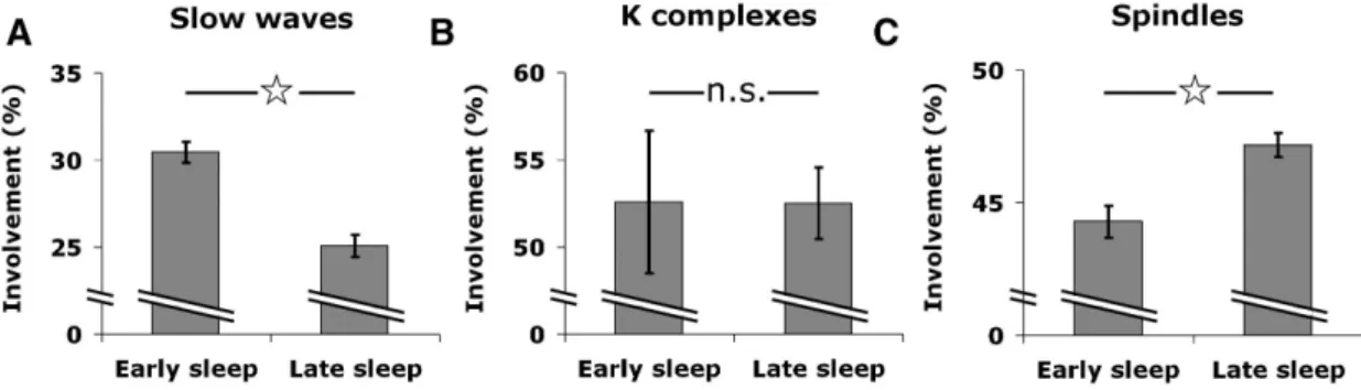
Figure 0-1 Presence of NREM and REM sleep across Vertebrates...3! Figure 0-2 Road sign in Australia ...4! Figure 0-3 Electrical activity of the brain across wake and sleep ...9! Figure 0-4 Hypnogram of a healthy young individual ... 10! Figure 0-5 Slow-waves and neuronal silencing ... 12! Figure 0-6 Sleep as a modulation of conscious contents and levels ... 24! Figure 1-1 Sleep studies and data overview ... 35! Figure 1-2 Example of EEG and single-unit activity during global sleep slow waves ... 37! Figure 1-3 Spiking activity underlying EEG slow waves... 38! Figure 1-4 Local sleep slow waves ... 40! Figure 1-5 Local sleep spindles... 42! Figure 1-6 Changes in spatial extent of slow waves and spindles between early and late
sleep ... 44! Figure 1-7 Sleep slow waves propagate across typical paths... 45! Figure 1-8 Afferent Information Predicts Occurrence and Timing of Activity Onsets in
Individual Slow Waves... 47! Figure 2-1 Data overview and spindle detection ... 80! Figure 2-2 Fast centroparietal spindles differ from slow frontal spindles ... 86! Figure 2-3 Fast centroparietal spindles precede slow frontal spindles ... 88! Figure 2-4 Association of spindles with slow wave up-states... 89! Figure 2-5 Local sleep spindles... 91! Figure 2-6 Deep sleep and high SWA are associated with lower spindle frequencies... 93! Figure 2-7 Unit discharges during sleep spindles... 94! Figure 3-1 Data overview and eye movement detection ... 112! Figure 3-2 REM-triggered averaging of neuronal activity... 115! Figure 3-3 Comparison of neuronal activity underlying visual and non-visual REMs ... 117! Figure 3-4 Comparison of transient spike-train properties in REM sleep and wakefulness
... 119! Figure 3-5 Reset of field oscillation phase following REMs and visual stimulation ... 120! Figure 4-1 Experimental paradigm ... 148! Figure 4-2 Motor preparation to semantic categories... 149! Figure 4-3 Motor preparation to lexical categories... 152! Figure 4-4 Memory test upon awakening ... 153! Figure 5-1 Semantic categorization task during full-night sleep... 172! Figure 5-2 Lateralized Readiness Potentials (LRPs) across sleep stages ... 177! Figure 5-3 Lateralized Readiness Potentials (LRPs) in light NREM and REM sleep for
viii!
Figure 5-4 Lempel-Ziv complexity across sleep stages and in relation to motor
preparation indexes ... 180! Figure 5-5 Local modulations of NREM sleep slow-waves and spindles in association to
stimuli ... 182! Figure 5-6 Bi-stability of neuronal responses in NREM-sleep gates sensory processing
... 183! Figure 5-7 Evoked responses to sounds correlate with LRP magnitude in REM sleep. 185! Figure 5-8 The ability to process information is dynamically modulated within sleep
cycles... 187! Figure 6-1 Experimental procedure... 199! Figure 6-2 Reconstruction accuracy and decoding performance across wake and NREM
sleep ... 206! Figure 6-3 Spatio-temporal integration of acoustic information... 209! Figure 6-4 Impact of sleep spindles, K-complexes and slow-waves on stimulus
reconstruction ... 211! Figure 7-1 Experimental procedure and evidences for complex information processing
during sleep... 231! Figure 7-2 First and second-order responses in the recognition test ... 237! Figure 7-3 Event-Related Potentials to wake, sleep and new words ... 239! Figure 7-4 Decoding stimulus category with the EEG signal ... 240! Figure 7-5 Alpha de-synchronization during memory recognition... 242! Figure 8-1 Experimental procedure and behavioral results ... 250! Figure 8-2 Electrophysiological Markers ... 252! Figure 8-3 Correlation of neural markers to behavioral performance for the compact
condition ... 254! Figure 8-4 Model simulations ... 256! Figure 9-1 Noise memory paradigm in wake and sleep... 282! Figure 9-2 Behavioral and electrophysiological indexes of perceptual learning in
wakefulness... 283! Figure 9-3 Brain responses to acoustic noise during sleep... 285! Figure 9-4 Evoked-activity to repeated noise snippets during sleep ... 286! Figure 9-5 Impact of prior exposure on behavioral performance upon awakening ... 288! Figure 9-6 Impact of prior exposure and sleep rhythms on phase coherence upon
awakening... 290! Figure 9-7 The learning index is dynamically correlated with slow-wave power in NREM
sleep ... 291! Figure 9-8 Tonic REM sleep is more favorable to learning than phasic REM sleep... 292! Figure 10-1 Slow-waves, from single neurons to cortical networks ... 321! Figure 10-2 The active consolidation hypothesis ... 336! Figure 10-3 The synaptic homeostasis hypothesis ... 338! Figure 10-4 A synthesis of active consolidation and synaptic homeostasis ... 340!
Table 2-1 Spindle occurrence across multiple regions in the human brain ... 85! Table 3-1 Bi-phasic single-unit modulation around REMs is more prevalent in the MTL
compared to frontal regions... 114! !
x!
Acknowledgements
On the first day of the entrance examination at the Ecole Normale Supérieure (ENS), I remember walking the (for some) mythical rue d’Ulm. On the pavement, footprints painted in a vivid orange headed to the ENS. I saw a sign in them. And indeed, during the 7 past years, I have found there the guidance I needed to become a scientist, and benefited from the counsel of many brilliant researchers and professors.
First and foremost, I want to thank my PhD supervisor: Sid Kouider. Not only Sid taught me what I needed to learn in order to perfect my training in research, but he also did so with the right method: he always took care that I developed a form of independence while always being there to rescue me when I was stuck and despaired. I am profoundly thankful for his care in giving me the impression I was climbing ahead while safeguarding me the whole way. I also had the chance to have a supervisor who always tried to highlight my research through various collaborations with other researchers and the participation to many conferences. Thanks to Sid, I have traveled a lot and met people whom I am sure will be part of my future. But most importantly, Sid set me on fantastic research tracks. I owe him the seeds of this doctoral work. Seeds that I remember considering quite crazy or even impossible at first. Nonetheless, I learnt to trust his instinct and (French) flair and the craziness happened not to be so crazy. After all those years however, I have not penetrated the mystery of Sid’s intuitions. I hope though that Sid and I will continue our work together, even from afar, and that, one day, I would have finally learnt how he manages to shoot for the moon without missing it.
I want also to thank the other mentors who accompanied me during this doctorate. At the ENS, I had the chance to benefit from the expertise of Daniel Pressnitzer, Alain de Cheveigné and Catherine Tallon-Baudry. Daniel, notably, was always there to check how our projects were progressing, with humor and good mood, and I am thankful for the efforts he made to put my work and myself under the spotlight. Within the Laboratoire de Sciences Cognitives et Psycholinguistique (LSCP), I had the chance to be able to reach researches who, even if we did not always share the same academic field, always brought their advises and experience when I needed it. In particular, I wish to thank Jérôme Sackur for his ever-pertinent insights and depth of thoughts. In the first months of my doctorate, I had the chance to meet and start collaborating with Damien Léger at the Hôtel-Dieu Hospital. I am deeply thankful for his genuine interest, solid counsel and unfailing trust. Over the three past years, I also had the opportunity to experience teaching for the first time and it has been a true revelation. I thank the professors who supervised me at the Université Pierre et Marie Curie (Mathilde Grassi) and at the Cogmaster (Sid, Jérôme Sackur and Claire Sergent). Lastly, during my doctoral studies and before, I had the chance to be mentored by dedicated professors (Laurent Bourdieu, Matthias Pessiglione and Anne Christophe) who always toke great care to orientate me in academia’s maze.
Intertwined with the research I conducted in Paris, I had the opportunity to collaborate with researchers abroad and particularly with Chiara Cirelli and Giulio Tononi in Wisconsin, and with Yuval Nir and Itzhak Fried in Tel-Aviv. The time I spent at the University of Wisconsin represents a key moment in my still-young academic career and I will always be in debt with Chiara Cirelli and Giulio Tononi for such opportunity. I will always be admiring their vision of science and I do hope that I will have the chance and honor to interact with them in the future. As for Yuval, I cannot tell how much he taught me about research and life in general, and I am proud to consider him (and be considered) more as a friend than anything else.
I focused so far on the tree of professors who help me, each in their own way, to grow up as a scientist. But I also benefited, beyond all possible expectations, from my comrades in research: the post-doctorate fellows, students or research assistants I had the opportunity to work with. At the LSCP, I had the chance to meet Leonardo Barbosa, who was always there to help me, to teach me and to cheer me up. His knowledge is hard to surpass and my doctorate would have been much harder without him. Next to me, as two horses on the same harness, Alexandre Cremers was a true brother-in-arms and his endless curiosity is one of the things that make me realize the chance I have to spend my time with such colleagues. Yue Sun shared with Alexandre,
to his capacity of adaptation and of always asking the right questions. I am sure he will progress smoothly in his academic dreams. I wish also to thank Louise Goupil, in a very special way. She is the one who started the sleep projects with Sid and she paved the way for my own work. Her humility and tenacity will always serve me as an example. I cannot describe all the reasons why I cherish my present and past colleagues at the LSCP but I hope they will know why they all have a special place in my mind: Nathan Faivre, Gabriel Reyes, Hernan Anllo, Jan Balaguer, Hielke Prins, Guillaume Legendre, Matthieu Koroma, Alya Vlassova, Sofie Gelskov, Hao Zhang, Andreas Poulsen, Lorna Le Stanc, Romain Trachel, Romain Grandchamp, Margaux Romand-Monnier... I want also to thank Michel Dutat, Vireak Ul and Cécile Girard for their assistance regarding my experimental work. Still at the ENS but some floors upstairs, I learnt from Trevor Agus the hidden beauty of white noise. At the Hôtel-Dieu hospital, I was saved many times by Maxime Elbaz and Stéphane Rio. I picked up a lot from their stories of veterans from the electroencephalography. Brice Faraut, Caroline Gauriau and Virginie Bayon have also been important figures during my doctoral work. I am much in debt with the people who helped me there when I was running my sleepless experiments (Livio de Sanctis and Audrey Dalbin) and especially with the ones who sometimes stayed with me the whole night (Hernan Anllo, Chiara Varazzani and Marlène). In Wisconsin, I had the opportunity to share my days with fantastic minds and, by now, great friends: Simone Sarasso and Ugo Faraguna (whose encyclopedic knowledge about Italian bestemie is of great use for me now), Michele Bellesi, Mélanie Boly, Bessie Hung, Stéphanie Maret, Aaron Nelson, Luisa de Vivo and Vlad Vyazovskiy. Back in France, I had the chance to interact with the members of the sleep research community through the VIFASOM group, whose members (and in particular the members of the Institut de Recherche Biomédicale des Armées such as Mounir Chenaoui) have been a second scientific family for me. I thank them for their precious inputs. The Société Française de Recherche et Médecine du Sommeil was also very supportive and I thank them for their interest in my work.
I know that this list is already long and yet not exhaustive. The success of this doctoral work is due to the people who helped me more than anything else. I wish to thank the examiners (Hélène Bastuji, Karim Benchenane, Jan Born, Marcello Massimini and Shihab Shamma) who agreed to evaluate this work and in particular my referees (Jan Born and Marcello Massinimi). Hélène Bastuji together with Lionel Naccache was instrumental in the progress of this work since they saw it building up and counseled me on the way. I wish to thank as well Isabelle Arnulf and Satoru Miyauchi, who shared with me their expertise on REM sleep and helped me in my endeavor in this new territory.
On a more personal level, I am surrounded by great friends, who are sometimes far from the research world but who have been always patient and curious of what I have been doing all these years. Some of them even participated to my experiments, for which I am very grateful. My numerous family (Dominique and Béatrice, my parents; Nicolas, Xavier, Vincent et Laure, my brothers and sister; Anne-Gaëlle, Veridiana, Anne-Laure et Jaime, their consorts; Élise, Louis, Martin, Philippe and Clotilde, my more than fantastic nieces and nephews) was also there for me in the good as in the bad times and I am proud to have grown up in such constellation. Il peut paraître étrange de remercier ses parents pour une modeste these, étant donné que je leur dois tant et plus, néanmoins je le fais avec joie. Une pensée spéciale pour ma grand-mère, qui garde malgré les ans toute sa jeunesse d’esprit. Sono adesso molto fortunato perché ho anche una seconda famiglia (Chiara, Carolina, Erminia e Massimo) che sta molto attenta a me. Spero che sappiano quanto la loro gentilezza sia preziosa per me. E poi, c’è questo topolino che si nasconde nelle pieghe della mia mente, e chi mi accompagna in ogni pensiero.
Finally, I would like to express the honor I had to meet so many bright minds and good friends. Sadly, not all of them are still alive to read these lines. Each in their own way, Julien Fussler and Sophie Thomain, to whom this doctorate is dedicated, are models I hope I will never fail. I have walked past and forth the still-mythical rue d’Ulm more than I can count. Enough to know it, in all its details; enough to have seen it changed, in its own immutable way. On the pavement, the footprints have disappeared. Maybe another sign that now, it is time for me to find my own
General Introduction
!
!
The!Sleeping!Gypsy! Henri!Rousseau!(1897)!
Introduction
2! !
What is sleep?
Sleep is ubiquitous
From our very first cry to our last breath, sleep marks the passage of days. Whatever its potential costs and whatever we think we could achieve in the meantime1, humans organize their lives and
societies around sleep. This centrality of sleep in existence may have inspired Shakespeare when he wrote these immortal verses2:
‘We are such stuff
As dreams are made on, and our little life Is rounded with a sleep.’
In Humans, sleep is characterized by the disappearance of the surrounding world. Aristotle in his rich essay on sleep (Aristotle, 350BC) describes it as a “privation of waking” that is to say the inability to “exercis[e] sense-perception”. Indeed, every night our minds dissolve at times into nothingness at times into a phantasmagoric inner world. Personal experience suggests that we cease to experience our environment while we sleep, since we cannot really tell what happened over these many hours. The direct consequence is that sleepers remain quite unresponsive to external stimulations, just as Gulliver when tied up by the Lilliputs. Such unresponsiveness can be objectively measured, providing a defining criterion for sleep that can be applied to both Humans and animals (Cirelli and Tononi, 2008).
Using the absence of responsiveness to probe sleep, it has been shown that sleep expands much further than the humankind (see Figure 0-1) and phenomena resembling human sleep have been described in all animal species studied so far, from fruit flies to platypuses (Cirelli and Tononi, 2008). Even animals for which sleep represents an ecological disadvantage do sleep. For example, migratory birds must fly for extended periods without landing. In this context, sleeping becomes both difficult to achieve and dangerous to maintain. Down the sea, dolphins need to periodically reach the surface in order to breathe, which implies that they cannot stop swimming in order to sleep. Hence, the ecological niches of these species put a huge pressure on sleep. But sleep remains, in an adapted way. In these species can be found what is called unihemispheric sleep (Rattenborg et al., 2000). Its principle is simple: one cerebral hemisphere only sleeps at a time, letting the other hemisphere deal with the environment and its potential dangers. Thus, even in the most constrained and unfavorable environments, animals do sleep. But how then can sleep be defined so as to take into account all its implementations?
!!!!!!!!!!!!!!!!!!!!!!!!!!!!!!!!!!!!!!!!!!!!!!!!!!!!!!!!
1 Plato for example considered sleeping too much as ‘shameful and unworthy’: ‘A sleeping man does not worth
anything, just as a dead man’ (Plato, Laws, 370-345 BC).
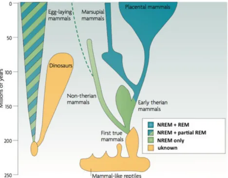
Figure 0-1 Presence of NREM and REM sleep across Vertebrates
All studied animals so far have been shown to sleep (even if some particular cases are still a matter of debate (Cirelli and Tononi, 2008)). But there are important differences across species. While placental mammals like primates or rodents show two types of sleep alternating within a night (NREM and REM sleep, see below), non-therian mammals do not show signs of sleep. Other species show REM-sleep only during the early stage of life. Crucially, NREM REM-sleep is present in all these species. Adapted from (Hobson, 2009).
Sleep as a state
Amongst human beings, the best way to know whether a person is asleep is simply to have a look. A still and curled posture, deep breathing, closed eyelids are well-known signs of sleep. If some doubt persists, asking is enough. An absence of response would probably mean that the person is asleep. Of course, such a definition lacks both specificity and sensitivity and can be hardly transferred to non-human species. But the core idea can be reprieved: sleep is a state in which the responsiveness to external stimuli is greatly diminished.
In 1862, Ernst Kohlschütter made the first detailed description of how sleepers get disconnected from their environment (Kohlschütter, 1862) by measuring the intensity a sound needed to awake a sleeper. This ‘perturbational’ approach that consists in determining the arousal threshold was later used to operationally characterize sleep in humans (Rechtschaffen et al., 1966). A similar approach is recommended to assess sleep in animals (Cirelli and Tononi, 2008). Such measure has the advantage to probe the most striking consequence of sleep at the subjective level in an objective manner. However, it supposes the ability to stimulate an organism in an appropriate way so as to record a behavioral response when the arousal threshold is exceeded. This is not always possible.
Introduction
4!
Let’s take the example of patients with disorders of consciousness (in a vegetative, minimally conscious or locked-in state). These patients cannot always interact with their surrounding environment. How can we possibly assess whether they sleep or not? It is obviously not possible to rely on behavioral markers. Instead, sleep (and wakefulness) can be defined at the neural level. Indeed, with the invention of the EEG in 1924 by Hans Berger (Berger, 1929) came quickly the discovery that the waking and sleeping brain had a very different electrical activity (Loomis et al., 1935). I will detail later the changes occurring during sleep, but the most striking is the replacement of low-amplitude desynchronized rhythms by high-amplitude synchronized oscillations (see Figure 0-3). Similar changes in neo-cortical activity can be observed in other mammals and birds. Nowadays, the EEG is routinely used in hospitals (and soon at home) to accurately monitor sleep without perturbing it.
EEG is also one of the key tools to investigate sleep, its mechanisms and functions. Accordingly, it is the technique I relied on for most of the work presented here. Using EEG, two different types of sleep have been clearly identified in mammals: Rapid Eye-Movement (REM) sleep (also called Paradoxical Sleep) and Non-Rapid Eye-Movement (NREM) sleep (often referred as Slow-Waves Sleep, SWS). They both occur while individuals are lying in a horizontal position, with eyelids closed, a reduced or absent muscle tone and increased arousal thresholds. However, NREM and REM sleep have also many different properties, stressing the variety of states that the term sleep encompasses.
Sleep as a process
Defining sleep as a state, surely, has an undeniable operational efficiency. But from a closer perspective, sleep seems much more than a mere condition. We sleep not necessarily for the enjoyment of it, but for a purpose and this purpose is to be found in our waking life. Indeed sleep has a restorative effect on the mind and body that will persist even after awaking. These long-term effects of sleep are things we look for when, for example, one takes a nap after a short night or because she got tired driving (Figure 0-2). Sleep can be therefore seen as a process: we sleep to realize a specific end. I will review in more details the many benefits of sleeping.
Figure 0-2 Road sign in Australia
Sleep is a process also in the sense that it is not a monolithic condition: sleep is a series of states that are laid out in a precise order. Indeed, our nights are structured in cycles, which vary in
numbers and durations across nights and individuals but share the same core properties (Figure 0-4). Each of these cycles starts with a gradual increase in the intensity and EEG markers of NREM sleep. Sleepers then stabilize in deep NREM sleep during which the brain activity is dominated by sleep slow-waves. Next, NREM sleep suddenly dampens and sleepers transit to a brain activity closer to wakefulness (REM sleep) although they do not wake up. After an episode of REM sleep, a new descent to NREM sleep starts again marking the beginning of a new cycle. Another reason to conceptualize sleep as a process rather than a state is that sleep does not appear randomly but is built upon the preceding wakefulness period. Scientific articles dealing with sleep often mention the term ‘sleep pressure’: the more one stays awake, the more she will get tired and the easier it will be to fall asleep (Borbely and Achermann, 1999). Thus, sleep can sometimes irrepressibly appear at the worst moment possible. Sleep also depends on the time of the day: it is easier to fall asleep right after lunch time (even if we don’t eat) than later in the afternoon even if the time spent awake has increased. These views have been succinctly summed up by Alexander Borbély in his “two process model of sleep” (Borbély, 1982). According to this model, sleep is regulated by two processes: (i) a circadian process which determines for example that we, humans, usually sleep at night while rodents sleep during the day, (ii) an homeostatic process which gradually builds up with time spent awake. These two processes interact in shaping sleep. For example, the proportion of Slow-Waves Sleep (SWS) increases with time spent awake prior to sleep illustrating the built up of the homeostatic pressure (Dijk et al., 1987; Friedman et al., 1979). Reversely, this pressure is dissipated during the night and the proportion of SWS within sleep cycles declines within a night (Riedner et al., 2007). This link between wake activity and sleep intensity is use-dependant and the circuits that have been solicited the most during the wake period get ‘tired’ first and sleep more soundly (Huber et al., 2004; Hung et al., 2013). Sleep can be therefore seen as a process reflecting and compensating our waking life, i.e. the price we pay for being awake (Tononi and Cirelli, 2014).
The function(s) of sleep
Seeing sleep as process comes down to attribute to sleep one or several functions. However, despite the elements briefly presented below, the search for the function of sleep is still one of the biggest challenges of sleep research (Tononi and Cirelli, 2014). Scientists have reach a quite good understanding of what is sleep, how it works and how it is regulated but less regarding its purpose. The problem is not really the lack of candidates but rather the multiplicity of functions that sleep seems to subserve and the difficulty to prove the specificity of sleep in their accomplishment3.
To gather clues about the potential function of sleep, one strategy consists in examining the effect of its absence, i.e. the effects of sleep deprivation. Acute sleep deprivation leads to death after few days, as evidenced in animals (Orzeł-Gryglewska, 2010). Sleep loss affects cognitive performance but also the immune system, triggers hormonal changes, obesity, alters cardio-vascular function. Sleep seems therefore very important to the correct functioning of the !!!!!!!!!!!!!!!!!!!!!!!!!!!!!!!!!!!!!!!!!!!!!!!!!!!!!!!!
3 Concerning this search for a function of sleep, Chiara Cirelli and Giulio Tononi reminded appropriately the words
of the mythical phoenix: “Che vi sia ciascun lo dice, dove sia nessun lo sa” (“that there is one they all say, where it may be no one
Introduction
6!
organism, not only the nervous system. Accordingly, the immune systems has been shown to benefit from SWS (Westermann et al., 2015). Sleep seems also very important for the conservation of energy (Benington and Heller, 1995), the cleansing of metabolic waste in the central nervous system (Xie et al., 2013), and the maintenance of the nervous system at the circuit or neuronal level (Maquet, 1995; Tononi and Cirelli, 2014).
Acute sleep deprivation impairs also a variety of cognitive abilities (Alhola and Polo-Kantola, 2007; Durmer and Dinges, 2005). The spectrum of cognitive processes affected goes from the simple reactivity to unpredicted stimuli to more complex processes such as attention allocation, memory, cognitive control, emotional balance, reasoning, creativity (De Gennaro et al., 2001; Doran et al., 2001; Gevers et al., 2015; Gujar et al., 2011; Harrison and Horne, 2000, 1999; Wimmer et al., 1992). At the neural level, sleep deprivation translates into the intrusion of sleep-like activity within the awake brain (Hung et al., 2013; Vyazovskiy et al., 2011). This has been interpreted as the fact that neurons cannot maintain their level of waking activity for an indeterminate amount of time and when neurons get ‘tired’, they ‘fall’ into sleep (Tononi and Cirelli, 2014; Vyazovskiy and Harris, 2013).
Sleep would thus be a process allowing the brain to recover its optimal functioning in the safest way, i.e., at a given time when the consequences of getting disconnected and unresponsive minimally impair survival. An extensive literature has also assessed the function of sleep in memory (Diekelmann and Born, 2010; P Peigneux et al., 2001; Rasch and Born, 2013; Tononi and Cirelli, 2014) (see also p20-21 and Discussion). Although the exact mechanisms are still unclear (see Chapter 3 and Discussion), sleep has an important role (i) in the consolidation of existing memories, (ii) in the ability to create novel associations between existing memories and (iii) in the ability to form new memories in ensuing wakefulness. The fact that the brain goes offline during sleep could be crucial to fulfill this memory function by suppressing potential external interferences during the consolidation and transfer of memories (Diekelmann and Born, 2010).
Can one or any of these functions be coined as ‘the function of sleep’? To conclude on this issue, scientists must firstly control for the circadian confound: sleep occurring at a certain phase of the circadian cycle, it is therefore easy to confound something co-occurring with sleep for something caused by sleep (Frank and Cantera, 2014). Functions of sleep should be specific to sleep. Among the functions listed above, which ones necessitate to be asleep, e.g. in a state of disconnection from the environment? Metabolic recovery or the consolidation of the immune memory does not need, a priori, such disconnected state. However, it has been argued that the neural homeostasis and memory consolidation functions would match this criterion since they benefit from the brain going offline (Diekelmann and Born, 2010; Tononi and Cirelli, 2014). Thus, according to Allan Hobson, “sleep is of the brain, by the brain and for the brain” (Hobson, 2005).
Another view, championed by Jeremy Siegel advances that there is no real function of sleep (Siegel, 2009). On the contrary sleep can be regarded as an adaptive phenomenon and a simple variant of the dormant state that spans both animal and vegetal kingdoms. In this view, sleep is the mere extreme of a continuum including rest. This argument is based on the failure to find (i) a function that necessitates being asleep and (ii) a function that would be common for all the species known to sleep. This argument also reckons on the variety of sleep properties across species and the absence of obvious boundary between resting and sleeping. While thought provoking, this approach falls short at explaining certain key features of sleep (e.g. a sensory
disconnection far more pronounced than during rest, the existence of REM sleep) and their striking preservation across species and biotopes.
The fact that NREM and REM sleep subserve different functions may account for the difficulty to identify ‘the function of sleep’. Indeed NREM sleep is the part of sleep that is most vital to animals since (i) not all animals experience REM sleep or an equivalent of it (see Figure 0-1), (ii) in Humans, REM sleep can be suppressed for long period of times, using for example anti-depressors (Mayers and Baldwin, 2005), without causing noticeable issues. On the other hand, acute sleep deprivation triggers REM sleep intrusion (animals falling into REM sleep directly from wake) stressing the importance of this state (Orzeł-Gryglewska, 2010).
In conclusion, sleep affects our brain but also our behavior and mind through the brain. As a ‘neuro-apprentice’, I am especially interested in the causal link between changes in brain activity and changes at the behavioral and phenomenological levels. Countless studies have confirmed the interest of such reductionist approach and the tight correlation between the transformation of brain dynamics at sleep onset and the loss of awareness or, on the contrary, its recovery during dreams (Nir and Tononi, 2010; Tononi and Massimini, 2008).
Introduction
8!
The physiology of sleep
As we have seen, sleep is a rich, complex and ubiquitous phenomenon that is hardly reduced to a unique definition due to the variety of its implementations in the animal kingdom. However, sleep has been mostly studied in vertebrates and especially mammals such as cats, rodents and humans. These species present a highly organized central nervous system whose activity follows the same common characteristics (Buzsáki, 2006). Among these shared properties is the impact of sleep on brain activity. I will give here few elements on sleep physiology, which will be recurrently mentioned throughout this manuscript.
NREM and REM sleep
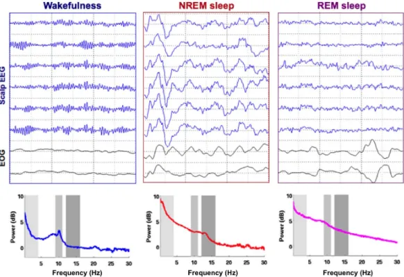
When examining EEG recordings across an entire night, brain activity clearly fluctuates between 3 drastically different states: (i) wakefulness, (ii) NREM sleep and (iii) REM sleep. Figure 0-3 shows representative snapshots of these 3 stages as well as their associated spectral profiles. During wakefulness, the EEG signal is dominated by low-amplitude fast rhythms such as alpha oscillations ([8, 12] Hz). The alpha rhythm is a resting oscillation, increasing when the eyelids are closed, potentially due to the disengagement of the visual cortex (Cantero et al., 2002). Interestingly, alpha oscillations slowly fade with drowsiness and mark the transition with light NREM sleep (NREM sleep stage 1, NREM1). Quickly after the disappearance of alpha oscillations, other rhythms appear, which are specific to NREM sleep. K-complexes for example are the biggest non-pathological brain response that can be recorded from the Human EEG. It consists in an isolated high-amplitude slow-wave ([0.5, 4] Hz) that can be either spontaneous or evoked by an external stimulation (Halasz, 2005). Another hallmark of NREM sleep are sleep spindles, a waxing-and-waving [10, 16] Hz oscillation, which is the central topic of Study 2 (Andrillon et al., 2011). The apparition of the first slow-waves and spindles mark the transition toward consolidated sleep and the NREM sleep stage 2 (NREM2). Then, slow oscillations tend to become more and more frequent, and to occur in trains rather than in isolation. The transition to an EEG signal dominated by slow-waves mark the onset of NREM sleep stage 3 (NREM3). Usually after an episode of continuous NREM3, sleep lightens again announcing the transition to REM sleep. REM sleep could be easily mistaken with wakefulness at first sight since the brain activity recovers in REM sleep properties similar to wakefulness (EEG activity is dominated again by low-amplitude desynchronized fast rhythms as in wakefulness). In REM sleep, NREM sleep attributes, such as slow-waves and sleep spindles, are no longer seen. Surprisingly, alpha oscillations are also absent despite the closed eyelids. More surprising is the apparition of rapid eye-movements in the electrooculogram (EOG) (Aserinsky and Kleitman, 1953), as if the recorded individual was awake in its bed, looking around. The contrast between a wake-like brain activity and the fact that the sleeper is profoundly asleep with a highly reduced if not absent muscle tone (as recorded through and electromyogram, EMG) inspired Michel Jouvet in calling it ‘paradoxical sleep’ (Jouvet, 1992), although the term of Rapid Eye-Movement (REM) sleep is now more widely used.
Figure 0-3 Electrical activity of the brain across wake and sleep
Top: Scalp electroencephalographic (EEG, blue curves) and electrooculographic (EOG, black curves) recordings performed in a healthy young adult during wakefulness with closed eyelids (left), NREM sleep (middle) and REM sleep (right). Note the drastic changes occurring in the different states. From wakefulness to NREM sleep, fast oscillations are replaced by slow-waves. REM sleep is characterized by a partial recovery of wake-like rhythms as well as the presence of saccades in the EOG (opposite deflections). Bottom: Power spectra corresponding to the different states obtained through a Fast Fourier Transform on the EEG signal displayed atop. Wakefulness is characterized by a peak in the alpha band (~10 Hz), NREM sleep by an increase in the delta (< 4 Hz) and spindle (11-16 Hz) bands.
The American Society of Sleep Medicine set a series of guidelines defining precisely these different stages and their neural signature (Iber et al., 2007). Here are some key points summing up sleep physiology:
Introduction
10!
The hypnogram and the structure of sleep
As already mentioned above, NREM and REM sleep are not randomly distributed within a night but follow a precise structure. The hypnogram depicts this structure by showing the succession of sleep stages across times. Figure 0-4 shows a typical example of hypnogram. The first sleep cycle has been highlighted in yellow.
Figure 0-4 Hypnogram of a healthy young individual
The hypnogram shows the succession of sleep stages across a night. A pattern can be quickly recognized: the succession of NREM and REM episodes. The first sleep cycle is highlighted in yellow. It is characterized by an initial descent from wakefulness to NREM3 via NREM1 and 2. Then NREM sleep dampens and a transition toward REM sleep appears. One typical night contains several cycles. The proportion of REM sleep within a cycle increases during the night and the proportion of NREM3 decreases. Awakenings are more frequent at the end of recordings. Note that here the large number of recordings may be due to the fact that sleepers were presented with auditory stimuli during the entire night (see Studies 4-7 and 9).
Each cycle starts first with NREM sleep, followed by REM sleep. Transitions may be accompanied by periods of wakefulness. It is worth noting that the proportion of REM sleep and awakenings tend to increase with the time spent asleep. For example the last cycle contains many awakenings from NREM2, and the individual did not enter the NREM3 stage. This illustrates the dissipation of the aforementioned homeostatic pressure (sleep gets lighter and more fragile toward the end of the night). On Figure 0-4, NREM1 was put in gray color to emphasize the specificity of this stage. Neither wake nor consolidated sleep, NREM1 usually spans the hypnagogic (i.e. sleep onset) period. It is a very rich ever-changing state (Hori et al., 1994; Ogilvie, 2001). However, such heterogeneity makes hypnagogia quite hard to study and its twilight nature places it out of scope of the present manuscript.
Macro and micro dynamics of NREM sleep
I have described so far sleep physiology as a series of snapshots rather than as a dynamic process. And indeed, sleep physiologists have mainly focused on stable states such as deep NREM sleep (usually called in rodents Slow-Waves Sleep, SWS) and REM sleep. Nonetheless, transitional states such as light NREM sleep, which marks the passage between wake and SWS
or SWS and REM sleep, are preponderant in Humans and amount for 50 % of the total amount of sleep in healthy young adults (25-35 years old, (Carskadon and Dement, 2011)). Thus, periods of continuous slow-wave trains are preceded by rather long episodes of light NREM sleep during which NREM hallmarks (slow-waves and spindles) appear only occasionally. It is therefore important not only to distinguish the different NREM stages but also to consider the broader context in which they occur, especially when considering sleepers’ ability to process external information. Light NREM is present at transition between states of deep stable sleep and represents a state in which the sleeping brain is more responsive to external stimuli (see Studies 4-6).
Light NREM sleep has also been seen as a forerunner which prepares the brain for deep NREM sleep (De Gennaro et al., 2000). Indeed, transiting from wakefulness to deep NREM sleep is not instantaneous since the apparition of slow-waves is the result of a profound change in the functioning of the brain. The larger the brain, the more complex such process is (Buzsáki et al., 2013), which could explain why Humans spend more time in transitional states than rodents. These changes are controlled by cholinergic neurons in the brainstem, which implies a rather slow dynamics (Mircea Steriade, 2003).
A recent study (Siclari et al., 2014a) has shown how NREM sleep hallmarks change through sleep onset. Siclari and colleagues propose to sort slow-waves into two types reflecting drastically different processes. Type-I slow-waves, that are predominant in light NREM sleep, could be approximated to K-complexes (i.e. isolated, usually evoked slow-waves occurring in light NREM sleep (Colrain, 2005; Loomis et al., 1938)). Rather global subcortico-cortical events, these type-I slow-waves affect a broad fronto-medial network and are associated to the activation of wake-promoting structures. Such properties may favor the processing of external information, as some of the data presented in Study 9 suggest. Through the nesting of other sleep rhythms such as sleep spindles and hippocampal ripples (Staresina et al., 2015), these type-I slow-waves could also organize the replay and consolidation of existing memories (Diekelmann and Born, 2010). On the other hand, type-II slow-waves (which can be approximated to NREM3 slow-waves) reflect local synchronization processes. The local nature of slow-waves will be presented in Study 1. By putting the brain into a state in which cortical regions become functionally independent (Massimini et al., 2005), the impact of type-II slow-waves on sensory processing could be diametrically different than for type-I slow-waves (see Discussion). Finally, Siclari et al. proposed that these type-II slow-waves are associated to the homeostatic recovery of neuronal networks (synaptic homeostatsis). They could therefore have an opposite effect on memory formation compared to type-I slow waves as well (see Study 9).
The micro-dynamics of sleep is very relevant when exploring how the brain processes information. As we have seen, slow-wave sleep is populated by slow-waves, which correspond to periods of neuronal silencing (OFF state or down-state) followed by a rebound activity (up-state) (Nir et al., 2011; Mircea Steriade, 2003) (see also Figure 0-5). This succession of down and up-states has been proposed to explain the lack of responsiveness and the loss of awareness during sleep (Tononi and Massimini, 2008). Indeed, off-states would cut any neural computation and break the temporal integration of information (Pigorini et al., 2015), which is necessary for the brain to perform complex processes. In study 5, I will present evidence showing that indeed down-states occurring right after stimuli are detrimental for integrated processes such as the preparation of a motor response.
Introduction
12!
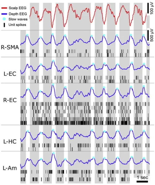
Figure 0-5 Slow-waves and neuronal silencing
Slow-waves recorded during NREM3 in an epileptic patient implanted with depth electrodes (Nir et al., 2011). The top red curves show the scalp EEG with its characteristic trains of slow-waves. Blue curves show the corresponding depth EEG across different brain regions (SMA: supplementary motor area, EC: entorhinal cortex, HC: hippocampus, Am: amygdala, R: right, L: left). Slow-waves span here very distant regions, which is quite rare. Slow-waves’ down-states are marked with a blue dot (negative peak on scalp EEG, positive peak in depth EEG). These down-states are accompanied by a silencing of neurons (black vertical ticks: each line shows a different neuron recorded in the same region as the depth EEG, each ticks correspond to a spike). Down-states are followed by an up-state (positive peak at the scalp level, negative in depth EEG), a rebound in units’ activity. Reproduced from (Nir et al., 2011) (see Study 1).
!
NREM sleep spatial dynamics
Sleep is not only a dynamic process in time but also in space, a dimension that had been rather overseen until recently (Nobili et al., 2012). This could be due to the focus on sleep stages that are stable across time, as mentioned above. It might also be due to the favored animal model for sleep physiology: rodents. Rodents indeed have a rather small brain (and are usually recorded with few electrodes), which makes the observation of regional differences more difficult than in larger brains such as in Humans. In the first chapter of this manuscript, I will present studies I have contributed to (Studies 1 and 2) that are part of a larger re-description of sleep as a more local phenomenon than previously thought (Andrillon et al., 2011; Nir et al., 2011).