Report
Long-term outcomes of breast cancer patients after endoscopic axillary lymph node
dissection: a prospective analysis of 52 patients
I. Langer
1, T. Kocher
3, U. Guller
1, J. Torhorst
2, D. Oertli
1, F. Harder
1, and M. Zuber
41
Department of Surgery; 2Institute of Pathology, University Hospital Basel;3Department of Surgery; Kantonsspital Baden;4Department of Surgery, Kantonsspital Olten, Switzerland
Key words: breast cancer, endoscopic axillary dissection, long-term outcomes
Summary
Background. Reports on long-term outcomes after endoscopic axillary lymph node dissection (ALND) of breast
cancer patients are still lacking in the medical literature. The objective of this prospective study was to assess the oncological and functional outcomes in breast cancer patients after endoscopic ALND.
Methods.Fifty-five breast cancer patients were prospectively enrolled, of whom 52 were available for follow-up with a median of 71.9 months (range 11–96). The following oncological and functional endpoints were evaluated during follow-up at several time points: occurrence of local, axillary and distant metastases, seroma or infection, shoulder mobility (range of motion), numbness, pain, presence of lymphoedema as well as restriction in activities of daily living.
Results.In 52 patients endoscopic ALND of level I and II was successfully performed. Two port-site metastases (2/52, 4%) occurred, one of which in a patient with negative axillary lymph nodes. The same patient suffered from the only axillary recurrence (1/52, 2%). Three patients (3/52, 6%) developed lymphoedema. No other functional adverse events (shoulder mobility, pain, numbness, hypertrophic scar) were noticed at the end of the observation period.
Conclusion.The present investigation with long-term follow-up after endoscopic ALND – the first one in the
literature – reveals minor morbidity, good functional and cosmetic results. In contrary to conventional surgery, the endoscopic procedure is associated with the occurrence of port-site metastases, not seen in the open approach. Axillary recurrences do not appear more frequently when compared with results after conventional ALND. In the meantime the less invasive sentinel lymph node (SLN) biopsy is the established standard technique in evaluating the axillary lymph node status.
Introduction
Axillary lymph node staging represents an integral part of the surgical procedure in breast cancer patients. It provides important prognostic information and deter-mines subsequent adjuvant therapy. Axillary clearance minimizes axillary recurrence rates [1]. Educational advertising, self-examination of the breast and screening mammography favour the detection of breast cancer at an early disease stage. Thus, the percentage of node-positive patients who could benefit from routine axillary lymph node dissection (ALND) is constantly decreasing [2]. The growing population of node-negative patients should be spared a routine ALND with its considerable short- and long-term sequelae [3]. Two different mini-mally invasive methods aiming to define the pathologi-cal lymph node status with minimal morbidity were evaluated in the nineties. Before the sentinel lymph node (SLN) technique was initiated at our institution, we adopted the endoscopic ALND of level I and II. The SLN procedure was validated and established at our
department in 1997 [4]. Although the SLN procedure as new axillary staging method in breast cancer patients has gained increasing importance over the past years, there are still surgeons and gynaecologists in Europe who perform level I and II ALND either by open or endoscopic approach. So far no long-term results after endoscopic ALND in breast cancer patients are avail-able. The objective of the present prospective study was to evaluate long-term oncological and functional out-comes in breast cancer patients after endoscopic ALND.
Patients and methods
Between January 1996 and June 1998, 55 women with primary breast cancer stage I and II were prospectively enrolled (Table 1). Exclusion criteria were: suspicious axillary lymph nodes and a tumour in the axillary tail in close vicinity to the axilla that was to be inflated. Written informed consent was obtained from all pa-tients.
Technique of endoscopic ALND
The technique of endoscopic ALND has been described elsewhere [5–8]. Briefly, the patients were placed in a supine position with the ipsilateral arm in a 90 degree abduction. The axillary fat pad was infiltrated with 250– 450 ml of liposuction fluid, depending on the amount of axillary fat. The liposuction fluid consisted of 500 ml of 0.45% sodium chloride with 10 ml of 4% lidocaine and 5 mg adrenaline. During the ongoing axillary lipolysis process, tumourectomy was performed. Thereafter, the liposuction canula was introduced through an incision of 10–15 mm in the middle axillary line. Aspiration was achieved with a vacuum of 800 mbar. A first 10 mm trocar was then introduced through the existing incision.
After insufflation of CO2gas with a pressure of 8 mmHg
two additional 5 mm trocars were placed, one in the anterior and another in the posterior axillary line. The 25 degree angled 10 mm optic was used. The axillary lymph nodes of level I and II were dissected and re-moved through the 10 mm trocar after changing to a 5 mm optic. The optic, all trocars, and the endoscopic instruments were reusable and taken from the laparos-copy instrumentation box. No specific adaptations were necessary. At the end of the intervention the liposuction fluid was filtered through a gauze and the retrieved lymph nodes were sent separately for histopathology. All operations were carried out by one surgeon (F.H.) Pathologic examination of lymph nodes
Excised and filtered lymph nodes were examined sepa-rately. Lymph nodes larger than 5 mm in diameter were
bisected, whereas lymph nodes less than or equal to 5 mm in diameter were entirely submitted. Lymph nodes were formalin-fixed and embedded in paraffin for his-tologic analysis. Two sections were performed on the larger lymph nodes (one section on each half) whereas the smaller nodes were cut only once. The sections were stained with Haematoxylin & Eosin (H&E). No step sections or immunohistochemistry were performed.
Adjuvant therapy
After breast-conserving surgery patients received post-operative radiation therapy with 45 Gray over 5 weeks with an additional boost of 10 Gray to the tumour site, which was intraoperatively marked with a titanium clip. No radiation therapy was applied to the axilla. Systemic
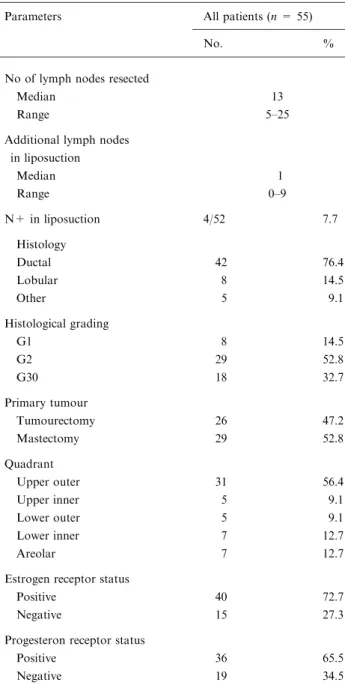
Table 1.Patients and tumour characteristics
Parameters All patients (n = 55)
No. % Age (Years) Median 58.0 Range 30–86 Menopausal status Premenopausal 12 21.8 Postmenopausal 43 78.2 Tumour size in mm Median 20 Range 5–50 T stage T1a 2 3.6 T1b 5 9.1 T1c 21 38.2 T2 27 49.1 N stage NO 37 67.3 N+ 18 32.7 Table 1. (Continued)
Parameters All patients (n = 55)
No. %
No of lymph nodes resected
Median 13
Range 5–25
Additional lymph nodes in liposuction Median 1 Range 0–9 N+ in liposuction 4/52 7.7 Histology Ductal 42 76.4 Lobular 8 14.5 Other 5 9.1 Histological grading G1 8 14.5 G2 29 52.8 G30 18 32.7 Primary tumour Tumourectomy 26 47.2 Mastectomy 29 52.8 Quadrant Upper outer 31 56.4 Upper inner 5 9.1 Lower outer 5 9.1 Lower inner 7 12.7 Areolar 7 12.7
Estrogen receptor status
Positive 40 72.7
Negative 15 27.3
Progesteron receptor status
Positive 36 65.5
treatment was based on the recommendations of the 1996 St. Gall Consensus Conference [9]. Adjuvant therapy consisted of hormonal treatment (Tamoxifen for 5 years: 20 mg daily) and/or chemotherapy (Adria-mycin + Cyclophosphamide or Epirubicin + Cyclo-phosphamide) every 3 weeks for a total of 12 weeks. In low-risk patients, elderly patients, and patients with contraindications for anthracyclines, six cycles of CMF
(Cyclophosphamide + Methotrexate + 5-FU) were
administered.
Postoperative follow-up
Postoperative follow-up consisted of a clinical exami-nation of the breast and axilla every 4 months for the first 3 years, every 6 months in the following 2 years and yearly thereafter. Subjective criteria such as pain, numbness, and restrictions in daily life activities due to scarring or complaints in the arm were assessed. Objective criteria included the pre- and postoperative measurements of (1) range of motion in all directions of the shoulder according to the neutral zero crossing method; (2) the circumferences of both upper extremities 15 cm above and 10 cm below the olecranon and (3) the dexterity to bind an apron at the neck and the back. A deficit in range of motion greater than 20 degrees to standard values was considered pathologic. The diag-nosis of lymphoedema was made if at least one of the following three criteria were present (subjective symp-toms, objective findings, and asymmetric ipsi- and con-tralateral measurements). Symptoms like swelling and heaviness of the affected arm were considered diagnostic as well as clinical findings like indentation following skin depression, a loss of skin folds and asymmetry of the extremities. A 2 cm increase in one of the circumferences compared with the ipsilateral baseline measurements was considered pathologic. Furthermore, a 2 cm in-crease in circumference compared to the contralateral side was regarded as pathologic. For this purpose the handedness was noted, a 1 cm preoperative difference in favour of the dominant arm was considered normal. Consequently, values exceeding 1 cm were calculated to the postoperative measurement results.
At 24 months of follow-up a standardized question-naire was sent to all patients assessing pain in the upper and lower arm, in the breast and chest wall, shoulder mobility, and restrictions in activities of daily living. Mammographies were performed annually. Breast ultrasound was only made for clarification of suspicious mammographic findings. Data were analyzed at 6 weeks after surgery (defined as early postoperative events) and during follow-up at a median of 24 and 71 months postoperatively.
Results
The patient characteristics are listed in Table 1. Endo-scopic ALND of level I and II was completed in 52 of 55
breast cancer patients (95%). Endoscopic ALND was converted to conventional open ALND in three patients due to bleeding of the thoracodorsal vein (n¼ 1), to an exceedingly fatty axilla not enabling proper
identifica-tion of important anatomical landmarks (n¼ 1), and to
fibrotic changes of the axillary fat pad after neoadjuvant
chemotherapy (n¼ 1). According to the
intention-to-treat principle these patients were also included in all analyses and remained tumour-free. Operating time for endoscopic ALND and removal of the primary tumour ranged from 95 to 240 min with a median of 135 min. We retrospectively compared the operating time of pa-tients undergoing endoscopic axillary dissection to a group of 52 breast cancer patients with matched char-acteristics regarding tumour size and age receiving conventional ALND. All operations were performed by the same surgeon (F.H.) 1 year prior to starting the endoscopic axillary dissection. The operating time for conventional ALND and removal of the primary tu-mour ranged from 40 to 205 min with a median of 80 min whereas the endoscopic approach required on average 55 more minutes. This difference in operating time was statistically highly significant (p < 0.0001; Mann–Whitney U-test).
A median number of 13 (range: 5–25) axillary lymph nodes were removed during the 52 successfully per-formed endoscopic procedures. Additionally, an average of one lymph node (range 0–9) per patient was found in the filtered liposuction fluid. Sixteen of 52 patients (30.8%) had lymph node metastases. Four patients had metastatic nodes in the filtered fluid. In two of them these nodes were the only metastatic ones.
Systemic adjuvant treatment consisted of hormonal therapy in 30/52 patients (57.6%), of chemotherapy only in 13/52 patients (25%) and of combined treatment in 8/ 52 patients (15.4%). One patient (2%) did not receive any systemic therapy.
At 6 weeks postoperative complications occurred in 10/52 (19%) patients. Eight patients (15%) developed an axillary seroma and underwent one or several needle aspirations. Injury to the thoracodorsal nerve resulted in a persistent winged scapula in one patient (2%). A low grade infection of the axilla occurring in one patient during adjuvant chemotherapy was successfully treated with antibiotics (2%). The patients were discharged after a median of 5.6 days (1–16 days).
Three patients were lost to follow-up who were ex-cluded from the analyses due to lack of information. Eight patients died at a median of 34.0 months (range 10.6 to 67.4 months) post surgery. Death was related to metastatic disease in four patients (bone, liver, brain and lung), two of whom were node-positive at the time of surgery, another patient showed a high grade pT2 tu-mour and the last was a premenopausal woman with a pT2 G2 carcinoma. One patient died of leukaemia, and three deaths were due to other, non-oncological causes. Furthermore, two patients developed bone metastases, one of them being initially node-positive. Local breast recurrence occurred in one patient (1/52, 2%).
In two patients histologically confirmed port-site metastases were detected at 24 and 49 months after endoscopic ALND, respectively (2/52, 4%). Diagnosis was histologically confirmed after excision of the af-fected axillary skin. One of these postmenopausal wo-men was node positive at the time of the initial operation (9 of 17 positive lymph nodes) staged as a pT2G2 invasive ductal carcinoma. After excision of the port-site metastasis at 24 months, distant metastatic disease to the liver occurred. The patient died one year later. In the second patient with pT2G3 invasive lobular carcinoma, all 11 endoscopically removed lymph nodes were free of metastases. Simultaneously with the port-site metastasis this patient developed the only axillary
recurrence (1/52, 2%) found in our study population. The recurrence was surgically removed. Three years la-ter, the patient is still alive and without evidence of recurrent local or metastatic disease.
The questionnaire was filled out by 52 patients at a median of 24 months (range 7–37 months) postopera-tively. On a scale between 1 (worst) and 10 (excellent) the patients noted median values for pain in the upper and lower arm, pain in breast and chest wall, shoulder mobility, and restrictions in activities of daily living between 9.2 and 9.9 (Table 2). At physical examination at the same time point two patients suffered from restricted shoulder mobility with an anteversion deficit of 20 degrees and abduction deficit of 30 degrees. Twelve of 52 patients (23%) indicated shoulder pain at abduction and anteversion movement and 9/52 patients (17%) superficial pain in the dorsomedial skin area of the affected upper arm. Three patients (6%) com-plained of numbness of the upper arm. No lymphoe-dema was detected at that stage (Table 3).
Median follow-up for the 52 patients was
71.9 months ranging from 11 to 96 months. Other than noted in the questionnaire no relevant numbness or hypertrophic scar were observed on clinical work-up.
Shoulder pain (n¼ 12) and pain in the upper arm
(n¼ 9), which were present at the first evaluation after a median of 24 months postoperatively, disappeared completely during further follow-up. No increase in functional restrictions of the shoulder regarding range of motion was noticed. At the last follow-up examination three patients (6%) had developed lymphoedema with an increase in circumference of 3–4 cm in the upper and/ or lower arm. None of the three patients suffered from recurrent axillary or distant disease, none complained of chronic pain or restriction in range of motion and in daily life. The number of lymph nodes removed in these patients were 12, 16 and 21, respectively. Two of these patients were node-positive at the time of operation with 1/21 and 3/12 positive lymph nodes, respectively (Table 3).
Discussion
The present investigation with long-term follow-up after endoscopic ALND – the first one reported in the liter-ature – reveals minor morbidity, good functional and cosmetic results after endoscopic ALND. The endo-scopic procedure shows a low axillary recurrence rate as one can expect after open level I and II ALND. But the endoscopic ALND carries a risk of port-site metastases. The clinical and prognostic impact of port-site metas-tases is unknown.
Our study provides compelling evidence that endo-scopic ALND is advantageous regarding long-term functional results such as shoulder mobility and chronic pain when compared with conventional ALND [10]. The occurrence of moderate lymphoedema in 6% in the present investigation is lower than in the majority of
Table 2.Self evaluation questionnaire (n = 51 patients) Subjective valuea
Median Range
Pain upper arm 9.4 4–10
Pain lower arm 9.9 6–10
Pain chest wall/breast 9.2 6–10 Shoulder range of motion 9.4 4–10 Use of arm in daily life 9.5 5–10
aScore 1–10: 1 – very bad; 10 – excellent.
Table 3.Morbidity and mortality at different time points (n = 52 patients)
Time points 6 weeks 24 (7–37) monthsa 72 (11–96) monthsa n % n % n % Axillary seroma 8 15.4 0 0 Winged scapula 1 1.9 1 1.9 1 1.9 Axillary infection 1 1.9 0 0 Shoulder restriction NA 2 3.8 0 Shoulder pain NA 12 23.1 0 Upper arm pain NA 9 17.3 0
Numbness NA 3 5.8 0 Hypertrophic scar NA 3 5.8 0 Lymphoedema 0 0 3 5.8 Loss to follow-up 0 1 1.9 3 5.8 Axillary recurrences 0 1 1.9 1 3.8 Port-Site metastases 0 1 1.9 1 1.9 Distant metastases 0 5 9.6 7 13.5 Death 0 3 5.8 8 15.4 NA – not assessed
investigations assessing the morbidity of conventional ALND. Indeed, the incidences of lymphoedema after open axillary dissection are described between 5 and 25% [1, 11–16]. Although there are generally accepted criteria but not well-established guidelines to diagnose
lymphoedema using circumferential measurements,
there are no universally applied methods to quantitatively define lymphoedema and no standard degree of enlarge-ment or consensus, what constitutes lymphoedema, thereby complicating interpretation of the literature [17]. There are reports citing that a greater than 2 cm difference from baseline (preoperative) measurements identifies lymphoedema [18]. On the other hand girth measurements do not always correlate with symptoms or quality of life [19]. A combination of symptom assessment and limb measurement may provide the best clinical assessment data for identifying changes associated with post-breast cancer lymphoedema [20, 21]. In a retrospective study from our institution including 390 breast cancer patients undergoing ALND lymphoedema of the upper and lower arm were found in 13.2 and 8.4%, respectively, after a median follow-up of 62 months [10]. In the afore-men-tioned investigation, numbness was seen in 28%, hyper-trophic scar in 17%, and shoulder pain in 15% of all patients, clearly more frequently than in the present study. Oncological safety might, however, be compromised as port-site metastases occurred in two patients (4%), one of whom suffered concomitantly from an axillary recurrence (2%). Current medical literature describes a similar ax-illary recurrence rate between 0 and 2% after open ALND [22]. In the above mentioned study from our institution an axillary recurrence rate of 1.3% after standard ALND of level l and II was observed [10]. Skin implantation metastasis after open ALND is an unknown adverse event and has never been published before. The prognostic significance of port-site metastases after endoscopic ALND for breast cancer is unclear. Port-site metastases are a known phenomenon in laparoscopic surgery for abdominal malignancies. Wexner reported an incidence of 4% in laparoscopic colorectal surgery for cancer [23]. Port-site metastases correlated with shortened survival in ovarian and gallbladder cancer [24].
Endoscopic ALND offers several advantages com-pared with conventional ALND. It provides an excellent view of the various important anatomical structures,
Figure 1. (a) Anatomical axillary landmarks and important structures after dissection: * long thoracic nerve; ** thoracodorsal nerve and vascular bundle. (b) Endoscopic axillary lymph node dissection after liposuction.
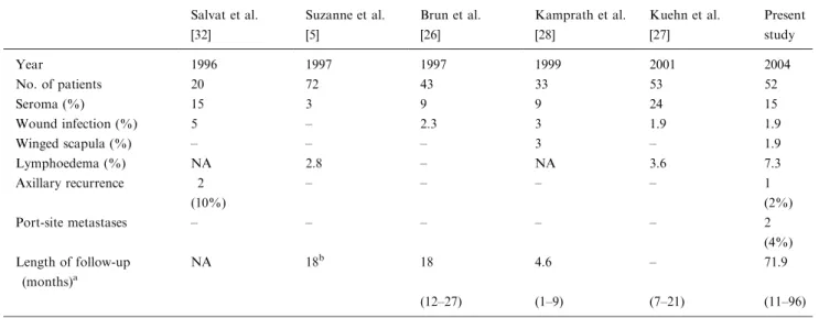
Table 4.Morbidity after endoscopic ALND – a comparison with the literature Salvat et al. [32] Suzanne et al. [5] Brun et al. [26] Kamprath et al. [28] Kuehn et al. [27] Present study Year 1996 1997 1997 1999 2001 2004 No. of patients 20 72 43 33 53 52 Seroma (%) 15 3 9 9 24 15 Wound infection (%) 5 – 2.3 3 1.9 1.9 Winged scapula (%) – – – 3 – 1.9 Lymphoedema (%) NA 2.8 – NA 3.6 7.3 Axillary recurrence 2 – – – – 1 (10%) (2%) Port-site metastases – – – – – 2 (4%) Length of follow-up (months)a NA 18b 18 4.6 – 71.9 (12–27) (1–9) (7–21) (11–96)
which can be easily preserved during the intervention (Figure 1a and b). Bleeding can be stopped precisely and incisions are smaller and thus the trauma to the tissue is reduced [8]. The number of retrieved lymph nodes is similar compared with open ALND and provides ada-equate information of the lymph node status [5, 10, 25– 27]. In the current literature, the occurrence of postop-erative seroma formation appears to be reduced (3– 24%), cosmetic results to be better and functional out-comes improved compared with conventional ALND [5, 8, 10, 28]. Maunsell et al. assessed 223 patients 3 months after open level I and II dissection. They reported that 82% of patients suffered from at least one arm problem, including swelling (24%), weakness (26%), limited arm movement (32%), stiffness (40%), pain (55%), and numbness (58%). No significant decrease of these symptoms was observed after 18 months post surgery [29]. Warmuth et al. found a 55% rate of patients suf-fering from at least one adverse outcome 2–5 years after conventional ALND of level I and II. Thirty-five per-cent reported numbness of the upper arm, 30% pain in the axilla, breast or chest, 15% arm swelling, and 8% limited shoulder or arm movement [30]. Roses et al. described a 76.5% rate of postoperative pain and numbness in his patient collective with a complete res-olution in 22% and an improvement in 57% of those cases after a median follow-up of 38.5 months [31]. These findings correspond well with the results of the present study, as pain in the shoulder joint and upper arm resolved completely during long-term observation.
Disadvantages of the technically demanding endo-scopic ALND compared with the open procedure in-clude longer operating times and higher material expenses [25, 27, 32]. Moreover, the significantly in-creased operating time, in our investigation almost 1 h, further contributes to higher costs. Material expenses in our group of patients were very low because reusable trocars and already available laparoscopic instruments were used and no specific adaptations were necessary. Finally, and most importantly, endoscopic ALND ig-nores the general surgical oncological principle of
en-blocresection of potentially tumour-involved lymph
nodes. The port-site metastases in two of our patients – the first description in the literature after endoscopic ALND – could result from violation of this principle. In other published series with shorter follow-up times (median follow-up shorter than or equal to 18 months) axillary recurrences after endoscopic ALND occurred between 0% [32] and 10% [5, 26, 27, 32] (Table 4). The two axillary failures published in the paper of Salvat et al. were both observed in patients with primarily extensive lymph node involvement [32].
Conclusion
Endoscopic ALND is associated with minor morbidity, good functional and cosmetic results. However, the endoscopic procedure carries the risk of port-site
metas-tases. The axillary recurrence rate is similar to the results after conventional open ALND. Based on previous assessments of our SLN data and the current literature, the SLN biopsy is a more adapted, less invasive technique and should be considered the preferred axillary staging procedure.
References
1. Ball AB, Waters R, Fish S, Thomas JM: Radical axillary dissection in the staging and treatment of breast cancer. Ann R Coll Surg Engl 74: 126–129, 1992
2. Gambazzi F, Zuber M, Oertli D, Marti W, Kocher T, Torhorst J, Harder F: Small breast carcinomas – less axillary surgery?.Swiss Surg 6: 116–120, 2000
3. Burak WE, Hollenbeck ST, Zervos EE, Hock KL, Kemp LC, Young DC: Sentinel lymph node biopsy results in less postoperative morbidity compared with axillary lymph node dissection for breast cancer. Am J Surg 183: 23–27, 2002
4. Langer I, Zuber M, Kochli OR, Kocher T, Muller-Brand J, Torhorst J, Harder F: Validation study of the sentinel lymph node (SLN) method in invasive breast carcinoma. Personal data and review of the literature. Swiss Surg 6: 128–136, 2000
5. Suzanne F, Emering C, Wattiez A, Bournazeau JA, Bruhat MA, Jacquetin B: Axillary lymphadenectomy by lipo-aspiration and endoscopic picking. Apropos of 72 cases. Chirurgie 122: 138–142, 1997
6. Kocher T, Zuber M, Harder F: Endoscopic axilla dissection in invasive breast carcinoma. Initial experiences with a new tech-nique. Zentralbl Chir 123 (Suppl 5): 98–99, 1998
7. Kocher T, Zuber M, Langer I, Harder F: Significance of endoscopic axillary dissection in invasive breast carcinoma after introduction of the ‘‘sentinel lymph node’’ method. Swiss Surg 6: 121–127, 2000
8. Zuber M, Kocher T, Langer I, Harder F: Endoscopic axillary dissection and its perspective in breast cancer. Acta Chir Austriaca (Eur Surg) 32: 110–114, 2000
9. Goldhirsch A, Wood WC, Senn HJ, Glick JH, Gelber RD: IX. International consensus conference on primary treatment of breast cancer. Recent Results Cancer Res 140: 325–335, 1996
10. Harder L, Harder F: Outcome and complications after standard axillary lymph node dissection in breast cancer. Thesis University Hospital Basel, 2002
11. Hladiuk M, Huchcroft S, Temple W, Schnurr BE: Arm function after axillary dissection for breast cancer: a pilot study to provide parameter estimates. J Surg Oncol 50: 47–52, 1992
12. Hoe AL, Iven D, Royle GT, Taylor I: Incidence of arm swelling following axillary clearance for breast cancer. Br J Surg 79: 261– 262, 1992
13. Ivens D, Hoe AL, Podd TJ, Hamilton CR, Taylor I, Royle GT: Assessment of morbidity from complete axillary dissection. Br J Cancer 66: 136–138, 1992
14. Keramopoulos A, Tsionou C, Minaretzis D, Michalas S, Aravan-tinos D: Arm morbidity following treatment of breast cancer with total axillary dissection: a multivariated approach. Oncology 50: 445–449, 1993
15. Kissin MW, Querci della RG, Easton D, Westbury G: Risk of lymphoedema following the treatment of breast cancer. Br J Surg 73: 580–584, 1986
16. Lin PP, Allison DC, Wainstock J, Miller KD, Dooley WC, Friedman N, Baker RR: Impact of axillary lymph node dissection on the therapy of breast cancer patients. J Clin Oncol 11: 1536– 1544, 1993
17. Petrek JA, Heelan MC: Incidence of breast carcinoma-related lymphedema. Cancer 83: 2776–2781, 1998
18. Bland KL, Perczyk R, Du W, Rymal C, Koppolu P, McCrary R, Carolin KA, Kosir MA: Can a practicing surgeon detect early lymphedema reliably? Am J Surg 186: 509–513, 2003
19. Rampaul RS, Mullinger K, Macmillan RD, Cid J, Holmes S, Morgan DA, Blamey RW: Incidence of clinically significant lymphoedema as a complication following surgery for primary operable breast cancer. Eur J Cancer 39: 2165–2167, 2003 20. Armer JM, Radina ME, Porock D, Culbertson SD: Predicting
breast cancer-related lymphedema using self-reported symptoms. Nurs Res 52: 370–379, 2003
21. Rockson SG, Miller LT, Senie R, Brennan MJ, Casley-Smith JR, Foldi E, Foldi M, Gamble GL, Kasseroller RG, Leduc A, Lerner R, Mortimer PS, Norman SA, Plotkin CL, Rinehart-Ayres ME, Walder AL: American Cancer Society Lymphedema Workshop. Workgroup III: Diagnosis and management of lymphedema. Cancer 83: 2882–2885, 1998
22. Fredriksson I, Liljegren G, Arnesson LG, Emdin SO, Palm-Sjovall M, Fornander T, Holmqvist M, Holmberg L, Frisell J: Conse-quences of axillary recurrence after conservative breast surgery. Br J Surg 89: 902–908, 2002
23. Wexner SD, Cohen SM: Port site metastases after laparoscopic colorectal surgery for cure of malignancy. Br J Surg 82: 295–298, 1995
24. Ziprin P, Ridgway PF, Peck DH, Darzi AW: The theories and realities of port-site metastases: a critical appraisal. J Am Coll Surg 195: 395–408, 2002
25. Harder F, Zuber M, Kocher T, Torhorst J: Endoscopic surgery to the axilla – a substitute for conventional axillary clearance? Recent Results Cancer Res 152: 180–189, 1998
26. Brun JL, Rousseau E, Belleannee G, de Mascarel A, Brun G: Axillary lymphadenectomy prepared by fat and lymph node suction in breast cancer. Eur J Surg Oncol 24: 17–20, 1998
27. Kuehn T, Santjohanser C, Grab D, Klauss W, Koretz K, Kreienberg R: Endoscopic axillary surgery in breast cancer. Br J Surg 88: 698–703, 2001
28. Kamprath S, Bechler J, Kuhne-Heid R, Krause N, Schneider A: Endoscopic axillary lymphadenectomy without prior liposuction. Development of a technique and initial experience. Surg Endosc 13: 1226–1229, 1999
29. Maunsell E, Brisson J, Deschenes L: Arm problems and psycho-logical distress after surgery for breast cancer. Can J Surg 36: 315– 320, 1993
30. Warmuth MA, Bowen G, Prosnitz LR, Chu L, Broadwater G, Peterson B, Leight G, Winer EP: Complications of axillary lymph node dissection for carcinoma of the breast: a report based on a patient survey. Cancer 83: 1362–1368, 1998
31. Roses DF, Brooks AD, Harris MN, Shapiro RL, Mitnick J: Complications of level I and II axillary dissection in the treatment of carcinoma of the breast. Ann Surg 230: 194–201, 1999 32. Salvat J, Knopf JF, Ayoubi JM, Slamani L, Vincent-Genod A,
Guilbert M, Walker D: Endoscopic exploration and lymph node sampling of the axilla. Preliminary findings of a randomized pilot study comparing clinical and anatomo-pathologic results of endoscopic axillary lymph node sampling with traditional surgical treatment. Eur J Obstet Gynecol Reprod Biol 70: 165–173, 1996
Address for offprints and correspondence:Igor Langer, MD, Depart-ment of Surgery, University Hospital Basel, Spitalstrasse 21, CH-4031 Basel, Switzerland; Tel.: +41-61-265-25-25; Fax: +41-61-265-72-50; E-mail: [email protected]