HAL Id: hal-02467603
https://hal.archives-ouvertes.fr/hal-02467603
Submitted on 5 Feb 2020
HAL is a multi-disciplinary open access
archive for the deposit and dissemination of
sci-entific research documents, whether they are
pub-lished or not. The documents may come from
teaching and research institutions in France or
abroad, or from public or private research centers.
L’archive ouverte pluridisciplinaire HAL, est
destinée au dépôt et à la diffusion de documents
scientifiques de niveau recherche, publiés ou non,
émanant des établissements d’enseignement et de
recherche français ou étrangers, des laboratoires
publics ou privés.
A Multiaction and Multitarget Ru(II)-Pt(IV) Conjugate
Combining Cancer Activated Chemotherapy and
Photodynamic Therapy to Overcome Drug Resistant
Cancers
Johannes Karges, Thirumal Yempala, Mickaël Tharaud, Dan Gibson, Gilles
Gasser
To cite this version:
Johannes Karges, Thirumal Yempala, Mickaël Tharaud, Dan Gibson, Gilles Gasser. A Multiaction and
Multitarget Ru(II)-Pt(IV) Conjugate Combining Cancer Activated Chemotherapy and Photodynamic
Therapy to Overcome Drug Resistant Cancers. Angewandte Chemie International Edition,
Wiley-VCH Verlag, In press, �10.1002/anie.201916400�. �hal-02467603�
A Multiaction and Multitarget Ru(II)-Pt(IV) Conjugate
Combining Cancer Activated Chemotherapy and
Photodynamic Therapy to Overcome Drug Resistant
Cancers
Johannes Karges,
[a]Thirumal Yempala,
[b]Mickaël Tharaud,
[c]Dan Gibson,*
[b]and Gilles
Gasser*
[a]Abstract: Cancer has emerged as one of the deadliest diseases worldwide. Pt(II) complexes are commonly used to treat this condition. To reduce their side effects and improve their pharmacological properties, Pt(IV) complexes are being developed as prodrug candidates that are activated by reduction in cancer cells. Concomitantly, photodynamic therapy (PDT) has received increasing attention over the last years. Among other compounds studied as photosensitizers (PSs), Ru(II) polypyridine complexes have gained much attention over the recent years due to their attractive characteristics. In this article, the first example of a novel Pt(IV)-Ru(II) conjugate, which combines cancer activated chemotherapy with photodynamic therapy, is presented. Upon entering the cancer cell, the Pt(IV) centre is reduced to Pt(II) and the axial ligands including the Ru(II) complex and phenylbutyrate are released. As each component has its individual target, the conjugate exerts a multitarget and multiaction effect with (photo-)cytotoxicity values upon irradiation up to clinically relevant 595 nm in the low nanomolar range in various (drug resistant) 2D monolayer cancer cells and 3D multicellular tumour spheroids.
[a] Dr. J. Karges, Dr. G. Gasser
Chimie ParisTech, PSL University, CNRS, Institute of Chemistry for Life and Health Sciences, Laboratory for Inorganic Chemical Biology, 75005 Paris, France.
gilles.gasser@chimieparistech.psl.eu; www.gassergroup.com [b] Dr. T. Yempala, Prof. D. Gibson
Institute for Drug Research, School of Pharmacy, The Hebrew University of Jerusalem, Jerusalem, Israel, 91120, dang@ekmd.huji.ac.il
[c] M. Tharaud
Université de Paris, Institut de Physique du Globe de Paris, CNRS, 75005 Paris, France.
Introduction
To date, the cure and treatment of cancer remains a major challenge in modern medicine. Cisplatin (cis-diamminedichloridoplatinum(II)) and its platinum based derivatives oxaliplatin and carboplatin are the clinically most frequently used anticancer drugs worldwide. Despite their enormous clinical success, the application of these compounds is limited due to severe side effects (e.g., kidney damage, nausea, vomiting and bone marrow suppression), low cancer cell selectivity and, more worryingly, an increasing number of platinum resistant tumours.[1] A known concept to improve
the therapeutic outcome of chemotherapy is to administer cisplatin and its derivatives in combination with other biologically active molecules such as histone deacetylase inhibitors (Vorinostat)[2], retinoid receptor regulators (vitamin D
derivatives)[3] or microtubule network disturbing compounds (e.g., paclitaxel, vinblastine)[4] to generate a drug mixture with
multiple targets and actions. Despite improvements in the treatment outcomes, the application of these drug mixtures is limited as the different components may reach the target after different circulation times leading to non-ideal drug doses.[5]
To overcome these drawbacks, research efforts are devoted towards the development of novel Pt(IV) based compounds, which can be activated on site. As the compound enters the cell, the metal centre can be reduced from Pt(IV) to Pt(II), releasing its axial ligands. To improve the efficiency of the treatment, the axial ligands themselves can be biologically active molecules such as targeting moieties, carbon nanotubes, protein-binding moieties or receptor-binding peptides.[6]
Recently, the use of phenylbutyrate (PhB), which is a histone deacetylase inhibitor that de-condenses chromatin to improve the interaction of cisplatin with DNA, was demonstrated.[7] As a consequence, Pt(IV) prodrugs with PhB as an
axial ligand were shown to have a highly improved therapeutic profile, causing DNA damage and epigenetic effects.[8]
Complementary to chemotherapy, cancer treatment using photodynamic therapy (PDT) has received increasing attention over the recent years. In PDT, a photosensitizer (PS) is either locally or systemically injected. Upon excitation at a specific wavelength, the PS acts as a photocatalyst to generate reactive oxygen species (ROS) and ultimately trigger cell death.[9] Worthy of note, Sadler and co-workers have recently reported the use of photoactivatable Pt(IV) complexes which can act by non-classical mechanism.[10] Among the investigated classes of compounds, metal complexes[11] and especially
Ru(II) polypyridine complexes have received increasing attention due to their attractive chemical and photophysical properties including high water solubility, high ROS production, chemical stability and photostability.[12] Despite recent research efforts, the majority of studied Ru(II) polypyridine complexes are excited using blue or UV-A light.[13] Due to light scattering effects and absorption by biological compartments, the light penetration depth in the tissue is poor at these wavelengths, limiting the treatment of deep seated or large tumors.[14] To tackle this issue, some of us have recently reported the DFT-guided design of Ru(II) polypyridine complexes with a strong absorption red shift towards the biological spectral window (600-900 nm). In this pursuit, the complex [Ru(4,7-diphenyl-1,10-phenanthroline)2
(4,4′-dimethyl-2,2′-bipyridine)]2+, which showed a phototoxic effect from 480 nm up to clinically relevant 595 nm, was unveiled.[15]
Herein, a novel Ru(II)-Pt(IV) conjugate (Ru-Pt), which combines cancer activated chemotherapy and PDT by acting on multiple targets inside the cancer cell with several biological mechanism to overcome drug resistances, is described. Upon entering cancer cells, the Pt(IV) metal centre is reduced to Pt(II) releasing PhB and the Ru(II) polypyridine complex. The Ru(II) complex was found to selectively accumulate in the Golgi apparatus where it is able to generate singlet oxygen (1O
2) upon irradiation at various wavelength ranging from 480 up to clinically relevant 595 nm.
Results and Discussion
To ensure that the photophysical and biological properties of each component are not influenced, the moieties were conjugated with a long aliphatic linker. The linking unit between the Ru(II) and the Pt(IV) complex was synthesised starting from 4,4´-dimethyl-2,2´-bipyridine, which was asymmetrically oxidised to the carboxylic acid using SeO2.[16] The generated
4´-methyl-2,2´-bipyridinyl-4-carboxylic acid was treated with SOCl2 to yield the acid chloride intermediate, which was used
to attach tert-butyl-N-(6-aminohexyl)carbamate by peptide bond formation.[17] The generated 2,2´-bipyridine derivative
was then coordinated to dichlorobis(4,7-diphenyl-1,10-phenanthroline)ruthenium(II), which was previously synthesised from 4,7-diphenyl-1,10-phenanthroline and RuCl3. Finally, the tert-butyloxycarbonyl protection group was removed using
trifluoracetic acid to give the Ru(II) polypyridine complex Ru. To facilitate the conjugation of Ru to the Pt-PhB-OH the axial OH group of the Pt(IV) was activated to form Pt-PhB-MSC (MSC=monosuccinimidylcarbonate), whose synthesis was prepared in a similar manner to the structurally related derivatives.[8b] Both carboxylates (link to PhB) and carbamate
(link to Ru) detach from the PtIV following reduction.[8c] The Ru complex was then coupled with Pt-PhB-MSC in presence
of diisopropylethylamine to yield the final Ru-Pt complex. The synthesis and characterisation of the complexes are described in the SI (Scheme S1, Figure S1-S8).
The photophysical properties of Ru were compared with Ru-Pt to evaluate its potential as a PDT PS and to investigate whether the conjugation to the Pt(IV) complex influenced them (Table S1). As expected, the absorption and emission spectra of Ru and Ru-Pt (Figure S9-S10) showed no significant differences. Importantly, the spectra showed an absorption tail towards the biological spectral window potentially enabling the treatment of deep-seated and large tumours as the light penetration depth directly correlates with the used wavelengths. Worthy of note, previous studies have shown that PSs can show a phototoxic effect although their extinction coefficients are below 100 M-1cm-1.[12a,15] The conjugate
was found to have a large Stokes shift, implying minimal interference between excitation and emission. The luminescence quantum yields of Ru an Ru-Pt were found to be similar (Φem, Ru = 2.0 %, Φem, Ru-Pt = 2.1 %), which is in the same range
as other published Ru(II) polypyridine complexes.[18] Both compounds were found to have excited state lifetimes in the
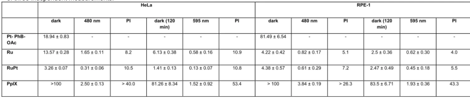
Table 1. IC50 values (μM) in the dark and upon irradiation at 480 (10 min, 3.1 J/cm2) and 595 nm (120 min, 22.5 J/cm2) for Pt-PhB-OAc, Ru and Ru-Pt in
comparison to Protoporphyrin IX (PpIX) in cancerous in human cervical carcinoma (HeLa) and non-cancerous retinal pigment epithelium (RPE-1) cells. Average of three independent measurements.
HeLa RPE-1 dark 480 nm PI dark (120 min) 595 nm PI dark 480 nm PI dark (120 min) 595 nm PI Pt- PhB-OAc 18.94 ± 0.83 - - - 81.49 ± 6.54 - - - - - Ru 13.57 ± 0.28 1.65 ± 0.11 8.2 6.13 ± 0.38 0.58 ± 0.16 10.9 4.22 ± 0.42 0.82 ± 0.17 5.1 2.5 ± 0.36 0.62 ± 0.30 4.0 RuPt 3.26 ± 0.07 0.31 ± 0.06 10.5 1.41 ± 0.13 0.13 ± 0.07 10.8 4.38 ± 0.57 0.61 ± 0.29 7.2 2.47 ± 0.49 0.45 ± 0.18 5.5 PpIX >100 2.50 ± 0.13 > 40.0 81.26 ± 8.34 1.52 ± 0.92 53.4 > 100 3.84 ± 0.19 > 26.3 83.5 ± 6.71 1.93 ± 0.36 43.3
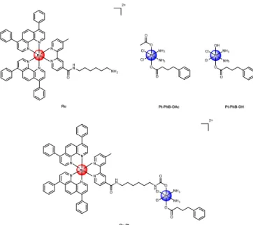
Figure 1. Chemical structures investigated in this work. Ru was isolated as a PF6- salt whereas Ru-Pt was isolated as a TFA- salt.
nanosecond range (Figure S11-S12) in an air saturated environment (147-183 ns) and in degassed environment (1003-1054 ns). Importantly, the presence of air had a drastic effect on the lifetime, indicating that the excited state of the complex can interact with molecular oxygen (3O
2) to generate singlet oxygen (1O2). For quantification of the amount of 1O
2 produced upon light irradiation, the singlet oxygen quantum yields were determined by two methods: 1) directly by
measuring the phosphorescence signal of 1O
2, 2) indirectly by capturing 1O2 with a reporter molecule and monitoring its
change by absorption spectroscopy.[19] The characteristic emission signal at 1270 nm in the infrared region using the
direct method confirmed the production of 1O
2 upon light exposure. Importantly, Ru and Ru-Pt are able to produce 1O2
upon irradiation at 450 and 540 nm. The measurements revealed high 1O
2 quantum yields of 45-64 % in CH3CN and of
3-6 % in an aqueous solution (Table S2). Overall, these studies showed that Ru-Pt has attractive photophysical properties for applications as a PDT agent and the conjugation to the Pt(IV) centre did not significantly influence the photophysical properties of the Ru(II) complex.
The stability of the conjugate was then investigated as previous studies have shown that this could be problematic for metal complexes.[20] The conjugate was incubated in the dark in H
2O at room temperature and physiological pH and the
absorption recorded in time intervals up to 48 h (Figure S13). Additionally, the complex was analysed by HPLC after a 48 h incubation in H2O (Figure S14). As no significant differences were observed, the stability in an aqueous solution is
indicated.
After a chemical and photophysical characterisation of the conjugate, its potential activity as a chemotherapeutic agent and PS for PDT was investigated. The cytotoxicity of cisplatin, Pt-PhB-OH, Pt-PhB-OAc, Ru and Ru-Pt towards non-cancerous retinal pigment epithelium (RPE-1) and non-cancerous human cervical carcinoma (HeLa) cells in the dark as well as upon irradiation at various wavelengths between 480 – 595 nm was determined (Table 1, Table S3). In both investigated cell lines the conjugate Ru-Pt (IC50, HeLa = 3.26 ± 0.07 μM, IC50, RPE-1 = 4.38 ± 0.57 μM) showed a higher
cytotoxicity in the dark than Ru (IC50, HeLa = 13.57 ± 0.28 μM, IC50, RPE-1 = 4.22 ± 0.42 μM) or the platinum based complexes
cisplatin, Pt-PhB-OH or Pt-PhB OAc (IC50, HeLa = 5.9 – 18.9 μM, IC50, RPE-1 = 27.7 – 81.4 μM) alone. All Pt(IV) prodrugs
and importantly Ru-Pt showed a 1.3 - 4.7 times higher cytotoxicity in the cancerous cell line in comparison to the non-cancerous cell line. This effect was previously described in the literature and is caused by the preferential reduction of the Pt(IV) centre to Pt(II) in cancer cells, which are hypoxic. Strikingly, upon irradiation at various wavelengths between 480 – 595 nm, the Ru-Pt conjugate is able to cause a phototoxic effect. Thanks to the additional PDT effect, the cytotoxicity improved drastically, with IC50 values in the nanomolar range (IC50, HeLa = 0.13 – 0.39 μM, IC50, RPE-1 = 0.45 – 0.61 μM).
Overall, these results indicate the beneficial effect of combing the properties of the Pt(II) complex as a chemotherapeutic agent and the Ru(II) complex as a PS as a conjugate. Interestingly, Ru-Pt showed a slightly higher cytotoxicity in cancerous cells in comparison to non-cancerous cells.
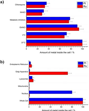
Capitalising on these preliminary results, the potential of Ru-Pt to overcome resistance was further investigated in depth in human ovarian carcinoma (A2780) cells and its cisplatin resistant line (A2780 cis) as well as its doxorubicin resistant line (A2780 ADR). The uptake mechanism of the Ru-Pt conjugate was studied in A2780 cells by blocking different cellular pathways and determining the amount of Ru and Pt inside the cells (Figure 2a) by inductively coupled plasma mass spectrometry (ICP-MS).[21] The incubation with the cationic transporter inhibitor (tetraethylammonium chloride) did not
significantly influence the cellular uptake, suggesting that this pathway does not majorly contribute to the internalisation of the compound. The incubation at lower temperature (4 °C) and with metabolic inhibitors (2- deoxy-D-glucose and oligomycin) decreased the uptake, indicating that the internalisation is energy dependent. In contrast, the incubation with endocytotic inhibitors (ammonium chloride or chloroquine) drastically decreased the uptake. Overall, these results indicate
that Ru-Pt is internalised primarily by an energy dependent endocytotic mechanism. Worthy of note, the recently reported Ru-Pt metallacage was internalised by the same pathway.[22] Importantly, similar levels of Ru and Pt were detected inside
the cells indicating that the Ru-Pt is stable in the cell culture medium and was internalised as a single moiety.
Following this, the localisation of the released Ru(II) complex and Pt(II) complex (cisplatin) were investigated by extraction of the major cellular organelles (nucleus, mitochondria, lysosomes, Golgi apparatus, endoplasmic reticulum) and determination of the amount of Ru and Pt inside of each by ICP-MS (Figure 2b). As expected, Ru and Pt were found in different cellular compartments, indicating that the Pt(IV) centre is reduced and the axial ligands (Ru and PhB) released and that the conjugate is acting
Figure 2. a) Cellular uptake mechanism study of Ru-Pt (10 μM) in A2780 cells in the presence of different inhibitors/conditions by determination of
the amount of Ru and Pt inside the cells by ICP-MS. Endocytic inhibition: NH4Cl (50 mM) or chloroquine (100 μM), metabolic inhibition:
2-Deoxy-D-glucose (50 mM) and oligomycin (5 μM), cation transporter inhibition: Et4NCl, low temperature: incubation at 4°C, control: incubation at 37°C. b)
Cellular localisation of Ru and Pt of the conjugate Ru-Pt (10 μM) in A2780 cells inside the major cellular organelles (endoplasmic reticulum, golgi apparatus, lysosomes, mitochondria, nucleus, whole cell) after 4 h incubation in the dark, extraction of their cellular organelles and determination of the amount of Ru and Pt inside each organelle by ICP-MS.
simultaneously in different cellular organelles. As anticipated, the vast majority of Pt was found in the nucleus. This finding is in agreement with the in-depth investigation of the anticancer drug cisplatin, which is targeting the nucleus and binding to DNA. On the contrary, the majority of Ru was found in the Golgi apparatus. Additionally, small amounts of Pt and Ru were also found in the lysosomes and the endoplasmic reticulum. Taken together, these results indicate that Ru-Pt is internalised through an engulfing mechanism, in which the cell membrane draws the complex from the outside to the inside by formation of intracellular transport vesicles. These can then interact with the endomembrane system consisting of the lysosomes, Golgi apparatus and endoplasmic reticulum, where they release their content. As small amounts of Ru and Pt were both found in the lysosomes and the endoplasmic reticulum, it tends to demonstrate that the Ru-Pt conjugate is majorly released in these organelles. In a further process, the Pt(IV) centre is reduced and the Ru and Pt moieties localise in their respective target organelles (Golgi apparatus and nucleus).
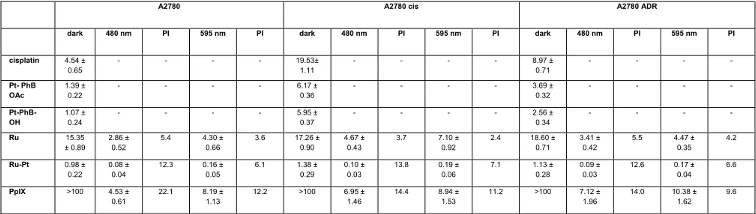
To study the efficiency of the conjugate against drug resistant cell lines, the cytotoxicity of cisplatin, Pt-PhB-OH, Pt-
PhB-OAc, Ru and Ru-Pt in the dark and upon irradiationat 480 (10 min, 3.1 J/cm2) and 595 nm (60 min, 11.3 J/cm2)
against A2780, A2780 cis and A2780 ADR was investigated. All complexes (Table 2) were found to have lower cytotoxic values in all ovarian cancer cell lines in comparison to the HeLa and RPE-1 cell lines (Table 1). These results show the enormous potential of the conjugate Ru-Pt with cytotoxicity values in the dark in the low micromolar range (IC50, dark= 0.98
– 1.38 μM). It is worth mentioning that a PS should be ideally non-toxic in the dark. The irradiation drastically improved the cytotoxic profile (IC50, 480nm= 0.08 – 0.10 μM, IC50, 595nm= 0.16 – 0.19 μM) with PI values between 6.1 – 13.8. While the
cytotoxicity of cisplatin, Pt-PhB-OH and Pt- PhB-OAc is lower in the resistant cell lines A2780 cis and A2780 ADR, the
Ru-Pt conjugate overcomes the resistance even in the dark and strikingly, by irradiation the cytotoxicity in all cell lines is
further improved by a factor of ten. Comparing the cytotoxicity of Ru and Ru-Pt under irradiation we see that Ru-Pt is significantly more potent than Ru in all A2780 cell lines. Taken together, these results confirm the beneficial effect of the conjugation of the Ru(II) and Pt(IV) complex to the conjugate Ru-Pt.
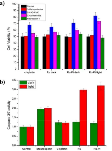
Encouraged by these promising results, the mechanism of action of Ru-Pt was further examined by determining its cell death pathway. For this study, the cytotoxic profile of cisplatin, Ru in the dark, Ru-Pt in the dark and upon irradiation at 480 nm (10 min, 3.1 J/cm2) at its IC
50 concentration in A2780 cells was measured in the presence of autophagy
(3-methyladenine), apoptosis (Z-VAD-FMK), paraptosis (cycloheximide) or necrosis (necrostatin-1) inhibitors (Figure 3a). The incubation with autophagy, paraptosis and necrosis inhibitors did not significantly influence the cell viability of
cisplatin, Ru and Ru-Pt in the dark, indicating that these pathways are not primarily accessed for triggering of their cell
death. In contrast, preincubation with an apoptosis inhibitor increased the survival of the cells treated with cisplatin, indicating that this mechanism is majorly contributing to the cell death. This result is in agreement with a study of cell death pathways of cisplatin in human ovarian carcinoma cells.[23] In addition, elevated survival levels for the preincubation
with an apoptotic inhibitor and treatment with Ru in the dark were detected, suggesting an apoptosis induced cell death mechanism. Therefore, it was not surprising to observe an apoptotic cell death pathway for Ru-Pt in the dark. Importantly, upon irradiation of Ru-Pt, the cell survival is highly increased with apoptotic and paraptotic inhibitors, indicating a cell death mechanism for the phototoxic effect involving both pathways. Worthy of note, several PDT agents were previously reported in the literature to cause cell death by apoptosis and paraptosis pathways.[24]
For further examination of the cell death mechanism, its dependency on caspase 3/7 pathways was investigated using a Caspase-Glo 3/7 assay. These caspases are known executers of extrinsic and intrinsic apoptosis mechanism.[25] The
caspase 3/7 activity was measured in A2780 cells for cisplatin, Ru and Ru-Pt in the dark and upon irradiation at 480 nm (10 min, 3.1 J/cm2) using half of their IC
50 concentration (Figure 3b). As a positive control for caspase activity, the kinase
inhibitor staurosporin was used. Cisplatin, Ru and Ru-Pt in the dark did not significantly increase caspase 3/7 activity, ruling out this pathway for cell death. The result for cisplatin is in agreement with a recent study in the same cell line that showed that apoptosis is induced by ERK/p53/PUMA activation.[26] In contrast to this, upon light irradiation and treatment
with Ru and Ru-Pt, the caspase 3/7 activity is highly elevated, suggesting that the phototoxic effect is caused by these pathways. Worthy of note, other Ru(II) polypyridine complexes were also found to generate a cytotoxic effect by this pathway.[27]
After evaluation of the (photo-)cytotoxicity on 2D monolayer cells, the effect of Ru-Pt on 3D multicellular tumour spheroids (MCTS) was investigated. This is of special interest as many anticancer agents have failed translation from 2D monolayer cells to in vivo models due to compromised drug delivery. MCTS can mimic the pathological conditions found in solid tumors such as proliferation gradients and its hypoxic centre and can therefore be used for the assessment of drug delivery.[28] A2780 MCTS with a diameter of about 400 μM were incubated with increasing concentrations of Ru-Pt for 12
h. After this time, the cytotoxic effect in the dark and upon irradiation at 480 nm (10 min, 3.1 J/cm2) and 595 nm (120 min,
22.5 J/cm2) was determined upon measurement of the ATP concentration of living cells inside the MCTS. Ru-Pt showed
a strong cytotoxic effect (IC50, dark= 7.32 ± 0.41 μM), indicating that the conjugate is able to completely penetrate the MCTS.
Importantly, as the cytotoxicity is shifting to the nanomolar range (IC50, 480nm= 0.49 ± 0.16 μM, IC50, 595nm= 0.68 ± 0.27 μM)
upon irradiation at 480 nm and 595 nm, the synergetic effect of the Pt(IV) complex as a chemotherapeutic agent and the Ru(II) complex as a PDT agent is demonstrated.
Table 2. IC50 values (μM) in the dark and upon irradiation at 480 (10 min, 3.1 J/cm2) and 595 nm (60 min, 11.3 J/cm2) for cisplatin, Pt-PhB-OH, Pt- PhB OAc, Ru and Ru-Pt in comparison to Protoporphyrin IX (PpIX) in the human ovarian carcinoma (A2780), human cisplatin resistant ovarian carcinoma (A2780 cis) and
human doxorubicin resistant ovarian carcinoma (A2780 ADR) cell lines. Average of three independent measurements.
A2780 A2780 cis A2780 ADR
dark 480 nm PI 595 nm PI dark 480 nm PI 595 nm PI dark 480 nm PI 595 nm PI
cisplatin 4.54 ± 0.65 - - - - 19.53± 1.11 - - - - 8.97 ± 0.71 - - - - Pt- PhB OAc 1.39 ± 0.22 - - - - 6.17 ± 0.36 - - - - 3.69 ± 0.32 - - - - Pt-PhB-OH 1.07 ± 0.24 - - - - 5.95 ± 0.37 - - - - 2.56 ± 0.34 - - - - Ru 15.35 ± 0.89 2.86 ± 0.52 5.4 4.30 ± 0.66 3.6 17.26 ± 0.90 4.67 ± 0.43 3.7 7.10 ± 0.92 2.4 18.60 ± 0.71 3.41 ± 0.42 5.5 4.47 ± 0.35 4.2 Ru-Pt 0.98 ± 0.22 0.08 ± 0.04 12.3 0.16 ± 0.05 6.1 1.38 ± 0.29 0.10 ± 0.03 13.8 0.19 ± 0.06 7.1 1.13 ± 0.28 0.09 ± 0.03 12.6 0.17 ± 0.04 6.6 PpIX >100 4.53 ± 0.61 22.1 8.19 ± 1.13 12.2 >100 6.95 ± 1.46 14.4 8.94 ± 1.53 11.2 >100 7.12 ± 1.96 14.0 10.38 ± 1.62 9.6
Figure 3. Cell death mechanism study in A2780 cells in the dark or upon irradiation at 480 nm (10 min, 3.1 J/cm2) a) upon treatment with the IC 50
value of the corresponding compound and in the presence of different inhibitors by determination of the cell viability. Autophagy inhibitor: 3-Methyladenine (100 μM), apoptosis inhibitor: Z-VAD-FMK (20 μM), paraptosis inihibitor: Cycloheximide (0.1 μM), necrosis inihibitor: Necrostatin-1 (60 μM). b) upon treatment with a concentration of half of the IC50 value of the corresponding compound by determination of the caspase 3/7 activity.
Conclusion
In summary, in this article, a novel conjugate (Ru-Pt) combining a Pt(IV) complex as a chemotherapy drug and a Ru(II) complex as a photosensitiser for long wavelength photodynamic therapy, which targets several cellular organelles and acts by various cytotoxic mechanism, is reported. The conjugate is able to enter cancerous cells by an energy depended endocytosis mechanism. While being stable in an aqueous solution, the Pt(IV) centre is reduced inside cancerous cells to Pt(II) and releases its axial ligands. Phenylbutyrate acts as a histone deacetylase inhibitor and can de-condense the chromatin, improving the binding of the Pt(II) complex (cisplatin) to DNA. The Ru(II) complex was found to accumulate at the Golgi apparatus. Next to the cytotoxic effect exerted by the anticancer properties of cisplatin, the Ru(II) complex can act as a photosensitizer to catalytically generate singlet oxygen from 480 up to clinically relevant 595 nm. Thanks to the multitargeting and multiaction mechanisms based on a combination of apoptosis and paraptosis pathways, (photo-)cytotoxicity values in the low nanomolar range were observed in various 2D monolayer cells as well as 3D MCTS. Importantly, the conjugate showed a synergetic effect and was able to overcome drug resistance, which is found in many clinically treated tumors. We strongly believe that this novel conjugate and the approach of combing a Pt(IV) complex as a chemotherapeutic drug and a Ru(II) complex as a photosensitizer for photodynamic therapy has a great potential for further development.
Acknowledgements
We thank Dr. Philippe Goldner for access to state-of-the-art laser apparatus. This work was financially supported by an ERC Consolidator Grant PhotoMedMet to G.G. (GA 681679), has received support under the program “Investissements d’ Avenir” launched by the French Government and implemented by the ANR with the reference ANR-10-IDEX-0001-02
PSL (G.G.). DG acknowledges the support of the Israel Science Foundation (grant 1002/18) and the Alex Grass Center for Drug Design and synthesis.
Keywords: Bioinorganic Chemistry • Medicinal Inorganic Chemistry • Metals in Medicine • Photodynamic Therapy
[1] a) A.-M. Florea, D. Büsselberg, Cancers 2011, 3, 1351-1371; b) C. Cullinane, G. B. Deacon, P. R. Drago, A. P. Erven, P. C. Junk, J. Luu, G. Meyer, S. Schmitz, I. Ott, J. Schur, L. K. Webster, A. Klein, Dalton Trans. 2018,
47, 1918-1932; c) Z. Wang, Z. Deng, G. Zhu, Dalton Trans. 2019, 48, 2536-2544; d) M. Hanif, C. G. Hartinger,
Future Med. Chem. 2018, 10, 615-617; e) C. G. Hartinger, in Advances in Bioorganometallic Chemistry (Eds.:
T. Hirao, T. Moriuchi), Elsevier, 2019, 157-172; f) D. Cirri, M. G. Fabbrini, A. Pratesi, L. Ciofi, L. Massai, T. Marzo, L. Messori, BioMetals 2019, 32, 813-817.
[2] C.-H. Pan, Y.-F. Chang, M.-S. Lee, B. C. Wen, J.-C. Ko, S.-K. Liang, M.-C. Liang, BMC Cancer 2016, 16, 857. [3] M. Milczarek, S. ROSIŃSKA, M. Psurski, M. Maciejewska, A. Kutner, J. Wietrzyk, Anticancer Res. 2013, 33,
433-444.
[4] Y. H. Kim, S. W. Shin, B. S. Kim, J. H. Kim, J. G. Kim, Y. J. Mok, C. S. Kim, H. S. Rhyu, J. H. Hyun, J. S. Kim,
Cancer 1999, 85, 295-301.
[5] a) S. Dasari, P. Bernard Tchounwou, Eur. J. Pharmacol. 2014, 740, 364-378; b) E. Boros, P. J. Dyson, G. Gasser, 2020 Chem 6, 41-60; c) A. Sharma, J. F. Arambula, S. Koo, R. Kumar, H. Singh, J. L. Sessler, J. S. Kim, Chem. Soc. Rev. 2019, 48, 771-813.
[6] a) T. C. Johnstone, K. Suntharalingam, S. J. Lippard, Chem. Rev. 2016, 116, 3436-3486; b) M. D. Hall, T. W. Hambley, Coord. Chem. Rev. 2002, 232, 49-67; c) W. H. Ang, I. Khalaila, C. S. Allardyce, L. Juillerat-Jeanneret, P. J. Dyson, J. Am. Chem. Soc. 2005, 127, 1382-1383; d) K. Suntharalingam, Y. Song, S. J. Lippard, Chem.
Commun. 2014, 50, 2465-2468; e) E. Petruzzella, R. Sirota, I. Solazzo, V. Gandin, D. Gibson, Chem. Sci. 2018, 9, 4299-4307; f) R. Ma, Y. Wang, L. Yan, L. Ma, Z. Wang, H. C. Chan, S.-K. Chiu, X. Chen, G. Zhu, Chem. Commun. 2015, 51, 7859-7862; g) M. V. Babak, Y. Zhi, B. Czarny, T. B. Toh, L. Hooi, E. K.-H. Chow, W. H.
Ang, D. Gibson, G. Pastorin, Angew. Chem. Int. Ed. 2019, 58, 8109-8114; h) J. X. Ong, C. S. Q. Lim, H. V. Le, W. H. Ang, Angew. Chem. Int. Ed. 2019, 58, 164-167; i) J. Z. Zhang, E. Wexselblatt, T. W. Hambley, D. Gibson,
Chem. Commun. 2012, 48, 847-849; j) L. Cubo, T. W. Hambley, P. J. Sanz Miguel, A. Carnero, C.
Navarro-Ranninger, A. G. Quiroga, Dalton Trans. 2011, 40, 344-347; k) L. Ma, N. Wang, R. Ma, C. Li, Z. Xu, M.-K. Tse, G. Zhu, Angew. Chem. Int. Ed. 2018, 57, 9098-9102; l) G. Thiabaud, L. Harden-Bull, Y.-J. Ghang, S. Sen, X. Chi, J. L. Bachman, V. M. Lynch, Z. H. Siddik, J. L. Sessler, Inorg. Chem. 2019, 58, 7886-7894; m) N. Muhammad, N. Sadia, C. Zhu, C. Luo, Z. Guo, X. Wang, Chem. Commun. 2017, 53, 9971-9974; n) X. Wang, X. Wang, S. Jin, N. Muhammad, Z. Guo, Chem. Rev. 2019, 119, 1138-1192.
[7] a) S. D. Gore, M. A. Carducci, Expert Opin. Inv. Drug. 2000, 9, 2923-2934; b) R. R. Rosato, S. Grant, Cancer
Biol. Ther. 2003, 2, 31-38.
[8] a) H. Kostrhunova, E. Petruzzella, D. Gibson, J. Kasparkova, V. Brabec, Chem. Eur. J. 2019, 25, 5235-5245; b) R. Raveendran, J. P. Braude, E. Wexselblatt, V. Novohradsky, O. Stuchlikova, V. Brabec, V. Gandin, D. Gibson,
Chem. Sci. 2016, 7, 2381-2391; c) J. J. Wilson, S. J. Lippard, Inorg. Chem. 2011, 50, 3103.
[9] a) D. E. Dolmans, D. Fukumura, R. K. Jain, Nat. Rev. Cancer 2003, 3, 380-387; b) S. Bonnet, Dalton Trans.
2018, 47, 10330-10343; c) F. Heinemann, J. Karges, G. Gasser, Acc. Chem. Res. 2017, 50, 2727-2736.
[10] a) C. Vallotto, E. Shaili, H. Shi, J. S. Butler, C. J. Wedge, M. E. Newton, P. J. Sadler, Chem. Commun. 2018,
54, 13845-13848; b) C. A. Wootton, C. Sanchez-Cano, A. F. Lopez-Clavijo, E. Shaili, M. P. Barrow, P. J. Sadler,
P. B. O'Connor, Chem. Sci. 2018, 9, 2733-2739; c) N. A. Kratochwil, J. A. Parkinson, P. J. Bednarski, P. J. Sadler, Angew. Chem. Int. Ed. 1999, 38, 1460-1463.
[11] a) L. K. McKenzie, H. E. Bryant, J. A. Weinstein, Coord. Chem. Rev. 2019, 379, 2-29; b) L. Zeng, P. Gupta, Y. Chen, E. Wang, L. Ji, H. Chao, Z.-S. Chen, Chem. Soc. Rev. 2017, 46, 5771-5804; c) C. Imberti, P. Zhang, H. Huang, P. J. Sadler, Angew. Chem. Int. Ed., 2020, 59, 61-73; d) V. Novohradsky, A. Rovira, C. Hally, A. Galindo, G. Vigueras, A. Gandioso, M. Svitelova, R. Bresolí-Obach, H. Kostrhunova, L. Markova, J. Kasparkova, S. Nonell, J. Ruiz, V. Brabec, V. Marchán, Angew. Chem. Int. Ed. 2019, 58, 6311-6315; e) J. Pracharova, G. Vigueras, V. Novohradsky, N. Cutillas, C. Janiak, H. Kostrhunova, J. Kasparkova, J. Ruiz, V. Brabec, Chem.
Eur. J. 2018, 24, 4607-4619; f) A. Zamora, G. Vigueras, V. Rodríguez, M. D. Santana, J. Ruiz, Coord. Chem.
Rev. 2018, 360, 34-76.
[12] a) S. Monro, K. L. Colón, H. Yin, J. Roque III, P. Konda, S. Gujar, R. P. Thummel, L. Lilge, C. G. Cameron, S. A. McFarland, Chem. Rev. 2019, 119, 797-828; b) A. Li, C. Turro, J. J. Kodanko, Acc. Chem. Res. 2018, 51, 1415-1421; c) J. Liu, C. Zhang, T. W. Rees, L. Ke, L. Ji, H. Chao, Coord. Chem. Rev. 2018, 363, 17-28; d) F. E. Poynton, S. A. Bright, S. Blasco, D. C. Williams, J. M. Kelly, T. Gunnlaugsson, Chem. Soc. Rev. 2017, 46, 7706-7756; e) M. Jakubaszek, J. Rossier, J. Karges, J. Delasoie, B. Goud, G. Gasser, F. Zobi, Helv. Chim. Acta
2019, 102, e1900104; f) J. Shum, P. K.-K. Leung, K. K.-W. Lo, Inorg. Chem. 2019, 58, 2231-2247; g) K. Qiu, Y.
Chen, T. W. Rees, L. Ji, H. Chao, Coord. Chem. Rev. 2019, 378, 66-86; h) J. Karges, M. Jakubaszek, C. Mari, K. Zarschler, B. Goud, H. Stephan, G. Gasser, ChemBioChem, 2019, accepted, DOI: 10.1002/cbic.201900419; i) Y. Ellahioui, M. Patra, C. Mari, R. Kaabi, J. Karges, G. Gasser, S. Gómez-Ruiz, Dalton Trans. 2019, 48, 5940-5951; j) B. S. Howerton, D. K. Heidary, E. C. Glazer, J. Am. Chem. Soc. 2012, 134, 8324-8327; k) J. D. Knoll, C. Turro, Coord. Chem. Rev. 2015, 282-283, 110-126; l) A. M. Palmer, B. Peña, R. B. Sears, O. Chen, M. E. Ojaimi, R. P. Thummel, K. R. Dunbar, C. Turro, Philos. Trans. R. Soc. A 2013, 371, 20120135; m) J. Karges, F. Heinemann, F. Maschietto, M. Patra, O. Blacque, I. Ciofini, B. Spingler, G. Gasser, Biorg. Med. Chem. 2019,
27, 2666-2675; n) S. A. McFarland, A. Mandel, R. Dumoulin-White, G. Gasser, Curr. Opin. Chem. Biol. 2020,
56, 23-27; o) W. Streciwilk, A. Terenzi, X. Cheng, L. Hager, Y. Dabiri, P. Prochnow, J. E. Bandow, S. Wölfl, B. K. Keppler, I. Ott, Eur. J. Med. Chem. 2018, 156, 148-161.
[13] a) H. Yin, M. Stephenson, J. Gibson, E. Sampson, G. Shi, T. Sainuddin, S. Monro, S. A. McFarland, Inorg.
Chem. 2014, 53, 4548-4559; b) J. Karges, O. Blacque, P. Goldner, H. Chao, G. Gasser, Eur. J. Inorg. Chem., 2019, 3704-3712, DOI: 10.1002/ejic.201900569; c) J. Karges, O. Blacque, M. Jakubaszek, B. Goud, P. Goldner,
G. Gasser, J. Inorg. Biochem. 2019, 198, 110752.
[14] a) K. Ogawa, Y. Kobuke, Anti-Cancer Agents Me. 2008, 8, 269-279; b) B. C. Wilson, W. P. Jeeves, D. M. Lowe,
Photochem. Photobiol. 1985, 42, 153-162.
[15] J. Karges, F. Heinemann, M. Jakubaszek, F. Maschietto, C. Subecz, M. Dotou, O. Blacque, M. Tharaud, B. Goud, E. V. Zahínos, B. Spingler, I. Ciofini, G. Gasser, ChemRvix, doi: 10.26434/chemrxiv.11336669.
[16] T. Banerjee, S. Rawalekar, A. Das, H. N. Ghosh, Eur. J. Inorg. Chem. 2011, 2011, 4187-4197.
[17] E. J. McLaurin, A. B. Greytak, M. G. Bawendi, D. G. Nocera, J. Am. Chem. Soc. 2009, 131, 12994-13001.[18] a) M. J. Cook, A. P. Lewis, G. S. McAuliffe, V. Skarda, A. J. Thomson, J. L. Glasper, D. J. Robbins, J.
Chem. Soc. Perkin Trans. 2 1984, 1293-1301; b) A. Juris, V. Balzani, F. Barigelletti, S. Campagna, P. l. Belser,
A. Von Zelewsky, Coord. Chem. Rev. 1988, 84, 85-277.
[19] a) J. Karges, P. Goldner, G. Gasser, Inorganics 2019, 7, 4; b) J. Karges, G. Gasser, Inorg. Chim. Acta 2019, 119196.
[20] a) U. Basu, J. Karges, F. Chotard, C. Balan, P. Le Gendre, G. Gasser, E. Bodio, R. Malacea Kabbara,
Polyhedron 2019, 172, 22-27; b) A. K. Renfrew, J. Karges, R. Scopelliti, F. D. Bobbink, P. Nowak-Sliwinska, G.
Gasser, P. Dyson, ChemBioChem 2019, 20, 2876.
[21] a) J. Karges, U. Basu, O. Blacque, H. Chao, G. Gasser, Angew. Chem. Int. Ed., 2019, 58, 14334-14340; b) J. Karges, O. Blacque, H. Chao, G. Gasser, Inorg. Chem. 2019, 58, 12422-12432.
[22] Z. Zhou, J. Liu, T. W. Rees, H. Wang, X. Li, H. Chao, P. J. Stang, Proc. Natl. Acad. Sci. 2018, 115, 5664-5669. [23] M. G. Ormerod, C. F. O'Neill, D. Robertson, K. R. Harrap, Exp. Cell Res. 1994, 211, 231-237.
[24] a) D. Kessel, Photochem. Photobiol. 2019, 95, 119-125; b) V. Rapozzi, F. D’Este, L. E. Xodo, J. Porphyr.
Phthalocyanines 2019, 23, 410-418.
[25] J. M. Adams, Genes Dev. 2003, 17, 2481-2495.
[26] H. Song, M. Wei, W. Liu, S. Shen, J. Li, L. Wang, Histol. Histopathol. 2018, 33, 73-79.
[27] a) C. Qian, J.-Q. Wang, C.-L. Song, L.-L. Wang, L.-N. Ji, H. Chao, Metallomics 2013, 5, 844-854; b) H. Huang, P. Zhang, B. Yu, Y. Chen, J. Wang, L. Ji, H. Chao, J. Med. Chem. 2014, 57, 8971-8983.
[28] a) P. A. Netti, D. A. Berk, M. A. Swartz, A. J. Grodzinsky, R. K. Jain, Cancer Res. 2000, 60, 2497-2503; b) J. Friedrich, C. Seidel, R. Ebner, L. A. Kunz-Schughart, Nat. Protoc. 2009, 4, 309.