AVIS
Ce document a été numérisé par la Division de la gestion des documents et des archives de l’Université de Montréal.
L’auteur a autorisé l’Université de Montréal à reproduire et diffuser, en totalité ou en partie, par quelque moyen que ce soit et sur quelque support que ce soit, et exclusivement à des fins non lucratives d’enseignement et de recherche, des copies de ce mémoire ou de cette thèse.
L’auteur et les coauteurs le cas échéant conservent la propriété du droit d’auteur et des droits moraux qui protègent ce document. Ni la thèse ou le mémoire, ni des extraits substantiels de ce document, ne doivent être imprimés ou autrement reproduits sans l’autorisation de l’auteur.
Afin de se conformer à la Loi canadienne sur la protection des renseignements personnels, quelques formulaires secondaires, coordonnées ou signatures intégrées au texte ont pu être enlevés de ce document. Bien que cela ait pu affecter la pagination, il n’y a aucun contenu manquant.
NOTICE
This document was digitized by the Records Management & Archives Division of Université de Montréal.
The author of this thesis or dissertation has granted a nonexclusive license allowing Université de Montréal to reproduce and publish the document, in part or in whole, and in any format, solely for noncommercial educational and research purposes.
The author and co-authors if applicable retain copyright ownership and moral rights in this document. Neither the whole thesis or dissertation, nor substantial extracts from it, may be printed or otherwise reproduced without the author’s permission.
In compliance with the Canadian Privacy Act some supporting forms, contact information or signatures may have been removed from the document. While this may affect the document page count, it does not represent any loss of content from the document.
Development of a pH-responsive liposomal drug
carrier using poly(phosphazenes)
par David Ghattas
Faculté de phannacie
Thèse présentée à la Faculté des études supérieures en vue de l'obtention du grade de Maître en sciences (M. Sc.)
en sciences phannaceutiques option technologies pharmaceutiques
Octobre, 2008
Cette thèse intitulée:
Development of a pH-responsive liposomal drug carrier using poly(phosphazenes)
présentée par: David Ghattas
a été évaluée par un jury composé des personnes suivantes :
Michel Lafleur, président-rapporteur Jean-Christophe Leroux, directeur de recherche
Résumé
Libérer un principe actif à son site d'action afin d'obtenir une formulation plus efficace et moins toxique; tel est l'objectif de la vectorisation, en particulier des vecteurs colloïdaux. Par exemple, le re1argage dans les compartiments acides de la cellule peut être réalisé grâce à des liposomes rendus sensibles au pH par des polyanions amphiphiles qui induisent une déstabilisation de la membrane à pH acide, et provoquent ainsi une libération contrôlée des agents encapsulés. Dans ce mémoire, des poly(organophosphazenes) (PPZ), polymères biodégradables et polyvalents, ont été modifiés pour obtenir cinq dérivés amphiphiles et ionisables. Différentes proportions de di-éthylène glycol éthyle éther (EEE), d'acide amino-butyrique (ABA) et de polyéthylène glycol octadécyl éther (CI8(EO)IO) ont été utilisées afin d'identifier
les proportions optimales pour la formulation des liposomes. La structure et composition des PPZ ont été déterminées par résonance magnétique nucléaire, titration acido-basique et chromatographie par exclusion de taille. Leur sensibilité à la température et au pH a été confirmée par calorimétrie différentielle et par turbidimétrie, respectivement. Il a d'abord été démontré que CI8(EO)1O permettait un
ancrage efficace des polymères dans la bicouche des vésicules. La protonation des ABA, quant à elle, a permis une libération contrôlée d'un marqueur encapsulé dans les PPZ-liposomes. Enfin, l'exposition des PPZ-liposomes au sérum humain a provoqué une diminution de la sensibilité au pH, même lorsque ces vésicules sont PEGylées. Bien que la libération puisse être contrôlée en modifiant la composition et la quantité de PPZ ancrée aux liposomes, une optimisation de la structure des polymères pourrait améliorer la libération en présence de sérum.
Mots-clés: Vectorisation du médicament, polyphosphazene, liposome, sensibilité au pH, LCST, amphiphile, ionisable.
Abstract
Colloidal drug carriers are currently being developed in order to achieve a safer and more efficient drug delivery than classical administration forms. Particularly, pH-responsive liposomes are being designed to specifically release their contents in acidic cellular compartments. Such vesicles can be generated by fixing amphiphilic polyanions to the surface of liposomes in order to induce acid-triggered membrane destabilization and release of encapsulated agents. Amphiphilic ionizable poly(organophosphazenes) (PPZ) have been proposed as a biodegradable polymer that can impart pH-sensitivity to liposomes. In this master's thesis, five PPZ have been synthesized with varying proportions of diethylene oxide ethyl ether (EEE) , amino butyric acid (ABA) and polyethylene glycol octadecyl ether (C1S(EO)1O) to identify the requirements for an optimal PPZ-liposome formulation. The structure and composition of the PPZ were determined by nuclear magnetic resonance, acid-base titrations and size exclusion chromatography. DifferentiaI scanniilg calorimetry and turbidimetry assays confirmed the temperature- and pH-sensitivity of the PPZ, respectively. It was shown that C1S(EO)1O allowed efficient fixation of PPZ to vesicles, while protonation of ABA induced acid-triggered release of an encapsulated marker from the PPZ-liposomes. Exposure to human serum, however, significantly reduced the acid-triggered release of the marker, ev en when the vesicles were PEGylated. Though release can be tuned by adjusting the composition and the amount of the PPZ anchored to liposomes, further optimization of the PPZ structure may be required to improve the release in the presence of serum.
Keywords: Drug delivery, polyphosphazene, liposome, pH-sensitive, LCST, amphiphilic, ionizable.
Table of contents·
Résumé ... iü
Abs~ract ... iv
Table of contents ... v
List of'tables ... viii
List of figures ... ix
Abbreviations ... xi
Acknowledgments ... xv
CHAPTER 1: Colloidal drug carriers ... 1
1-1 Introduction ... 1 1-2 Drug-Polymer Conjugates ... 4 1 -3 Polymerie N anoparticles ... 8 1-4 Polymerie Micelles ... ~ ... 10 1-5 Liposomes ... 13 1-5.1 Liposome composition ... 14 1-5.2 Liposome preparation ... 17
1-5.3 Liposomes for drug delivery ... 18
CHAPTER II: Poly(phosphazenes) - Polyvalent polymers ... 22
11-1 Introduction to poly(phosphazenes ) ... 22
11-2 Poly(phosphazene) synthesis ... 23
11-2.1 Thennal ring-opening polymerization ... 24
11-2.2 Condensation polymerization ... 26
11-2.2.1 Condensation ofphosphorus pentachloride and ammonia ... 26
1I-2.2.2 Synthesis and polymerization ofChP=NP(O)Clz ... 27
1I-2.2.3 Synthesis and polymerization of (trimethylsilyl)phosphoranimine ... 28
II-2.3 Post-polymerization modifications ... 30
11-3 Applications of poly(phosphazenes) ... 31
11-3.1 II-3.2 Industrial applications of poly(phosphazene) materials ... 31
11-3.2.1 Biomedical poly(phosphazene) materials ... 33
II -3.2.2 Pharmaceutical applications of poly(phosphazenes) ... 34
11-4 Research hypothesis and objectives ... 36
CHAPTER III: Amphiphilic ionizable poly(phosphazenes) for the preparation of pH-responsive liposomes ... ' ... 38
III -1 Abstract ... 39
111-2 Introduction ... 40
111-3 Materials and Methods ... 45
111-3.1 Materials ... 45
III-3.2 Synthesis and characterization ... 46
III-3.2.1 III-3.2.2 III-3.2.3 Phosphoranimine synthesis ... 46
Synthesis of poly( dichlorophosphazene) ... 47
Synthesis of poly( organophosphazenes) ... ; ... 47
III-3.3 Physical characterization of pH-responsive polymers ... 48
III-3.4 Analysis of pH-sensitive liposomes ... 49
III-3.4.1 III-3.4.2 Incorporation of poly( organophosphazenes) into liposomes ... 49
In vitro release kinetics ... 50
111-4 Results and Discussion ... 51
III-4.1 Synthesis and characterization of pH-sensitive poly( organophosphazenes) ... 51
III-4.1.1 III-4.1.2 III-4.1.3 Synthesis ... : ... 51 Physical characterization ... 53 Biodegradation study ... 55
III-4.2 Characterization of pH-responsive liposomes ... 56
III-4.2.1 III-4.2.2 Incorporation of poly( organophosphazenes) into liposomes ... 56
In vitro release kinetics of pH-responsive liposomes ... 57
111-5 Conclusion ... 61
111-6 Acknowledgments ... 62
111-7 References ... 62
CHAPTER IV: Discussion ... 68
IV-l Synthesis and characterization of pH-sensitive poly( organophosphazenes) ... 68
IV -1.1 Synthe sis ... : ... 68
'Iv -1.2 Physical 'characterization ... .,.. ... 70
IV -1.3 Biodegradation study ... 72
IV -2 Characterization of pH-responsive liposomes ... 74
IV -2.1 Incorporation' of poly( organophosphazenes) into liposomes ... , ... 74
IV -2.2 In vitro release kinetics of pH-responsive liposomes ... 75
Chapter V: Conclusion and research perspectives ... 79
List of tables
Table 1-1: Classificl;ltion ofnaturallipids used in liposome preparation ... 15
Tabl~ 1-2: Phospholipid geometry and aggregation morphology. Adapted with
permission from [Dowhan and Bogdanov, 2002]. ... 16
Table II1-l: Summary of pH-sensitive copolymers investigated for liposomes ... 43 Table 1II-2: Characteristics of synthesized poly(organophosphazenes) ... , ... 52
List of figures
Figure 1-1: The EPR. effect. ... 2
Figu~e 1-2: Schematic representation of Ringsdorfs model of a drug-polymer eonjugate and examples of different structures that can arise from conjugation. Haag Rand Kratz F.: Polymer therapeutics: Concepts and applications. Angew Chem Int Ed. 2006.45. 1198-1215. Copyright Wiley-VCH Verlag GmbH & Co. KGaA. Reproduced with permission ... 5
Figure 1-3: Schematic representation for the preparation of MLV, LUV and SUV. Reproduced with permission from [Lasic, 1997] ... 17
Figure 1-4: Schematic representation of the four major categories of liposomes Reproduced with permission from [Storm and Crommelin, 1998]. ... 19
Figure II-l: The general structure ofPPZ ... 22
Figure II-2: Hexachlorocyclotriphosphazene ... 23
Figure II-3: Mechanism of thermal ring-opening polymerization ofHCTP ... 25
Figure II-4: Preparation of short PPZ oligomers ... 27
Figure II-5: Synthesis ofChP=NP(O)Ch ... 27
Figure II-6: The optimized synthesis pathway for ChP=NSiMe3 ... 28
Figure II-7: PDCP synthesis by the "living" cationic polymerization of ChP=NSiMe3 . ... 29
Figure II1-1: Mechanisms of intracellular targeting. Upon endocytosis, the acidification of the endosomal lumen induces one of three possible release mechanisms: destabilization and pore formation of both liposome and endosome (A), destabilization of the liposome and passive diffusion of the active agent (B) or fusion between liposomal and endosomal lamella (C). Adapted with permission from [Simoes et al., 2004] ... 41
Figure III-3: pH-dependent phase transition of PPZ ArP6 (circ1es), A9-PS.S (triangles)
and AWPl6 (squares) as determined by turbidimetry in PBS at 371°C. Mean ±
SD (n=3) ... 54 Figure III-4: Percent HPTS released from EPC/Chol (3:2 mol/mol) liposomes
(120-180 nm) prepared with 1mol% PPZ ArP6 (A), A9.5-P7.5 (B) and AWPl6 (C) at 37
Oc
and pH 7.4 (sol id triangles), 6.0 (open circ1es) and 5.0 (solid circ1es). Mean ± SD (n=3) ... 58 Figure III-5: Percent HPTS released from EPC/Chol (3:2 mol/mol) liposomes (ca.120 nm) at 37 oC prepared with PPZ A9.5-P7.5 and 5.5 mol% PEG2ooo-DSPE. Re1ease performed at pH 7.4 (solid triangles), 6.0 (open circ1es) and 5.0 (solid circ1es). Mean ± SD (n=3) ... 60 Figure III-6: Percent HPTS released after 30 min at 37 oC from pH-sensitive EPC/Chol (3:2 mol/mol) A9.5-P7.5-1iposomes (ca. 120 nm) prepared without (A)
and with (B) 5.5 mol% PEG-DSPE. pH-sensitivity was evaluated before and after 1-h incubation with 50:50 (v/v) human serum at pH 6.0 (solid bars) and 5.0 (open bars). Mean ± SD (n=3) ... 61
Figure IV -1: Complete synthesis of amphiphilic ionizable PPZ ... 69 Figure IV-2: Comparison ofraw DSC thermograms for ArP6 at pH 7.4 (A) and 5 (B) ... 71 Figure IV- 3: Possible mechanisms for PPZ hydrolysis, adapted with permission from
[ Allcock et al., 1994] ... 72
Figure IV-4: Percent HPTS released after 30 min at 37 oC from pH-sensitive EPC/Chol (3:2 mol/mol) A9-Ps.5-liposomes (ca. 120 nm) prepared without (A)
and with (B) 5.5 mol% PEG-DSPE. Percent released is relative to complete HPTS released from lysed liposomes. pH-sensitivity was evaluated before and after 1-h incubation with 50:50 (v/v) human serum at pH 7.4 (solid bars), 6.0 (open bars) and 5.0 (grey bars). Mean ± SD (n=3) ... 78
Abbreviations
ABA CDC Chol Chol-BODIPY cmc DCM DMPE DODA DOPE Dox DPX DSC Dtxl EAB EAB·HCl EEE EPC EPRAmino butyric acid
Polyethylene glycol octadecyl ether
Colloidal drug carriers
Cholesterol
,
Cholesteryl 4,4-difluoro-5,7-dimethyl-4-bora-3a;4a-diaza-s-indacene-dodecanoate
Critical micelle concentration
Dichloromethane
Enthalpy of lower critical solution temperature transition Dimyristoyl-N-[ [4-(maleimidomethyl)cyclohexyl]carbonyl] phosphatidyl-ethanolamine Dioctadecylamide Dioleoyl phosphatidylethanolamine Doxorubicin p-xylene-bis-pyridinium bromide
DifferentiaI scanning calorimetry
Docetaxel
Ethyl 4-aminobutyrate
Ethyl 4-:aminobutyrate hydrochloride
Diethylene oxide ethyl ether
Egg phosphatidylcholine
Enhanced permeation and retention
GOV Giant oligomeric vesicles
Gly Glycine acrylamide
HBS 4-(2-hydroxyethyl)-I-piperazineethanesulfonic acid ) buffer saline
HCTP Hexachlorocyclotriphosphazene
HEPES 2-[ 4-(2-hydroxyethyl)-I-piperazinyl]ethanesulfonic acid
HPTS 8-hydroxypyrene-l,3,6-trisulfonic acid
i.v. Intravenous
LCST Lower critical solution temperature
LMWD Low molecular weight drug
LMWS Low molecular weight surfactants
LUV Large unilamellar vesicles
MAA Methacrylic acid
MCF-7 Human breast adenocarcinoma cellline
MEEP Poly(bis-methoxyethoxyethoxy-phosphazene)
MES 2-N-( morpholino )ethanesulfonic acid
MLV Multilamellar vesicles
Mn Number-average molecular weight
MPS Mononuclear phagocyte system
Mw Weight-average molecular weight
NCS N eocarzinostatin
NIPAM N-isopropylacrylamide
NP Polymeric nanoparticles
P(NIP AM-co-MAA) Poly(N-isopropylacrylamide-co-methacrylic acid) P(NIP AM -co-MAA-co-ODA) Poly(N-isopropylacrylamide-,co-methacrylic
acid-co-octadecyl acrylate)
PB Phosphate buffer
PCL Poly( E-caprolactone)
PC PP Poly[ die carboxylatophenoxy)phosphazene]
PDCP Poydichlorphosphazene
PEAA Poly(2-ethylacrylic acid)
PEG Polyethylene glycol
PEG2ooo-DSPE N-[methoxy(polyethylene glycol) 2000] carbonyl-I,2-distearoyl-sn-glycero-3-phosphoethanolamine, sodium salt
PEO Poly( ethylene oxide)
PF AP Poly(fluoroalkoxyphosphazenes)
PG Decylamine-succinylated poly(glycidol)
PGA Poly(glycolic acid)
PHPMA Poly-N-(2-hydroxypropyl)methacrylamide
PHPMA-Dox Doxorubicin-conjugated
poly-N-(2-hydroxypropyl)methacrylamide
PH SM pH-sensitive mixed micelles
PHSM/f Folate-conjugated pH-sensitive mixed micelles
PIC Polyion complex
PK2 Doxorubicin-conjugated
poly-N-(2-hydroxypropyl)methacrylamide with galactosamine targeting residue
PLA Poly(D,L-lactic acid)
PLGA PM PNIPAM polyHis polyHis-block-PEG PPZ PVP SEC SMA SMANCS SUV TEA THF VP Poly(lactide-co-glycolic acid) Polymerie micelles Poly(N-isopropylacrylamide) Po ly( L-histidine)
Poly(L-histidine )-block-polyethylene glycol
Poly(phosphazenes) and poly( organophosphazenes)
Poly(N~vinyl-pyrrolidone )
Size exclusion chromatography
Polystyrene-maleic anhydride
Polystyrene~maleic anhydride neocarzinostatin
Small unilamellar vesicles
Triethylamine
Glass transition temperature
Tetrahydrofuran
Acknowledgments
First, l must thank Professor Jean-Christophe Leroux for your belief in me when taking on this project. Your remarkable insight and work ethic are exemplary to aU who surround you. Inspite of the difficulties along the way, it has truly been an opportunity for personnal growth and the expansion of intagiable qualities that l will never forget. Again, thank you.
To Professors Michel Lafleur and Suzanne Giasson, l thank you for your close reading and your enlightening remarks in the review of this thesis. It lias permitted me to criticaUy further my leaming although the work had been essentiaUy completed. l would like to add a special thank you to professor Lafleur for your welcoming attitude during sorne of the trouble shooting with the DSC experiments and the discussions thereaftei'. Though you were busy with your own research and students, you took the time to sit and discuss the matter. It is much appreciated.
l would like to aknowledge the several people who have taken the time to proofread this manuscript, even if only in part. Namely, thanks to: Jeanne Leblond, Marie-Christine Jones, Marie-Hélène Dufresne, Pierre Simard, Geneviève Gaucher, Nicolas Bertrand and Mahmoud Eisabahy.
Special thanks to aU those whom l met in the Leroux lab, past and present. It has been truly a joy to work with such inspiring people. You created a wonderful working experience, fiUed with sharing and camaraderie. Amongst the members of the lab, l would like to particularly thank Marie-Christine Jones and Marie-Hélène Dufresne. Like captains on a sports team, you lead by exemple: hard working, passionate and reàdy to help by sharing your experience and knowledge. Thank you also to Geneviève Gaucher for always lending me your ear, whether in discussing science or just life aU together. Thanks to Pierre Simard and François Plourde for your technical and moral support.
The completion of this thesis would not have been possible without the tremendous support from my friends. To the gang from Virgin Mary's Coptic Orthodox Church, my c10sest friends since my youth, whom l consider my brothers and sisters, thank you. Special gratitude must be given to Fr. Tadros EI-Masry and Mr. George Shokry for your continuous concem and your particular support through the challenges.
Last, but definetly not least, to my loving family, who always believed that l can do anything: one million thank yous. Sandra, you have been the c10sest and best friend l can ask for. You experienced all the highs and lows with me, and without you, l would not have survived. Andrew and Mariam, l am grateful for your commitment to sharing the joy in your lives, highlighted lately in the birth of my beautiful niece, Marissa, who reminds me of all that is good and pure in life. And to my mOIp., Angele, this accomplishment may be as much yours as it is mine, since you have endured everything leading to this point. So thank you, and congratulations.
, My sincerè apologies to anyone l may have forgotten. Just know that no act of kindness shall ever be forgotten.
1-1 Introduction
Colloidal drug carriers (CDC) are dispersed systems, typically of nanometric particle size
«
1 !lm in diameter), intended to selectively deliver therapeutics to their target. The development of such formulations is particularly important for medicines with poor clinical efficiency due to their physicochemical properties. For instance, severallow molecular weight drugs (LMWD) are hydrophobic and need solubilizers to prevent drug precipitation in the bloodstream and ensure adequate bioavailability. Furthermore, many drugs are subject to premature degradation and/or elimination by the system's metabolic pathways. Consequently, LMWD that are intravenously administered (i. v.) require high dosages to attain therapeutics levels at the intended site and frequently involve adverse effects at other sites. In order to circumvent these obstacles, CDC have been developed to lengthen the circulation time of drugs and to reach diseased tissues by both passive and active targeting. A variety of CDC has been designed for the delivery of different types of bioactives, that include both water soluble and insoluble LMWD, as well as hydrophilic macromolecules, such as peptides [Bickel et al., 2001; Seong et al., 2006] and genetic material [Masson et al.,2004].
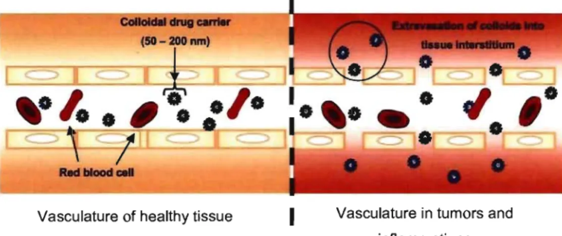
CDC can be designed to achieve targeting of specific tissue, especially solid tumors, using. the enhanced permeation and retention (EPR) effect [Maeda et al.,
2000]. Compared to healthy tissue, sol id tumors are characterized by porous, leaky vasculature (Figure 1-1). Thereby, suitable sized vectors (50-200 nm) are able to extravasate through the fenestrations of these blood vessels and reach the tissue interstitium. Moreover, the poor lymphatic drainage observed in the tumors will ensure that the vector remains in the vicinity of the diseased tissue where it might
release its payload. Therefore, the EPR effect not only allows for the drug to be targeted to diseased cells but also ensures local concentration of the active agent.
Vasculature of healthy tissue
Figure 1-1: The EPR effect.
1
Vasculature in tumors andinflammations
In order to efficiently benefit from the EPR effect, the CDC has to remain in blood circulation for extended periods of time. But free drug administration is often limited by the half-life of the drug in blood circulation. So the role of a vector is to transport the active drug, protect it during its circulation, and prevent it from early elimination, particularly by the mononuclear phagocyte system (MPS). In this aim, a CDC should present specific properties conceming its size and surface properties. For the former, the vector diameter should ideally lie between 50 nm and 200 nm. Smaller CDC are more likely taken up through the fenestrations of the hepatic sinusoidal endothelium [Braet et al., 1995], while colloids above 200 nm are often trapped in the spleen [Moghimi et al., 2001]. To avoid elimination by the MPS, CDC can be coated using biocompatible, flexible, hydrophilic and non-charged polymers, such as polyethylene glycol (PEG). PEG forms a highly hydrated and steric shield against protein adsorption and recognition by cells of the MPS [Allen et al., 2002]. Although PEG remains the most widespread polymer for the preparation of long-circulating colloids [Owens III and Peppas, 2006], several other polymers, such as
poly-N-(2-hydroxypropyl)methacrylamide (PHPMA) [Duncan et al., 2001] and poly(N-vinyl-pyrrolidone) (PVP) [Lukyanov and Torchilin, 2004], have been developed for the same purpose. Long-circulating CDC have also been conceptually proposed to serve as drug containing reservoirs in the bloodstream [Moghimi et al., 2001].
Along with the EPR effect, intracellular drug accumulation can be increased by active targeting to specified cells. This can be performed by associating a targeting residue, or ligand, to the vector. The ligand is able to recognize andfix to a distinct receptor on the surface of the target cells, inducing internalization of the CDC. This has been exploited for the targeting of tumors known to over express specifie surface' receptors, such as folate-receptor [Chung et al., 1993]. Various targeting moieties have been investigated inc1uding galactosal!line [Seymour et al., 2002; Haag and Kratz, 2006], transferrins [Sahoo et al., 2004], antibodies [Allen et al., 1994; Kocbek
et al., 2007] and aptamers [Nutiu and Li, 2005].
Site-specifie concentration of the drug can also be promoted by designing "intelligent" CDC that react to an externally applied stimulus, such as heat [Kono, 2001], ultrasound [Kost et al., 1989] or a magnetic field [Vyas and Jain, 1994], to provoke a localized release. Although such techniques were successful, they often require sophisticated equipment. In contrast, sorne physiological variations within the organism, like changes in pH [Yessine et al., 2003], can be used to drive the discharge of the therapeutic agent without external assistance. pH-sensitive CDC are formulated to retain the therapeutic while in circulation in the blood, which is neutral (pH = 7.4), and release their contents in more acidic compartments, such as tumor interstices and acidic organelles [Schmaljohann, 2006]. Regardless of the stimulus, drug release can be induced by two principal mechanisms: (i) if the drug is covalently bound to the colloid, environmental changes induce hydrolysis of the bond [Seymour
et al., 2002]; (ii) if it is physically entrapped within the vector, leakage results from a
sudden destabilization of the CDC [Connor et al., 1984; Lee, Shin et al., 2003]. The type of colloidal drug delivery system is chosen according to the drug's physico-chemical properties and the intended mechanism of delivery. The following
chapter presents an overview of various CDC that exploit these concepts culminating with a discussion of liposomes, which have been extensively used in this ,work.
1-2 Drug-Polymer Conjugates
Drug-polymer conjugates consist of biologically active agents, including peptides and LMWD, covalentlybound to a polymer. They have encountered a certain success, since several formulations have reached the market and many more are currently in clinical trial [Duncan, i006]. While other CDC are usually designed to physically incorporate the therapeutic, conjugation enhances the pharmacokinetics of the drug by creating new "chemical entities" [Duncan, 2003]. Conjugation generally enhances ~mor targeting, limits toxicity and increases circulation times of the active agent. Furthermore, the administration of hydrophobic LMWD can be facilitated by associating them to hydrophilic polymers, thus vastly improving their water-solubility.
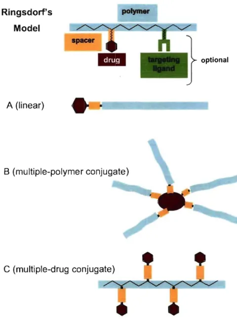
, The first model of drug-polymer conjugate was proposed by Ringsdorf in 1975 (Figure 1-2) and consists ofthree basic elements: the polymer, the linker and the bioactive substance. Optionally, a targeting ligand can be conjugated for cell-specific intemalization [Duncan et al., 2001; Seymour et al., 2002]. According to the number and localization of sites for conjugation, several structures could be envisaged (Figure 1-2).
Ringsdorf's Model A (Iinear) polymer B (multiple-polymer conjugate)
C
(multiple-drug conjugate) optionalFigure 1-2: Schematic representation of Ringsdorfs model of a drug-polymer conjugate and examples of different structures that can arise from conjugation. Haag R and Kratz F.: Polymer therapeutics: Concepts and applications. Angew Chem Int Ed. 2006. 45. 1198-1215. Copyright Wiley-VCH Verlag GmbH & Co. KGaA.
Polymer properties are crucial to impart appropriate pharmacokinetics to the drug conjugate. First, a neutral, water-soluble and flexible macromolecular structure, like PEG, can create a barrier protecting the complex from immunogenic response. Secohdly, the molecular weight of the polymer should be high enough to pro long blood circulation and promo te the EPR effect. AlI the while, depending on the nature of the polymer, the molecular weight should be sufficiently low to aIlow renal clearance after drug delivery, since most polymers used in conjugation so far are not biodegradable [Duncan, 2006]. As for the drug itself, therapeutics such as peptides, oligonucleotides and LMWD are aIl potential candidates as long as they possess a functional group permitting conjugation. The linker may be a simple covalent bond or a spacer molecule. Polypeptide drugs are typicaIly conjugated directly to the polymer through amino acid residues bearing nucleophilic groups, such as cysteine and arginine, as weIl as at the amino and carboxylic termini of the peptidic backbone [Veronese, 2001]. On the other hand, LMWD could be conjugated to the polymer via
cleavable spacers, such as acid labile peptide sequences [Duncan, 2007] and _ hydrazone linkages [Lee et al., 2006]. In such cases, the spacer is employed to release
the LMWD at the site of action.
Most polymer-peptide conjugates are formed using PEG to increase blood circulation times of therapeutic proteins (Figure 1-2A). One of the' first proteins conjugated to PEG was the enzyme L-asparaginase. Treatment of lymphoblastic leukemia necessitates frequent intramuscular administration of L-asparaginase in high doses, which causes allergic and toxic reactions. PEGylated L-asparaginase (Pegaspargase, commercialized by Enzon as Oncaspar~ demonstrated dràsticaIly improved anti-Iymphoma activity over the native enzyme [Graham, 2003]. In pharmacokinetic studies, Pegaspargase demonstrated a half-life of 357 ± 243 h, which was significantly longer than that of L-asparaginase (20 ± 6 h) [Ho et al., 1986].
Since Pegaspargase, several other PEGylated-peptides have been marketed for their capacity to increase plasma residence times of the peptide therapeutics, which include interferon alfa-2a (Pegasys® and Peginterferon®) and 2b (peg-Intron®), recombinant
methionyl human granulocyte colony stimulating factor (Neulasta®) and adenosine deaminase (Adagen~[Duncan et al., 2005; Hamidi et al., 2006].
Though peptide conjugation should ideally involve water-soluble polymers, one particular exception must be noted. The anti-tumor protein neocarzinostatin (NCS) was conjugated to the hydrophobie polymer polystyrene-maleic anhydride (SMA) to form SMANCS [Maeda et al., 1984]. Two chains of SMA were bound to the 1 st and 20th amino acids ofNCS, 1eading to a double polymer conjugate (Figure 1-2B). In aqueous media, SMA presents a globular structure due to the c1ustering effect of the hydrophobie residues, pushing the carboxylate groups to the surface of the globule. Due to the hydrophobie nature of the complex, SMANCS was formulated with the lipid contrast medium Lipidol® and has been approved for the treatment of hepatocellular carcinoma in Japan [Duncan, 2006].
Since linear PEG possesses only two sites of conjugation, at each terminus, it often carries insufficient payload to meet therapeutic requirements for LMWD. A greater loading capacity can be achieved by using polymers with multiple linking sites along the chain (Figure 1-2C) [Duncan, 2006]. Incidentally, it also allows for the conjugation of a targeting moiety [Thatte et al., 2005]. This is the case with PHPMA which displays similar non-toxic and non-immunogenic character as PEG, but additionally possesses several sites of conjugation by substitution of the 2-hydroxypropyl groups. Several anti-cancer drugs have been conjugated to PHPMA and are currently 111 clinical trials, including pac1itaxel, camptothecin, diaminocyc1ohexane palatinate and doxorubicin (Dox) [Duncan et al., 2001; Duncan, 2006]. The latter was conjugated to the PHPMA backbone via a Gly-Phe-Leu-Gly linker, which is hydrolyzable in the acidic medium of lysosomes [Duncan, 2007]. A liver targeting moiety; galactosamine, was also linked to the terminus of the copolymer. The final complex, called PK2, was then tested for the treatment of hepatocellular carcinomas. In a phase 1 study, it was determined that 15-20% of injected dose accumulated in the liver of cancer patients, which was significantly higher than the localization of the non-targeted conjugate [Seymour et al., 2002].
Depending on the initial dose, the accumulation of Dox in hepatic tumor was 12 to 50-fold higher for PK2 than for the free drug, thus achieving notable tissue targeting.
Drug-polymer conjugates have formed the framework upon which other CDC might be developed. In their simplicity, they have exploited the beneficial properties of polymers to keep therapeutic agents in circulation long enough to promote passive and active targeting. Their development has thus been adapted to generate advanced CDC with more complex structure and that will potentially allow more efficient formulation, administration and delivery of CUITent and future medicines.
1 -3 Polymerie N anopartieles
Polymeric nanoparticles (NP) are defined as sol id dispersions that can be categorized as either nanospheres or nanocapsules [Mohanraj and Chen, 2006]. The first are·matrix-like systems within which a drug can be dispersed whereas the second are vesicular structures made of a polymer membrane that confines the drug within an aqueous or oily core [Brigger et al., 2002]. NP have been developed for the delivery of various therapeutics including genetic material [Mao et al., 2001; Yang et al.,
2008], proteins [Watnasirichaikul et al., 2000; Sanchez et al., 2003] and LMWD [Gaucher et al., 2007; Haley and Frenkel, 2008].
NP offer several advantages for the delivery of therapeutics. With a wide range of materials and preparation methods, these versatile systems can improve the pharmacokinetics of bioactive compounds [Soppimath et al., 2001]. The release
i
kinetics of the drug can be controlled by the porosity of the polymeric network [Sant
et al., 2005] as weil as influenced by the biodegradability and erosion of the polymers [Soppimath et al., 2001]. With the help of additives that prevent precipitation, they can form relatively stable dosage forms [Pinto Reis ét al., 2006]. Furthermore, NP
.
can be functionalized with ligands for active targeting [Farokhzad et· al., 2006;
Kocbek et al., 2007]. It is a1so possible to PEGylate the NP to increase blood circulation and reduce recognition by the MPS, which is especially important since NP are often formed with hydrophobic polymers [Li et al., 2001].
Several methods can be used for the preparation of NP [Pinto Reis et al.,
2006]. Nanocapsules are prepared by either interfacial polymerizatiQn or by the .polymer condensation method. For instance, nanocapsules with aqueous cores have been prepared by the polymerization of alkylcyanoacrylates at the interface of water-in-oil emulsions [Lambert et al., 2000; Watnasirichaikul et al., 2000]. Stable emulsions are first prepared with an aqueous solution of the active agent, an oily phase and stabilizing emulsifiers. Afterwards, a solution of the mono mer is added to the mixture which is stirred until complete polymerization, leading to the formation of films around the droplets. The aqueous core makes such systems interesting candidates for the encapsulation of most biological macromolecules [Lambert et al.,
2000; Watnasirichaikul et al., 2000]. Similarly, nanocapsules with hydrophobie cores can be prepared by first forming oil-in-water emulsions in order to encapsulate lipophilic molecules [Al Khouri Fallouh et al., 1986].
The preparation of nanospheres is often based on oil-in-water emulsions followed by solvent evaporation. This method is especially used to prepare nanospheres with hydrophobie polymers, such polY(D,L-lactic acid) (PLA), poly(glycolic acid) (PGA), poly(lactide-co-glycolic acid) (PLGA) and pcilY(E-caprolactone ) (peL) [Soppimath et al., 2001; Pinto Reis et al., 2006]. NP consisting of such polymers have the added advantage of being biodegradable over time in aqueous media [Park, 1994; Sanchez et al., 2003]. The size of the NP can be controlled using emulsifiers, such as surfactants and block copolymers. The latter can additionally be designed to remain attached to the surface to generate long-circulating formulations with well hydrated surfaces [Gaucher et al., 2007]. The NP can be collected by dialysis, filtration or ultra centrifugation, which also helps remove excess emulsifiers and to wash the partic1es.
Another method to prepare nanospheres is nanoprecipitation of hydrophobie polymers. In one particular study, PEGylated NP were formed with amphiphilic block
copolymers with large PLGA chains and shorter PEG segments [Farokhzad et al., 2006]. PLGA-block-PEG was first dissolved with the drug, Docetaxel (Dtxl), in acetonitrile and added dropwise into water. Without the need of surfactants, the
diffusion of the organic phase allowed the PLGA block to precipitate, thus producing PEGylated nanospheres (d:::::: 153 nm) loaded with Dtx1. Furthermore, each PEG chain possessed a carboxylic acid at the terminus that permitted conjugation of aptamers to the NP for active-targeting. Targeted NP improved tumor regression for mice bearing prostate cancer cells with a 100% survival rate, whereas only 50% survival was observed in mice treated with non-targeted NP and ev en less when free Dtxl was administered [Farokhzad et al., 2006].
Hydrophilic polymers, such as chitosan [Calvo et al., 1997b; Obara et al.,
2005] and dextran [Kim et al., 2000] have been used to prepare nanospheres due to their biocompatibility. They are particularly used for the vectorization of hydrophilic bioactives like peptides [Soppimath et al., 2001]. For instance, protein-Ioaded NP have been formed with positively charged chitosan by the neutralization of its charges using a counter-ion, tripolyphosphate [Calvo et al., 1997a, b] .. Genetic material, which is negatively charged, can be incorporated into chitosan NP by similar electrostatic interactions
There is
a
wide range of biodegradable and biocompatible materials available for the preparation of NP. Though many NP preparation methods lead to the vectorization different bioactive compounds, the size and polydispersity of the particles are sometimes difficult to control, especially for NP produced by nano-precipitation [Mohanraj and Chen, 2006]. AIso, a marked burst release of the drug is noticed when it is located close to the surface of the NP [Mohanraj and Chen, 2006]. Nonetheless, NP often exhibit greater drug loading efficiency and stability than the following CDC, i.e. polymerie micelles.1 -4 Polymerie Micelles
Micelles are colloidal structures formed by the spontaneous self-association of low molecular weight or polymerie amphiphiles in a solvent that is selective for either moi et y [Torchilin, 2001]. These molecules exist separately below their critical micelle concentration (cmc), above which they assemble into core-shell structures
[Torchilin, 2001; Torchilin, 2007]. Polymerie micelles (PM) are made of amphiphilic
block copolymers which possess an inert hydrophilic block to form ,the hydrated corona and another segment to form the core. Depending on the composition of the core-forming segment, micellization can be driven by hydrophobie or electrostatic cohesive forces. In addition, the length and structure of the segments can' influence the size and stability of the micelles. Though PM aregenerally spherical in shape, the length of the polymer blocks [Zhang and Eisenberg, 1995; Zhang and Eisenberg, 1996] and the solvent conditions [Shen et al., 1999; Choucair and Eisenberg, 2003] may impose other morphological arrangements, such as rods, tubules and lamellae. In non-polar organic solvents, it is also possible to form reverse-micelles, which consist of hydrophobie corona surrounding a hydrophilic core. However, the following discussion will be limited to spherical PM formed in aqueous media as they are the most applied in drug delivery research.
PM offer several advantages, such as their capacity to solubilize or incorporate bioactives, their size that allows for efficient passive targeting, and the biocompatibility of available polymers [Yokoyama, 2005; Torchilin, 2007]. The hydrophilic chains in the corona can also prevent secondary aggregation of the micelles due to their hydration, while also stabilizing core formation. Finally, PM are good candidates for active targeting since they can be formed of stimuli-sensitive polymers and can often be functionalized by chemical conjugation of ligands on the surface of the corona. PM have b~en successfully designed for the encapsulation of poorly water-soluble drugs [Ramaswamy et al., 1997; Cavallaro et al., 2004; Huh et
al., 2005; Elsabahy et al., 2007], genetic material [Kataoka et al., 1999; ltaka et al.,
2003; Dufresne et al., 2004] and proteins alike [Harada and Kataoka, 1998, 1999]. Amphiphilic copolymers typically possess cmc values around 10-6_10-7 M, whereas those of low molecular weight surfactants (LMWS) usually lie between 10-3 and 10-4 M [Bae and Kataoka, 2005]. PM are therefore considerably more stable against dissociation than LMWS-micelles upon dilution. Hydration of the hydrophilic block also imparts a steric stabilization that prevents aggregation of the PM.
In the same manner as for NP, organic solvents can be used to introduce hydrophobic LMWD into the core. The poorly water-soluble drug and the amphiphile are first dissolved in an organic phase. The latter is then slowly removed from the medium, either by dialysis against water or by evaporation after addition of the aqueous phase. These methods allow the hydrophobic chains to associate and entrap the drug. Another approach is to covalently conjugate the poorly water-soluble drugs to the core-forming block of the polymers [Y okoyama et al., 1990; Y okoyama et al.,
1991]. This method increases loading efficacy, . ensures the delivery of high doses and prevents the agent from leaking out. In such instances, it is sometimes preferable that the bonds are cleaved when the vector reaches the target site to improve therapeutic activity.
PM designed for the delivery of charged bioactives are called polyion complex (PIC) micelles and associate via electrostatic interactions [Harada and Kataoka, 1998; Kataoka et al., 1999; ltaka et al., 2003; Dufresne et al., 2004]. PIC micelles are formed of copolymers possessing a neutral hydrophilic segment and a polyionic block. The latter can complex with charged bioactives and then self-associate. For instance, negatively charged genetic material can be associated to block
copolymers with a polycationic segment to form the core of the PM. PIC micelles have been shown to protect therapeutic oligonucleotides from nuclease activity [Katayose and Kataoka, 1998] and improve their pharmacokinetics [Harada-Shiba et al., 2002].
The following example highlights the principal advantages of PM as CDC. Lee et al. (2003) prepared Dox-Ioaded, pH-sensitive mixed micelles (PHSM), targeted or not with folate ligand (PHSM/f) against a malignant breast cancer cell line (MCF-7) [Lee, Na et al., 2003]. The authors synthesized block copolymers consisting ofpoly(L-histidine) (polyHis) and PEG, which micellized under basic pH [Lee, Shin
et al., 2003]. The polyHis block that formed the hydrophobic core of the micelles had two roles. First, it provided pH-sensitivity to the PM since protonation of the histidine groups under mildly acidic conditions made the core-forming block water-soluble, thus prompting disassembly of the micelles and the release of Dox [Lee, Shin et al.,
2003]. Secondly, polyHis was selected for its endosomolytic properties and was expected to promotei intracellular drug delivery by destabilization of the endosome
[Lee, Na et al., 2003]. PolyHis-block-PEG was formulated with 25 wt%
PLA-block-PEG' to lower the dissociation pH from 7.6 to 7.0 [Lee, Na et al., 2003]. This proportion of PLA-block-PEG was necessary to enhance micelle stability at pH 7.4 and yet permit controlled release ofthe incorporated contents [Lee, Na et al., 2003].
PHSM and PHSM/fwere administered by i.v. injection into mice grafted with MCF-7, where Dox accumulation was 5 times greater than in the tumors of mice treated with free Dox [Lee et al., 2005]. For mice bearing a drug resistant MCF -7 strain, PHSM/f maintained Dox delivery to the tumor cells, while Dox accumulation was 50% lower after PHSM-treatment and undetectable after administration of the free drug [Lee et al., 2005].
As compared to nanospheres, the size of PM can be more easily controlled due to the way amphiphilic block copolymers self-assemble. Second, micelle formation is govemed by a dynamic structure that gives PM the potential for improved triggered release. Stimuli-responsive polymers can be employed to control disassembly and delivery of the therapeutic. In the final section of this chapter, we discuss liposomes, which are also formed by self-association of amphiphiles and can likewise be designed to respond to stimuli.
1-5 Liposomes
Liposomes are c10sed micro- or nanoparticulate vesic1es of one or more lamellae that are formed by the self-assembling of phospholipids [Lasic and , Templeton, 1996]. As the latter are typically found in the biological membranes of all living organisms, their biocompatibility makes liposomes good CDC candidates. Although liposomes are not composed of synthetic polymers, polymer chemistry has played a role in the design of liposomes for drug delivery. The following sections will begin with a synopsis of the structures and dynamics involved in liposome formation, followed by a brief review of the different types of liposomal vectors.
1-5.1 Liposome composition
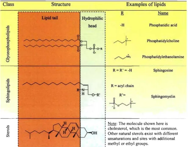
One of the principal benefits of liposomes is that the y can be prepared using natural amphiphiles. There are three major classes of natural lipids, as de~cribed in Table 1-1. The first class consists of phospholipids, specificaUy glycerophospholipids, which are fonned of a glycerol molecule bonded to a phosphate group to fonn the polar head group, and one or two fatty acids via ester linkages. The fatty acids generaUy vary in chain length (12, 14, 16 and 18 carbons) and unsaturation (l, 2 or 3). The second class are the sphingolipids, since they are derivatives of the base structure, sphingosine. Sphingomyelin, which is often used for liposome preparation, may also be categorized as a phospholipid because it, too, possesses a phosphate group. The third type of naturaUy occurring lipids employed for liposomes is sterols, of which cholesterol (Chol) is the most commonly used. AU three classes of lipids have in common a lipidic domain and a hydrophilic head. These two regions account for the attraction of the hydrophobie tails and electrostatic repulsion of the hydrophobie heads. The sum of the attractive and repulsive forces results in the. self-assembly of the amphiphiles in aqueous media.
Table 1-1: Classification of naturallipids used in liposome preparation Class {j :ë-ë3 ..c:: 0. '" o ..c:: 0. o <1> Ü ;>, 6 Structure Hydrophilic head ~-O-R b--R' OH
L
_
_____
__
_
_
_____
_
___
_________ _
Examples of lipids -H R = R' =-H R = acyl chain R'= ~1
.
- P ...-... ~J'I,.'-J'o' o '-/1 Phosphatidic acid Phosphatidylc ho 1 ine Phosphatidylethanolarnine Sphingosine SphingomyelinNote: The molecule shown here is cholesterol, which is the most common. Other natural sterols exist with different unsaturations and sites with additional methyl or ethyl groups.
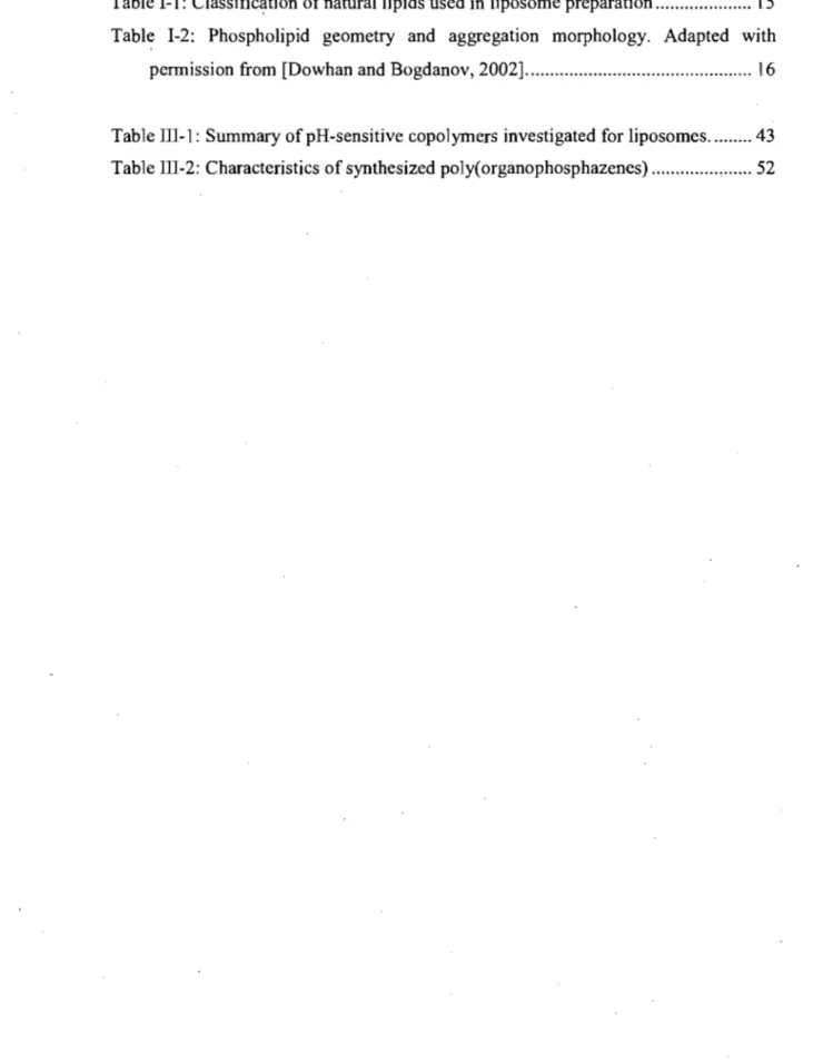
The molecular geometry of these lipids dictates their arrangement in aqueous media (Table 1-2). Phospholipids that possess a cylindrical shape associate into lipid bilayers. This arrangement is the most thermodynamically stable conformation since it allows minimal contact of water with the lipophilic chains. Conversely, lipids that possess a single alkyl chain per molecule are said to have an inverted conical shape and preferentially associate to form micelles. Finally, phospholipids with smaller head groups and unsaturated fatty acid chains possess a conical shape and self-associate into a hexagonal phase, which can be illustrated as reverse micelle rods. The physical properties of liposomal membranes are typically controlled by lipid composition and can be influenced by the environmental conditions (temperature, pH,
etc.). Based on the desired membrane properties, synthetic lipids can also be designed
to complement or replace naturally existing lipids.
Table 1-2: Phospholipid geometry and aggregation permission from [Dowhan and Bogdanov, 2002].
Molecular shape Phase Cylinder Bilayer 1nverted Cone Micellar morphology. Adapted
fj
Cone Hexagonal (Hn) withWhile liposomes are sometimes c1assified according to the method of preparation [Simard et al., 2007], it may be more intuitive to compare them by size
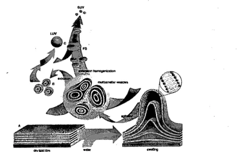
and lamellarity [Sharma and Sharma, 1997; Simard et al., 2007]. When dried lipids
are rehydrated, they form a heterogeneous dispersion of multilamellar vesic1es (ML V), ranging in size from 0.1 to 10 Ilm [Ulrich, 2002]. These vesic1es generally contain multiple lipid bilayers of concentric spheres [Deamer and Uster, 1983] and can also be called giant oligomeric vesic1es (GOV) [Simard et al., 2007]. Large
unilamellar vesic1es (LUV) are between 100 and 400 nm in diameter and small unilamellar vesic1es (SUV) between 40 and 100 nm [Simard et al., 2007]. Sometimes,
the term medium sized unilamellar vesic1es (MUV) is used for liposomes with a size distribution overlapping LUV and SUV [Simard et al., 2007].
1-5.2 Liposome preparation
Several met~ods exist to prepare liposomes, depending on the desired size and lamellarity. The first preparation of ML V was described by Bangham et al. (1965).
,
The lipids were solubilized in an organic solvent, like chloroform, which was removed by evaporation to obtain a dry. lipid film. Then, slow hydration under moderate agitation lead to ML V formation. The size of ML V can be tuned by vigorous vortexing, brief sonication or extrusion [Szoka and Papahadjopoulos, 1981].
Figure 1-3: Schematic representation for the preparation of MLV, LUV and SUV. Reproduced with permission from [Lasic, 1997].
LUV can be generated by several methods. First, extrusion of ML V with multiple passages through polycarbonate filters results in LUV with narrow size distribution [Deamer and Uster, 1983]. The second method is reverse-phase evaporation [Szoka and Papahadjopoulos, 1981], which consists of preparation of inversed micelles by first forming water-in-oil emulsions with lipids as emulsifiers and then sonication to homogenize the drop lets. Subsequent evaporation of the organic phase under reduced pressure allows the lipids to coalesce to form LUV [Deamer and Uster, 1983]. A last commonly used method is the detergent removal
method [Deamer and Uster, 1983]. Lipids and detergent are co-suspended 10 a concentrated aqueous phase. Dilution, often by gel filtration, removes the detergent and allows vesicle formation.
Conceming SUV, there are primarily two methods used for their preparation: sonication and solvent-injection. The former consists of sonicating MLV su'spensions either using a probe or bath sonicator. Though the probe can provide more power and produces SUV in a few minutes, it might degrade the lipids and contaminate the , vesicles with metal impurities [Szoka and Papahadjopoulos, 1981]. Conversely, bath sonication allows for better control of the temperature and the lipid suspension may be manipulated in an inert atmosphere [Szoka and Papahadjopoulos, 1981]. The solvent-injection method implies that phospholipids, dissolved in a small volume of organic solvent like ethanol, are injected through a narrow syringe into a large aqueous medium where they self-assemble [Szoka and Papahadjopoulos, 1981]. The solvent is then subsequentlyremoved by filtration. A similar method replaces ethanol with diethyl ether. The aqueous medium is heated so that, as the organic solutionis
inject~d, the ether is removed by evaporation and the SUV are formed.
In this work, we used the extrusion method to produce LUV smaller than. 200 nm in size. Amphiphilic polymers were included in the lipid composition in order to incorporate them into the bilayer.
1-5.3 Liposomes for drug delivery
The application of liposomes in drug delivery first arises from the possibility of encapsulating hydrophilic molecules in the aqueous inner compartment of the vesicles. It has also been established that hydrophobie drugs and amphiphilic molecules can be trapped into the lipid bilayer [Sharma and Sharma, 1997]. Incorporating amphiphilic polymers into the membrane has been particularly useful for the modification of liposome surface properties. Pharmaceutical liposomes can thus be classified on the basis of their composition, which places them in at least one of the following categories (Figure 1-4): conventionalliposomes, cationic liposomes,
targeted liposomes, 10ng-circu1ating (stealth) liposomes [Storm and Cromme1in, 1998]. Each of these categories has its particularly intended applications [Storm and Crommelin, 1998], yet recent developments are seeking to combine these technologies.
Conventional
Targeted
Figure 1-4: Schematic representation of the four major categories of liposomes Reproduced with permission from [Storm and Crommelin, 1998].
Conventional liposomes are by far the simp1est type, constituted main1y of bi1ayer-forming phospholipids. These are the basis for the deve10pment of aU other types of liposomes. Their application in drug de1ivery has been 1imited by uptake by the MPS [Storm and Crommelin, 1998], yet the rate of elimination is dependent on the 1ipids selected for the formulation [Gabizon and D, 1988]. On the other hand, cationic liposomes, or liposomes formed with positively charged lipids, are particu1arly efficient for the complexation and formulation of genetic materia1 [Lasic and Temp1eton, 1996; Huang, 2008]. As for aU the CDC seen so far, targeted liposomes can be formed by fixing recognition ligands to the phospho1ipid surface. For instance, antibodies can be grafted to the surface of the liposome, forming
immunoliposomes [Torchi1in, 2006; Khaw et al., 2007]. To overcome premature elimination, 10ng-circulating liposomes can be generated by the addition of
PEG-conjugated lipids to the liposome composition [Ulrich, 2002]. Several long-circulating liposome' formulations are on the market or in clinical studies for the passive targeting of cancers [Sharma and Sharma, 1997].
, The inherent contradiction with long-circulating liposomes for drug delivery is that the contents are required to be released for therapeutic activity. Stimuli-sensitive liposomes have thus gained noteworthiness for the triggered discharge of encapsulated agents [Kono et al., 1994; Kono et al., 1999; Drummond et al., 2000;
Simoes et al., 2004; Ishida et al., 2006; Karanth and Murthy, 2007; Huang, 2008]. pH-sensitive liposomes can use natural physiological changes in pH to release the contents. The first pH-sensitive vesicles were generated using positively charged phospholipids with an inversed conical shape, dioleoyl phosphatidylethanolamine (DOPE), that were stabilized by mildly acidic amphiphiles to form bilayers [Connor
et al., 1984]. When the medium was acidified, the amphiphiles were protonated, destabilizing the hydrophobie interactions within the bilayer. The liposomes thus became fusogenic and the encapsulated material was released [Ellens et al., 1984,
1985]. Other pH..:responsive liposomes have since been developed, employing various mechanisms, such as fusogenic peptides [Subbarao et al., 1987], pH-sensitive polymers [Yong-Hee et al., 1994] and acid-labile bonds [Guo and Szoka, 2001; Boomer et al., 2003]. These are discussed in more depth in the fourth chapter of this thesis.
More recently, advanced liposomal CDC have beendeveloped, combining the different types of liposomes. For instance, it was possible to formulate vesicles with both pH-sensitive polymers and PEG-lipids in order to produce ~H-responsive, long-circulating liposomes [Roux et al., 2004]. Others prepared vesicles by stabilizing DOPE with lipids conjugated to PEG using acid labile bonds [Hong et al., 2002].
Cleavage of PEG induced liposomal fusion and leakage of the contents. Long-circulating immunoliposomes have also been developed by decorating the surface of the vesicles with both PEG and monoclonal antibody conjugates [Allen et al., 1995;
Maruyama et al., 1997]. Currently, liposomal delivery systems are being developed bearing all three features: long-circulation, target-mediated and stimuli-induced
release [Mastrobattista et al., 1999]. In addition to this, numerous polymers are being investigated for either their capability to shield liposomes or their response to stimuli. Indeed, we propose poly(phosphazenes) for the pH-induced drug release from liposomes. In the following chapter, we review the history and principles of phosphazene chemistry, as well as the various applications of poly(phosphazenes), including drug delivery.
polymers
11-1 Introduction to poly(phosphazenes)
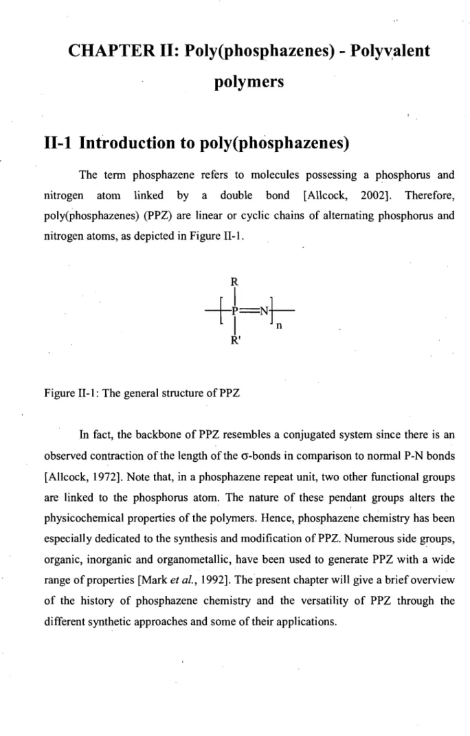
The term phosphazene refers to molecules possessing a phosphorus and nitrogen atom linked by a double bond [Allcock, 2002]. Therefore, poly(phosphazenes) (PPZ) are linear or cyclic chains of altemating phosphorus and nitrogen atoms, as depicted in Figure II-l.
Figure II-l: The general structure ofPPZ
In fact, the backbone of PPZ resembles a conjugated system since there is an observed contraction of the length of the cr-bonds in comparison to normal P-N bonds [Allcock, 1972]. Note that, in a phosphazene repeat unit, two other functional groups are linked to the phosphorus atom. The nature of these pendant groups alters the physicochemical properties of the polymers. Rence, phosphazene chemistry has been especially dedicated to the synthesis and modification of PPZ. Numerous side groups, organic, inorganic and organometallic, have been used to generate PPZ with a wide range ofproperties [Mark et al., 1992]. Thepresent chapter will give a brief overview of the history of phosphazene chemistry and the versatility of PPZ through the different synthetic approaches and sorne of their applications.
11-2 Poly(phosphazene) synthesis
The history 'of phosphazene chemistry dates back to the 19th century, before Lewis proposed his theory on bonding [Lewis, 1916], and well before the term polymer was universally accepted [Staudinger, 1920]. In 1850, Laurent proposed that the white precipitate formed by heating phosphorus pentachloride (PCls) with ammonia (NH3) should simply be CbPN [Laurent, 1850]. Over ten years later,
Gladstone confirmed that the product more likely possessed the empirical formula P3N3Cl6 [Gladstone. and Holmes, 1864], or three times the formula proposed by
Laurent. Near the end of the century, Stokes obtained sorne groundbreaking results, isolating and characterizing a series of four phosphonitrilic chlorides, [CbPNh-7, also
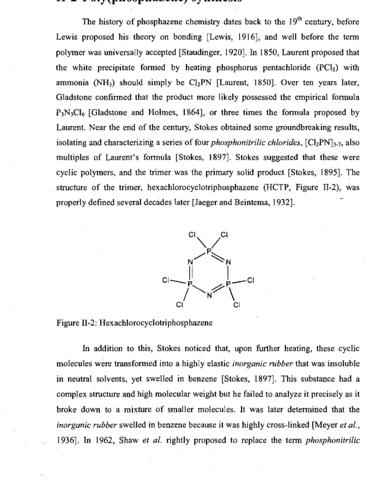
multiples of Laurent's formula [Stokes, 1897]. Stokes suggested that these were cyclic polymers, and the trimer was the primary solid product [Stokes, 1895]. The structure of the trimer, hexachlorocyclotriphosphazene (HCTP, Figure 11-2), was properly defined several decàdes later [Jaeger and Beintema, 1932].
Figure II-2: Hexachlorocyclotriphosphazene
In addition to this, Stokes noticed that, upon further heating, these cyclic molecules were transformed into a highly elastic inorganic rubber that was insoluble
in neutral solvents, yet swelled in benzene [Stokes, 1897]. This substance had a complex structure and high molecular weight but he failed toanalyze it precisely as it broke down to a mixture of smaller molecules. 1t was later determined that the
inorganic rubber swelled in benzene because it was highly cross-linked [Meyer et al.,
chlaride for phasphazene because of the double bond between phosphorus and nitrogen [Shaw et al., 1962]. In the mid-1960's, Allcock et al. finally fine-tuned Stokes' polymerization procedure to ob tain high molecular single chains of polydichlorophosphazene (PDCP), where the side groups are chlorides. Today, PDCP is a precursor to most functional PPZ by the substitution of the chlorine side-groups. In the following section, we shall first discuss the conclusions of Allcock's initial work which has led to the vastly employed "thermal ring-opening polymerization. We will then examine the evolution of the alternative condensation polymerization procedures as well as the post-polymerization modifications that impart stability and functionality to these polymers.
11-2.1
Thermal ril1g-opening polynlerization
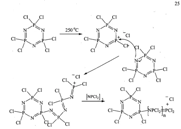
The major problem with Stokes's polymer was its hydrolytic instability, due to the reactivity of the phosphorous-chlorine bonds. Stabilization of the polymer required the synthesis of a linear PDCP that wou Id be soluble in a suitable solvent for side-group substitution. Allcock et al. (1964) first noted that, unlike condensation reactions, there was no leaving group in this polymerization as the empirical formula essentially remained the same. Conductivity data obtained during polymerization suggested that heating induced dissociation of a chloride ion, which permitted nucleophilic attack by the nitrogen from a neighboring HCTP [Allcock and Best, 1964]. Hence, the mechanism for the ring-opening polymerizationwas elucidated, as depicted in Figure 11-3. Later, careful control of the reaction time, temperature and the purity of HCTP yielded linear PDCP that was soluble in several aprotic solvents (e.g.
THF) and could subsequently be substituted with various nucleophiles [Allcock and Kugel, 1965, 1966; Allcock et al., 1966]. These reactions will be further discussed with other post-polymerization modifications.
Figure II-3: Mechanism of thermal ring-opening polymerization ofHCTP.
, Thermal ring-opening polymerization is usually performed in the bulk state in closed tubes. Trace impurities such as water [Allcock et al., 1975] and Lewis acids [Hergenrother et al., 1986; Sohn et al., 1995] can catalyze the reactions. Cyclotriphosphazenes bearing good leaving groups, such as other halogens, can be polymerized in the same way. However, the temperature of the reaction should be adjusted accordingly. For example, hexafluorocyclotriphosphazene polymerizes at 350 oC instead of 250 oC because of the strength of the phosphorus-fluorine bond [Seel and Langer, 1956].
The thermal ring-opening polymerization of HCTP possesses several advantages. First, polymers of very high molecular weight, over 100,000 g morl, can be generated by this method. Regularly altemating repeat units can also be formed by partially substituting sorne of the chlorines on HCTP prior to polymerization [Allcock
et al., 1978; Allcock et al., 1990]. To this end, no more than 2 or 3 chlorides should be replaced to prevent over-stabilization of the trimeric unit, unless trans-annular -organic or ferrocenyl species are substituted on the ring [Allcock et al., 1991]. It is
believed that the strain imposed by these substituents on the cyclic monomer promotes ring-opening upon heating. The lower reactivity of substituted-cyclotriphosphazenes can generally help limit the rate of chain elongation wh en desired. The principal disadvantages of the ring-opening procedure are the high polydispersity of the resulting PPZ (> 2) and the difticulty to control the molecular weight.
11-2.2
Condensation polymerization
Although the ring-opening polymerization is the most popular synthesis pathway for the preparation of PDCP, condensation polymerization offers several advantages that are particularly interesting for the preparation of CDC. Sorne of the methods could be simply transposed in industrial settings while others give access to polymers with controlled chain length and low polydispersity. Furthermore, they may lead to PPZ with different and precise structures. The following will describe the synthesis and polymerization of three types of condensation monomers, presenting for each their synthetic advantages and disadvantages.
11-2.2.1
Condensation of phosphorus pentachloride and ammonia
The tirst condensation polymerization is based on the same starting blocks as for the preparation of HCTP. However, Hombaker et al. (1980) developed an
industrial process for the synthesis of short oligomers. Since manipulation of PCIs is delicate on large scales, it was prepared in situ prior to the addition of ammonia.
Liquid PCb was tirst fed along with gaseous chlorine (Ch) into a chlorobenzene tilled reactor set between 100 and 140 oC, followed by addition of ammonia or ammonium chloride (Figure 1I-4). Controlling the feed to have a slight excess in PCb yielded linear PPZ with 2 to 9 repeat units [Hombaker and Li, 1980].
NH3(g) +
-PCI3(l) + CI2(g)
- - - 1
...
CI-tPCI2NtPCI3 PCl6 + HCIChlorobenzene 2-9
100-140 Oc
Figure IIA: Preparation of short PPZ oligomers
After oligomerization, the temperature can be increased to 140-160 oC and NH4CI added to the reactor to link the short chains together, forming PPZ up to 6900
g mOrl [Pettigrew et al., 1983]. This method offers the advantages ofusing affordable reactants and modest polymerization temperatures (160 vs. 250 oC for ring-opening).
Nevertheless, this technique leads to considerable losses as cyclic side-products and short polymer chains.
11-2.2.2
Synthesis and polymerization of CI
3P=NP(O)Ch
CbP=NP(O)Ch is a moi sture sensitive, inorganic solid that could be polymerized to form PPZ with higher molecular weight than the previous method. It
was most efficiently obtained by heating PCIs with ammonium sulfate (Figure II-5).
Figure II -5: Synthesis of ChP=NP(O)Ch
This reaction achieved a 100%, high purity yield since all of the side products are gaseous and readily removed from the reaction medium [Emsley et al., 1971]. The monomer polymerizes by heating above 240 oC and is monitored by the removal of gaseous OPCh from the reaction medium [D'Halliun and De Jaeger, 1989]. When ChP=NP(O)Ch was heated to 245 oC, OP Ch production was no longer detected in the vessel after 7.7 h and 95% completion of the reaction [D'Halliun et al., 1992].
![Table 1-2: Phospholipid geometry and aggregation permission from [Dowhan and Bogdanov, 2002]](https://thumb-eu.123doks.com/thumbv2/123doknet/7657790.238178/33.919.77.812.164.1112/table-phospholipid-geometry-aggregation-permission-dowhan-bogdanov.webp)
![Figure 1-4: Schematic representation of the four major categories of liposomes Reproduced with permission from [Storm and Crommelin, 1998]](https://thumb-eu.123doks.com/thumbv2/123doknet/7657790.238178/36.919.156.798.83.585/figure-schematic-representation-categories-liposomes-reproduced-permission-crommelin.webp)