HAL Id: tel-02308916
https://tel.archives-ouvertes.fr/tel-02308916
Submitted on 8 Oct 2019HAL is a multi-disciplinary open access archive for the deposit and dissemination of sci-entific research documents, whether they are pub-lished or not. The documents may come from teaching and research institutions in France or
L’archive ouverte pluridisciplinaire HAL, est destinée au dépôt et à la diffusion de documents scientifiques de niveau recherche, publiés ou non, émanant des établissements d’enseignement et de recherche français ou étrangers, des laboratoires
Induction of atrial endothelial senescence by angiotensin
II and thrombin : role of oxidative stress and
characterization of pro-thrombotic, pro-adhesive,
proteolytic and pro-fibrotic phenotype
Hira Hasan
To cite this version:
Hira Hasan. Induction of atrial endothelial senescence by angiotensin II and thrombin : role of ox-idative stress and characterization of pro-thrombotic, pro-adhesive, proteolytic and pro-fibrotic phe-notype. Cardiology and cardiovascular system. Université de Strasbourg, 2018. English. �NNT : 2018STRAJ122�. �tel-02308916�
Induction de la sénescence endothéliale
auriculaire par l’angiotensine II et la thrombine :
Rôle du stress oxydant et caractérisation du
phénotype pro-thrombotique, pro-adhésif,
protéolytique et pro-fibrotique
UNIVERSITÉ DE STRASBOURG
ÉCOLE DOCTORALE DES SCIENCES DE LA VIE ET DE LA SANTE
INSERM UMR 1260 Nanomédecine régénérative
THÈSE
présentée par :Hira HASAN
soutenue le : 19 NOVEMBRE 2018
pour obtenir le grade de :
Docteur de l’Université de Strasbourg
Discipline/ Spécialité: Pharmacologie
Directeur de Thѐse :
Mr. MOREL Olivier PUPH, Université de Strasbourg
Co-directeur de Thѐse :
Mme. SCHINI-KERTH Valérie B. PU, Université de Strasbourg
RAPPORTEURS externes : Mr. RICHARD Vincent Mme. BOULOUMIE Anne
PUPH, Université de Rouen
DR, INSERM UMR 1048, Université Paul Sabatier Toulouse
PUPH, Université de Strasbourg RAPPORTEURS interne :
Mr. GENY Bernard
Mr. MANSOURATI Jacques
Invitée
DEDICATION
I would like to dedicate my thesis to my parents, Ch Ilyas Hasan and Batool Ilyas, my husband
Muhammad Umar and my siblings Ch Raza Hasan, Sarah Hasan, Fareeha Hasan and Maryam Nawal Hasan whose continuous support made this journey easier and possible.
ACKNOWLEDGMENT
First, I would like to thank my parents who supported and facilitated me in every aspect to continue the journey towards doctor of philosophy. Then, Higher education commission of Pakistan for the financial support to attain this degree. From the core of my heart, I would also like to thank my supervisor OLIVIER Morel and co-supervisor SCHINI-KERTH Valerie B. for the continuous support throughout the last three years. There is so much I learnt which will definitely enlighten the future pathways for me.
I would also like to thank MOREL JESEL Laurence for the precious discussions and for teaching me. I am also grateful to all researchers, students and teachers that I met on my way during these years. I am also grateful to all my Pakistani colleagues who made this journey easier by their valuable advices, support and help. I would also like to thank Professor AKHTAR Shoaib and ALAMGEER, who always wanted me to continue the research and were always there for guidance. I would also like to thank all my tecahers who taught me at different stages of my life as they gave me much valuable advices, comments and suggestions.
I am really thankful to my husband MUHAMMAD Umar, who always believes in me and who is always there for me without whom everything seems impossible. Thanks my love, I always feel honoured to have you on my side and I am really thankful to GOD who chose you for me. At the end I would also like to thanks my father- in law and mothe-in-law, who always kept praying for me for my successful and healthy journey of Life.
Table of contents
Dedication
i
Acknowledgement
ii
Table of contents
iii
List of abbreviations
viii
List of figures
x
List of tables
xii
Resume-French version
1
Resume-English version
7
Scientific contribution
12
Introdution
15
1 ATRIAL FIBRILLATION (AF)
16
1.1 Introduction and definition
16
1.2 Classification of AF
16
1.3 Epidemiology and impact for patients
17
1.3.1 Incidence and prevalence of atrial fibrillation
17
1.3.2 Morbidity, mortality and health care burden of atrial fibrillation18
1.4 Atrial fibrillation risk factors
19
1.4.1 Ageing
19
1.4.2 Hypertension
20
1.4.3 Heart failure and coronary artery diseases
21
1.4.4 Diabetes
23
1.4.5 Thyroid dysfunction
24
1.4.6 Alcohol
25
1.4.8 Post-operative atrial fibrillation
27
1.4.9 Genetic risk factors
27
1.4.10 Others
27
2. ATRIAL FIBRILLATION PATHOPHYSIOLOGY
29
2.1 Electrophysiological remodeling
31
2.2 Atrial stretch and mechanical remodeling
33
2.3 Structural remodeling and fibrosis
33
2.3.1 Angiotensin II
37
2.3.2 Transforming growth factor beta-1
38
2.3.3 Platelet-derived growth factor39
2.3.4 Connective tissue growth factor39
2.4 Coagulation cascade components and atrial fibrillation
39
2.5 Neural autonomic remodeling
41
2.6 Anatomic factors
42
2.6.1 Role of specific structures
42
2.6.2 Regional ion current differences43
2.7 Atrial fibrillation and inflammation
43
2.8 Atrial fibrillation and oxidative stress
46
2.9 Atrial fibrillation, stroke and microparticles
47
2.10 Management of atrial fibrillation
47
2.10.1 Rate control therapy in atrial fibrillation
47
2.10.2 Rhythm control therapy in atrial fibrillation
48
2.10.3 New antiarrhythmic drugs
48
2.10.4 Catheter ablation of atrial fibrillation
49
2.10.6 Interventional approaches to stroke prevention
50
2.10.7 Upstream therapy
50
3. ENDOCARDIAL ENDOTHELIAL CELLS
51
3.1 Introduction
51
3.2 Strutural characteristics of endocardial endothelial cells
52
3.3 Physiological role of endocardial endothelial cells
52
3.3.2 Vasostatin-1 and endothelin-1
58
3.3.3 Prostaglandins
61
3.3.4 Angiotensin II
61
3.3.5 Reactive oxygen species
62
3.3.6 Peptide growth factors
64
3.4 Endocardial endothelial dysfunction
65
3.5 Endocardial endothelial dysfunction and heart diseases
66
3.6 Endocardial endothelial dysfunction and atrial fibrillation
68
4. ENDOTHELIAL SENESCENCE
71
4.1 Cellular senescence
72
4.2 Biomarkers and features of senescence
72
4.2.1 Morphological characteristics
72
4.2.2 Cell cycle arrest
73
4.2.3 Senescence-associated beta galactosidase activity
73
4.2.4 Senescence-associated heterochromatin foci73
4.2.5 Secreted factors
74
4.3 Mechanism of senescence
74
4.3.1 Replicative senescence
74
4.3.2 Premature senescence
75
4.3.3 Molecular machinery of cellular senescence
76
4.3.4 Reactive oxygen species and senescence79
4.4 Senescence and endothelial dysfunction
81
4.5 Local angiotensin system and senescence
82
AIMS OF THE STUDY
85
RESULTS
88
Article 1
89
Article 2
124
DISCUSSION AND PERSPECTIVES
156
List of Abbreviations
AF Atrial fibrillation
AECs Atrial endothelial cells
Ang II Angiotensin II
AT1R Angiotensin type 1 receptor
ACE Angiotensin-converting enzyme
APD Action potential duration
ANP Atrial natriuretic peptide
CAD Coronary artery disease
CABG Coronary artery bypass graft
DTI Direct thrombin inhibitor
ECG Electrocardiogram
eNOS Endothelial nitric oxide synthase
HF Heart failure
ICAM-1 Intercellular adhesion molecule-1
MCP-1 Monocyte chemoattractant protein-1
NO Nitric oxide
PVs Pulmonay veins
PDGF Platelet-derived growth factor
PAR Protease-activated receptor
PAI-1 Plasminogen activator inhibitor-1
PKC Protein kinase C
ROS Reactive oxygen species
RAAS Renin angiotensin aldosterone system
SASP Senescence-associated secretory phenotype
VCAM-1 Vascular cell adhesion molecule-1
VEGF Vascular endothelial growth factor
LIST OF FIGURES
Figure 1: Rhythm of healthy heart and fibrillating heart.
Figure 2: Histological analysis of a rabbit’s adult and aged (age >2 years) left atrium (LA) after Masson trichome staining.
Figure 3: Association between AF and HF.
Figure 4: Management of AF-related risk factors by life style modification. Figure 5: Risk factors and proposed mechanisms associated with AF. Figure 6: Schematic illustration of AF.
Figure 7: AF mechanisms and relationship to clinical forms. Figure 8: AF mechanism.
Figure 9: Link between types of AF and magnitude of atrial fibrosis. Figure 10: Profibrotic and proremodeling responses of angiotensin II.
Figure 11: Cardiomyocyte-fibroblast crosstalk. Humoral and mechanical stimuli are amplified by various autocrine and paracrine mechanisms which lead to tissue fibrosis.
Figure 12: Cellular locations of PAR1 and PAR2 and the effects observed by thrombin- or factor Xa-mediated PAR acivation on the heart and vasculature, which leads to atherosclerosis and AF. Figure 13: AF trigger points.
Figure 14: Sources of inflammation in AF patients.
Figure 15: Association of inflammatory pathways in cardiac fibrosis. Figure 16: Oxidative stress in cardiac fibrosis.
Figure 17: Paracrine relations between cardiac endothelial cells and cardiomyocytes. Figure 18: Various functions of endothelium.
Figure 19: Endothelial cells and physiological functions.
Figure 20: Relaxing/dilating and constrictive signals send by endothelial cells to the smooth muscle cells.
Figure 21: Atheroprotective characteristics of nitric oxide generated by endothelial nitric oxide synthase.
Figure 22: Biosynthesis of nitric oxide and opposing roles of eNOS and nNOS in modulating heart contraction.
Figure 23: Autocrine and paracrine nitric oxide modulation of cardiomyocytes. Figure 24: Possible role of endothelin-1.
Figure 25: Transverse section of healthy and aged artery. Figure 26: Dual actions of endothelial NOX4.
Figure 27: eNOS uncoupling.
Figure 28: Characteristics of senescent cells.
Figure 29: Various inducers have the ability alone or in combination to move the cells into the senescent cell fate via pathways involving p16INK4a/Rb, p53/p21, and likely other pathways.
Figure 30: ROS can possess both endogenous and exogenous sources. Figure 31: Level of H2O2 associated with mROS formation.
LIST OF TABLES
Table 1: Morbidity and mortality linked with AF.
Table 2: Risk scheme for CHADS2 and CHADS2-VAS2.
RESUME (Version French)
La fibrillation auriculaire (FA) représente un enjeu mondial de santé publique. Les estimations suggèrent que son incidence devrait doubler d'ici 20 ans. Bien que les progrès accomplis dans le diagnostic et le traitement de la FA soient considérables, la pathologie reste associée à une morbidité et une mortalité importantes notamment en raison de son rôle dans la survenue des accidents vasculaires cérébraux d’origine ischmiques. Il est généralement reconnu que la fibrose est un processus central qui contribue au remodelage pathologique du massif auriculaire, conduisant au développement et au maintien de la FA. Cependant, les mécanismes sous-jacents de la fibrose et du rtemodelage tissulaire dans la FA restent mal connus. La prévalence de la FA est liée à l'âge. Le raccourcissement télomérique, caractéristique du vieillissement, est corrélé positivement à l'incidence de la FA indiquant qu'il s'agit d'un facteur de risque majeur de développer la pathologie.
La sénescence est une réponse cellulaire caractérisée par un arrêt de croissance associé à l’acquisition d’un phénotype pro inflammatoire. Elle joue un rôle dans le développement normal, le maintient de l'homéostasie tissulaire et limite la progression tumorale. Cependant, la sénescence est également considérée comme une cause majeure de maladie liée à l’âge. La sénescence peut être induite par différents stimuli, notamment un dysfonctionnement télomérique, suite à une exposition à des rayonnements ionisants, les espèces réactives de l’oxygène (ROS), de fortes concentrations en glucose ou l’exposition des cellules à des cytokines pro inflammatoires. Il est bien établi que l’arrêt du cycle cellulaire est médié par p21 et p16, deux inhibiteurs de la kinase dépendante des cyclines (CDK). De plus, les dommages persistants de l’ADN sont rapportés pour être à la base du phénotype caractéristique inflammatoire et tumorigène des cellules sénescentes. On parle du phénotype sécrétoire associé à la sénescence (SASP). Ce SASP, largement dépendant de la signalisation NF-κB, est caractérisé par l’exposition de molécules d’adhérence, l’activation de métalloprotéinases (MMP) ainsi que la sécrétion de nombreuses cytokines. Il a également été montré que la sénescence des cellules endothéliales (CE) pouvait se produire dans différents contextes pathologiques in vivo. En effet, des niveaux élevés de sénescence vasculaire ont été observés dans l'arc aortique de rats spontanément hypertendus et dans l'aorte de rats diabétiques. Des études sur des cellules en culture ont montré que la sénescence des CE était associée à une diminution de l’expression de la monoxyde d'azote synthase endothéliale (eNOS), à l'induction
de tumeur p53 est responsable d’un dysfonctionnement endothélial et d’une biodisponibilité réduite du monoxyde d’azote (NO) dans des aortes de rat ex vivo et des CE en culture.
Le stress oxydatif a été suggéré comme un contributeur majeur au développement de la dysfonction endothéliale et à l’hyper contractilité artérielle liée au vieillissement. Ceci en réduisant la biodisponibilité de facteurs vasodilatateurs comme le NO et le facteur hyperpolarisant dépendant de l'endothélium, mais également par l'induction de réponses contractiles dépendantes de l'endothélium. Le stress oxydatif implique plusieurs sources de ROS, notamment la nicotinamide adénine dinucléotide phosphate (NADPH) oxydase, les mitochondries, les eNOS non couplés et les cyclooxygénases. En outre, des études antérieures ont montré que l'apocynine, un inhibiteur de la NADPH oxydase, restaurait les relaxations dépendantes de l'endothélium altérées au cours du vieillissement et ce, aussi bien dans les micro-vaisseaux humains, que dans les modèles animaux. La vitamine C également, par son action antioxydante, permet de restaurer l’augmentation de flux sanguin induite par l'acétylcholine, réduite chez les sujets âgés.
De nombreux travaux suggèrent que le système rénine angiotensine (RAS) est acteur majeur du dysfonctionnement endothélial lié au vieillissement. En effet, l'enzyme de conversion de l'angiotensine (ACE) et les récepteurs AT1R de l’angiotensine II (Ang II) sont augmentés dans paroi artérielle d’individus âgés. De plus, les traitements par les inhibiteurs de l’ACE ou les antagonistes des récepteurs de l’Ang II préviennent le dysfonctionnement endothélial lié au vieillissement. En outre, des études antérieures ont démontré un rôle clé de l'Ang II dans la pathogenèse de la FA. Le récepteur AT1R est connu pour activer les protéines kinases activées par les mitogènes (MAPK), qui favorisent le remodelage auriculaire en induisant la prolifération des fibroblastes, l'hypertrophie cellulaire et l'apoptose.
On sait depuis longtemps que la FA a été associée à l'activation de facteurs de coagulation locaux et circulants. Cette hypercoagulabilité augmente considérablement le risque de formation de caillots et d'accident vasculaire cérébral chez les patients atteints de FA. Cependant, le rôle potentiel de cette hypercoagulabilité dans le remodelage du tissu auriculaire et plus spécifiquement le rôle de la thrombine ou du facteur Xa (FXa) sont inconnus. Outre ses effets hémostatiques, il a également été démontré que la thrombine induisait des effets cellulaires par l’activation des récepteurs activés par les protéases (PAR).
Les récepteurs PAR constituent une famille de récepteurs couplés aux protéines G qui s'activent par clivage protéolytique du domaine N-terminal, révélant un nouveau ligand captif qui se lie de
manière intramoléculaire pour induire une transduction de signal intracellulaire. Quatre membres de la famille PAR sont identifiés, PAR-1 à 4. PAR-1 étant principalement activé par la thrombine, alors que PAR-2 est principalement activé par la trypsine et les protéases analogues. De nombreux types de cellules sont activés par l’action de la thrombine sur les PAR, notamment les plaquettes, les cellules musculaires lisses (CML) vasculaires, les lymphocytes et les CE, liant ainsi la coagulation à l'inflammation. Les récepteurs PAR-1 et PAR-2 sont exprimés dans le cœur. PAR- 1 est exprimé par les myocytes, les fibroblastes, les CE et les CML. Bien que PAR-2 soit également exprimé par les myocytes, les CE et les CML, son expression par les fibroblastes n'a pas été confirmée.
La thrombine entraine l'expression de P-sélectine à la surface des plaquettes et des CE, de molécules d’adhésion telles que ICAM-1, VCAM-1 et la E-sélectine à la surface des fibroblastes, des CML et des CE ainsi que l'expression de diverses cytokines telles que l'interleukine-6 et la chimiokine MCP-1. Ces mécanismes conduisent au recrutement de leucocytes dans la paroi vasculaire, à leur diapédèse, et contribue aux processus inflammatoires et fibrotiques. De plus, la thrombine induit l'apoptose des CE par l'activation de NF-κB et des caspases, et augmente la perméabilité vasculaire en modifiant la structure et les adhérences endothéliales. Finalement, lla démonstration récente que certains anticoagulants oraux directs, comme le dabigatran, puissent limiter la progression du remodelage auriculaire suggère un rôle direct de la thrombine dans ce processus.
Des effets directs du FXa ont également été rapportés. Le FXa augmente l'expression des récepteurs PAR et de médiateurs inflammatoires dans des sections d’oreillette humaines. D’autres travaux ont établi que la tachyarythmie par elle même, était capable d’augmenter l'expression du récepteur PAR-1. D’une façon générale, il est probable que l’activation des récepteurs PAR contribuent au remodelage structural des oreillettes, caractérisées notamment par une fibrose et une dilatation. Ces modifications de substrat jouent un rôle majeur dans la perpétuation de la fibrillation auriculaire. Par ailleurs, la réaction inflammatoire, la fibrose tissulaire et l'hypertrophie cellulaire contribuent de manière significative à la perte de conductivité électrique entre les myocytes et par conséquent majorent les perturbations de conduction dans les oreillettes pathologiques. Par conséquent, au vu de l’efficacité des inhibiteurs spécifiques de la coagulation dans la prévention des modifications cellulaires arythmogènes, le rôle des récepteurs PAR et/ou
Cette étude vise à caractériser les changements phénotypiques associés à la sénescence des CE auriculaires et à déchiffrer le lien entre vieillissement et thrombogénicité. En outre, nous avons évalué la contribution de facteurs de la cascade de coagulation, tels que la thrombine, dans l'induction de la sénescence prématurée des CE auriculaires et l’acquisition d'un profil pro- thrombotique et pro-fibrotique.
Pour mener à bien cette étude, un modèle original de culture primaire de CE auriculaires a été mis au point. Les cultures ont été obtenues après digestion à la collagénase d’oreillettes de porcs. La sénescence endothéliale a été évaluée par la mesure de l’activité bêta-galactosidase (SA-β-gal) par cytométrie en flux, par l’expression de protéine par Western blot, par la mesure de l’agrégation plaquettaire, par la mesure d’acteurs du remodelage de la matrice extracellulaire par zymographie (MMP matricielles) et par mesure du stress oxydatif (sonde dihydroéthidium). Une sénescence réplicative a été induite par le passage des CE auriculaires de P1 à P4 et une sénescence prématurée par l'exposition à un inhibiteur de la eNOS (L-NAME), le peroxyde d'hydrogène, la thrombine ou l’Ang II.
La sénescence des CE auriculaires est caractérisée par une augmentation de l'activité de la SA-β- gal, une augmentation d’un régulateur de la sénescence cellulaire, la protéine p53, et d’inhibiteurs clés de la CDK, p21 et p16. L'exposition des CE auriculaires à la thrombine entraîne une augmentation concentration-dépendante de l'activité de la SA-β-gal, à un niveau similaire à celui induit par l’Ang II et le peroxyde d'hydrogène. La réponse pro-sénescence à la thrombine a également été associée à une expression accrue de p16, p53 et p21. De plus, le phénotype des CE auriculaires sénescentes était caractérisé par: (i) une thrombogénicité cellulaire accrue via une augmentation de l'expression du facteur tissulaire, une diminution de la eNOS et un potentiel antiagrégant plaquettaire réduit, (ii) une augmentation des protéines d’adhésion cellulaire comme ICAM-1, (iii) une protéolyse matricielle et un remodelage pro fibrosant attestée par l’expression accrue des MMP-2 et 9 et du TGF-β1, et (iv) l’activation du SRA local par l’expression accrue des récepteurs AT1R et de l’ACE. Le losartan, un antagoniste des récepteurs AT1R comme le Perindoprilat, un inhibiteur de l'ACE, empêchent la sénescence des CE auriculaires. Tout comme l'Ang II, la thrombine provoque un stress oxydatif et cet effet est bloqué par la N-acétylcystéine, un antioxydant, par l'inhibiteur de la NADPH oxydase le VAS-2870, par l'inhibiteur de la cyclooxygénase, l’indométacine et par les inhibiteurs de la chaîne respiratoire mitochondriale (roténone, myxothiazol et KCN), ainsi que par le losartan et le périndoprilat. De plus, nous avons
également des données préliminaires suggérant un effet similaire du facteur Xa sur l'induction de la sénescence et l'augmentation du stress oxydatif dans les CE auriculaires.
Ainsi, à partir de cette étude, nous pouvons conclure que la sénescence de l'endothélium auriculaire favorise la thrombogénicité, l'inflammation, le remodelage matriciel et la régulation positive du SRA local. Les présents résultats indiquent en outre que la thrombine est un puissant inducteur de sénescence prématurée des CE auriculaires caractérisée par une altération de la voie protectrice du NO et par l'induction de réponses pro-inflammatoires et pro-fibrotiques. Ils mettent en évidence l'implication du SRA local et suggèrent qu’un ciblage de la voie Ang II / AT1R pourrait constituer une stratégie thérapeutique prometteuse pour limiter les effets délétères du vieillissement endothélial auriculaire.
RESUME (English version)
Atrial fibrillation (AF) has become a serious epidemic health problem across the world, and the incidence is expected to double within the next 20 years. Although there is considerable progression in the diagnosis and treatment of AF, it is associated with increased morbidity and mortality. It is generally known that atrial fibrosis contributes to atrial structural remodeling, leading to the development and maintenance of AF. However, the underlying mechanisms of fibrosis in AF remain unclear. Whilst numerous epidemiological studies have demonstrated the close link between AF and ageing, the description of precise mechanisms is still lacking. Among Pioneering study has demonstrated that short telomere length, a hallmark of aging and senescence, was associated with the incidence of AF suggesting that senescence per se could pave the way to AF onset. Senescence is a cellular response characterized by a stable growth arrest and other phenotypic alterations that include the acquisition of a proinflammatory secretome. Senescence plays a role in normal development, maintains tissue homeostasis, and limits tumor progression. However, senescence has also been implicated as a major cause of age-related disease. Senescence can be induced by a plethora of stimuli, including ionizing radiation telomere dysfunction, ROS, high glucose concentrations or inflammatory cytokines. It has been established that the underlying cell cycle arrest is mediated by p21 and p16, two cyclin-dependent kinase inhibitors, and that persistent DNA damage signaling drives the hallmark - inflammatory and tumorigenic - phenotype of senescent cells, termed the senescence-associated secretory phenotype (SASP). This SASP, which prominently involves NF-κB signaling, comprises adhesion molecules, metalloproteinases, and many cytokines. Endothelial cell senescence has also been shown to occur in vivo in several types of pathological arteries. Indeed, high levels of vascular senescence have been observed in the aortic arch of spontaneously hypertensive rats and in the aorta of diabetic rats. Studies with cultured cells have indicated that endothelial cell senescence is associated with the down-regulation of endothelial nitric oxide synthase (eNOS), the induction of a proinflammatory state, and DNA damages. In addition, it was previously established that the overexpression of endothelial p53, a mediator of endothelial senescence, induced endothelial dysfunction and decreased nitric oxide (NO) bioavailability in rat aortic sections and the down- regulation of eNOS in cultured endothelial cells.
Oxidative stress has been suggested to be a major contributor to the development of aging-related endothelial dysfunction by reducing the bioavailability of both the endothelial NO and the endothelium-dependent hyperpolarization response, and possibly also by induction of endothelium-dependent contractile responses. Indeed, a high level of oxidative stress is observed throughout the aged arterial wall, which has been suggested to involve several sources of ROS including nicotinamide adenine dinucleotide phosphate (NADPH) oxidase, mitochondria, uncoupled eNOS, and cyclooxygenases. The major relevance of this pathway was emphasized by previous studies demonstrating (i) that the NADPH oxidase inhibitor apocynin improved aging- related blunted endothelium-dependent relaxations in human microvessels, mice aortas, and in the rat mesenteric artery, a(ii) that the antioxidant vitamin C enhanced the blunted acetylcholine- induced forearm blood flow in old subjects.
Several lines of evidence have suggested that the angiotensin system is a major contributor to the aging-related endothelial dysfunction. Indeed, both angiotensin-converting enzyme (ACE) and AT1 receptors are upregulated within the old arterial wall, and treatments with either an ACE inhibitor or an AT1 receptor antagonist prevented aging-related endothelial dysfunction. Also, previous studies have demonstrated a key role of Ang II in the pathogenesis of AF. Ang II type 1 receptor (AT1) activation is known to induce the activation of mitogen-activated protein kinases, (MAPK) which, in turn, favors atrial remodeling through fibroblast proliferation, cellular hypertrophy and apoptosis.
It is known for many decades that AF has been associated with the activation of local and circulating coagulation factors (hypercoagulability). This AF-related hypercoagulability significantly enhances the risk of clot formation and stroke in patients with AF. However, little has been described about the potential role of this AF-related hypercoagulability in atrial tissue remodeling and predominantly the role of thrombin or factor Xa. Apart from its haemostatic effects, thrombin has also been shown to induce cellular effects that have been mediated by protease-activated receptors (PARs).
PARs constitute a family of G protein-coupled receptors that become activated by proteolytic cleavage of the N-terminal domain, revealing a new tethered ligand that binds intramolecularly to
family are identified, PAR-1 to 4. PAR-1 being mainly activated by thrombin, whereas PAR-2 being primarily activated by trypsin and trypsin-like proteases. Numerous cell types are activated by thrombin, including platelets, vascular smooth muscle cells (VSMCs), lymphocytes and endothelial cells (ECs) through the PARs activation, thus linking coagulation with inflammation. Both PAR-1 and PAR-2 are found in the heart. PAR-1 is chiefly expressed by myocytes, fibroblasts, endothelial cells, and SMCs. Although PAR-2 is also expressed by myocytes, endothelial cells, and SMCs, its expression by fibroblasts has not been confirmed.
Thrombin activates proinflammatory signaling pathways, which lead to the expression of adhesion molecules and P-selectin on the membrane of platelets and ECs, as well as expression of various cytokines such as interleukin -6 and chemokines MCP-1 and adhesion molecules such as ICAM- 1, VCAM-1 and E-selectin from fibroblasts, VSMCs and ECs, leading to leukocyte recruitment to the vessel wall and contributing to inflammatory and fibrotic processes. Moreover, thrombin induces ECs apoptosis through the activation of nuclear factor κB (NF-κΒ) and caspases, regulates prosctacyclin and NO production leading to ECs shape change and to the enhancement of barrier permeability. Thrombin contributes in formation of left atrial remodeling and it has been known that direct oral anticoagulants, such as the direct thrombin inhibitor dabigatran, can prevent its progression.
Moreover, direct signaling effects of factor Xa (FXa) have also been noted. FXa increased the expression of PARs and inflammatory molecules in human atrial tissue slices. The cellular effects of this stimulation have been likely to contribute to structural remodeling in fibrillating and dilated atria. Inflammatory changes, tissue fibrosis, and cellular hypertrophy significantly contribute to loss of electrical conductivity between myocytes leading to conduction disturbances in fibrillating and dilated atria. Overall, the role of PAR activation and hypercoagulability in the development of AF might be important for the notion that specific coagulation inhibitors may prevent not only structural tissue remodeling but also arrhythmogenic cellular changes favouring AF maintenance.
Thus, this study aims to characterize phenotypical changes associated with atrial endothelial cells (AEC) senescence and to depict the link between ageing and thrombogenicity. In addition, we also
evaluate the possibility that coagulation cascade-derived factors such as thrombin could induce premature AEC senescence leading to the acquisition of a pro-thrombotic and pro-fibrotic profile.
To conduct the study, an original model of primary cell culture of atrial endothelial cells was established in the laboratory. In this model, atrial endothelial cells were obtained after collagenase digestion of porcine atria freshly sacrificed and cultured (primary cultures). Endothelial senescence was assessed by senescence-associated beta-galactosidase activity (SA-β-gal), using flow cytometry, protein expression by Western blot analysis, platelet aggregation using an aggregometer, extracellular matrix remodeling by gel zymography and oxidative stress using the redox-sensitive probe dihydroethidium. Replicative senescence was induced by passaging AECs from passage P1 to P4, and premature endothelial cell senescence by exposing AECs at passage P1 to L-NAME, an endothelial NO synthase (eNOS) inhibitor, H2O2, thrombin and angiotensin II.
AEC senescence was characterized by an increase in SA-β-gal activity and an up-regulation of p53, a key regulator of cellular senescence, and of p21 and p16, key cyclin-dependent kinase inhibitors. Exposure of AECs to thrombin caused concentration-dependent increased in SA-β-gal activity to a similar level as that induced by the pro-senescence inducers Ang II and hydrogen peroxide. The pro-senescence response to thrombin was also associated with an increased expression of p16, p53 and p21. Moreover, senescent AECs phenotype was characterized by (i) cell thrombogenicity through an up-regulation of tissue factor expression, eNOS down-regulation and reduced NO-mediated inhibition of platelet aggregation, (ii) cell adhesion through up- regulation of ICAM-1, (iii) proteolysis and fibrosis remodeling through MMP-2, 9 and TGF-ß1 expression, and (iv) up-regulation of the local Ang II system through enhanced AT1 receptors (AT1R) and angiotensin-converting enzyme (ACE) expression. Losartan, an AT1 receptor antagonist, and Perindoprilat, an ACE inhibitor, prevented atrial endothelial cell senescence. Thrombin induced oxidative stress in the same extent to Ang II and this effect was prevented by the antioxidant N-acetylcysteine, the NADPH oxidase inhibitor VAS-2870, the cyclooxygenase inhibitor indomethacin and by inhibitors of the mitochondrial respiratory chain (rotenone, myxothiazol and KCN), and also by the AT1R antagonist losartan and perindoprilat. Moreover, preliminary findings may suggested that factor Xa, another marker of hypercoagulability may contribute to the induction of senescence and increased oxidative in atrial endothelial cells (data
Altogether, the present data demonstrates the existence of a new paradigm linking atrial endothelial senescence to thrombogenicity, inflammation, matrix remodeling and the up- regulation of the local Ang II system. The present findings further indicate that thrombin is a potent inducer of premature senescence in atrial endothelial cells leading to an endothelial dysfunction with the down-regulation of the protective NO pathway and the induction of pro-infiltrative and pro-fibrotic responses. They further suggest the involvement of the local angiotensin system and that targeting the Ang II/AT1R pathway may be a promising therapeutic strategy to delay atrial endothelial ageing and subsequent atrial tissue remodeling.
PUBLICATIONS SCIENTIFIC CONTRIBUTIONS
H. Hasan, M. Abbas, C. Auger, S.H. Park, B. Marchandot, P. Ohlmann, M.A. Farooq, E. Belcastro, F. Toti, V. Schini-Kerth, O. Morel, L. Jesel-Morel “Atrial endothelial cells senescence promotes
thrombogenicity, inflammation and extra-cellular matrix remodeling: Role of the Ang II / AT1 receptor/ oxidative stress pathway” – in prepaparation
H. Hasan, S.H. Park, C. Auger, K. Matsushita, B. Marchandot, G. Kauffenstein, P. Ohlmann H.H. Lee, A. W. Qureshi, F. Toti, V. Schini-Kerth, O. Morel, L. Jesel-Morel ‘’Thrombin induces
oxidative stress and atrial endothelial cells senescence : Impact on pro-thrombotic, pro- inflammatory, pro-fibrotic and pro-remodeling patterns’’ – in preparation
S.H. Park, E. Belcastro, H. Hasan et al., “Angiotensin II induced oxidative stress-mediated
upregulation of sodium-glucose cotransporters 1 and 2 (SGLTs) expression in cultured coronary artery endothelial cell”- in preparation.
L. Jesel-Morel, M. Abbas, M. kindo, H. Hasan, Z. Niazi, C. Auger, S. Park, P. Ohlmann, J. Mazzucotelli, V. Schini-Kerth, O. Morel, F. Toti, "Impact of atrial fibrillation progression on
human atrial senescence burden as determined by p53 and p16 expression“- submitted to the
Archives of Cardiovascular Diseases.
H.H. Lee*, K. Sharma*, H. Hasan, D.S. Kong, M.H. Oak, ‘’Particulate matter 10 induces
endothelial senescence by the activation of the redox-sensitive local angiotensin system’’-in
preparation), * equal contribution.
A. Qureshi, R. Altamimy, A. El Habhab, L. Amoura, M. Kassem, S. Khemais, M. Farooq, H. Hasan, P. Sin-Hee, F. El-Ghazouani, C. Auger, L. Kessler, V. Schini-Kerth, F. Toti ‘’Treatment of
POSTER PRESENTATIONS
H. Hasan, M. Abbas, C. Auger, E. Belcastro, M.A. Farooq, S.H. Park, P. Ohlmann, F. Toti, V. Schini-Kerth, O. Morel, L. Jesel-Morel “Atrial endothelial cells senescence promotes
thrombogenicity, inflammation and extracellular matrix remodeling: role of the local Ang II / AT1 receptor pathway”. Round table presentation at Printemps de la Cardiologie Recherche
Fondamentale & Clinique, 4-6 April, Montpellier, 2018.
A. Qureshi, R. Altamimy, A. El Habhab, L. Amoura, M. Kassem, S. Khemais, M. Farooq, H. Hasan, P. Sin-Hee, F. El-Ghazouani, C. Auger, L. Kessler, V. Schini-Kerth, F. Toti ‘’Treatment of
rats with the omega fatty acid 3 formulation EPA:DHA 6:1 decreases the leukocyte microparticles- induced endothelial pro-inflammatory responses and senescence’’ Oral presentation at
International Meeting On Ischemic Reperfusion Injury (IMIRT), 19-20 April, Poitiers, 2018.
H. Hasan, M. Abbas, C. Auger, E. Belcastro, M.A. Farooq, S.H. Park, P. Ohlmann, F. Toti, V. Schini-Kerth, O. Morel, L. Jesel-Morel “Atrial endothelial cells senescence promotes
thrombogenicity, inflammation and extracellular matrix remodeling: role of the local Ang II / AT1 receptor pathway” Poster Presentation at Printemps de la Cardiologie Recherche Fondamentale
& Clinique, 4-6 April, Montpellier.
H. Hasan, M. Abbas, C. Auger, E. Belcastro, M.A. Farooq, S.H. Park, P. Ohlmann, F. Toti, V. Schini-Kerth, O. Morel, L. Jesel-Morel “Atrial endothelial cells senescence promotes
thrombogenicity, inflammation and extracellular matrix remodeling: role of the local Ang II / AT1 receptor pathway” Poster presentation at Ecole Doctorale school days 8 & 9 March, 2018.
H. Hasan, M. Abbas, C. Auger, E. Belcastro, M.A. Farooq, S.H. Park, P. Ohlmann, F. Toti, V. Schini-Kerth, O. Morel, L. Jesse-Morel “Atrial endothelial cells senescence promotes
thrombogenicity, inflammation and extracellular matrix remodeling: role of the local Ang II / AT1 receptor pathway” Poster presentation at Journée du Campus D'Illkirch 2018 (JCI), 8 & 9 May
2018 Illkirch.
S.H. Park, E. Belcastro, H. Hasan, C. Auger, V. Schini Kerth “Angiotensin II induced oxidative
stress-mediated upregulation of sodium-glucose cotransporters 1 and 2 (SGLTs) expression in cultured coronary artery endothelial cell” Poster presentation at WCP 2018, July 1- 6 2018,
ATRIAL FIBRILLATION AND RISK
FACTORS
1 ATRIAL FIBRILLATION 1.1 Introduction and definition
Atrial fibrillation (AF) is the most common sustained cardiac arrhythmia observed by the clinician and constitutes the most important cause of embolic stroke. AF is characterized by a rapid and high rate (400–600 beats/minute) of asynchronous atrial cell depolarization of the atria without discrete P waves on the surface electrocardiogram (ECG) causing a loss of atrial contractile function and irregular ventricular rates (Calvo et al., 2018; Wijffels et al., 1995). It affects more than 33 million people worldwide (Chugh et al., 2014) and is also the number one cause of hospitalization for arrhythmias (Heijman et al., 2014; Miyasaka et al., 2006). Prevalence increases with advancing age and so are its associated comorbidities, like heart failure (Sardar et al., 2016). The pathophysiology of AF is complex, involving dynamic interactions among several factors, including substrate, triggers, and perpetuators, and the therapeutic approaches/strategies aims at targeting each steps of the disease progression from atrial tissue remodeling, initiation of the abnormal electrical rhythm, perpetuation of arrhythmia, thrombus formation and stroke.
1.2 Classification of atrial fibrillation
AF is classified as first detected or diagnosed AF which is independent of its duration, and presence or absence of any symptoms; paroxysmal AF constituting episodes lasting less than 24–48 h that terminate spontaneously but may also last up to7 days; persistent AF that is sustained beyond 7 days requiring termination by either direct electrical cardioversion or pharmacological intervention; long-lasting persistent AF consisting of episodes lasting longer than one year mainly present the cases where a rhythm control strategy is recommended mostly to consider catheter ablation of AF; permanent AF in which the presence of the AF is accepted by the patient and physician, and no further attempts will be made either to restore or maintain sinus rhythm; lone AF consisting of patients below age 60 with clinically detectable structural cardiovascular disease, which could be paroxysmal, persistent, permanent; AF burden which can be defined as the proportion of time patients are in AF (Calkins et al., 2017; Calvo et al., 2018; Kirchhof et al., 2016a; Shenasa et al., 2014).
Figure 1: Rhythm of healthy heart and fibrillating heart (Shenasa et al., 2014)
1.3 Epidemiology and impact for patients
1.3.1 Incidence and prevalence of atrial fibrillation
The ratio of men and women with AF worldwide, in 2010, is about 1.5:1 (Chugh et al., 2014). This represents, at the global level in 2010, an estimated number of 20.9 million men and 12.6 million women with AF. Developed countries show greater number of frequency and presence of AF (Colilla et al., 2013). It is estimated that about 25% i.e. 1 in every 4 middle-aged adults in Europe and US will suffer from AF (Go et al., 2001a; Heeringa et al., 2006; Lloyd-Jones et al., 2004). The risk of AF becomes twofold every 10 years of life. The growing frequency of AF is highlighted by the anticipated numbers of AF patients – by 2030, the number of AF patients will be between 14-17 million with 120,000 – 215,000 new AF patients every year (Colilla et al., 2013; Krijthe et al., 2013; Zoni-Berisso et al., 2014). The higher occurrence of AF is observed especially in older individuals and in patients with certain health conditions such as hypertension, heart failure, coronary artery disease (CAD), valvular heart disease, obesity, diabetes mellitus, or chronic kidney disease (CKD) (Ball et al., 2013a; Chiang et al., 2012; Kannel et al., 1998; McManus et al., 2012; Nguyen et al., 2013; Oldgren et al., 2014; Zoni-Berisso et al., 2014). Generally, the estimated numbers approximately show that 3% of adults aged 20 years or older suffer from AF (Bjorck et al., 2013; Haim et al., 2015). Better diagnosis of AF has contributed towards AF prevalence. Better
detection of silent AF (Kishore et al., 2014; Sanna et al., 2014; Wang et al., 2003) coupled with increasing age and health conditions susceptible for AF (Schnabel et al., 2015) have all contributed towards the higher incidence and prevalence of atrial fibrillation.
1.3.2 Morbidity, mortality, and health care burden of atrial fibrillation
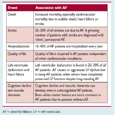
AF is independently responsible for higher risk of death in women as opposed to men. Studies suggest that AF, as an existing condition, doubles the mortality risk for women and results in a 1.5-fold increase in mortality risk for men (Andersson et al., 2013; Benjamin et al., 1998; Stewart et al., 2002) (Table 1). Certain conditions can be treated to mitigate the risk of death, for instance, anticoagulation can reduce the likelihood of death by stroke but the current evidence suggests that cardiovascular deaths due to heart failure and sudden death are still frequent even in AF patients under anticoagulant therapy (Kotecha et al., 2014). AF is linked to higher occurrence of heart failure and stroke i.e. it results in increased morbidity (Krahn et al., 1995; Stewart et al., 2002; Wolf et al., 1991). Recent studies indicate the diagnoses of AF in 20-30% of patients of ischaemic stroke either prior to, during or after the first occurrence (Grond et al., 2013; Henriksson et al., 2012; Kishore et al., 2014). 10-40% of AF patients are hospitalized every year (Kirchhof et al., 2014; Kotecha et al., 2014; Steinberg et al., 2014). Poor lifestyle resulting in decreased quality of life (Marzona et al., 2012; Thrall et al., 2006) and depression (von Eisenhart Rothe et al., 2015) are common in AF patients. The cognitive impairment and white matter lesions in the brain (Ball et al., 2013b; Knecht et al., 2008; Ott et al., 1997) are also very frequently observed in such patients. AF has and will continue to significantly result in increased healthcare costs unless it is prevented and treated effectively. The direct costs associated with AF-related treatments and complications are approximately 1% of total healthcare expenditure in the UK and between 6-26 billion US dollars in United States for year 2008 (Kim et al., 2011; Stewart et al., 2004).
.
Table 1: Morbidity and mortality linked with AF (Kirchhof et al., 2016a) 1.4. Atrial fibrillation risk factors
1.4.1 Ageing
AF is strongly age dependent, and it affects approximately 1%, 4% and 15% at 50, 65 and 80 years, respectively (Andrade et al., 2014). Aging plays a critical role in the genesis of AF and also increases the risks of cardiac dysfunction and stroke in AF patients. Clinical and laboratory evidence indicates that aging is significant in the creation of atrial electrical and structural remodeling that leads to increased susceptibility to AF occurrence. Aging is commonly associated with cardiovascular comorbidities, oxidative stress, calcium dysregulation, atrial myopathy with apoptosis, and fibrosis, which all contribute to the genesis of AF (Lin et al., 2018). Mounting evidence suggests that extracellular matrix (ECM) and perivascular fibrosis were increased progressively with age, leading to cardiac remodeling and dysfunction in elderly individuals (Horn and Trafford, 2016; Sahin et al., 2011). Besides, telomere attrition affects mitochondrial function, thus promoting aging (Sahin et al., 2011), and short telomere length is considered to be a hallmark of aging (Lopez-Otin et al., 2013). Recently, Carlquist et al found that AF subjects had shorter
telomeres compared without a history of AF subjects (Carlquist et al., 2016). Such finding suggests that aging, and/or replicative senescence, may contribute to the development and maintenance of AF (Xie et al., 2017). Koura and colleagues (Koura et al., 2002) demonstrated that with aging, the amount of interstitial fibrosis and fatty infiltrates increases, predisposing the atrial muscle to electrical impulse conduction disturbances. These disturbances, such as the so-called zigzag electrical impulse propagation aberrancy, are considered to be “trigger” events that lead to AF initiation and maintenance. At the cellular level, several anomalies may also contribute to age related AF initiation. For example, atrial myocytes in an aged atrium exhibit a prolonged action potential duration (APD). Moreover, evidences were provided that a larger APD heterogeneity exist across the atrium (Xu et al., 2013).
Figure 2: Histological analysis of a rabbit’s adult and aged (age >2 years) left atrium (LA) after Masson trichome staining. (A) showing marked fibrosis associated with cell loss and cardiomyocytes hypertrophy accompanied with increased fiber diameter and large-volume nuclei in the aged rabbit LA when compared with adult rabbit LA (B) with normal cardiomyocytes (Lin et al., 2018; Tsai et al., 2014).
1.4.2 Hypertension
to hypertension (Benjamin et al., 1994; Rogers et al., 2018). Hypertension increases sympathetic output which may lead to increased left atrial pressure and volume, as well as renin–angiotensin– aldosterone system (RAAS) activation, thereby leading to atrial fibrosis, structural and electrical atrial remodeling, and promotion of AF (Brandes et al., 2018; Lau et al., 2012). Long-term longitudinal studies from Framingham Heart Study1 and Women’s Health Study revealed both high systolic and diastolic BP increase the risk of developing AF (Tedrow et al., 2010). Moreover, in spontaneously hypertensive rats, the inducibility of atrial tachycardia was increased, accompanied by a rise in atrial fibrosis (Choisy et al., 2007). In a sheep model of long-standing elevated blood pressure induced by prenatal corticosteroid exposure multiple proarrhythmic abnormalities were seen: increased AF stability, reduced conduction velocities, (Hong and Glover, 2018; Shenasa et al., 2014) and increased fibrosis with myocyte hypertrophy and myolysis (Kistler et al., 2006).
The first evidence that optimal treatment of hypertension may prevent AF and improve outcomes came from intervention trials in hypertensive patients. In the Losartan Intervention for Endpoint Reduction in Hypertension (LIFE) study, which compared the use of ARB losartan with the beta- blocker atenolol, losartan prevented more cardiovascular morbidity and death than atenolol for a similar reduction in BP (Dahlof et al., 2002). A post-hoc analysis from this trial showed that the greatest reduction (40 %) in risk of incident AF occurred in patients who achieved optimal systolic BP levels of <130 mmHg, compared to those with systolic BP ≥142 mmHg. Moreover, incident AF occurred less frequently in patients treated with losartan than in those treated with atenolol, although there was no significant difference in BP reduction (Wachtell et al., 2005). A Danish nationwide nested case-control study also found less new-onset AF in patients with hypertension treated with ARBs or ACE inhibitors compared to beta-blockers or diuretics (Marott et al., 2014). These findings suggest that inhibition of the renin–angiotensin system itself might have a beneficial effect on the reduction of incident AF besides BP control (Brandes et al., 2018)
1.4.3 Heart failure and coronary artery disease
AF may be caused by any cardiac condition with, however, a predominance of heart failure (HF) and coronary artery disease (CAD) (Benjamin et al., 1994; Benjamin et al., 1998; Kannel et al., 1983; Kannel and Benjamin, 2008; Krahn et al., 1995; Roy et al., 2009). HF represents major AF risk factor as HF patients are associated with approximately 5-fold increased risk of AF onset
(Kannel et al., 1998). The risk of AF increases with the severity of HF clinical symptoms (Jais et al., 2000; Maisel and Stevenson, 2003; Tsang et al., 2002). Atrial fibrosis is markedly increased in the setting of HF, similar to that observed with aging and hypertension-related AF. Moreover, in HF too, the formation of atrial interstitial fibrosis plays a strong determinant of the occurrence of AF (Cha et al., 2004; Shinagawa et al., 2002; Tanaka et al., 2007). Specifically, the spatial distribution of atrial fibrosis could be an indicator of AF electrophysiologic mechanisms—reentry or spontaneous focal discharges—and of the exact locations of AF electrical sources. Therefore, understanding of fibrosis or scar distribution could be an asset in the performance of tailored AF ablation procedures (Trayanova, 2014). Thus, HF-related fibrosis formation has been one of the main targets of so-called upstream therapies, such as inhibitors of the renin-angiotensin- aldosterone system (Savelieva et al., 2011).
Acute and chronic CAD has emerged as a substantial risk factor of AF onset (Miyasaka et al., 2006) and perpetuation (Goldberg et al., 2002; Kannel et al., 1983; Wong et al., 2000). Although AF after ventricular myocardial infarction might be also triggered by an increase in intra-atrial pressure in the context of acute ventricular dysfunction, (Moller et al., 2003; Tsang et al., 2001) various works have shown that isolated atrial infarction is common. It was documented that the pathophysiologic role of atrial ischemia/infarction in AF onset has been greatly underestimated. Understanding of the pathophysiology linking CAD and AF has benefited from experimental studies. These works have highlighted several atrial ischemia/infarction related electrophysiologic changes. Spontaneous discharges have been significantly more numerous in cells bordering the infarcted region. Atrial ischemia/infarction had also been shown to reduce atrial refractory periods, to increase AF inducibility and adversely modulate regional electrical impulse propagation, and finally lead to an acceleration of atrial drivers (Anumonwo and Kalifa, 2016).
Figure 3: Panel (A) depicts the association between AF and HF cycle whereas, panel (B) HF-AF cycle. (ANP, atrial natriuretic peptide; BNP, brain natriuretic peptide; EDP, end diastolic pressure; LAP, left atrial pressure; LVD, left ventricular dilation) (Shenasa et al., 2014).
1.4.4 Diabetes
The Veterans Health Administration Hospitals study showed that the prevalence of AF in patients with diabetes mellitus was 14.9%, this being pointedly higher than that of hypertension. Thus, diabetes mellitus represents a strong and independent risk factor for the occurrence of AF, with an odds ratio of 2.13 (Pb.0001) (Lin et al., 2013; Movahed et al., 2005). The proposed mechanisms linking diabetes mellitus and AF include autonomic remodeling, structural remodeling, electrical remodeling, and insulin resistance. With respect to structural remodeling, in a diabetes mellitus rat model Kato et al. has demonstrated fibrosis in the atria with formation of anchoring points for reentry circuits and changes in the forward propagation of fibrillatory wavelets, thus resulting in atrial fractionated potentials and conduction delay (Kato et al., 2006). The atrial tissue collected
from diabetes mellitus patients which were then biopsied during coronary artery bypass graft surgery, displayed mitochondrial dysfunction causing oxidative stress which could also be involved in the formation of hyperglycemia-associated AF substrates, which ultimate lead to atrial interstitial fibrosis (Anderson et al., 2009). It is documented that the advanced glycation end products (AGEs) and AGE receptors (RAGEs) (both constituting the AGERAGE system) enable the interstitial collagen deposition in atrial myocardium of diabetes mellitus rats by encouraging up-regulation of the expression of connective tissue growth factors, and, as a consequence, result in myocardial structural remodeling (Koektuerk et al., 2016). Interestingly, in the atrial myocardium of rats with induced diabetes mellitus, the expression of Cx43 was found to be increased whereas its phosphorylation was decreased, thus leading to disorders of intercellular electrical coupling and consequent atrial arrhythmia (Mitasikova et al., 2009).
1.4.5 Thyroid Dysfunction
In the setting of hyperthyroidism, AF has been considered as one of the most frequent rhythm disturbance with its occurrence ranging from 2% to 20% (Klein and Danzi, 2007). When compared with a population with normal thyroid function and a 2.3% prevalence of AF, the incidence of AF in overt hyperthyroidism has been 13.8%. Shimizu et al. demonstrated that in a cohort of patients with hyperthyroidism studied for age distribution, AF incidence increased stepwise in each decade, climaxing at about 15% in patients N70 years, thus showing that hyperthyroidism-related AF has been more common with advancing age (Auer et al., 2001; Shimizu et al., 2002). Various potential mechanisms of AF in hyperthyroidism are proposed constituting elevation of left atrial pressure as a result of increased left ventricular mass and impaired ventricular relaxation, enhanced atrial ectopic activity, and ischemia secondary to raised resting heart rate (Bielecka-Dabrowa et al., 2009; Fazio et al., 2004; Sgarbi et al., 2003). Interestingly, it is recently documented that both hypothyroidism and hyperthyroidism cause increased AF vulnerability in a rat thyroidectomy model (Zhang et al., 2013). In fact, hypothyroidism and hyperthyroidism, while inducing opposite electrophysiological changes in heart rates and atrial effective refractory period, both pointedly increase AF susceptibility (Weltman et al., 2015) .
The connection of episodic heavy alcohol (ethanol) use with the onset of AF is termed as “holiday heart syndrome”. However, more recently, it is proposed that even habitual heavy alcohol consumption could be linked with a risk of AF (Balbao et al., 2009). Related to alcohol consumption and AF, Kodama et al. showed a meta-analysis of studies to summarize the estimated risk of AF associated to alcohol. It was found that the pooled estimate for AF for highest vs. lowest alcohol intake in individual investigations was 1.51 and a positive relationship between AF risk and heavy alcohol intake had been consistently found in all stratified analyses (Kodama et al., 2011). Over the last few years, a number of mechanisms by which alcohol consumption could be linked to the development of AF have been suggested: a direct toxic effect on cardiac myocytes, a hyperadrenergic state which has been reached during both drinking and withdrawal of alcohol, an impaired vagal tone, an increase in intra-atrial conduction time (which is also testified by a P-wave prolongation) (Corradi, 2014b).
1.4.7 Pericardial fat and obesity
A considerable portion of the epicardial surface in large mammals is normally covered by adipose tissue, and fat cells (adipocytes) may be participating in myocyte-adipocyte cross talk significant in the normal function of the myocardium. Obesity considerably increases plasma levels of free fatty acids as well as overall visceral and epicardial adiposity in the studies comprising humans and animal models. With obesity, extensive fatty infiltration leads to elevated levels of biofactors. These biofactors have been potentiated by paracrine and vasocrine signaling pathways and overload the myocardium resulting in deterioration of the myocardial function and also lead to abnormal impulse initiation mechanisms and myocyte atrophy. In obese patients, it is also documented that steatosis of the myocardium well correlates with epicardial fat behaves as an independent contributing factor to myocardial dysfunction. Additionally, experimental studies in isolated myocytes reveal that excess epicardial adiposity, or its biofactors, lead to abnormality in myocardial electrical excitation (Anumonwo and Kalifa, 2014; Shenasa et al., 2014).
Figure 4: Management of AF-related risk factors by life style modification (Hong and Glover, 2018).
1.4.8 Post-operative atrial fibrillation
The most common arrhythmia after cardiac surgery is AF occurring in approximately 20–50% of patients depending on the type of surgery performed. AF occurs in 30–40% of patients post coronary artery bypass graft (CABG) and up to 60–70% of patients with combined CABG and valve surgery. Majorly, post-op AF converts to sinus rhythm spontaneously in the first 24–48 h, but if it takes longer than 48 h it increases the risk of stroke and prolongs hospitalization and associated expenses. Post-operative AF affects both early and late mortality after isolated CABG. Most of the complications being related to stroke thus, post-operative surveillance and long-term management with antiarrhythmic agents and antithrombotic management have been warranted. Preoperative treatments with beta-blockers have been shown to effectively decrease the risk of AF. Colchicine as well as statins has been effective in prevention of this arrhythmia (Deftereos et al., 2013; Omae and Kanmura, 2012; Shenasa et al., 2014).
1.4.9 Genetic risk factors
Genetic predisposition also cannot be ignored (Kirchhof et al., 2016b) A considerable portion of AF occurring in younger ages has been known to be associated with a genetic predisposition than the accompanying disease. Some studies conducted report that more than 30% of AF have a common genetic variation. Among the various known genetic factors, the most important variants have been located close to the paired-like homeodomain transcription factor 2 gene on chromosome 4q25 (Gudbjartsson et al., 2007). This genetic variation has been known to be associated with up to a 7-fold increase the incidence of AF. In addition to the genetic variants that are likely to cause AF itself, gene mutations that contribute to the atrial remodeling process and electrophysiological changes described above may also mediate the occurrence of AF (Cha, 2018).
1.4.10 Other risk factors
Chronic kidney disease and smoking are accepted as independent AF risk factors but their respective importance is still debated. An example of a controversial risk factor is exercise. Although moderate physical activity may decrease AF incidence, a cumulative life practice of more than 1500 h is associated with 3-fold AF risk. Pathophysiologic mechanisms are still unclear, but the role of an increased vagal tone seems to be accepted (Anumonwo and Kalifa, 2014).
ATRIAL
2. ATRIAL FIRILLATION MECHANISM AND PATHOPHYSIOLOGY
The lack of reliable experimental models resembling this complex arrhythmia presents one of the major problems in understanding the mechanism leading to AF. However, an increased awareness of the role of “atrial remodeling” over the past 10 to 15 years has significantly increased our understanding of AF pathophysiology. Atrial remodeling constitutes any persistent change in atrial structure or function. Many forms of atrial remodeling promote the occurrence or maintenance of AF by acting on the fundamental arrhythmia mechanisms (Nattel et al., 2008) as illustrated in Figure below. AF requires both a trigger and a susceptible substrate (Dobrev and Nattel, 2010; Iwasaki et al., 2011b; Schotten et al., 2011). The trigger for initiation and maintenance of AF is mostly related to an enhanced electrical activity of the pulmonary vein cardiomyocyte sleeves, while non-pulmonary vein sources also become more important as AF continues to persistent form. Thereafter, AF is often sustained by a primary “driver” mechanism, which may be either focal ectopic sources or rapid local re-entry in a vulnerable substrate. Re-entry also involves both a substrate (a modified atrium or a portion of it) and a trigger (often an ectopic beat) (Nattel et al., 2008). The excitation seems to propagate through the susceptible substrate with a circular or spiral wavefront, mentioned as a rotor, thereby sustaining the AF and the alteration of the structure of the atrium (substrate).
Atrial remodeling has the ability to enhance the probability of ectopic or reentrant activity through a multitude of potential mechanisms (Khaji and Kowey, 2017; Nattel et al., 2008)
Figure 7: AF mechanisms and relationship to clinical forms (A) represents local ectopic firing, (B) represents single circuit reentry, (C) represents multiple-circuit reentry. (D) represents various AF clinical forms in relation to mechanisms. Paroxysmal forms involve local triggers/drivers mainly from pulmonary veins (PVs). Reentry substrates (initially functional and then structural) become prominent as AF continues towards permanent. Where, RA: right atrium; SVC: superior vena cava; LA: left atrium; IVC: inferior vena cava (Nattel et al., 2008).
2.1 Electrophysiological remodeling
Sustained AF with atrial rhythms as high as 350 to 600 bpm, in turn, results in electrophysiological remodeling, which consists of mainly the outward K+ current (Ito), the ultra-rapid delayed rectifier
K+ current (I
and the slow component of the delayed rectifier K+ current (IKs). The consequence of these various
alterations in currents leads to shortening of the action potential and effective refractory period (ERP), and thus maintenance of AF (Nattel et al., 2008). Significantly, this electrophysiological remodeling may also be accompanied with abnormal Ca2+ handling and enhanced propensity of potentially proarrhythmic Ca2+ release events from sarcoplasmic reticulum during diastole, which has the potential to compromise atrial contractility and show an exasperating role in the initiation and maintenance of ectopic (triggered) activity (Beavers et al., 2013; Chelu et al., 2009; Dobrev et al., 2011; Hove-Madsen et al., 2004; Neef et al., 2010; Voigt et al., 2014; Voigt et al., 2012). Electrophysiological remodeling is vague when the heart is in sinus rhythm, and is less recurrent in paroxysmal AF, mainly due to reversibility during AF-free intervals (Dobrev and Nattel, 2010; Voigt et al., 2014; Voigt et al., 2013). Remodeling can occur within h, days, or weeks of the onset of arrhythmia. It is linked with a higher incidence of delayed after depolarizations (DADs) and triggered activity (Voigt et al., 2014).