HAL Id: hal-01063205
https://hal-univ-rennes1.archives-ouvertes.fr/hal-01063205
Submitted on 12 Sep 2014
HAL is a multi-disciplinary open access archive for the deposit and dissemination of sci-entific research documents, whether they are pub-lished or not. The documents may come from teaching and research institutions in France or abroad, or from public or private research centers.
L’archive ouverte pluridisciplinaire HAL, est destinée au dépôt et à la diffusion de documents scientifiques de niveau recherche, publiés ou non, émanant des établissements d’enseignement et de recherche français ou étrangers, des laboratoires publics ou privés.
Natural and synthetic poly(malic acid)-based derivates:
A family of versatile biopolymers for the design of drug
nanocarriers.
Pascal Loyer, Sandrine Cammas-Marion
To cite this version:
Pascal Loyer, Sandrine Cammas-Marion. Natural and synthetic poly(malic acid)-based derivates: A family of versatile biopolymers for the design of drug nanocarriers.. Journal of Drug Targeting, Informa Healthcare, 2014, 22 (7), pp.556. �10.3109/1061186X.2014.936871�. �hal-01063205�
1
Natural and synthetic poly(malic acid)-based derivates: A family of
versatile biopolymers for the design of drug nanocarriers.
Pascal Loyer1, Sandrine Cammas-Marion2,*
1. Inserm UMR S-991; Foie, Métabolismes et Cancer; Université de Rennes 1; Fédération de Recherche Biosit; CHU Rennes; 35 033 Rennes, France.
2. UMR 6226 CNRS; Institut des Sciences Chimiques de Rennes; Université de Rennes 1; Ecole Nationale Supérieure de Chimie de Rennes; Avenue du Général Leclerc; CS 50837; 35 708 Rennes Cedex, France.
*Corresponding author: Tel: +33 2 23 23 81 09
Mail: sandrine.marion.1@ensc-rennes.fr
In memory of Professor Philippe Guérin
Keywords: degradable polymers, drug delivery systems, drug-polymer conjugates,
2
Abstract
The field of specific drug delivery is an expanding research domain. Besides the use of liposomes formed from various lipids, natural and synthetic polymers have been developed to prepare more efficient drug delivery systems either under macromolecular prodrugs or under particulate nanovectors. To ameliorate the biocompatibility of such nanocarriers, degradable natural or synthetic polymers have attracted the interest of many researchers. In this context, poly(malic acid) (PMLA) extracted from microorganisms or synthesized from malic or aspartic acid was used to prepare water-soluble drug carriers or nanoparticles. Within this review, both the preparation and the applications of PMLA derivatives are described emphasizing the in vitro and in vivo assays. The results obtained by several groups highlight the interest of such polyesters in the field of drug delivery.
Introduction
Since the term Magic Bullet was coined by Paul Ehrlich nearly a century ago [1], the concept of efficient drug delivery systems for therapy in Humans is becoming a reality. Indeed, owing to the quest for more effective treatments of several diseases such as cancers, numerous researches have been and continue to be performed on the development of systems able to deliver high amount of drugs at a specific site of action while decreasing non-specific distribution and toxicity. As a consequence of such considerable research works, some of the developed nanovectors, especially liposome and nanoparticles, have already received FDA approval or are in the preclinical or clinical phases [2-6]. However, even if undeniable progresses have been achieved in the design of the ideal drug carriers, several properties, such as the biocompatibility, drug loading capacity, and site-specificity of drug delivery as well as a reduced immunogenicity of the carrier following its administration in the body need further improvement.
Among drug delivery systems designed to address these objectives, polymer based carriers have attracted much interest mainly because of the versatile characteristics of polymers allowing adjustment of the physico-chemical and biological properties of the corresponding nanocarriers. Consequently, several families of polymers have been developed to formulate multifunctional drug carriers with properties adjusted to the considered application [7]. However, when designing a polymer family, one must keep in mind that the resulting materials are prepared to be used in vivo. Therefore, it is necessary to develop polymers corresponding to very strict specifications among which the following ones are of
3
great importance: (i). biocompatibility at any stage of the material life and until its complete excretion from the body, (ii). the lack of toxicity, (iii). the stealth and reduced immunogenicity, (iv). ability to carry large amount of drug(s), (v). the drug delivery at a specific site in a sustainable way.
Besides these essential properties, the (bio)degradability of the polymers constituting the drug delivery systems can be of interest in the sense that the carrier can be eliminated from the body as a result of its (bio)degradability. However, this means that the degradation process must lead to non-immunogenic, biocompatible and/or (bio)assimilable low molecular weight molecules that can be eliminated through bile and/or urine.
In the field of drug delivery systems, several stable or (bio)degradable polymers have been developed and formulated under either macromolecular linear prodrugs or nanoparticulate forms [7-10].
As described by Ringsdorf in 1975, a macromolecular linear prodrug, called also polymer-drug conjugate, is constituted by a (bio)degradable or biostable polymer backbone on which several functionalities are introduced: (i). water or lipid solubilizer as comonomer units or blocks, (ii). biologically active molecules linked to the backbone through a degradable bound, (iii). targeting agents and body distribution modulator groups [11]. Since the Ringsdorf’s model, several systems have been developed [3]. Recently, Pang et al. have published a review on the present state and future perspectives of polymer-drug conjugates: the most studied polymer-drug conjugates were described in terms of structures and nature of the conjugated drug as well as the possible applications and the ongoing clinical trials if any, etc. [12]. However, even if such carriers have been widely investigated, further improvements are required to minimize the material’s heterogeneity by optimizing the synthesis, to find versatile linkers for site-specific controlled release and to decrease the immunogenicity of the polymer-drug conjugates.
Besides such linear drug delivery systems, nanoparticulate carriers have emerged following the pioneer works realized by Professor Kataoka’s team on macromolecular micelles based on amphiphilic block copolymers of hydrophilic poly(ethylene glycol), PEG, and hydrophobic poly(amino acids) blocks [13]. In the meantime, Kabanov’s group developed nanoparticulate carriers via the work performed on Pluronic® micelles [14].
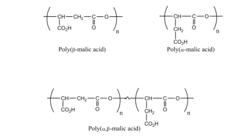
In this context, the design of (bio)degradable polymers, whose physico-chemical and biological properties can be adjusted either by copolymerization, chemical modifications or both, are of great importance [15,16]. Poly(malic acid) (PMLA) (Figure 1) is a very good candidate due to the presence of carboxylic acid pendant groups, which can be chemically
4
modified to introduce molecules of interest, and ester cleavable bounds within the polymer backbone, allowing the degradation of the polymer main chain into biocompatible metabolites [17].
Figure 1. Structure of the different forms of poly(malic acid).
Moreover, following the synthesis of a large monomer family of malolactonates (MLAR) and their homo- and/or co-polymerization, numerous PMLA derivatives are now accessible with physico-chemical and biological properties depending on the nature of the repeating units forming the polymer [18-20]. Besides these synthetic macromolecular materials, natural poly(malic acid) can be obtained from the plasmodia extracts and from the culture medium of Physarum polycephalum [21]. The adjustment of physico-chemical and biological properties of the natural PMLA can be realized only by chemical modifications of the naturally available polyester.
In this article, we present an overview on the researches conducted on both natural and synthetic PMLA derivatives for designing multifunctional drug carriers under forms of either drug-polymer conjugates or nanoparticles. In a first part, preparation and characterization of natural PMLA derivatives will emphasize the in vitro/in vivo assays. The second part will focus on both the synthesis of the PMLA’s derivatives and the applications of such synthetic polymers as drug carriers.
Drug delivery systems based on natural PMLA
At the end of the 1980s, Holler et al. have isolated an unusual polyanion from plasmodium extracts and from the culture medium of P. polycephalum [21]. They have shown that this polyanion inhibits the DNA polymerase α of this organism and that it exhibited a poly(malate)
5
structure [21]. Few years later, we demonstrated that this natural PMLA compound is the β -ester form of the poly(β-malic acid) obtained by chemical synthesis [22]. The natural polyester has attracted the attention of researchers as a result of its potential application in the drug delivery system field. The natural PMLA has been indeed used, either unaltered or following chemical modifications (Figure 2) to formulate nanoparticles [23-28] or as nanoplatforms called Polycefins™ [29-50].
Figure 2. Structures and characteristics of Nat-PMLA derivatives.
Nanoparticles based on natural PMLA
Muñoz-Guerra et al. have developed derivatives of natural PMLA with the goal of elaborating ionic complexes as well as nano- and micro-particles for drug delivery [23-28]. Ionic complexes were obtained by mixing aqueous solutions of natural PMLA and alkyltrimethylammonium sulfates [23,26].
The degradation of such complexes occurred by hydrolysis with a rate that can be modulated by changing the chemical structure of either the main chain or the surfactant alkyl chain [23,26]. Moreover, up to 30 wt% of the drug erythromycin can be homogenously entrapped within the ionic matrix and the kinetics of release followed the hydrolysis profile
O O CO2H CO2R O O O C O NHR1 C O NH C O OR2 C O NHR3 PEG5000 O O n O CO2- n O CH3(CH2)n-1N+(CH3)3 O O
R: methyl, L-leucine ethyl ester or L-phenyl alanine methyl ester
O O O C O
NHR4
O
n
R1: Dox, TMZH or AONs
R2: fluorescence probe (Alexa fluor 680) R3: mAbs
R4: endosomal escape units (valine, L-leucine ethyl ester, tri-leucine
Ionic complexes
Erythromycin encapsulation [22,25]
Nano and microparticles Nanoplatforms
Encapsulation of protein, antibiotic or anti-cancer drugs In vitro assays [23,24,26,27,49]
Polycephin™ nanoplatforms
Studies of cytotoxicity in vitro, interactions with membranes, mechanism of membrane permeation Vectorization of Dox, TMZH or AONs
6
regardless of the loaded amounts [23,26]. Despites these interesting observations, to the best of our knowledge no result of cell based assays have been reported using these biomaterials.
Besides this work, Muñoz-Guerra et al. have also synthesized derivatives of natural PMLA containing hydrophobic groups and prepared the corresponding nano- and micro-particles [24,25,27,28]. These derivatives have been obtained by reacting diazomethane [24,27,28] or by grafting hydrophobic amino acids [25] on natural PMLA.
Two hydrophobic amino acids have been grafted onto natural PMLA, the L-leucine ethyl ester and the L-phenyl alanine methyl ester, in proportions of 30%, 60% and 90% without dramatically affecting the molecular weights and polymolecularities of the obtained derivatives [25]. Nanoparticles have been prepared from natural PMLA containing 60% of amino acid (L-leucine ethyl ester or L-phenyl alanine methyl ester) using the precipitation-dialysis method. Spherical nanoparticles with a diameter ranging from 70 to 230 nm were obtained [25] and cytotoxicity assays realized with two kinds of nanoparticles on various cancer cell lines have evidenced a concentration-dependent cytotoxicity but only for polymer concentrations above 125 µg/ml. Muñoz-Guerra et al. concluded that such dose-dependent cytotoxicity was related to the binding of polymer to cell membranes leading to irreversible membrane dys-organization [25]. However, they did not report any experiment evidencing such nanoparticles-cell membrane interactions. Finally, they evaluated the use of such nanoparticles as drug carriers by studying loading and release with two drugs, Temozolomide (TMZ) and Doxorubicin (Dox) [25]. The drug encapsulation was quite low with a fast release for TMZ and a slower release for Dox [25]. Unfortunately, no cell cytotoxicity assays and in
vitro drug delivery of the drug carriers have been conducted.
Muñoz-Guerra et al. mainly studied the synthesis, characterization and formulation of methyl ester of natural PMLA [24,27,28]. Beginning from either 75% or 100% methylated natural PMLA, they formulated nano- or micro-particles, by precipitation-dialysis or emulsion-evaporation methods with the goal to use these systems as proteins or drugs carriers. The micro-sphere diameters were in the range of 1 to 20 µm [28] while the size of nanoparticles formulated with the same polymers varied from 100 to 350 nm [24,27]. Several proteins [27], an antibiotic molecule [28] and two anti-cancers drugs [24] have been used to evaluate the potential of these biodegradable nano- and micro-particles as drug carriers. The encapsulation efficiency was shown to be dependent on the nature of the loaded molecule while the kinetics of drug release seem to be determined by polymer degradation rate [24,27,28]. Finally, cytotoxicity assays using various cancer cell lines have been realized with nanoparticles formulated with fully methylated natural PMLA. After one hour of incubation
7
with nanoparticles at a polymer concentration up to 1 mg/ml, the cell viability was not affected; however, it decreased significantly after 12 hours of incubation at all the polymer concentrations tested. Muñoz-Guerra et al. estimated that such toxicity came from the methanol released during polymer degradation [24].
Such research has highlighted the key role of the nature of the ester side groups which not only allows the adjustment of physico-chemical properties of the biopolymer but also affects its biocompatibility and its interactions with the biological environment.
Nanoplatforms based on natural PMLA
In the early 2000s, Ljubimova et al. have developed a new type of nanoconjugates based on natural PMLA and called Polycefins™ [29-50]. As mentioned above for other nanovectors, the natural PMLA has been harvested from the culture broth of P. polycephalum microplasmodia [21]. The crude PMLA was purified and characterized with Mw between 50 and 100 kDa with a polydispersity index of 1.3 before being subjected to a series of chemical modifications leading to the synthesis of an all-new set of polymers and the corresponding nanoplatforms [41,42].
Taking advantage of the carboxylates’ lateral groups, Ljubimova et al. have set up a method allowing a highly controlled chemical coupling of various molecules of interest to the polymeric PMLA backbone [29-50]. The carboxylate lateral groups were first activated under N-hydroxysuccinimidyl esters [41-43,45], then the different molecules of interest, called “modules” [43], were chemically coupled to the activated PMLA following a particular sequence mainly defined by the molecule sensitivity and the reactive groups [43,45].
The first series of chemical coupling consisted in the addition of the more stable molecules through amide bonds:
- The PEG module (m-PEG5000-NH2) for protecting the nanoplatforms against
unspecific degradation and scavenging [29-31,33-49].
- Endosome escaping modules which can be activated in response to acidic pH in mature endosomes/lysosomes. Different modules have been tested: the valine [35,41], the leucine ethylester (LeuEt) [29-31,33,34,36-40,43-48] and the tri-leucine (TriLeu) [29,31,33,34,40,45-48]. The evaluation of the membrane lysis by natural PMLA substituted by various amino acid derivatives has demonstrated that: (i) natural PMLA derivatives having a pH-dependent membrane lysis property, such as natural PMLA substituted by the TriLeu, allowed safe and efficient cytoplasmic delivery [32-34,46], and that (ii) natural PMLA derivatives with pH-independent membrane lysis properties, such as natural PMLA
8
substituted by the LeuEt, were cytotoxic at high concentrations for several cell lines. Nevertheless, some of these pH-independent natural PMLA derivatives can be used for trans-membrane drug delivery under certain circumstances [32-34,46]. Moreover with the goal to design nanoplatforms with optimal cytoplasmic delivery efficiency and reduced systemic toxicity, the mechanisms of membrane permeation for two endosome escaping modules, LeuEt and TriLeu have been studied [33]. Natural PMLA bearing TriLeu had a pH-response membrane disruption by the “barrel-stave” mechanism (formation of aggregates in solution followed by insertion into the membrane forming trans-membranal pores leading to membrane permeation) and was less cytotoxic with a more efficient cytoplasmic delivery than the nanoplatforms bearing LeuEt, characterized by a “carpet” mechanism (binding to the membrane with high affinity leading to membranolysis in a cooperative manner) and a pH-independent membrane disruption [33].
- A thiol-containing spacer module allowing the chemical coupling of other molecules such as targeting agent, drugs or fluorescent dyes [29-31,33-36,38,40-46]. The bioactive molecules were then chemically coupled to natural PMLA derivatives through thioether bonds for monoclonal antibodies [mAbs] and disulfide bonds for anti-sense oligonucleotides (AONs), the disulfide bonds being cleaved by cytoplasmic gluthatione to release free AONs [29-31,34-37,39-46]. A fluorescence dye, mainly the Alexa Fluor 680, was also introduced before masking the non-reacted thiol groups for in vitro and in vivo imaging [29-31,34-37,39-46].
Two anti-cancer drugs, the Temozolomide hydrazide (TMZH) [47,48] and the Dox [49], have also been chemically bound to natural PMLA nanoplatforms using labile bounds, hydrazide bound for TMZH and hydazone linker for Dox.
From the various natural PMLA based nanoplatforms, Ljubimova et al. performed in
vitro and in vivo assays in order to evaluate the potential of their nanoplatforms as
personalized nanomedecine in the field of anti-cancer drug delivery [29-50].
In collaboration with Ljubimova’s team, Muñoz-Guerra et al. have prepared and characterized nanoplatforms based on partially methylated natural PMLA (25% and 50% of methylation) obtained by reaction of diazomethane on natural PMLA [50]. These authors have shown that degradation of methylated natural PMLA nanoplatforms was faster in human plasma than in phosphate buffered solution (PBS) and faster for low methylated derivatives [50]. Several cell based in vitro assays using nanoplatforms based on either 25% or 50% methylated natural PMLA have been realized in order to validate their use as biodegradable and biocompatible drug delivery systems. They evidenced that only the nanoplatforms
9
constituted by 50% of methylated natural PMLA have a membrane disruption activity, property that can be useful to deliver drugs into cells [50]. Besides, in vitro cytotoxicity assays conducted on several cancer cell lines have shown that methylated natural PMLA exhibited an increasing toxicity with the degree of methylation related to the release of methanol occurring during the degradation of the corresponding methylated nanoconjugates [50]. In addition, cellular uptake studies realized with Rhodamine-labeled copolymers showed a homogenous fluorescence repartition within cells with a higher intensity observed for polymers having a higher methylation degree [50].
Besides nanoplatforms formulated with methylated natural PMLA, the other systems developed by Ljubimova et al. lead to encouraging in vitro and in vivo results. First of all, concerning in vitro evaluation of Polycefins™ carrying low molecular weight anti-cancer drugs (TMZH and Dox) have been coupled [47-49], the major conclusions are the following:
- The half-life of the conjugated TMZH has been significantly increased compared to that of free TMZH and the degradation of the nanoconjugates bearing the TMZH drug was faster at 37°C than at 4°C and in human plasma versus PBS [47,48].
- The release of Dox from the nanoconjugate Nat-PMLA/PEG5000(5%)/GH-Dox(5%)
was pH-dependent with a fast release at pH5 (about 75% of release after 10 hours of incubation) and almost no Dox release at pH7 [49].
- Membrane destabilization studies have evidenced that substitution of around 40% of pendant natural PMLA carboxylates of the platform by LeuEt (pH-independent) or TriLeu (pH-dependent) lead to good candidates for endosome disruption [47,48].
- Cell viability studies, realized on human glioma cells U87MG (TMZH & Dox), T98G (TMHZ) and U251 (Dox) and on invasive breast carcinoma cells, MDA-MB-231 and MDA-MB-468 (TMZH & Dox), have evidenced that: (i). the Nat-PMLA/PEG5000(5%)/GH(5%), the Nat-PMLA/PEG5000(12%)/TriLeu(40%) and the
Nat-PMLA/TriLeu(40%) were well tolerated by all cell lines within the studied concentration [47-49]; (ii). the Nat-PMLA/PEG5000(12%)/LeuEt(40%) decreased significantly the cell viability
and was not further considered [47,48]; (iii). the Nat-PMLA/PEG5000(5%)/GH-Dox(5%)
followed a dose dependent response similar to that of free Dox [49]; in this case, the authors suggested adding to the nanoplatform monoclonal antibodies as targeting agent to improve in
vivo targeted delivery of Dox [49]; (iv). the leads compounds
Nat-PMLA/TriLeu(40%)/TMZH(17%) and Nat-PMLA/PEG5000(2%)/TriLeu(40%)/TMZH(17%)
on which an anti-human transferrin monoclonal antibody (HuTfR mAb, 0.25%) has been grafted showed a significant decrease in the studied tumor cell viability (human glioma and
10
human breast cancer cell lines) [47,48]; in this case also, the authors proposed to move to in
vivo assays with these two Polycefins™.
The major part of the research conducted by Ljubimova’s group is devoted to the design of Polycefins™ grafted with PEG5000, sense oligonucleotides [AONs] as
anti-cancer drugs, one or two monoclonal antibodies [mAbs] as targeting agents, L-valine/LeuEt/TriLeu as membrane disrupting units and, in some cases, a fluorescent dye [29,33-37,39,41-46]. They performed several in vitro studies using various human glioma cell lines and human breast cancer cell lines, as well as in vivo assays with mice or rats bearing either human breast cancer or human glioma cancer. They have shown that the Nat-PMLA/PEG5000(5%)/AON1(2.5%)/AON2(2.5%)/PEG3400-mAb-OX26(0.23%)/valine(49%)
entered U87MG cells via a transferrin receptor-mediated endocytosis and that conjugated AONs efficiently inhibited target protein expression. After intra-venous injection of Polycefin™ labeled with Alexa Fluor 680 dye in nude mice in which human brain cancer cells were implanted, the in vivo imaging experiment showed that Polycefin™ accumulated in the tumors because of the ability of this polymeric nanoconjugate to cross the Brain-Tumor-Barrier (BTB) [41]. After its intracranial injection in nude rats bearing human U87MG glioblastoma cells, a reduction of the expression of laminin-8 (α4 and β1) chains and of the tumor vessel density along with an increase in animal survival have been observed without significant effect on other tissues and organ morphology [35].
A Polycefin™ bearing a mouse monoclonal anti-human transferrin receptor (TfR) antibody was shown to accumulate preferentially in brain and breast tumors after intra-venous injection into nude mice and in a lesser extent into kidney and liver leading the authors to conclude that Polycefins™ might be of great interest as anti-cancer treatment [44].
To improve direct tumor targeting, they prepared a new Polycefin™ derivative bearing PEG chain, AONs, LeuEt and two monoclonal antibodies with different specificities: the mouse monoclonal human TfR antibody and the mouse monoclonal tumor-specific anti-nucleosome antibody 2C5 [36]. The authors demonstrated that this Polycefin™ was internalized in U87MG cells in vitro thank to the presence of the 2C5 antibody able to recognize the nucleosomes bound to an unknown surface receptor on the glioma cells [36]. After its intra-venous injection into nude mice intracranially implanted with human glioma cell line U87MG, the highest drug accumulation was observed with Polycefin™ bearing both mTfR and 2C5 mAbs: the anti-mouse TfR antibody allowed transporting drug through the BTB by binding specifically to the endothelium-expressed TfR and the anti-nucleosome 2C5 antibody allowed targeting human tumor cells by binding to a tumor cell surface antigen [36].
11
In their attempt to improve their nanoplatform, Ljibumova et al. have modified the targeting agents and the endosome escaping module. For that purpose, they chemically linked to the Polycefin™ backbone two monoclonal antibodies, one targeting the BTB (mouse TfR) and one targeting tumor cells (human TfR), together with AONs targeting the laminin and the development of the angiogenesis within the tumor, and a pH-activated TriLeu as endosome escape module [30,31]. Results have shown that:
- AONs were efficiently delivered by Nat-PMLA/TriLeu/AON/human TfR into the cytoplasm of U87MG and T986 glioma cells and inhibited the synthesis of Laminin-411 α4 and β1 chains (Western blot analysis). Confocal analysis with double-labeled nanoplatform confirmed the entry of the nanoplatform and AONs in the U87MG and T986 glioma cells and its escape from the endosome pathway [30,31].
- The Nat-PMLA/TriLeu/AONs/mouse TfrR/human TfR showed the highest accumulation 24 hours after its intra-venous injection in human U87MG cells forming brain tumors implanted in mice. It should be noted that some of the drug was found in liver and kidney. These results suggested that this nanoplatform might be able to pass through the BTB and be efficiently internalized into the tumor cells [30,31].
- In these experiments, the tumor growth was significantly decreased after systemic injection of the nanoplatform in mice bearing intracranial human glioma U87MG cells,. Indeed, the tumor was 90% smaller in nanovector treated mice than in mice treated with PBS. Observation of brain sections of nanoplatform-treated mice evidenced mainly tumor remnants with significant necrosis and decreased tumoral angiogenesis [Ding, 2009, 2010]. The immunostained tumor sections showed a significant decrease in the staining intensity for both laminin chains with vessels having sizes almost similar to those in normal brain [30,31].
In view of these results, the Polycefin™ nanoplatforms seem to be very promising candidates to treat brain diseases.
Recently, Ljubimova’s group evaluated the Polycefin™ nanoplatforms for breast cancer treatments [29,33,37-39,46]. In a first study, they have prepared a Polycefin™ nanoplatform conjugated with: (i) AON directed against HER2/neu mRNA to block new HER2/neu receptor synthesis, (ii) anti-HER2/neu antibody trastuzumab [Herceptin] to target breast cancer cells and inhibit receptor activity, (iii) transferrin receptor antibody to target the tumor vasculature and mediate nanoplatform delivery through the endothelial system, (iv) the LeuEt (40%) as endosomal escape module to achieve cytoplasmic delivery of the AON and (v) a PEG5000 chain (5%) to improve nanoplatform blood-stream stability [37,38]. They first
(MDA-12
MB-231 and MDA-MB-435) HER2/neu levels. They showed that the Nat-PMLA/HER2/neu AON/LeuEt/Herceptin/human TfR/PEG5000 nanoplatform was able to inhibit both the growth
of all the breast cancer cell lines and the phosphorylation of serine/threonine kinase (Akt). Moreover, Ljubimova et al. showed that their Polycefin™ was able to triggered apoptosis in both high and low HER2/neu expressing cell lines in vitro [37,38]. Based on these encouraging results, they investigated the therapeutic effect of their developed Polycefin™ after intra-venous injection in nude mice bearing subcutaneous human breast tumor xenografts (BT-474 cell lines). The treatments were well tolerated by mice upon administration of the Polycefin™. The treatment induced tumor growth inhibition with a synergic effect of the presence of HER2/neu AON and Herceptin, and appearance of necrotic areas with little tumor tissue remaining [37,38]. Western-blot analysis of tumors after treatments showed that Polycefin™ induced strong and sustained inhibition of HER2/neu tumor expression and phosphorylation of Akt, as well as the induction of apoptosis [37,38]. These results constitute a proof of concept for the use of natural PMLA nanoplatforms for cancer therapy.
With the aim to improve the treatment of HER2/neu positive breast cancer, Ljubimova and coworkers have slightly modified the nanoplatform described above. Because Interleukin-2 (IL-Interleukin-2) can be considered as a potential cancer treatment with, however, a low therapeutic efficacy due to its rapid blood clearance and severe side effects [51], they designed a natural PMLA nanoplatform bearing, among other modules, a modified human IL-2, which was obtained by its fusion to the C-terminus of the heavy chains of an HER2/neu IgG3, anti-HER2/neu IgG3-(IL-2) [29,34]. This Polycefin™, called PMLA-fusion nanobioconjugate, containing 5% of mPEG5000 chains, 40% of LeuEt, 0.25% of anti-human-HER2/neu
IgG3-(IL-2) to target the tumor and function as immunomodulator while 2.5% of AONs trigger the inhibition of both human and mouse Laminin expression (α4 and β1 chains) [34]. After showing that IgG3-(IL-2) conjugated to natural PMLA retained the HER2/neu targeting properties of the body and that the biological activity of the IL-2 within the complex anti-HER2/neu IgG3-(IL-2) was maintained, Ljubimova et al. evaluated, by confocal microscopy, cellular uptake of the PMLA-fusion nanobioconjugate by BT-474 human breast cancer cells expressing high level of HER2/neu. Their results suggested that the presence of anti-HER2/neu IgG3-(IL-2) on the nanoplatform allowed both the cell surface binding and the cellular uptake of the nanobioconjugate [29,34]. In vivo biodistribution studies were realized on athymic mice subcutaneously injected with BT-474 human breast cancer cells and after intra-venous injection of PMLA-fusion nanobioconjugate without AONs. The
13
nanobioconjugate preferentially accumulated at the tumor site and in a lesser extent in the liver [29,34]. Anti-cancer activity and immunoactivation by the PMLA-fusion nanobioconjugate were also evaluated after intra-venous injection in a syngeneic model of immunocompetent BALB/c mice implanted with D2F2/E2 cancer cells [29,34]. Results evidenced that only treatment with the PMLA-fusion nanobioconjugate increased the survival of treated mice and the serum level of anti-HER2/neu IgG1 and IgG2a. Such results suggested an enhanced of the humoral and cellular immune responses [29,34].
Furthermore, Ljubimova et al. adapted their Polycefin™ nanoplatform to the treatment of triple-negative breast cancer (TNBC) [39,46]. In a first study, they chemically bound to natural PMLA nanoplatform 5% of m-PEG5000, 40% of LeuEt, 2% of epidermal growth factor
receptor (EGFR) AON to inhibit the synthesis of EGFR, protein highly expressed in TNBC, 0.12% of anti-tumor nucleosome-specific monoclonal antibody mAb [2C5] to target breast cancer cells and 0.12% of anti-mouse transferrin receptor (TfR) antibody for delivery through the endothelium of blood vessels [39]. Western-blot analysis performed on EGFR expressing cells (MDA-MB-468) and cells expressing low EGFR levels (SKBR-3) demonstrated that Polycefin™ derivative efficiently inhibited EGFR expression in both cell lines. To confirm the tumor targeting property of mAb 2C5, a variant of Polycefin™ containing mAb 2C5 and EGFR-AON was injected intra-venously into athymic mice bearing MDA-MB-468 human breast cancer cells. The biodistribution study showed that this nanobioconjugate accumulated mainly in tumor with a non negligible accumulation in kidney and liver. The introduction of the second antibody, the TfR, on the Polycefin™ nanoplatform allowed to a more important drug accumulation into implanted tumor an a reduced accumulation into kidney and liver [39].
Finally, the Nat-PMLA/PEG5000/EGFR-AON/mAb 2C5/TfR/LeuEt was intra-venously
injected into MAD-MB-468 tumor-bearing mice: this Polycefin™ induced the most pronounced anti-cancer effect with a strong tumor growth inhibition, a significant necrotic effect in treated tumor, a high inhibition of EGFR expression and of phospho-Akt levels in tumor [39]. In view of their results, the authors concluded that this new generation of nanobioconjugate might represent a powerful way to treat TNBC.
Before engaging Polycefins™ into clinical trials, Ljubimova and colleagues have performed a detailed evaluation of the toxicity and immunogenicity in vitro and in vivo of the Polycefin™ designed for TNBC treatment [46]. For this study, they have prepared a Polycefin™ bearing AONs targeting EGFR, Laminin α4 and/or Laminin β1 protein synthesis, LeuEt modules, mPEG5000, MsTfR mAb targeting mouse endothelial cells in tumor vessels
14
and HuTfR mAb for binding to human tumor cells and nanodrug internalization. The investigators have shown that chronic exposure of nude mice to nanobioconjugates of natural PMLA at both low and high doses did not induce toxic changes in blood cell count or metabolic effects. PMLA nanoplatforms were well tolerated in vitro and in vivo in extreme dosage without toxicity and immune reactions. It is important to note that only low complement activation has been observed at very high dosage of the nanobioconjugate (1mg/kg) [46]. In addition, Polycefin™ nanoplatforms inhibited the growth of EGFR-positive triple negative breast tumor cells in nude mice after intra-venous multiple injections. For both treatments, the Nat-PMLA/AONEGFR,α4,β1/MsTfr/HuTfR nanoconjugate showed a significant
tumor growth inhibition. These results lead the authors to conclude that this dual-action nanodrug significantly inhibited the TNBC growth and angiogenesis [46]. From the data, Polycefin™ derivatives might be considered as versatile nanoplatforms for treatment of various cancers, such as breast and brain cancers, because of the possibilities to chemically couple several molecules of interest and the low toxicity and immunogenicity of the native natural PMLA backbone.
Drug delivery systems based on synthetic poly(malic acid)
Besides the use of natural PMLA derivatives for the design of drug delivery systems, synthetic poly(β-malic acid) has been the first polymer synthesized to be formulated as water-soluble drug carrier [52]. The chemical synthesis of PMLA has been conducted from two natural molecules, the malic acid [53] or the aspartic acid [54,55], and by different polymerization techniques, ring opening polymerization [54-56], polycondensation [57] or enzymatic polymerization using lipases [58]. The synthetic PMLA (Figure 1) is found under the β-form (ring opening and enzymatic polymerizations), the α-form (ring opening polymerization) or a mixture of both α- and β-forms (polycondensation). These PMLAs have been successfully used to formulate of various drug delivery systems by several research teams.
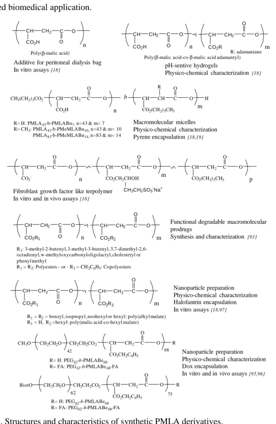
First, applications of synthetic PMLA derivatives as macromolecular prodrugs are developed and summarized in Figure 3.
15
Figure 3. Structures and characteristics of water soluble synthetic PMLA derivatives.
In the early 1990s, Ouchi and coworkers have synthesized the poly(α-malic acid) by ring-opening polymerization (ROP) of malide benzyl ester and have grafted the 5-fluorouracil (5FU) on this water soluble polyester anti-cancer drugs, [59-61] or the Dox [62-64].
The 5FU has been covalently linked to the carboxylic acid lateral group through ester, amide or carbanoyl bounds using classical coupling reactions [59-61]. Five saccharide
CH C O
O
n
CH2
CO2H
Poly(α-malic acid)
CH C O
O CH2
CO2R
R: 5FU, Dox, Saccharide residues
C CH O O CH2 C O O CH CH2 CH3 O C CH2 O CH C O O C CH O OH CH2 C O O CH CH2 CH3 O OR
Malic acid propane 1,2 diol copolyesters
O CH2 CH2 CH2 CH2 CH2 C O O CH CH2 C C O O OCH2CH2OC O C CH3 CH2 n HEMA grafted poly(ε-caprolactone-co-RS-β-malic acid)
CH C O O n CH2 CO2H PMLA-based hydrogel CH CH2 CO2R R: HEMA or mPEG C O O CH C O O CH2 CO2R CH CH2 CO2H C O O CH C O O n CH3
Poly(lactide-co-β-malic acid)
CH CH2
CO2R
R: H, hydroxyl arms, extented carboxyl arm or GRGDS
C O O p CH CH2 CO2R R: H or RGD C O O CH2 O CH CH2 CO2R C O O MMA MMA b CH2 b n m PEGMLAAc
+ PEGDAc + methylacrylated O-carboxychitosan Water soluble drug carrier
In vitro degradation
In vitro and in vivo assays [58-61, 103]
Tissue engineering
Physico-chemical characterization In vitro assays[70-76]
Enteric coating materiels for tablets containing sodium diclofenac Physico-chemical characterizations [63-65]
Micropattened tissue engineering scaffolds[66]
Degradable hydrogel
Physico-chemical characterizations Dox encapsulation
In vitro assays [67,68]
16
residues have been also chemically coupled on poly(α-malic acid)-5FU in order to evaluate the cell specific targeting of the resulting conjugate [60]. Ouchi et al. have first studied the degradation of their macromolecular conjugates in vitro in order to determine the release of 5FU from poly(α-malic acid)-5FU conjugates [59-61]. They evidenced that the hydrolysis rates of the main-chain ester bonds of the poly(α-malic acid)were much slower than those of lateral ester, amide and carbamoyl bonds leading to a slow release of 5FU [59-61]. The anti-tumor activity of poly(α-malic acid)-5FU conjugates has been evaluated after intra-peritoneal injections into female CDF1 mice bearing P388 lymphocytic leukemia cells [59,61]. The poly(α-malic acid)-5FU conjugates lead to an improved survival rate with no acute toxicity at a dose of 200-800 mg/kg [59,61]. The conjugates bearing saccharide residues as targeting agent also seemed to have significant anti-tumor activities with no acute toxicity in the studied dose ranges after intra-peritoneal injection in the same animal model [60]. Ouchi et al. concluded that the 5FU side effects were significantly decreased by its attachment to poly(α -malic acid) backbone bearing or not saccharide residues as targeting agent [59-61].
Interestingly, the nature of the saccharide affected the in vitro growth inhibition in SK-Hep-1 and HLE human hepatoma cells and HeLa uterocervical carcinoma cells [60]. The poly(α-malic acid)-5FU-galactosamine conjugate had the highest growth inhibitory effect on SK-Hep-1 and HLE cells while no effect has been observed on HeLa cells. Blocking the galactose receptors of human hepatoma cells with the trimer of galactosamino-saccharide induced a decrease in the growth inhibitory effect of poly(α-malic acid)-5FU-galactosamine conjugates demonstrating that the galactosamide allowed targeting of 5FU to hepatoma cells via galactose-receptor [60].
The same group has also prepared a poly(α-malic acid) derivate on which the Dox molecule was linked through ester or amide bounds [62]. As expected, the release rate of Dox conjugate to poly(α-malic acid) through ester bounds was faster than compared to that of Dox conjugated to poly(α-malic acid) through amide bounds [62]. Cytotoxicity assays have been realized in vitro using P388 D1 lymphocytic leukemia cells. While the Dox linked to poly(α -malic acid) through ester bond induced a cytotoxicity similar to free Dox, the Dox bound to poly(α-malic acid)through amide bound had a lower cytotoxicity [62]. Such results have led Ouchi’s group to conjugate onto their poly(α-malic acid)-amide-Dox various saccharide units as targeting agents and to study both the Dox release and the in vitro cytotoxicity of these conjugates on cancer cell lines [63,64]. As previously observed, free Dox was released faster from the poly(α-malic acid) conjugates when Dox was linked through ester bonds and under acidic conditions compared to the poly(α-malic acid)-amide-Dox [63]. The cytotoxicities of
17
Dox linked to poly(α-malic acid) were lower than with the free Dox [64]. The introduction of saccharide units seemed to have no influence on cytotoxicities of the corresponding Dox-conjugates on AZ521, KNS and Hela cells. In contrast, the presence of galactosamine as saccharide units on the poly(α-malic acid)-amide or ester-Dox had led to higher cytotoxicities on HLE and HepG2 hepatoma cells than those observed for non-glycosylated poly(α-malic acid)-amide or ester-Dox and for free Dox [63,64]. The authors concluded that such higher activity resulted from galactose receptor-mediated uptake of the conjugate into hepatoma cells but further studies are still needed to confirm the interest of such poly(α-malic acid)-Dox-galactosamine conjugates.
In a different context, copolyesters containing malic acid and propane 1,2-diol units have been also prepared with the goal to use them as enteric coating materials for tablets containing sodium diclofenac [65-67]. Bakr et al. have shown that the physico-chemical properties of the copolyesters remained unchanged in simulated gastric fluid (pH1.2) but these polymers gradually degraded in simulated intestinal fluid (pH7.4). These authors have evidenced that 80% of the sodium diclofenac were released within 45 min at pH7.4 and concluded that the release patterns of malic acid copolyesters coated tablets might correspond to the British Pharmacopea drug release profile of enteric coated tablets [65-67]. Despites such results and to the best of our knowledge, no additional data have been published for these applications.
In 2005, Chan-Park et al. have synthesized and characterized a 2-hydroxylethyl methacrylate (HEMA) grafted on poly(ε-caprolactone-co-RS-β-malic acid) with the goal to prepare functionalized and photopatternable biodegradable polyester for applications in micropatterned tissue engineering scaffolds [68]. These authors have concluded that their liquid copolymer possessed the required properties for UV microembossing, a liquid micromodeling technique, [68]. However, to date neither in vitro nor in vivo assays have been reported.
In the domain of the preparation of biocompatible materials based on PMLA derivatives, some research works have been realized on the synthesis and characterization of PMLA-based hydrogel [69-71]. He et al. prepared a poly(α,β-malic acid) by polycondensation of malic acid, on which they grafted HEMA molecules using classical coupling reactions. The resulting poly(α,β-malic acid)-graft-HEMA was then UV-crosslinked in presence of Irgacure 2959 as photoinitiator in water thus leading to the expected hydrogel [69]. These authors realized only physico-chemical characterization of the obtained hydrogel: water uptake, which varied in function of the HEMA content, and degradation behavior. They found that
18
degradation was related to HEMA content and occurred simultaneously at the surface and in the bulk [69]. He and colleagues al. further modified poly(α,β-malic acid)-based hydrogel prepared a supramolecular injectable hydrogel by mixing methylated-poly(ethylene glycol)-graft-poly(α,β-malic acid) [mPEG-g-poly(α,β-malic acid)], obtained by coupling mPEG on poly(α,β-malic acid) [70]. The polyrotaxanes obtained by mixing mPEG-poly(α,β-malic acid) and α-cyclodextrin (α-CD) in water were shown to act as physical cross-linked hydrogel with properties (gelation condition, hydrogel morphology, etc.) depending on their composition especially the content of copolymer and α-CD and the molecular weight of mPEG. Dox-HCl has been encapsulated within the network with amounts varying between 0.015 and 0.034 wt% depending on the network composition. The Dox-HCl release in PBS at 37°C was quite fast within the first hours of incubation and was shown to be dependent on the hydrogel composition [70]. Finally, the anti-tumor effect of the released Dox-HCl was evaluated in
vitro on U87MG glioma cancer cells. The variation of cell morphologies and the growth
inhibition of cancer cells led the authors to conclude that their injectable hydrogel might be a new drug delivery system injectable in the vicinity of the tumor with minimal invasiveness [70].
Finally, Chan-Park and coworkers have prepared and characterized biodegradable hydrogel based on PMLA derivatives as drug delivery systems [71]. A block copolymer PMLA-b-PEG-b-PMLA was synthesized by polymerization of benzyl malolactonate in presence of PEG and modified by addition of methacrylate groups on both chain ends (PEGMLAAc). These authors mixed methacrylated O-carboxychitosan (OCMCS), PEGMLAAc and PEG diacrylate (PEGDAc) in various proportions in water. These solutions were able to form hydrogels after photopolymerization of acrylate groups [71]. On the PMLA block of the hydrogel, the carboxylic acid lateral groups were used to chemically link the Arg-Gly-Asp (RGD) peptide. The RGD-grafted hydrogels were used as coating matrix for smooth muscle cell (SMCs) that have grown over time with no apparent toxicity. These hydrogels were shown to be degradable with a moderated release of malic acid at physiological pH. Therefore, such PMLA-based hydrogels might be considered as promising coating substrate for long-term cell culture and proliferation [71].
In the design of biodegradable materials for tissue engineering, poly(lactide-co-malic acid) copolymers were synthesized by ring-anionic polymerization of lactide (L or DL emantiomer) and RS-benzyl malolactonate [72-78]. The authors studied the properties of such biodegradable polymers in terms of degradation rates, thermal properties and morphology of the block copolymers. All these parameters varied upon the copolymer composition and the
19
synthesis temperature [72,73]. The various synthesized copolymers were then evaluated for cell colonization and biocompatibility as well as for tissue engineering [74-78]. He and coworkers have tested the cell adhesion on films prepared from poly(L-lactide-co-β-malic acid) using 3T3 mice fibroblasts. Their study evidenced that these fibroblasts could grew well on both the surface and inside the scaffolds [74].
With the aim of designing biocompatible matrices that would support cell culture, the authors have also evaluated the potential of the amorphous biodegradable poly(L-lactide-co-β -malic acid) grafted or not with bioactive RGD peptide to allow the culture of primary umbilical artery smooth muscle cells [75]. The cell viability was improved and their proliferation and spreading were shown to be better on films made by poly(L-lactide-co-β -malic acid) with or without RGD peptide than on films prepared from PLA [75]. Poly(L-lactide-co-β-malic acid) polymers also support the culture of SMCs leading the authors to conclude that their functionalized copolymers might be of interest for vascular tissue engineering [76].
Few years later, another research team evaluated the uses of films based on poly(L-lactide-co-β-malic acid) on which they grafted hydroxyl or extended carboxyl arms [78] or Gly-Arg-Gly-Asp-Ser (GRGDS) peptide [77] for vascular tissue engineering. In the search of an ideal biocompatible material for vessel engineering, Wang et al. synthesized a PMLA-based copolymer with extended carboxyl arms to enhance cell adhesion of endothelial cells and improve hemocompatibility [78]. They compared the properties of this new copolymer to the PMLA-based copolymers with hydroxyl arms and PLA in terms of degradation rates, water uptakes, surface morphologies, platelet adhesion, hemacompatibilities and Human umbilical vein endothelial cells (HUVECs) attachment and proliferation [78].
The results of their studies showed that the degradation rates of poly(lactide-co-β -malic acid) with pendant hydroxyl arms (PMLA-HE) and poly(lactide-co-β-malic acid) with extended carboxyl arms (PMLA-ECA) were similar with slow degradation rates during the first month accelerating thereafter. The intrinsic and extrinsic coagulation times representing the hemocompatibility were not influenced by the presence of both copolymers. The PMLA-ECA reduced, however, significantly the adhesion of blood platelets without activating them while allowing the best HUVECs attachment and proliferation [78].
Finally, Wang and colleagues evaluated the blood compatibility of
poly(L-lactide-co-β-malic acid) on which a GRGDS peptide has been grafted [77]. This copolymer, containing 9% of malic acid units and 2.9 mol% of carboxyl residues linked to GRGDS peptide, was shown to: (i) possess a quite high hydrophilicity; (ii) activate neither the intrinsic nor the
20
extrinsic coagulation pathway; (iii) significantly decrease the adhesion and activation of blood platelets; (iv) promote the adhesion and proliferation of HUVECs [77]. Coagulation assays and platelet adhesion were also studied on the HUVECs plated on the copolymers: no difference was observed before and after cell seeding. From their results, the authors concluded that the cells cultured on their films showed a good anti-thrombogenicity and that these biodegradable films might be of interest for vascular tissue engineering [77].
All these results have highlighted that it is possible to modulate physico-chemical properties (degradation rate, hydrophilicity, biocompatibility, etc.) of PMLA derivatives by either grafting adapted lateral groups or by copolymerization with other monomers such as LA.
Besides the design of macromolecular water soluble prodrugs, hydrogels and films, numerous studies have been conducted on the elaboration of biodegradable nanoparticulate systems for site-specific delivery of drugs (Figure 4).
In this field, Stolnik et al. published the first results on the preparation and characterization of biodegradable colloidal particles prepared from amphiphilic PMLA derivatives [79-81]. Different poly(β-malic acid-co-benzyl malate) copolymers were synthesized by anionic ROP of benzyl malolactonate followed by partial catalytic hydrogenolysis. These authors performed only physico-chemical characterization of the corresponding particulate systems and showed that their size, surface charge and surface chemistry varied considerably with the copolymer composition: the presence of malic acid units resulted in the formation of larger nanoparticles with a significant decrease in zeta potential [79,80]. The degradation rates of such nanospheres were slow and most likely varied with copolymer composition: the presence of benzyl ester functions reduced the hydrolysis rate of ester bonds in the polymer main chains [81]. Even though such systems seemed to be of interest as drug delivery systems, no in vitro and in vivo assays have yet been published. Over a decade ago, Osanai and colleagues have studied the effects of the complexation between liposomes and PMLA on aggregation and encapsulation behavior with the goal to prepare pH-sensitive liposomes [82]. The authors prepared a poly(sodium-β -DL-malate-co-octyl-β-DL-malate) by copolymerization of benzyl malolactonate and octyl malolactonate followed by catalytic hydrogenolysis. This copolymer was then mixed with the soy lecithin L,α-dipalmitoyl phosphatidylcholine (DPPC) to formulate liposomes with a polymer corona [82]. The obtained liposomes were shown to have a spherical structure with diameters in the range of 30 to 55 nm, the octyl groups introduced on the PMLA working as anchors stabilizing therefore the complexation between lipid and PMLA [82]. Osanai et al. studied the
21
influence of the pH on aggregation and fusion of liposomes as well as on the calcein release from the liposomes. Through their study, they showed that the aggregation and fusion were low at neutral pH and increased under acidic pH conditions. The same behavior was observed for calcein release, low at neutral pH and faster under acidic conditions [82]. These nanovectors have unfortunately not been further characterized despite these interesting features.
Figure 4. Structures and characteristics of synthetic PMLA derivatives under nano-object forms. CH CH2 CO2H C O O n CH CH2 CO2CH2C6H5 C O O m Poly(β-malic acid-co-benzyl malate)
CH CH2 CO2-Na+ C O O n CH CH2 CO2(CH2)7CH3 C O O m Poly(sodium β-DL-malate-co-octyl β-DL-malate)+ DPPC
CH C O
O m
CH3
MePEG-b-Poly(lactide-co-β-malic acid)
CH CH2 CO2R CH2 CH2 O n H3CO C O O b O C CH2 O CH CO2H O C O O m CH O R1 C CH O R2 b R1= R2= CH3: PMLA-b-poly(lactide) -- R1= R2= -(CH2)5CH3: PMLA-b-PDiHeLA R1= -(CH2)5CH3,R2= CH3: PMLA-b-PMoHeLA CH C O O m CH3 CH CH2 CO2H C O O n CH C O O CH2 CO2H
Poly(lactide-co-α,β-malic acid)
+ magnetic fluorescent magnetite
C CH2 CH2 m O CH O CO2R2 CH2 CH2 O n R1 b C (CH2)5 O O R1= CH3,R2= C12H25: PEG-b-Poly(β -dodecyl malate-co-ε-caprolactone)
R1= Folic acid, R2= Dox: PEG-b-Poly(β -malic acid (Dox)-co-ε-caprolactone)
Nanoparticule formation
Physico-chemical characterization [77-79]
Liposome fromulation Encapsulation of calcein
Physico-chemical characterizations In vitro calcein release [80]
Nanosphere preparation In vitro degradation studies [82]
Composite nanoparticles for stem cell labeling
In vitro assays [83]
Micelle preparation Nile Red encapsulation Degradation studies[81]
Nanocapsule preparation SiRNA loading
In vitro and in vivo assays [85]
Targeted micelles Dox loading In vitro assays [86] CH C CO2H C O O n CH C CO2R C O O m
Randomly hydrophobized poly(dimethyl malate)
H3C CH3 H3C CH3
R: hexyl or decyl
Nanoparticle formation
Amphotemicin or clofazimine loading In vitro degradation studies [92]
Chitosan modified by linoleic acid and PMLA Nanoparticle formation
Paclitaxel or DNA loading In vitro and in vivo assays [87,88]
Chitosan modified by poly(lactide-co-α,β-malic acid)
Polyelectrolyte complex Dox encapsulation In vitro assays [89]
PEG-b-poly(L-β-malic acid) + PEG-b-poly(D-β-malic acid)
Sterocomplex micelles Physico-chemical characterization [90]
Poly(α,β-malic acid) Hollow nanoparticles [91]
22
Several studies also report studies realized with poly(lactide-co-β-malic acid) derivatives for the design of degradable nanoparticles [83-85]. Wang et al. have synthesized an amphiphilic di-block copolymer, the MePEG-b-poly(lactide-co-β-malic acid) by hydrogenolysis of the MePEG-b- poly(lactide-co-β-benzyl malate) obtained by ring-opening copolymerization of DL-lactide and RS-benzyl malolactonate using MePEG as the initiator and stannous octoate as the catalyst [84]. The corresponding nanospheres were prepared by the solvent evaporation method The images obtained by TEM showed that the nanospheres prepared from the protected block copolymers had a compact spherical morphology while those prepared from the deprotected block copolymers had an incompact spherical morphology. Moreover, the in vitro experiments evidenced that the degradation rates increased with the amounts of hydrophilic malic acid units [84]. Unfortunately, no in vitro or
in vivo assays have been realized so far on such nanospheres.
Recently, Nottelet et al. have prepared and characterized micelles based on the fully degradable block copolymers, the PMLA-b-poly(dihexyllactide), the PMLA-b-poly(monohexyllactide) and the PMLA-b-PLA. The first step consisted in the ring opening polymerization of benzyl malolactonate generating the PMLABe block which was used in the 2nd step for the ring opening polymerization of dihexyllactide (DiHeLA), monohexyllactide (MoHeLA) or LA. The last step, catalytic hydrogenolysis of the PMLABe blocks, led to the expected amphiphilic block copolymers [83].
The corresponding micelles were prepared by adding an acetone solution of block copolymers into water or PBS at physiological pH under sonication. The resulting micelles varied in size with diameter ranging from 21 to 60 nm and their critical micellar concentration [cmc] between 7 and 100 mg/L [83]. Stability and loading capacity of these micelles evaluated with Nile Red fluorescence probe evidenced that the PMLA-b-PDiHeLA polymeric micelles presented: (i) an acceptable stability at room temperature, (ii) a reasonable stability during the first two weeks at pH7.4 and 37°C followed by a quite fast degradation, and (iii) a faster degradation at pH5 and 37°C. The authors concluded that the properties of PMLA-b-PDiHeLA polymeric micelles might be of interest for controlled drug release in vivo [83].
The last study we wish to present here was conducted by Wang and coworkers in 2010 on biodegradable magnetic fluorescent magnetite/poly(lactide-co-α,β-malic acid) composite nanoparticles for stem cell labeling [85]. The block copolymers were synthesized by two successive polycondensations: the DL-lactic acid was first polycondensated to give PLA on which DL-malic acid was polycondensated compound leading to the expected block copolymers. FITC was then grafted on some of the carboxylic acid lateral groups of the
23
poly(α,β-malic acid) block. Magnetite nanoparticles were then modified with the prepared fluorescent block copolymer and the resulting FITC-poly(lactide-co-α,β-malic acid)-MNPs nanoparticles were subjected to several tests. First the magnetic properties of these nanoparticles have been evaluated and the results showed that they were suitable as magnetic resonance imaging T2-contrast agent. Moreover, these particles were shown to be non toxic towards RAW macrophages, 3T3 fibroblasts and human mesenchymal stem cells (hMSCs). In addition, FITC-poly(lactide-co-α,β-malic acid)-MNPs were shown to be rapidly internalized into hMSCs and they had adverse effect on the osteogenic and adipogenic differentiation potentials of hMSCs. Finally, such interesting results led the authors to conclude that the magnetite nanoparticles they developed might be excellent magnetic fluorescent tracking agents for stem cells [85]. It is important to note that the use of PMLA, obtained by enzymatic polymerization using lipases, for surface coating of magnetic nanoparticles with applications as medical resonance imaging was patented [86].
With the goal to administrate new specific siRNAs able to target estrogen receptor alpha (Erα) without affecting ERβ, Bouclier et al. studied the possibilities to load these siRNAs into nanocapsules constituted by PEG-b-poly(ε-caprolactone-co-dodecyle malate) block copolymers [87]. In this report, the authors described the synthesis and the physicochemical characterization of siRNA loaded nanocapsules as well as results of several
in vitro and in vivo assays. The PEG-b-poly(ε-caprolactone-co-dodecyle malate) based nanocapsules, prepared by double emulsion technique in presence or in absence of siRNAs, had a diameter of 105 to 118 nm across with a slightly negative zeta potential and a siRNA charge yield of 72% [87]. The PEG-b-poly(ε-caprolactone-co-dodecyle malate) based nanocapsules were not toxic towards MCF7 cells in vitro. The authors investigated the time course of siRNA release from the nanocapsules and showed that the presence of proteins increased siRNAs release rate, and that a burst effect was observed followed by a period of slower release, phenomenon explained by the nanocapsules morphology [87]. Before moving to in vivo assays, the authors showed that siRNA loaded into PEG-b-poly(ε -caprolactone-co-dodecyle malate) nanocapsules significantly decreased the expression of Erα into MCF7 cells
in vitro proving the efficient uptake of siRNA into cells [87]. Finally, siRNA loaded
PEG-b-poly(ε-caprolactone-co-dodecyle malate) nanocapsules was intravenously injected into nude mice bearing estrogen-dependent brain cancer cell xenografts. Under the experimental conditions, the authors observed a significant decrease in Erα expression in tumors and a slight decrease in tumor growth [87]. The authors concluded that the formulations need to be
24
optimized to lead to more significant effects on tumor regression but these nanocapsules, however, present a real potential as nucleic acids and drug carriers.
To prepare site-specific macromolecular micelles, Yang and colleagues utilized similar block copolymers of PEG-b-poly(ε-caprolactone-co-malic acid), on which they introduced folic acid (FA) at the free chain end of the PEG block as targeting agent and Dox on lateral carboxylic acid lateral functions of the malic acid units through the pH sensitive hydrazone linker [88]. The obtained micelles had a diameter of about 25 nm with 14% of encapsulated Dox. At pH7.4, almost no Dox was released while the Dox release was increased at lower pH values. The FA-conjugated micelles were shown to be effectively uptaken by 4T1 cells in vitro leading to the induction of cytotoxicity through the release of Dox [88].
Other interesting studies concern the design of either nanoparticles prepared from chitosan modified by linoleic acid and poly(β-malic acid) [89,90] or polyelectrolyte complex (PEC) nanoparticles obtained by mixing poly(lactide-co-α,β-malic acid)) with chitosan [91]. Yin et al. double grafted on chitosan the hydrophobic linoleic acid and a hydrophilic poly(β -malic acid) having at one chain end lactic acid obtained by ring opening polymerization of benzyl malolactonate in presence of lactic acid [89,90]. In a first study, Yin et al. prepared nanoparticles by a sonication method, from the synthesized chitosan derivative. The obtained nanoparticles had diameters in the range of 190330 nm with negative zeta potential (around -10 mV) while the critical aggregation concentration (CAC) was found to be dependent on the hydrophilic/hydrophobic ratio controlled by the number of linoleic acid and PMLA molecules grafted on the chitosan [89]. Paclitaxel (PTX) has been encapsulated into nanoparticles during the preparation step by sonication with loading efficiencies of around 70 w%. The PTX release profile from all the prepared nanoparticles in vitro (PBS pH7.4 containing 0.1% of tween 80) showed a very fast PTX release within the first hour (burst effect) followed by a slightly slower PTX release. Almost 80% of encapsulated PTX were released after 12 hours of incubation. The in vitro hemolytic effects of the nanoparticles, representative of blood compatibility in vivo, were quite low, indicating that the prepared formulations might be suitable for intravenous injections and hematological applications [89]. Minimal acute toxicity has been observed for one of the formulations. Indeed, when this formulation was injected intravenously in mice at a dose of 625 mg/kg, no sign of toxicity and death of animals have been observed. Moreover, a higher dose (1250 mg/kg) induced only moderate and transient signs of toxicity, which disappeared after 24 hours [89]. Eight days after the intravenous injection of this formulation loaded with PTX, tumor volume and weight were significantly
25
decreased, meaning that PTX encapsulated into the chitosan based formulations was more effective than PTX alone [89].
In addition, Yin and colleagues have also prepared and characterized chitosan based formulation showing a positive surface charge for the encapsulation of DNA with the goal to develop degradable gene delivery systems. As described above, they synthesized amphiphilic linoleic acid and PMLA double grafted chitosan with substitution degree lower than those of the previous study [89] allowing them to obtain, by sonication of a buffered solution at pH5.5 containing the copolymer, nanoparticles with diameters varying from 170 to 260 nm and zeta potential between +30 to +50 mV [90]. They prepared the nanocomplexes by mixing these nanoparticles with the plasmid pEGFP encoding the Green Fluorescent Protein (GFP). Stable and uniform nanoparticle/pEGFP nanocomplexes were obtained at pH5.5 with diameter lower than 300 nm. These researchers demonstrated that hydrophobic linoleic acid and hydrophilic PMLA substitutions allowed the suppression of non-specific adsorption, the reduction of interactions within the nanoparticle/pEGFP nanocomplexes and enhanced DNA release. On the other hand, they demonstrated that cell adsorption and uptake of the nanocomplexes were promoted by higher content of linoleic acid and lower PMLA substitution degree [90]. In vitro transfection assays on HEK 293 cells demonstrated that the nanocomplexes with a weight ratio of 12 had a high gene expression level with 34 % of GFP positive HEK cells detected. This nanocomplexe has been selected for intramuscular transfection assay. It exhibited in vivo transfection capability superior to that observed for chitosan/pEGFP and poly(ethylenimine)/pEGFP nanocomplexes indicating that PMLA substituted chitosan might be of interest in the drug and gene delivery field [90].
In 2014, Wang and coworkers developed a PEC based on interactions between chitosan and the poly(lactide-co-α,β-malic acid) which was obtained by polycondensation of L-malic acid and DL-lactic acid [91]. The authors observed that: (i) stable spherical nanoparticles were formed when the pH of the solution was in the range of 3 to 6, (ii) their diameters were influenced by the composition of chitosan and the copolymers and were in the range of 300 to 600 nm, (iii) the prepared PEC micelles had a negative surface charge of about -30 mV, and (iv) their CAC varied from 0.009 to 0.105 mg/ml [91]. As a drug model, Dox was loaded into these PEC micelles and slightly increased the micelle diameter [91]. The drug release at different pH evidenced an initial burst effect within the first hours followed by a slower release regardless of the pH value [91]. In vitro cytotoxicity assays realized with the PEC nanoparticles on L929 cells showed that such nanovectors were not cytotoxic [91].