HAL Id: hal-02400057
https://hal-univ-rennes1.archives-ouvertes.fr/hal-02400057
Submitted on 8 Jul 2020
HAL is a multi-disciplinary open access
archive for the deposit and dissemination of sci-entific research documents, whether they are pub-lished or not. The documents may come from teaching and research institutions in France or abroad, or from public or private research centers.
L’archive ouverte pluridisciplinaire HAL, est destinée au dépôt et à la diffusion de documents scientifiques de niveau recherche, publiés ou non, émanant des établissements d’enseignement et de recherche français ou étrangers, des laboratoires publics ou privés.
Shared detection of Porphyromonas gingivalis in
cohabiting family members a systematic review and
meta-analysis
M. Bennani, H. Rangé, V. Meuric, F. Mora, P. Bouchard, M.C. Carra
To cite this version:
M. Bennani, H. Rangé, V. Meuric, F. Mora, P. Bouchard, et al.. Shared detection of Porphyromonas gingivalis in cohabiting family members a systematic review and meta-analysis. Journal of Oral Microbiology, Taylor & Francis, 2020, 12 (1), pp.1687398. �10.1080/20002297.2019.1687398�. �hal-02400057�
Full Terms & Conditions of access and use can be found at
https://www.tandfonline.com/action/journalInformation?journalCode=zjom20
Journal of Oral Microbiology
ISSN: (Print) 2000-2297 (Online) Journal homepage: https://www.tandfonline.com/loi/zjom20
Shared detection of Porphyromonas gingivalis in
cohabiting family members: a systematic review
and meta-analysis
Maha Bennani, Hélène Rangé, Vincent Meuric, Francis Mora, Philippe
Bouchard & Maria Clotilde Carra
To cite this article: Maha Bennani, Hélène Rangé, Vincent Meuric, Francis Mora, Philippe
Bouchard & Maria Clotilde Carra (2020) Shared detection of Porphyromonas�gingivalis in
cohabiting family members: a systematic review and meta-analysis, Journal of Oral Microbiology, 12:1, 1687398, DOI: 10.1080/20002297.2019.1687398
To link to this article: https://doi.org/10.1080/20002297.2019.1687398
© 2019 The Author(s). Published by Informa UK Limited, trading as Taylor & Francis Group.
View supplementary material
Published online: 07 Nov 2019. Submit your article to this journal
Article views: 859 View related articles
Shared detection of Porphyromonas gingivalis in cohabiting family members:
a systematic review and meta-analysis
Maha Bennania,b, Hélène Rangéa,b,c, Vincent Meuricd, Francis Moraa,b, Philippe Boucharda,b,c
and Maria Clotilde Carra a,b,e
aDepartment of Periodontology, Service of Odontology, Rothschild Hospital, Paris, France;bU.F.R. of Odontology, Université de Paris,
Paris, France;cEA 2496 Laboratory Orofacial Pathologies, Imaging and Biotherapies, Faculty of Dental Surgery, Paris Descartes
University, Montrouge, France;dMicrobiology UPRES-EA 1254, Université Européenne de Bretagne, Université of Rennes 1, Rennes,
France;eInserm, Population-based Epidemiologic Cohorts Unit, Villejuif, France ABSTRACT
Introduction: Periodontitis is an inflammatory dysbiotic disease. Among putative dysbiosis causes, transmission of Porphyromonas gingivalis between individuals of the same family remains unclear. The aim of this systematic review and meta-analysis is to assess the like-lihood of shared detection of Porphyromonas gingivalis among cohabiting family members. Methods: A literature search was conducted on different databases up to September 2018. Articles assessing the presence of P.gingivalis between members of the same family were screened. Only English literature was retrieved, whereas no limits were applied for bacterial sampling and detection methods.
Results: Overall, 26 articles published between 1993 and 2017 met the inclusion criteria. Of these, 18 articles were used for meta-analyses. Based on bacterial culture, the likelihood of an intra-familial transmission of P.gingivalis once a member of the family harbors the bacterium is estimated at 63.5% (n = 132 pairs of family members); this drops to 45% when pooling together culture and Polymerase-Chain-Reaction (n = 481 pairs), whereas it is estimated at 35.7% when genotyping is applied (n = 137 pairs).
Conclusion: Pooled results suggest that the likelihood of detecting P.gingivalis within within family members is moderately frequent. Personalized periodontal screening and prevention may consider intra-familial co-occurrence of P.gingivalis as feasible.
ARTICLE HISTORY Received 15 July 2019 Revised 18 September 2019 Accepted 17 October 2019 KEYWORDS Periodontitis; periodontal pathogens; Porphyromonas gingivalis; family members; proband; spouses; microbiota
Introduction
Periodontitis is a multifactorial inflammatory disease associated with dysbiotic subgingival microbiota and characterized by progressive destruction of the
tooth-supporting apparatus [1]. The transition from
period-ontal health to periodperiod-ontal disease is associated with the shift from a symbiotic microbial community, mostly composed of facultative anaerobic bacterial genera like Actinomyces and Streptococci, to a dysbiotic microbial community composed of anaerobic genera from the
phyla Firmicutes, Proteobacteria, Spirochaetes,
Bacteroidetes and Synergistetes [2,3]. This shift is likely induced by pathogenic bacteria able to trigger quantita-tive and qualitaquantita-tive alterations in the commensal com-munities and consequently initiate the destructive inflammatory processes at the level of the periodontium. The most studied‘keystone pathogen’ for periodontitis is Porphyromonas gingivalis (P. gingivalis), a highly virulent Gram-negative asaccharolytic bacterium [4,5].
Thus, colonization of the mouth by putative patho-gens may lead to periodontal disease in susceptible recipients. The infectious etiology of periodontitis is
consistent with the familial clustering of the disease, although the classical meaning of infection transmis-sion cannot be applied in periodontitis given the com-plexity of the multifactorial aspect of oral microbiome and host interactions [6,7]. However, the possibility of an inter-individual transfer of oral microbiota remains relevant and investigating how and how often the main periodontal pathogens can be transmitted may provide further information to assess the patient’s risk profile.
The present systematic review and meta-analysis aims to answer the following question: what is the likelihood of shared detection of P. gingivalis among cohabiting family members?
Materials and methods
Study design
This is a systematic review of studies focusing on the intra-familial co-occurring detection of P. gingivalis between spouses, parents-infants, and among sib-lings. The PRISMA statement checklist was followed in the reporting of this systematic review.
CONTACTMaria Clotilde Carra mclotildecarra@gmail.com Department of Periodontology, Service of Odontology, Rothschild Hospital, AP-HP, Paris and University of Paris Diderot, U.F.R. of Odontology, 5, rue Garanciere, Paris 75006, France
Supplemental data for this article can be accessedhere. JOURNAL OF ORAL MICROBIOLOGY
2019, VOL. 12, 1687398
https://doi.org/10.1080/20002297.2019.1687398
© 2019 The Author(s). Published by Informa UK Limited, trading as Taylor & Francis Group.
This is an Open Access article distributed under the terms of the Creative Commons Attribution License (http://creativecommons.org/licenses/by/4.0/), which permits unrestricted use, distribution, and reproduction in any medium, provided the original work is properly cited.
Eligibility criteria for study inclusion
Clinical trial, longitudinal studies (retrospective or pro-spective), cross-sectional studies, case–control studies, case-series (reporting about at least four families cases) were eligible for inclusion. Review articles, case report, and studies treating of a possible intrauterine transmis-sion of periodontal pathogens or transmistransmis-sion between humans and animals were not considered. Studies
focusing on periodontal bacteria other than
Porphyromonas gingivalis were also excluded.
The eligibility criteria by applying the PICO fra-mework were the following:
Population: Family members living together with at least one member carrying P. gingivalis (so-called proband), with or without periodontitis. Intervention: All microbiological detection
meth-ods were considered (e.g. culture, polymerase chain reaction (PCR), serotyping, genotyping, ribotyping).
Comparator: Not applicable
Outcomes: Likelihood of intra-familial detection of P. gingivalis.
In the majority of the studies, the co-occurring detec-tion of P. gingivalis between two family members (con-cordance rate) was used to support the hypothesis of a direct intra-familial transfer, namely horizontal trans-mission when occurring between spouses and vertical transmission when occurring between parent and infant. In the present study, we avoid to talk about bacterial transmission, we rather estimate the likelihood of a simultaneous detection of the bacterium in the proband (carrier of Porphyromonas gingivalis) and his/ her family relative.
Information sources
The literature search for the present systematic review was conducted on the following online available
data-bases: MEDLINE (through PubMed), EMBASE,
Cochrane Oral Health Group Specialized Register, ProQuest Dissertations and Thesis Database. A grey literature search was also performed by searching the OpenGrey database. Studies meeting the selection cri-teria were reviewed if written in English. The study protocol was begun in November 2017; literature search was performed on September 2018. The systematic review protocol was registered in Prospero on 17 April 2018 (registration number: ID = CRD42018092737).
Search strategy
A specific research equation was formulated for each different database, using the following keywords and/or
MeSH terms: transmission, aggregation, Porphyromonas gingivalis, family/families, spouse, periodontitis, oral bac-terial colonization, oral bacteria, microbiome. In addi-tion, the reference lists of eligible studies and relevant review articles (not included in the systematic review) were crosschecked to identify other relevant studies.
Study selection and quality assessment
Studies were selected by two independent reviewers (M.B. and M.C.C.). At first, the titles and abstracts of the retrieved studies were independently and blindly screened for relevance. To enhance sensitivity, records were removed only if both reviewers excluded them at the title/abstract level. Subsequently, both reviewers performed a full-text analysis of the selected articles. Disagreements about inclusion or exclusion of a study were resolved by consensus. The two reviewers independently assessed the risk of bias, using appropriate tools according to the study design. Most of the study were observational studies, thus the quality assessment was carried out by using the star template of the Newcastle-Ottawa Scale (NOS) tool. The NOS scores of 1 to 3, 4 to 6, and 7 to 9 were judged for low, moderate, and high quality of studies, respectively.
Data extraction and analysis
Data from the selected studies were processed for quality synthesis. Relevant findings and outcomes were extracted from the original studies and summar-ized in tables. Extracted data included first author, year of publication, patient numbers, study design, periodontal status of the probands, bacteria sampling, detection methods, transmission rate. Whenever the transmission rate was not provided or not estimable from the article data, corresponding authors were contacted by email to obtain data on the proportion
of family members sharing P. gingivalis.
Consequently, whenever possible, studies were
grouped by detection method and type of familial relationship, and a proportion meta-analysis was run. The meta-analysis and forest plots were derived by using MedCalc software (version 17.9 for Windows). The pooled proportion of likelihood of co-occurrence of P. gingivalis was analyzed with the estimation of 95% confidence interval (CI). Random
effects model (DerSimonian and Laird method) [8]
was used. Heterogeneity of the studies was tested by Cochran’s Q statistic, and I2. Sensitivity analysis was
also performed by sequentially excluding studies that may be responsible for heterogeneity. Funnel plots were used to examining the presence of publication bias.
Results
Article search and selection
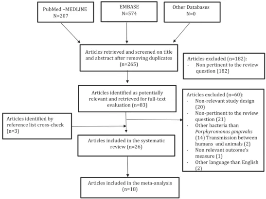
Overall, 265 articles were initially identified; of these, 182 were rejected upon titles and abstract because not relevant for the review topic. The remaining 83 arti-cles were screened at the full-text level; 26 were selected for the systematic review. Of these, 18 articles were used for pooled data analyses. Overall, the selected articles were published between 1993 and 2017. The flow chart of the study selection process
is shown inFigure 1.
Study characteristics
All the selected studies were observational studies, including 7 cross-sectional, 10 case series, 5 case-control, and 4 cohort studies. Co-occurrence of P. gingivalis between spouses was explored in 18 studies, including 334 couples; co-occurrence between sib-lings was explored in 3 studies. Detection concor-dance was explored between parent and infant(s) in 14 studies, including 625 pairs of parents-children. Details of study characteristics according to the type of familial relationship are displayed inTables 1–3.
Regarding the clinical periodontal status of the pro-band, the classical periodontal parameters, i.e. period-ontal pocket depth, clinical attachment level, gingival inflammation, and bleeding were recorded in some studies. In few of them, radiographic parameters were also considered. Classification of periodontitis according to the AAP Classification 1999 defining chronic or
aggressive periodontitis was used in three studies [9–
12]; adult periodontitis was described in four studies,
whereas advanced periodontitis in three. Several studies did not provide a clear definition of the periodontal disease used.
Bacterial sampling was carried out on supra-gingival and/or subgingival plaque, stimulated saliva, or sampled from dorsum of the tongue, buccal mucosa, or tonsillar area. Detection methods included culture in 10 studies (38.4%), and Polymerase Chain Reaction (PCR) in 11 studies (42.3%). DNA restriction enzyme analysis (REA-DNA) was used in 4 studies (15.3%), pulsed field gel electrophoresis (PFGE) in 1 study, arbi-trarily primed polymerase chain reaction (AP-PCR) in 2 (7.6%) studies, amplified fragment length polymorph-ism (AFLP-PCR) in 3, strain-specific identification of P. gingivalis I Isi 1126 PCR in 1, and Fim A genotyping in 3 studies. Serotyping characterization and ribotyping were reported in 2 studies, respectively.
Quantitative analyses
Forest plots were built pooling together intra-familial data according to the detection method used. Concerning the co-occurring detection between adult partners or spouses living together, the meta-analysis showed that the likelihood of detecting P. gingivalis in the partner/spouse when a proband was harboring P.
gingivalis was 53.58% (95%CI: 44.98%-62.95%; I2: 0%; n
= 99 pairs of partner/spouse) by culture, and 58.88%
(95%CI: 48.02%-69.32%; I2: 39.6%; n = 142 pairs) when
considering culture + PCR.
Figure 1.Flowchart of the literature search and article selection.
Table 1. Summary of the included studies assessing the simultaneous detection of Porphyromonas gingivalis (Pg) between adult couples of the same family. Author and year Study design Number of couples Periodontal status of the proband Periodontal status of the spouses Bacteria sampling Detection methods Proportion of intra-familial transmission Petit et al. 1993 Case series 4 Adult periodontitis Adult periodontitis ● 4 periodontal pockets ● Mucous membrane ● Dorsum of the tongue ● Culture ● DNA-REA Culture: 3/4 DNA-REA: 0/4 Van Steenbergen et al. 1993a Case series 18 Untreated adult severe periodontitis (at least 10 pockets of ≥ 5 mm and at least one pocket with ≥ 4m mo f attachment loss and sub-gingival presence of Pg) 5/18 spouses diagnosed as periodontitis patients ● 4 deepest pockets (by paper points) ● Buccal mucosal ● Dorsum of the tongue ● Tonsillar area ● Saliva ● Culture ● DNA-REA Culture: 8/18 DNA-REA: 6/18 Van Steenbergen et al. 1993 Case series 8 Severe adult periodontitis and subgingival presence of Pg ● 4 deepest pockets (by paper points) ● Buccal mucosa ● Dorsum of the tongue ● Tonsillar area ● Saliva ● DNA-REA ● AP-PCR ● Ribotyping DNA-REA: 6/8 AP-PCR: 6/8 Ribotyping: 6/8 Saarela et al. 1993 Case series 4 Advanced periodontitis and subgingival presence of Pg Advanced periodontitis and subgingival presence of Pg ● Deepest, the most inflamed per-iodontal pockets (by curette) ● Stimulated saliva ● Serotyping ● Ribotyping Ribotyping: 2/4 (married couples for at least 10 years) Von Troil Lindén et al. 1995 Case-control 10 vs. 10 Advanced periodontitis probands vs. periodontally healty controls Not specified ● 6 deepest and most inflamed periodontal pockets (by sterile curette) ● Stimulated saliva ● Culture Probands: 6/10 on subgingival samples 2/10 on salivary samples Controls: 1/10 on subgingival samples 0/10 on salivary samples Asikainen et al. 1996 Case series 15 Adult periodontitis (AAP Classification 1989) Adult periodontitis (85% of spouses) ● 4 to 6 deepest periodontal pock-ets (by curette) ● Dorsum of the tongue ● From healthy subjects: mesial subgingival sites of the first molars. ● Culture ● AP-PCR Culture: 9/15 AP-PCR: 2/15 Van der Velden et al. 1996 Longitudinal study Time frame: 1987 – 1994 22 No specific definition (based on clinical parameters: plaque index, calculus, probing depth, bleeding on probing, loss of attachment) Not specified ● 4 periodontal pockets (paper points) ● Culture Culture: 9/22 Tuite-McDonnell et al. 1997 Cross-sectional 43 17% of individuals had at least one site with an attachment level or PPD ≥ 5.5 mm Not specified ● all teeth (paper point) ● PCR PCR: 32/43 Spouses were significantly more frequently colonized by Pg (RR: 4 [95%CI: 2.3 –7]) than what would be expected if Pg was randomly distributed in the study population. No relationship was observed between the length of time a couple had been married and their concordance of colonization. (Continued )
Table 1. (Continued). Author and year Study design Number of couples Periodontal status of the proband Periodontal status of the spouses Bacteria sampling Detection methods Proportion of intra-familial transmission Von Troil Linden et al. 1997 Longitudinal experimental study Six month follow up study 8 Advanced periodontitis 3 spouses: moderate periodontitis undergone a mechanical treatment combined with metronidazole. 7 spouses: mild to moderate periodontitis were untreated ● 6 deepest and most inflamed pockets ● Paraffin-stimulated saliva ● Culture Culture: 4/8 Van Winkelhoff et al. 1999 Longitudinal study. Time frame: 1987 – 1994 19 No selection on the periodontal status Not specified ● 4 sub-gingival samples using 1 paper point per pocket ● Culture ● Serotyping Culture: 15/19 Serotyping: 0/19 Asano et al. 2003 Cross-sectional 25 Periodontitis defined as at least 4 pockets with PPD > 4 mm and radiographs showing the presence of at least one intrabony defect > 4 mm. Not specified ● 3 deepest pockets (by paper points), or gingival sulci ● Culture ● PFGE ● FimA gen-otyping Culture: 14/25 PFGE: 6/14 The frequency of FimA type II in isolates from the 6 couples with identical PFGE patterns was 83.3%, which was higher than for the periodontitis unrelated patients (46.8%) (positive for Pg ). Park et al. 2004 Case series 16 Chronic and aggressive periodontitis Not specified ● 3 sterile paper points into the mesial sulcus of diseased sites of the probands and the deepest sites of their family members ● PCR (Isi 1126 spe-cific pri-mer) PCR (Isi 1126): 5/16 Rijnsburgurger et al. 2007 Case series 6 Untreated severe periodontitis Not specified ● Subgingival samples ● Dorsum of the tongue ● Buccal mucosa, ● Tonsils ● Saliva ● REA ● AFLP- PCR REA: 4/6 AFLP-PCR: 4/6 (showing a similarity >85%) Van Winkelhoff 2007 Cross-sectional 13 Population without regular dental care Not specified ● Deepest bleeding sites with the greatest amount of attachment loss ● AFLP- PCR AFLP-PCR: 0/13 Van Winkelhof 2008 Longitudinal study. Time frame: 1994 – 2002 27 Population without regular dental care Not specified ● Deepest bleeding sites with the greatest amount of attachment loss ● Culture ● AFLP- PCR Culture: 1994: 13/27 2002: 18/27 AFLP-PCR: 1994: 0/27 2002: 3/27 Martelli et al. 2012 Case series 9 Untreated periodontitis Untreated periodontitis ● One pocket per quadrant (by paper point) ● PCR ● FimA gen-otyping PCR: 6/9 FimA type II: 6/9 Feng et al. 2015 Case series 10 Aggressive periodontitis Not specified ● Subgingival samples ● Saliva ● PCR ● FimA gen-otyping PCR: 7/10 FimA genotyping: 7/10 AP-PCR stands for arbitrarily primed polymerase chain reaction; BOP for bleeding on probing; PPD for probing pocket depth; PI for plaque index; Pg for Porphyromonas gingivalis. Aa for Agregatibacter actinomycetemcomitans. Pi for Prevotella intermedia . AFLP: Amplified fragment length polymorphism, DNA REA: Deoxyribo Nucleic Acid Restriction Endonuclease Analysis; Fim A: Fimbriae A.
Table 2. Summary of the included studies assessing the simultaneous detection of Porphyromonas gingivalis (Pg) between parents and children of the same family. Author and year Study design Number of children Periodontal status of the parents Periodontal status of the children Bacteria sampling Detection methods Main findings (Number of positive/Total) Petit et al. 1993 Case series 10 Adult periodontitis Severe generalized juvenile periodontitis (GJP) with presence of Aa and Pg ●4 periodontal pockets
●Mucous membrane ●Dorsum
of the tongue ●First molar if no PPD ●Culture ●DNA-REA Culture: 7/10 DNA REA: 5/10 Petit et al. 1994 Case series 36 Untreated adult severe periodontitis (at least 10 pockets of ≥ 5 mm and at least one pocket with ≥ 4m mo f attachment loss and subgingival presence of Pg or Aa or Pi) Not specified ●4 periodontal pockets
●Mucous membrane ●Dorsum
of the tongue ●Tonsilla area ●Saliva ●First molar if no PPD ●Culture Culture: 1/36 Asikainen et al. 1996 Case series 19 Adult periodontitis (AAP Classification 1989) 5% of children
exhibited periodontal destruction
●4 to 6 deepest periodontal pockets (by curette) ●Dorsum of the tongue ●First molar if no PPD ●Culture ●AP PCR Culture: 1/19 No AP PCR (not enough samples) Tuite-McDonnell et al. 1997 Cross sectional 103 oldest children with their mothers 101 oldest children with their fathers 98 adult children with their mothers 69 adult children with their fathers 17% of individuals had ≥ 1 site with an attachment level or PPD ≥ 5.5 mm Not specified ●All teeth (Paper points) ●PCR PCR (Oldest child with the mother): 20/103 PCR (Oldest child with the father): 17/101 PCR (Adult child with the mother): 25/98 PCR (Adult child with the father): 14/69 Umeda et al. 2004 Cross sectional 56 Only 4 parents had PPD > 4 mm Not specified ●Saliva + supra-gingival plaque (Children) ●Saliva (Parents) ●PCR PCR (Parent): 54.8% PCR (Children): 7.1% Okada et al. 2004 Cross sectional 28 1 healthy individual 4 gingivitis 7 periodotnitis 6 healthy individuals 6 gingivitis 6 periodontitis ●Supra-gingival plaque collected with sterile tooth brushes (1 min) ●PCR PCR: 6/28 (21.4%) Park et al. 2004 Case series 14 Chronic and aggressive periodontitis Not specified ●3 Sub-gingival samples (Paper points) ●PCR with Isi 1126 specific primer PCR Isi 1126: 11/14 (Continued )
Table 2. (Continued). Author and year Study design Number of children Periodontal status of the parents Periodontal status of the children Bacteria sampling Detection methods Main findings (Number of positive/Total) Tamura et al. 2006 Cross sectional 106 Not specified. All patients were recruited from the Periodontics Clinic of Osaka University Hospital. No children had periodontitis ●Saliva samples ●PCR PCR (Mothers): 42.5% PCR (Children): 2.7% Kobayachi et al. 2008 Cross sectional 78 Not specified No children had moderate or severe gingivitis
●Sub-gingival plaque ●(first
molar for children; first and second molars for mothers) ●PCR PCR (Mothers):17.6% PCR (Children): 9% Belcheva et al. 2012 Case control 11 Group of diseased parents and their children Group of healthy parents and their children Not specified ●Deepest pockets ●(First molars and lower incisors) ●Sulci between premolars and molars when healthy ●Culture ●PCR Culture (Children): No detection PCR (Children): 2/11 The study did not find statistically significant difference (p > 0.05) in the prevalence of Pg between children with healthy parents and children whose parents presenting with periodontal disease Monteiro et al. 2014 Case control 15 Generalized aggressive periodontitis (GAP) vs healthy Not specified
●Stimulated saliva ●(without
dental hygiene-no brushing 2 hours before sampling) ●PCR PCR (Periodontitis parent): 50% PCR (Infant): 30% Parents with GAP showed statistically higher Pg salivary concentration than periodontally healthy parents (p < 0.05). Children of GAP families, however, exhibited similar Pg concentration than healthy children (p > 0.05). Monteiro et al. 2015 Case control 15 Generalized aggressive periodontitis vs healthy Not specified
●Stimulated saliva ●(without
dental hygiene-no brushing 2 hours before sampling) ●PCR PCR (Periodontitis parent): 80% PCR (Infant): 60% Feng et al. 2015 Case series 10 Aggressive periodontitis Not specified
●Saliva ●Sub-gingival samples ●(First
molars) ●PCR with Fim A genotype ●Specific pri-mers PCR (Fim A genotyping): 7/10 Al Yahyoufi. 2017 Case series 11 Chronic and Aggressive Periodontitis Not specified ●4 Sub-gingival samples ●(Paper point in each quadrant) ●DNA probe DNA probe: 7/11 AP-PCR stands for arbitrarily primed polymerase chain reaction; Pg for Porphyromonas gingivalis. Aa for Aggregatibacter actinomycetemcomitans. Pi for Prevotella intermedia . AFLP: Amplified fragment length polymorphism; Fim A for Fimbriae A; DNA REA for Deoxyribo Nucleic Acid Restriction Endonuclease Analysis.
By applying genotyping, the likelihood of detecting the same strain of P. gingivalis between the members of the couple dropped at 29.02% (95%CI:
17.68%-41.87%; I2: 43.2%; n = 91 pairs). Using Fim A
geno-typing method, the same Fim A was retrieved in
57.15% (95%CI: 40.87%-72.68%; I2: 0.2%; n = 33
pairs) of the cases. Overall, the same ribotype of P. gingivalis was found in 64.7% (95%IC:
38.83%-86.63%; I2: 0%; n = 12 pairs) of couples when both
adults were positive for P. gingivalis.
Concerning the parent-infant co-occurrence,
when at least one parent was colonized by P. gingi-valis, the likelihood that also one child harbored P. gingivalis was estimated at 20.15% when considering three studies using culture (95%CI: 0.31%-58.89%; I2: 90.4%, n = 65 pairs of parent-infant) [13–15], and 5% when considering two studies (95%CI:
0.91%-12.11%; I2: 0%; sensitivity analysis on 55 pairs)
[13,15]. The detection of P. gingivalis using PCR
amplification showed a likelihood of 22.46% (95%
CI: 17.46%-27.90%; I2: 0%, n = 240 pairs), whereas
when combining both detection methods (culture + PCR), the estimated likelihood was 24.05% (95%CI:
13.41%-36.64%; I2: 80.5%, n = 316 pairs) [13–19].
Based on two studies only, the same P. gingivalis genotype of the parents was shared in 64.95% of children harboring P. gingivalis (95%CI: 37.73– 87.74%; I2: 50.5%; n = 24 pairs) [12,14].
By pooling together all data about shared detection both between-spouses and parent-infant, the likeli-hood of detecting P. gingivalis once a family member harbors the bacterium is estimated at 63.54% by
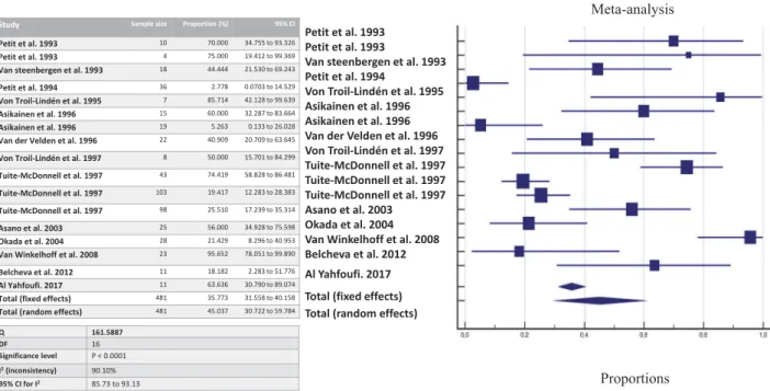
cul-ture (Figure 2), 45.03% by culture + PCR (Figure 3),
and 35.71% by genotyping (Figure 4).
Study quality assessment
Two reviewers (M.B. and M.C.C.) scored the metho-dological qualities of the included studies. The NOS score varies from 3 to 5, and over 9; thus, the studies were qualified as low (n = 17) and moderate (n = 9) quality studies. Detailed information regarding the quality assessment of the included studies is reported in Supplement Table 1.
Discussion
The present systematic review and meta-analysis focuses on the likelihood of detecting one of the major periodontal pathogens, namely P. gingivalis, among cohabiting family members, and demonstrates that the co-occurrence of P. gingivalis among couples, children, or siblings ranges between 42% and 62% when a family member of is carrying P. gingivalis (the so-called proband). However, only in 35% of cases, these members are sharing the same genotype of the bacterium. Table 3. Summary of the included studies assessing the simultaneous detection of Porphyromonas gingivalis (Pg) between siblings. Author and Year Study Design Number of Subjects Periodontal Status of the Probands Periodontal Status of the Siblings Bacteria Sampling Detection Method Main Findings Van winkelhoff et al. 1999 Longitudinal study 1987 –1994 29 sibships No selection on the periodontal status Not specified 4 sub-gingival samples using 1 paper point per pocket ●Culture ●Serotyping Culture: 26/29 Serotyping: 3/29 Van Winkelhoff. 2007 Cross sectional study 10 sibships 30 siblings Population without regular dental care Not specified Deepest bleeding site with the greatest amount of attachment loss ●AFLP typing 2/10 Sibships 13/30 Siblings Van Winkelhoff. 2008 Longitudinal study 1994 –2002 23 sibships 54 siblings Population without regular dental care Not specified Deepest bleeding site with the greatest amount of attachment loss ●Culture ●AFLPT typing Culture (1994): 22/23 Culture (2002): 22/23 AFLP PCR (1994): 6/23 AFLP PCR (2002): 6/23 Pg stands for Porphyromonas gingivalis . AFLP for Amplified fragment length polymorphism.
Study Sample size Proportion (%) 95% CI
Petit et al. 1993 4 75.000 19.412 to 99.369
Petit et al. 1993 10 70.000 34.755 to 93.326
Van steenbergen et al. 1993 18 44.444 21.530 to 69.243
Von Troil-Lindén et al. 1995 7 85.714 42.128 to 99.639
Asikainen et al. 1996 15 60.000 32.287 to 83.664
Van der Velden et al. 1996 22 40.909 20.709 to 63.645
Von Troil-Lindén et al. 1997 8 50.000 15.701 to 84.299
Asano et al. 2003 25 56.000 34.928 to 75.598
Van Winkelhoff et al. 2008 23 95.652 78.051 to 99.890
Total (fixed effects) 132 63.125 54.598 to 71.089
Total (random effects) 132 63.543 48.360 to 77.459
Q 25.6820 DF 8 Significance level P = 0.0012 I2(inconsistency) 68.85% 95% CI for I2 37.64 to 84.44 Petit et al. 1993 Petit et al. 1993
Van steenbergen et al. 1993 Von Troil-Lindén et al. 1995 Asikainen et al. 1996 Van der Velden et al. 1996 Von Troil-Lindén et al. 1997 Asano et al. 2003 Van Winkelhoff et al. 2008 Total (fixed effects) Total (random effects)
Figure 2.The likelihood of co-occurrence of P. gingivalis once a family member harbors P. gingivalis assessed by culture.
Petit et al. 1993 Petit et al. 1993
Van steenbergen et al. 1993 Petit et al. 1994
Von Troil-Lindén et al. 1995 Asikainen et al. 1996 Asikainen et al. 1996 Van der Velden et al. 1996 Von Troil-Lindén et al. 1997 Tuite-McDonnell et al. 1997 Tuite-McDonnell et al. 1997 Tuite-McDonnell et al. 1997 Asano et al. 2003 Okada et al. 2004 Van Winkelhoff et al. 2008 Belcheva et al. 2012 Al Yahfoufi. 2017 Total (fixed effects) Total (random effects)
Proportions Meta-analysis
Study Sample size Proportion (%) 95% CI Petit et al. 1993 10 70.000 34.755 to 93.326 Petit et al. 1993 4 75.000 19.412 to 99.369 Van steenbergen et al. 1993 18 44.444 21.530 to 69.243 Petit et al. 1994 36 2.778 0.0703 to 14.529 Von Troil-Lindén et al. 1995 7 85.714 42.128 to 99.639 Asikainen et al. 1996 15 60.000 32.287 to 83.664 Asikainen et al. 1996 19 5.263 0.133 to 26.028 Van der Velden et al. 1996 22 40.909 20.709 to 63.645 Von Troil-Lindén et al. 1997 8 50.000 15.701 to 84.299 Tuite-McDonnell et al. 1997 43 74.419 58.828 to 86.481 Tuite-McDonnell et al. 1997 103 19.417 12.283 to 28.383 Tuite-McDonnell et al. 1997 98 25.510 17.239 to 35.314 Asano et al. 2003 25 56.000 34.928 to 75.598 Okada et al. 2004 28 21.429 8.296 to 40.953 Van Winkelhoff et al. 2008 23 95.652 78.051 to 99.890 Belcheva et al. 2012 11 18.182 2.283 to 51.776 Al Yahfoufi. 2017 11 63.636 30.790 to 89.074 Total (fixed effects) 481 35.773 31.558 to 40.158 Total (random effects) 481 45.037 30.722 to 59.784
Q 161.5887
DF 16
Significance level P < 0.0001
I2(inconsistency) 90.10%
95% CI for I2 85.73 to 93.13
Figure 3.The likelihood of co-occurrence of P. gingivalis once a family member harbors P. gingivalis assessed by culture and PCR.
Study Sample size Proportion (%) 95% CI
Petit et al. 1993 4 0.000 0.000 to 60.236
Petit et al. 1993 10 50.000 18.709 to 81.291
Van Steenbergen et al. 1993 18 33.333 13.343 to 59.007 Asikainen et al. 1996 15 13.333 1.658 to 40.460
Asano et al. 2003 14 42.857 17.661 to 71.139
Park et al. 2004 16 31.250 11.017 to 58.662
Park et al. 2004 14 78.571 49.202 to 95.342
Rijnsburger et al. 2007 6 66.667 22.278 to 95.673
Van Winkelhoff et al. 2008 18 16.667 3.579 to 41.418
Van Winkelhoff et al. 2008 22 27.273 10.729 to 50.222 Total (fixed effects) 137 34.979 27.306 to 43.272 Total (random effects) 137 35.718 23.165 to 49.358
Q 25.1557 DF 9 Significance level P = 0.0028 I2(inconsistency) 64.22% 95% CI for I2 29.50 to 81.84 Petit et al. 1993 Petit et al. 1993
Van Steenbergen et al. 1993 Asikainen et al. 1996 Asano et al. 2003 Park et al. 2004 Park et al. 2004 Rijnsburger et al. 2007 Van Winkelhoff et al. 2008 Van Winkelhoff et al. 2008 Total (fixed effects) Total (random effects)
Meta-analysis
Proportions
Figure 4.The likelihood of co-occurrence of P. gingivalis once a family member harbors P. gingivalis assessed by genotyping. JOURNAL OF ORAL MICROBIOLOGY 9
These data quantify the likelihood of co-occurring detection and support the hypothesis that an intra-familial bacterial transfer may occur. However, this appears to be highly variable both between spouses or couples and between parents and children despite their intimate cohabitation for long periods of time. The observed variability could be explained in several ways. First of all, bacterial transfer and detection does not necessarily mean persistent colonization up to the detectable levels of the pathogens but could be caused
by repeated inoculation [6]. Indeed, bacterial
trans-mission results from a combination of a sufficiently large and concentrated inoculum enabling bacterial survival during colonization, with a favorable oral environment of the recipient, which is dependent on the resident microbiota and the host defenses
and characteristics [6,20]. Moreover, several
beha-vioral and environmental patterns may influence the likelihood of bacterial colonization, such as hygiene habits, proximity, and intimacy differences among family members. However, as observed for other body sites, like skin or fecal microbiota, the family unit have a strong effect on human microbial com-munity composition: indeed, family membership may explain a large proportion of the variability in bacter-ial diversity, with family members tending to harbor similar microbiota [7,21,22]. Indeed, the chance of sharing the same bacterial genotype appears to be greater among related individuals than unrelated ones or if P. gingivalis was randomly distributed in the population [13,19,23].
Sampling and detection of porphyromonas gingivalis
Although the possible ways for transferring of P. gingivalis remains unclear, the role of saliva as a vehicle of bacterial spread is probable and it is sup-ported by the fact that P. gingivalis can be cultured from salivary samples, indicating that this bacterium survives in the saliva during transportation to a new
host [24]. Indeed, the probability of inoculation
appears to be directly related to the salivary bacterial load, with a greater risk of colonization in the
reci-pient for greater bacterial loads [20,24]. Then, P.
gingivalis is able to spread intra-orally and colonize supra-gingival and sub-gingival plaque at sites with and without periodontal attachment loss, although, it is more likely to find P. gingivalis in deep pockets
rather than shallow ones [25]. Consequently, the
eventual eradication (i.e., a bacterial load under the detection level) of this pathogen by efficacious peri-odontal treatments may prevent its spread among individuals [6,24].
It must be noted that the detection of periodontal pathogens is drastically influenced by the methods applied to sample and recover them (e.g., culture,
PCR, DNA probe checkerboard) [26]. As deemed
from Tables 1 and 2, the sampling and detection
methods applied in the studies included in the pre-sent systematic review are highly heterogeneous. This implies to analyze data by sub-groups of comparable detection methods while avoiding global compari-sons. However, no distinction could be made accord-ing to the bacterial samplaccord-ing site (e.g. saliva, subgingival plaque) because almost all studies pro-vided pooled results of all samples examined (although multiple sites were sampled in most cases). Moreover, we must highlight that the included studies were conducted in a time span of 24 years during which microbiological techniques have drasti-cally evolved as well as our knowledge about the role of specific periodontal pathogens and the complexity of the oral microbiota [27].
Several studies used culture to detect and quantify P. gingivalis, but no distinction between clones was made. The sensitivity of bacterial culturing is rather
low, with detection limits averaging at 103–104
bac-terial cells [26,28]. On the contrary, methods based
on immune diagnosis (serotyping) and molecular analysis, such as PCR and ribotyping are highly sen-sitive and specific [26]. No study to date investigated the concordance of bacterial colonization and the likelihood of sharing the same periodontal microbiota between family members by applying modern meth-ods, such as high throughput sequencing methods.
Clinical implications of familial porphyromonas gingivalis sharing
If an intra-familial transmission of P. gingivalis is possible and probable, the available studies present a design and an overall level of quality that do not allow to conclude about ta specific intra-familial transmission pattern. Nevertheless, they provide evi-dence about the likelihood of a shared detection of P. gingivalis among family members.
A key question is whether the sharing of P. gingi-valis can affect the periodontal health of the indivi-duals becoming colonized. Some studies found that spouses of patients with advanced periodontitis have a worse periodontal status than spouses of
period-ontal healthy individuals [29]. Others did not
demon-strate that the periodontal condition of the spouse
was influenced by that of the partner [15,19]. When
the relationship between the duration of marriage and the chance of transmission was explored, the study of Tuite-Mcdonnel et al. found no relationship between the frequency of co-occurring detection and the length of marriage, suggesting that cross-coloni-zation likely occurred in the early years of marriage
and remains stable over time [19].
When the frequency of presence of P. gingivalis in spouses of colonized proband was compared to
unrelated patients, the spouses were significantly more frequently colonized by P. gingivalis than what would be expected if P. gingivalis was randomly
dis-tributed in the population [19,23]; but this was not
observed in all studies [13].
Indeed, transfer of P. gingivalis from an individual to another does not necessarily translate into stable colonization and periodontal breakdown. It may per-sist an equilibrium between the host and the resident microbiota despite the repeated inoculation of P. gin-givalis, especially in periodontally healthy individuals. However, we may consider P. gingivalis colonization as a potential risk factor, as it is known that this bacter-ium is causal in periodontitis initiation, progression, recurrence, as well as in peri-implantitis [5,30].
P. gingivalis can be possibly shared between parents and children. This raises another important question: does an early inoculation increase the chances of a per-manent colonization and development of periodontitis later on in children? Early studies using culture probably underestimated the prevalence of P. gingivalis in young subjects, seldom detecting it before puberty [31,32]. Later on, studies relying upon more sensitive techniques, such as DNA-based technologies, demonstrated the presence of P. gingivalis in a large fraction of young subjects and showed it to be equally common in children of all ages [19]. Indeed, P. gingivalis can be detected in children aged of 20 days as of 18 years [33–35]. An epidemiolo-gical study, using DNA probe checkerboard assay, found that 71% of the 18- to 48-month-old children were infected with at least one periodontal pathogen [36]. P. gingivalis detection rates was estimated at 68.8% in chil-dren. A study evaluating the influence of mother’s peri-odontal clinical status on the prevalence of periperi-odontal pathogens in newborns (aged of 3 months) showed that P. gingivalis was the most prevalent pathogen followed by others periodontal bacteria (Prevotella intermedia,
Tannerella forshythia, Campylobacter rectus,
Aggregatibacter actinomycetemcomitans) [37]. They
con-cluded that the maternal clinical periodontal status is a significant indicator of the oral microbiota composition in the newborn children.
If the colonization in young children occurs, it appears to be composed by the same P. gingivalis strain of the parent(s) only transitory, whereas it becomes more stable during teenage years, possibly
as deeper pockets develop [33]. Indeed, the presence
of P. gingivalis is favored by the presence of deep probing depths, and thus it is likely that transmission from a proband infected individual to a periodontally healthy one is possible in terms of transient presence in the oral cavities, but in the absence of a permanent niche, such as a deep pocket, it may no longer survive. It is noteworthy that the exposure and chance of colonization of P. gingivalis may vary in relation to the proband status, the severity of the periodontal disease, and the administration of a successful periodontal
treatment. Moreover, bacterium-specific virulence fac-tors may facilitate the familial sharing of P. gingivalis. Particularly, the fimbriae A (FimA), a specific compo-nent of the cell surface [38], seems to play a strategic role in the colonization and invasion of the periodontal tissues [39,40]. The FimA gene has been classified into six types (I to V and Ib) [41]. A higher rate of type II FimA was detected in couples who shared the same strains of P. gingivalis [9,23,42], being this strain mainly
associated with severe periodontitis [43]. Conversely,
the type IV FimA was not found in couples with identical PFGE patterns [23].
The present systematic review has some limita-tions. Retrieved data are heterogeneous: different methods for bacterial sampling, detection, and quan-tification were applied, requiring subgroup analyses. Findings are based upon a low-to-moderate level of evidence coming mainly from retrospective, cross-sectional studies or case series with a small sample size, which hamper any conclusion on the specific colonization patterns or on the eventual deterioration of periodontal health after the occurrence of P. gingi-valis sharing within the family. Moreover, it should be stressed that a shared detection of pathogens (or colonization concordance) in cohabiting family mem-bers does not necessarily prove the transmission of these pathogens. Finally, the majority of the available
studies were published in the ‘90. Due to relevant
technical progress in microbiology in the last decade, the presented findings should be replicated and updated in lights of more recent knowledge about the diversity and richness of the oral microbiota [44].
Conclusion
The present systematic review and meta-analysis sup-ports the co-occurring detection of P. gingivalis within family members. Due to the role of P. gingi-valis in the etiology of periodontitis, the likelihood of sharing P. gingivalis should be considered in the assessment of patient’s periodontal risk.
Acknowledgments
The authors would like to thank the entire team of the European Postgraduate Program in Periodontology and Implant Dentistry of the University of Paris for their support.
Author contributions
M.B.: study conception, literature search, data extraction, data analysis, manuscript drafting, and manuscript final ver-sion approval. H.R.: study conception, critical appraisal, manuscript revisions, and manuscript final version approval. V.M.: critical appraisal, manuscript revisions, and script final version approval. F.M.: critical appraisal, manu-script revisions, manumanu-script final version approval. P.B: study conception, critical appraisal, manuscript revisions, and JOURNAL OF ORAL MICROBIOLOGY 11
manuscript final version approval. M.C.C.: Reviewer and supervisor, study conception, data analysis, manuscript revi-sions, and manuscript final version approval.
Disclosure statement
No potential conflict of interest was reported by the authors.
ORCID
Maria Clotilde Carra http://orcid.org/0000-0002-5717-3274
References
[1] Papapanou PN, Sanz M, Buduneli N, et al. Periodontitis: consensus report of workgroup 2 of the 2017 World workshop on the classification of periodontal and peri-implant diseases and conditions. J Periodontol.2018;89(Suppl 1):S173–S182.
[2] Darveau RP. Periodontitis: a polymicrobial disruption of host homeostasis. Nat Rev Microbiol. 2010;8:481– 490.
[3] Palmer RJ. Composition and development of oral bacterial communities. Periodontol 2000.2014;64:20– 39.
[4] Hajishengallis G. Periodontitis: from microbial immune subversion to systemic inflammation. Nat Rev Immunol.2015;15:30–44.
[5] Hajishengallis G, Darveau RP, Curtis MA. The key-stone-pathogen hypothesis. Nat Rev Microbiol.
2012;10:717–725.
[6] Van Winkelhoff AJ, Boutaga K. Transmission of per-iodontal bacteria and models of infection. J Clin Periodontol.2005;32(Suppl 6):16–27.
[7] Kilian M, Chapple IL, Hannig M, et al. The oral microbiome - an update for oral healthcare profes-sionals. Br Dent J.2016;221:657–666.
[8] DerSimonian R, Laird N. Meta-analysis in clinical trials. Control Clin Trials.1986;7:177–188.
[9] Feng X, Zhu L, Xu L, et al. Distribution of 8 period-ontal microorganisms in family members of Chinese patients with aggressive periodontitis. Arch Oral Biol.
2015;60:400–407.
[10] Monteiro MDF, Casati MZ, Taiete T, et al. Periodontal clinical and microbiological characteris-tics in healthy versus generalized aggressive period-ontitis families. J Clin Periodontol.2015;42:914–921.
[11] Monteiro MF, Casati MZ, Taiete T, et al. Salivary carriage of periodontal pathogens in generalized aggressive periodontitis families. Int J Paediatr Dent.
2014;24:113–121.
[12] Park O-J, Min K-M, Choe S-J, et al. Use of insertion sequence element IS1126 in a genotyping and trans-mission study of Porphyromonas gingivalis. J Clin Microbiol.2004;42:535–541.
[13] Asikainen S, Chen C, Slots J. Likelihood of transmit-ting Actinobacillus actinomycetemcomitans and Porphyromonas gingivalis in families with periodon-titis. Oral Microbiol Immunol.1996;11:387–394.
[14] Petit MD, van Steenbergen TJ, Scholte LM, et al. Epidemiology and transmission of Porphyromonas gingivalis and Actinobacillus actinomycetemcomitans
among children and their family members. A report of 4 surveys. J Clin Periodontol.1993;20:641–650. [15] Petit MD, van Steenbergen TJ, Timmerman MF, et al.
Prevalence of periodontitis and suspected periodontal pathogens in families of adult periodontitis patients. J Clin Periodontol.1994;21:76–85.
[16] Al Yahfoufi Z. Prevalence of periodontal destruction and putative periodontal pathogens in the same leba-nese family. J Contemp Dent Pract.2017;18:970–976. [17] Belcheva MD, Kiselova-Yaneva A, Krasteva A. Transmission of Porphyromonas gingivalis from care-givers to children. J Imab.2012;18:157–162.
[18] Okada M, Hayashi F, Soda Y, et al. Intra-familial distribution of nine putative periodontopathogens in dental plaque samples analyzed by PCR. J Oral Sci.
2004;46:149–156.
[19] Tuite-McDonnell M, Griffen AL, Moeschberger ML, et al. Concordance of Porphyromonas gingivalis coloniza-tion in families. J Clin Microbiol.1997;35:455–461.
[20] Asikainen S, Chen C. Oral ecology and person-to-person transmission of Actinobacillus actinomycetem-comitans and Porphyromonas gingivalis. Periodontol 2000.1999;20:65–81.
[21] Song SJ, Lauber C, Costello EK, et al. Cohabiting family members share microbiota with one another and with their dogs. Elife.2013;2:e00458.
[22] Stahringer SS, Clemente JC, Corley RP, et al. Nurture trumps nature in a longitudinal survey of salivary bacterial communities in twins from early adolescence to early adulthood. Genome Res.2012;22:2146–2152. [23] Asano H, Ishihara K, Nakagawa T, et al. Relationship
between transmission of Porphyromonas gingivalis and fimA type in spouses. J Periodontol.2003;74:1355–1360.
[24] von Troil-lindén B, Torkko H, Alaluusua S, et al. Salivary levels of suspected periodontal pathogens in relation to periodontal status and treatment. J Dent Res.1995;74:1789–1795.
[25] Socransky SS, Haffajee AD, Ximenez-Fyvie LA, et al. Ecological considerations in the treatment of Actinobacillus actinomycetemcomitans and Porphyromonas gingivalis periodontal infections. Periodontol 2000.1999;20:341–362.
[26] Sanz M, Lau L, Herrera D, et al. Methods of detection of Actinobacillus actinomycetemcomitans, Porphyromonas gingivalis and Tannerella forsythensis in periodontal microbiology, with special emphasis on advanced mole-cular techniques: a review. J Clin Periodontol.
2004;31:1034–1047.
[27] Teles R, Teles F, Frias-Lopez J, et al. Lessons learned and unlearned in periodontal microbiology. Periodontol 2000.2013;62:95–162.
[28] Socransky SS, Haffajee AD, Smith GL, et al. Difficulties encountered in the search for the etiologic agents of destructive periodontal diseases. J Clin Periodontol.1987;14:588–593.
[29] Von Troil-Lindén B, Torkko H, Alaluusua S, et al. Periodontal findings in spouses. A clinical, radio-graphic and microbiological study. J Clin Periodontol.1995;22:93–99.
[30] Zhuang L-F, Watt RM, Mattheos N, et al. Periodontal and peri-implant microbiota in patients with healthy and inflamed periodontal and peri-implant tissues. Clin Oral Implants Res.2016;27:13–21.
[31] Könönen E, Asikainen S, Jousimies-Somer H. The early colonization of gram-negative anaerobic bacteria in edentulous infants. Oral Microbiol Immunol.
[32] Könönen E, Kanervo A, Takala A, et al. Establishment of oral anaerobes during the first year of life. J Dent Res. 1999;78:1634–1639.
[33] Lamell CW, Griffen AL, McClellan DL, et al. Acquisition and colonization stability of Actinobacillus actinomyce-temcomitans and Porphyromonas gingivalis in children. J Clin Microbiol.2000;38:1196–1199.
[34] Mättö J, Saarela M, Alaluusua S, et al. Detection of Porphyromonas gingivalis from saliva by PCR by using a simple sample-processing method. J Clin Microbiol. 1998;36:157–160.
[35] McClellan DL, Griffen AL, Leys EJ. Age and preva-lence of Porphyromonas gingivalis in children. J Clin Microbiol. 1996;34:2017–2019.
[36] Yang EY, Tanner ACR, Milgrom P, et al. Periodontal pathogen detection in gingiva/tooth and tongue flora samples from 18- to 48-month-old children and per-iodontal status of their mothers. Oral Microbiol Immunol.2002;17:55–59.
[37] Aquino DR, Franco GCN, Cortelli JR, et al. The impact of the maternal’s periodontal status on the detection of periodontal pathogens in newborn chil-dren. Revista Odonto Ciência.2010;25:333–338.
[38] Travis J, Potempa J, Maeda H. Are bacterial proteinases pathogenic factors? Trends Microbiol.1995;3:405–407. [39] Amano A, Kuboniwa M, Nakagawa I, et al. Prevalence
of specific genotypes of Porphyromonas gingivalis fimA and periodontal health status. J Dent Res.
2000;79:1664–1668.
[40] Nakagawa I, Amano A, Kuboniwa M, et al. Functional differences among FimA variants of porphyromonas gin-givalis and their effects on adhesion to and invasion of human epithelial cells. Infect Immun.2002;70:277–285. [41] Amano A, Nakagawa I, Okahashi N, et al.
Variations of Porphyromonas gingivalis fimbriae in relation to microbial pathogenesis. J Periodontal Res. 2004;39:136–142.
[42] Martelli FS, Mengoni A, Martelli M, et al. Comparison of periodontal microbiological patterns in Italian spouses. Ig Sanita Pubbl.2012;68:589–599.
[43] Olsen I, Chen T, Tribble GD. Genetic exchange and reassignment in Porphyromonas gingivalis. J Oral Microbiol.2018;10:1457373.
[44] Schwarzberg K, Le R, Bharti B, et al. The personal human oral microbiome obscures the effects of treat-ment on periodontal disease. PloS One.2014;9:e86708. JOURNAL OF ORAL MICROBIOLOGY 13