R
EWARD-
RELATED BEHAVIORAL AND BIOLOGICAL RESPONSES ASSOCIATED WITH EATING – INSIGHTS FROM ANOREXIANERVOSA AND OBESITY RESEARCH
R
ÉPONSESB
IO-
COMPORTEMENTALES LIÉES À LA RÉCOMPENSE–
A
PERÇUS DE LARECHERCHE EN ANOREXIE MENTALE ET EN OBÉSITÉ
Pereira Picolo Ribeiro, Mayron
d’Arapongas, Brésil
2020
Thèse de doctorat cumulative présentée à la Faculté des lettres et des sciences humaines de l’Université de Fribourg (Suisse)
3
SUMMARY
FIGURE LIST ... 6 TABLE LIST ... 8 ACKNOWLEDGEMENTS ... 11 ABSTRACT ... 15 INTRODUCTION ... 17 THEORETICAL BACKGROUND ... 23 CHAPTER 1 ... 25THE REGULATION OF EATING BEHAVIORS ... 25
1. EATING AS A COMPLEX BEHAVIOR ... 27
2. ORGANIC SYSTEMS REGULATING EATING ... 31
2.1. THE HOMEOSTATIC SYSTEM ... 31
2.2. THE REWARD SYSTEM ... 34
2.2.1. FOOD AS A REWARD ... 36
2.2.2. DOPAMINE AND EATING ... 40
2.2.3. FASTING AND FOOD AS A REWARD ... 42
2.3. THE ENDOCANNABINOID SYSTEM: HOMEOSTASIS AND REWARD PROCESSING44 CHAPTER 2 ... 47
FROM EATING BEHAVIORS TO EATING DISTURBANCES ... 47
1. ANOREXIA NERVOSA ... 50
1.1. DIAGNOSTIC CRITERIA OF ANOREXIA NERVOSA ... 50
4
1.3. ANOREXIA NERVOSA AND REWARD PROCESSING ... 52
1.3.1. ANOREXIA NERVOSA AND MONETARY REWARDS ... 54
1.3.1.1. BEHAVIORAL RESPONSES TO MONETARY REWARDS IN AN ... 56
1.3.1.2. SELF-REPORTED RESPONSES TO MONETARY REWARDS IN AN ... 57
1.3.1.3 NEURAL RESPONSES TO MONETARY REWARDS IN AN ... 58
2. OBESITY ... 66
2.1. OBESITY AND COMORBIDITIES ... 67
2.2. OBESITY AND FOOD REWARD ... 68
2.3. OBESITY AND MONETARY REWARD ... 70
2.3.1. BEHAVIORAL RESPONSES TO MONETARY REWARDS IN OBESITY ... 87
2.3.2. SELF-REPORTED RESPONSES TO MONETARY REWARDS IN OBESITY ... 90
2.3.3. NEURAL RESPONSES TO MONETARY REWARDS IN OBESITY ... 90
3. ANOREXIA NERVOSA, OBESITY AND MONETARY REWARDS ... 91
EMPIRICAL RESEARCH ... 93
CHAPTER 3 ... 99
FOOD VS MONEY? EFFECTS OF HUNGER ON MOOD AND BEHAVIORAL REACTIVITY TO REWARD IN ANOREXIA NERVOSA ... 99
1. INTRODUCTION ... 103
2. METHODS ... 107
2.1. ETHICS ... 107
2.2. PARTICIPANTS ... 107
2.3. PROCEDURE ... 108
2.4. THE WHEEL OF FORTUNE TASK (WOF) ... 109
5
3. RESULTS ... 111
3.1. DEMOGRAPHICS AND DESCRIPTIVE STATISTICS ... 111
3.2. SELF-REPORTED HUNGER ... 111
3.3. SELF-REPORTED AFFECT (PANAS) ... 113
3.4. RESPONSES TO REWARD ... 114
4. DISCUSSION ... 121
CHAPTER 4 ... 129
EFFECTS OF HUNGER ON MOOD AND BEHAVIORAL REACTIVITY TO REWARD IN WOMEN WITH OBESITY ... 129
1. INTRODUCTION ... 133
2. METHODS ... 139
2.1. ETHICS ... 139
2.2. PARTICIPANTS ... 139
2.3. THE WHEEL OF FORTUNE TASK (WOF) ... 140
2.4. PROCEDURE ... 142
2.5. DATA ANALYSIS AND STATISTICS ... 143
3. RESULTS ... 145
3.1. HYPOTHESIS 1 ... 145
3.2. HYPOTHESIS 2 ... 146
3.3. HYPOTHESIS 3 ... 146
3.4. HYPOTHESIS 4 ... 147
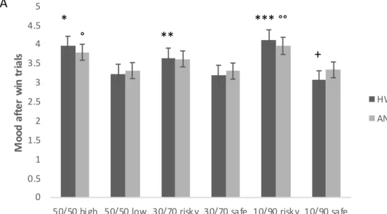
3.4.1. MOOD AFTER WIN TRIALS. ... 147
3.4.2. MOOD AFTER NO-WIN TRIALS ... 148
6 4. DISCUSSION ... 150 CHAPTER 5 ... 159 1. INTRODUCTION ... 162 2. METHODS ... 164 2.1. ETHICS ... 164 2.2. PARTICIPANTS ... 164 2.3. PROCEDURE ... 165 2.4. ENDOCANNABINOID MEASUREMENTS ... 166
2.5. DATA ANALYSES AND STATISTICS ... 169
3. RESULTS ... 170
3.1. SELF-REPORT OF HUNGER ... 170
3.1.1.1. ACUTE PHASE OF AN ... 170
3.1.1.2. COMPARISON OF ECB BETWEEN THE ACUTE AND WEIGHT-RESTORED PHASES 171 4. DISCUSSION ... 174
5. ADDITIONAL ANALYSES ... 181
5.1. WHEEL OF FORTUNE AND CIRCULATING ECB CORRELATION ... 181
CONCLUSION ... 183
REFERENCES ... 189
CURRICULUM VITAE ... 245
F
IGURE
L
IST
Figure 1. Reward and homeostatic integration in eating behaviors. ... 407
Figure 2. PRISMA flow diagram of studies involving “anorexia nervosa” and “money OR monetary reward”. ... 56 Figure 3. PRISMA flow diagram of studies involving “obese OR obesity” and “money OR monetary reward”. ... 71 Figure 4. General procedure. ... 97 Figure 5. Means and standard error for self-reported hunger under fasted and fed (A) and self-reported affect across time (B) in healthy women (HW) and anorexia nervosa (AN). Both HW and AN reported stronger hunger mood under fasted compared to fed, while HW reported by AN. ... 113 Figure 6. Positive mood after winning trials (A) and negative mood after non-winning trials (B) across sessions. ... 117 Figure 7. Positive mood after winning trials (A) and negative mood after non-winning trials (B) across groups and decision categories. ... 118 Figure 8. The wheel of fortune, a computerized task used to measure responses to monetary rewards under risky, safe and uncertain conditions. ... 142 Figure 9. Affect ratings measured by the PANAS across groups and timepoints. .... 147 Figure 10. Mood ratings. Mood reactivity to winning and not winning in the 10/90 conditions. ... 149 Figure 11. Study procedure. ... 166 Figure 12. Self-report of hunger in healthy controls (HC) and patients with anorexia nervosa (AN). Endocannabinoid measurements ... 170 Figure 13. Circulating endocannabinoids across time points (Fasting, 120-min and 240-min after eating a standard meal). ... 173
8
Figure 14. Total circulating levels of endocannabinoids across groups and anorexia nervosa phase. ... 174
T
ABLE
L
IST
Table 1. Studies excluded from review after screening and reason for exclusion in anorexia nervosa. ... 60 Table 2. Studies identified through search on PubMed investigating monetary reward processing in anorexia nervosa. ... 60 Table 3. Studies excluded from review after screening and reason for exclusion in obesity. ... 71 Table 4. Studies identified through search on PubMed investigating monetary reward processing in obesity. ... 73 Table 5. Demographic and descriptive data of study population. Descriptive data are given as mean ± standard errors. ... 112 Table 6. Reaction time in response to reward in the Wheel of Fortune in Anorexia Nervosa (AN) and healthy women (HW) means and standard error (SE) during Fasting and Fed States. ... 119 Table 7. Mod ratings in response to reward in the Wheel of Fortune in Anorexia Nervosa (AN) and healthy women (HW) means and standard error (SE) during Fasting and Fed states. ... 120 Table 8. Description of the study population. ... 140 Table 9. Mood ratings in response to reward in the Wheel of Fortune in women with obesity (OB) and healthy controls (3) during Fasting and Fed States. ... 155
9
Table 10. Description of study population. Body mass index, the Beck Depression Inventory (BDI) scores, and the State-Trait Anxiety Inventory (STAI) scores for healthy controls (HC) patients with anorexia nervosa during the acute phase (AN), and weight-restored patients at the end of treatment (ANR). ... 179 Table 11. Means and standard errors for plasma levels of endocannabinoids (pmol/ml) in healthy controls (Pattullo et al.), patients with anorexia nervosa during the acute phase (AN), and weight-restored patients at the end of treatment (ANR). ... 179 Table 12. Correlation between mood ratings to winning and not winning under different categories of the wheel of fortune for patients with anorexia nervosa and healthy controls. ... 181
11
A
CKNOWLEDGEMENTS
“In every victory, let it be said of me: ‘my source of strength, my source of hope is Christ alone’”.
I was given so much! And I’m so thankful for that, for I’m sure it’s God’s grace that’s brought me this far.
Obrigado aos meus pais, Ivanilde e Itamar, por irem além, e se esforçarem mais do que podiam pra que eu chegasse aqui. É por vocês! Obrigado à minha irmã, Nayara, por me ensinar a cada dia a ter um coração ‘selfless’. Com a minha mãe aprendi a ser guerreiro e lutar pelo que quero, com meu pai aprendi a buscar um coração bom, e com minha irmã aprendi o significado da palavra ‘selfless’. Vocês me inspiram a ser melhor. Sempre!
Obrigado à minha família como um todo. Em especial ao meu avô, João Evangelista (in memoriam), e minhas avós Hilda e Cida (in memoriam). Vocês me embalaram com tanto amor que crescer foi fácil. Um obrigado especial às minhas tias, Tania, Ivone, Neide e Tuca, que me mimam até hoje! E, lógico, aos meus primos, que sempre me encorajaram e me apoiaram em meio a esse e todos os outros processos da minha vida.
My special gratitude goes to Prof. Dr. Martin Sölch, Chantal, who has given all necessary conditions for this dissertation to come through. During my first years of PhD, the nearly weekly mentorship was essential to set the path for this PhD and allowed me to advance this work in a nice pace. Similarly, thank you to Prof. Dr. Milos, Gabriella, and Prof. Dr. Kristin Javaras who agreed to join us in this journey and has
12
brought their expertise to what we have done as part of this work. Professionals like you are inspiration to the coming generation of scientists, doctors, and professors. My special thanks to Gabriella again for allowing me to work with the data presented here. Equally, I would like to thank Dr. Michael Fried, the principal investigator of the study which allowed the inclusion of the experiments developed in this thesis.
Romina, Christophe, Matthias, Claudie, Tanya, Dany Laure, Clément, Fiona, Phillipe, Michael, thank you so much for making this road unlonely! I want you to know how much I appreciate each one of you, and the time spent together, whether it was at “La Tour Vagabonde”, at Regina Mundi or Pérolles! Seriously, I couldn’t have asked for better people to walk alongside during this time in Switzerland. I will never forget any of you, and I will always cherish these years in a special way in my heart. ALWAYS! You were, without a doubt, the best part of this whole experience. We HAVE to work on our Hollywood plot! This is the next step J.
To all of the team of Clinical and Health Psychology at the University of Fribourg. Thank you for contributing in a unique way for my formation and thank you for always making lunch more interesting! You inspire me! Also, thank you for the other professors who crossed my way during these years, sharing knowledge and laughter, and always great advice: Prof. Valerie Camos, Prof. Pierre Barrouillet, Prof. Roberto Caldara, Prof. Jurgen Sauer, and Prof. Dominique Schöbi. Similarly, I would like to thank all the staff in the Department of Psychology for welcoming me so nicely to the team.
My warmest gratitude to two people who were as “cold water in a hot day”, Vera and Vincent. I must have done something right for deserving people like you! You made my life in Fribourg and Marly so much better, that I can’t even imagine
13
what it would have been without both of you. Thank you for every little thing you have done, and I hope one day I will be able to do the same.
I am very grateful for the Federal Swiss Confederation, and the Swiss Excellence Scholarship, and for Ms. Nast, at the International Relations Office. I would have never been able to get to this point without this scholarship. The scholarship has allowed me to fully focus on my doctoral work. Similar gratitude goes to the Swiss National Science Foundation for the DocMobility grant awarded to me, which has led me to live some of the most incredible experiences of my whole life. I hope to pay back in scientific production and honor these institutions being the best scientist I can be.
Similarly, I would like to thank Prof. Diego Pizzagalli and Prof. Kristin Javaras again, who gladly welcomed me during my research stay at McLean Hospital, Harvard Medical School, and expanded my knowledge and research field. Kristin, you have shown me how balancing research and clinical practice is possible, and I am very grateful for every lesson taught.
I am thankful for all of my friends, the ones I knew before going to Switzerland, and all of those I got to know in Fribourg and in Boston/Cambridge. You have given me strength and joy to carry on.
Lastly, thank you to all of my students at UniFr. Working close to you was one of the best parts of these years!
15
A
BSTRACT
Research on clinical populations with eating disturbances has shown the involvement of reward processing in the maintenance of disease-associated symptoms. Investigation on food as a reward indicates the metabolic or feeding state (i.e. fasted or fed state) as a modulator of food as a reward, but little is known about how these states influence other types of reward. This is important because responses to rewards other than food are also altered in several disorders, including those involving eating disturbances. Therefore, the aim of this dissertation was to investigate behavioral and biological responses to monetary reward during fasting and fed states in healthy women, women with anorexia nervosa and women with obesity. For the behavioral responses, we used an experimental gambling task, the wheel of fortune, to evaluate responses to winning or losing a monetary reward involving different probabilities. The patients with anorexia nervosa (AN) repeated the same procedures after weight-restoration treatment. This procedure resulted in empirical studies 1 and 2. The biological responses consisted of circulating levels of endoncannabinoids (eCB) in the participants, and investigation of the correlation between eCB and the responses to reward were reported in empirical study 3. In summary, our results show higher positive mood to winning monetary rewards during fasting for normal-weight women, but not women with anorexia nervosa and more negative mood to not winning during the fed state in normal-weight women and women with AN. No difference regarding mood between feeding states was observed for women with obesity. Regarding affect before and after the task, independently of the feeding state, women with AN and women with obesity reported more negative affect than normal-weight women, with positive affect improving in normal-normal-weight women and women
16
with AN after the task, but not in women with obesity. Patients with AN’s mood reactivity to losing monetary rewards was blunted in comparison to normal-weight controls. Patients with obesity also showed blunted activity to losing low-risk monetary rewards, but also showed less mood reactivity to winning high-risk rewards than controls. Reaction times to categories that involved equal chance (50% of chance of either option) in AN were longer than controls and other categories. With regards to the biological responses, no significant correlations were found between eCB and behavioral responses to monetary rewards. Anandamide, and eCB, was found to be significantly lower during fasting in comparison to the fed state. In patients with AN, levels of anandamide were significantly lower than in healthy controls and remained unchanged following weight-restoration treatment. These results indicate that some types of responses to monetary rewards can be modulated by the feeding state. The applicability of the results is discussed.
17
19
Eating is perhaps one of the most common behaviors across species (Rozin & Todd, 2015), and it has developed as an important behavior to guarantee the survival of humans and animals, as a way to generate energy (Zucoloto, 2011). One of the mechanisms responsible for maintaining eating behaviors is the rewarding potential found in food (Enax & Weber, 2016; Lutter & Nestler, 2009; Saper, Chou, & Elmquist, 2002). One of the reasons humans and animals go back to food is because of the rewarding effects it produces (Berridge, 2009). And finding pleasure in food has become easier with the development of processing techniques and of the food industry. This complex relationship between humans and food has led to the development of several disorders associated with eating (Zucoloto, 2011). In that vein, the general aim of this dissertation is to investigate reward-related behavioral and biological responses associated with eating, bringing insights from research on anorexia nervosa and obesity.
The processing of reward is one of the dimensions involved in the new framework developed for investigating the pathophysiology of mental disorders (Insel et al., 2010). Anything people and animals put effort into acquiring may be considered a reward (Schultz, 2010), and, many times, reward comes associated with sensorial pleasure (Berridge & Kringelbach, 2008). For instance, primary needs in humans and animals (e.g. food, sex) are considered primary rewards, due to their direct association with the survival of the species (Schultz, 2010). Other non-primary rewards (e.g. gambling, money), are not directly related to the survival of the species, but, in the long-term, they work to strengthen primary rewards (Schultz, 2010).
Research has shown similar brain structures involved in the processing of primary and non-primary rewards. Regions like the orbitofrontal cortex, ventromedial
20
prefrontal cortex, nucleus accumbens (NAcc), ventral pallidum, and even the brainstem are involved in processing rewards (Berridge, 2009). Many of these areas have been associated with behavioral measures such as valuation of food as pleasant or not (Zald, 2009), and stimulation of these areas can increase or decrease food intake depending on the type of stimulation (Bakshi & Kelley, 1993; Tuulari et al., 2017).
The metabolic state (i.e. fasted or fed states) also plays an important role in modulating the rewarding potential of food (Kringelbach, 2004). Besides the reward system, the homeostatic system is also involved in controlling food intake by maintaining a stability related to body fat and eating (Enax & Weber, 2016). It involves several hormones and ligands that circulate peripheral regions and communicate with brain structures, which are then responsible for suppressing or increasing food intake (Lutter & Nestler, 2009) and modulating, through hunger, reward processing (Briers, Pandelaere, Dewitte, & Warlop, 2006). One of the systems working in the intersection food x reward is the endocannabinoid (A. M. Monteleone et al., 2015; P. Monteleone et al., 2005; B. A. Watkins & Kim, 2014). Greater concentrations of these endocannabinoids have been associated with food initiation, also modulating the rewarding effect of food (Cooper, 2004; Jarrett, Limebeer, & Parker, 2005; Kirkham & Williams, 2001). The aspects of food as a reward, reward processing, homeostatic signaling, and the impact of the metabolic state on food as a reward will be introduced in chapter 1.
In the same sense, research has shown that alterations in these regions in relation to reward are implicated in the development and maintenance of several disorders, both at behavioral and neural levels (Blum et al., 2006), from mental (Barbato, Fichele, Senatore, Casiello, & Muscettola, 2006; Bergen et al., 2005; Davis &
21
Woodside, 2002; Ehrlich et al., 2014; Piazza et al., 1993; Wagner et al., 2007) to metabolic disorders (Ho, Kennedy, & Dimitropoulos, 2012; Kenny, 2011; Rothemund et al., 2007). Anorexia nervosa (Wagner et al., 2008) and obesity (Kenny, 2011), for instance, have been associated with several dysfunctions related to reward processing of primary and non-primary rewards, at neural, behavioral, and self-reported levels. The second chapter will focus on the relationship of these disorders and the processing of reward, and more focus will be given to the processing of money as a non-primary reward. More specifically, Chapter 2 will examine how monetary reward is processed in anorexia nervosa and obesity.
Since the value of food as a reward is modulated by the metabolic state of the individual (Cassidy & Tong, 2017; Kringelbach, 2004), and research has shown similarities between the processing of primary and non-primary rewards, it may be important to evaluate how the metabolic state influence other types of reward, which have also shown to be processed differently across disorders involving eating. In that sense, the following section of this dissertation will focus on empirical research developed to evaluate behavioral (including mood and affect) and biological responses to monetary rewards in healthy women, women with anorexia nervosa, and women with obesity, during a fasted and a fed condition. We used a computerized task, the wheel of fortune (Ernst et al., 2004), designed to measure mood and affect to winning vs losing monetary rewards in different decision categories. Chapter 3 shows the study in individuals with anorexia nervosa (Piccolo, Milos, Bluemel, Schumacher, Muller-Pfeiffer, et al., 2019), and chapter 4 reports the same procedure done with individuals with obesity without the presence of any mental disorder (Piccolo, Milos, et al., 2020). The fifth and last chapter aimed at evaluating how fasting modulated
22
circulating endocannabinoid levels in patients with anorexia nervosa during the underweight phase, and following weight-restoration treatment (Piccolo, Claussen, et al., 2020), and how that was associated with processing of monetary reward.
23
THEORETICAL
BACKGROUND
25
C
HAPTER
1
T
HE
R
EGULATION OF
E
ATING
B
EHAVIORS
“Let food be thy medicine and medicine be thy food”
27
1. EATING AS A COMPLEX BEHAVIOR
Eating, or food intake, is a common behavior across species. It has been
suggested that knowledge related to eating is part of the most important information
one can obtain about humans (Rozin & Todd, 2015). In most species, eating starts with
a physiological stress in response to deprivation, followed by a search phase, involving
the questions of what and where to search, mostly depending on availability and
nutritional potential (Kearney, 2010). Then, a stage of capture of food precedes its
preparation and, finally, consumption happens, mostly with the purpose of generating
energy (Rozin & Todd, 2015). In humans, however, due to different phylogenetic and
cultural aspects, this process may not be exactly as it once was, and eating has evolved
to be something more than a simple source of energy intake, influenced by several
aspects, such as religion, familiarity, and sociopolitics, for instance (Zucoloto, 2011).
Although humans and animals have very similar nutritional needs, eating
patterns vary not only from humans to animals, but also among humans, with a great
range of variability (Zucoloto, 2011). Animals obtain their food mostly from nature. In
contrast, due to the advance of technology and culture, humans can also obtain food
from secondary sources, being even able to fabricate different types of food (via
28
which exclusively depended on availability – since food could become scarce due to
climatic conditions, for instance – to one that involves abundance of choices.
Besides processing, a social meaning was added to food with the
development of language. According to some evolutionists, early human ancestors
would gather around fire (once fire control had been obtained) to better make use of
food by eliminating possible toxins. Moreover, since operant control of the vocal cords
had also been acquired, meaning they could then talk, it is very likely that this context
also consisted of a social gathering for early humans (Zucoloto, 2011). One can
hypothesize that the environment changed, but humans continue to gather around
food, and this could also be due to the rewarding properties that eating socially has
acquired. A recent study investigated the connection between social eating and
happiness in 2000 participants (Dunbar, 2017). The results showed that those who
frequently eat socially feel more emotionally supported and also better about
themselves than those who eat socially less often. In ancient times, social gatherings
around food also warded off predators (Zucoloto, 2011), and social gatherings around
food were, and still remain, especially rewarding.
The positive effects of food and social gatherings may exist even in the
29
properties by looking at the paradigm of classical conditioning. Pavlov (1897/1902)
realized that after a number of associations of an unconditional stimulus (US) to a
neutral one, the latter became a conditioned stimulus (CS) that would, in turn, elicit
similar reflexive responses (conditioned responses) to those that before only the UC
produced, even the absence of it. In his experiments, he observed that when food was
presented for a dog, the dog salivated. Following that, he introduced a bell, which, at
first, was considered a neutral stimulus to the response of salivation. After some
conditioning associations (bell + food à salivation), only the bell, which then would
be considered a CS, could produce salivation in the dog, even in the absence of the
food. Because of the rewarding effects of social eating, food alone could evoke similar
feelings experienced when in the presence of people, even in the absence of the social
crowd. This classically-conditioned response to food-related cues can be considered
one aspect of emotional eating (Cardi, Leppanen, & Treasure, 2015), but there are a
number of other aspects of emotional eating since human’s relationship to food is
much more complex.
Specifically, Macht (2008) proposes a model that involves five different ways
that emotion can influence eating behaviors. The first type of relationship involves
30
is that intense emotions may suppress eating to some extent, mainly among
normal-weight people. Finally, the author suggests that moderate emotions affect eating
depending on the motivation in three different ways. For example, restrained eaters (i.e.
those who intentionally restrict food intake motivated by weight gain or loss) mostly
increase food intake in reaction to negative and positive emotions. According to
Mach’s theory, for emotional eaters (i.e. those who increase food intake in response to
negative emotions, leading to a high risk of overweight), there is a tendency to
increased intake of caloric and sweet foods in response to negative emotions. Besides
the conception that emotional eating can also refer to increased food intake in response
to positive emotions (Cardi et al., 2015), a recent review showed that, although there
is some evidence supporting that theory, most studies show contrasting or unclear
results (Bongers & Jansen, 2016). And, lastly, among normal eaters (i.e. those within the
considered normal weight, with emotional and restrained eating scores falling in the
average), amount of food intake is related to the type of emotion it follows. Happiness,
for instance, would be associated with a maximized ability to perceive stimuli, thus,
enhancing food intake, while sadness would cause the opposite. In summary,
31
emotional states. It is important to note that weight (high or not) does not necessarily
define disturbances in eating behaviors.
2. ORGANIC SYSTEMS REGULATING EATING
The emotional system controlling food intake is supported by biological
systems (Fig. 1). Research suggests that food intake in humans is regulated by at least
two systems: the homeostatic and the reward systems (Enax & Weber, 2016; Lutter &
Nestler, 2009; Saper et al., 2002).
2.1. THE HOMEOSTATIC SYSTEM
The homeostatic system works in a way to maintain a stable balance between
food intake and exergy expenditure, which enables a stability between body fat and
weight (Enax & Weber, 2016). This involves peripheral hormones that work to
communicate with the central nervous system in order to inform available energy
stores which will, in turn, regulate food intake, mostly by modulating the motivation
to eat (Lutter & Nestler, 2009). One important region in this process is the
hypothalamus. In early studies, Hetherington and Ranson (1940) reported that rats
with hypothalamic lesions presented altered feeding patterns, leading to accumulated
fat. More recently, Bagnasco, Dube, Kalra, and Kalra (2002) clarified this relationship
32
(ARC) were responsible for the inhibition of food intake. On the other hand, lesions in
the lateral hypothalamus (LH) would be responsible for promoting food intake
(Anand & Brobeck, 1951). These early studies showed that the hypothalamus was
associated with hunger and satiety. However, recent studies have brought new
interpretation to such findings. The hypothalamus is believed to serve to functions,
among others, related to the energetic metabolism, and, thus, alterations such as
lesions and/or electrical stimulations in its lateral and/or ventromedial parts may lead
to weight increase or loss (Pinel, 2011). This occurs mostly in response to two different
hormones: leptin and ghrelin (Lutter & Nestler, 2009). Research has shown that leptin
is mainly responsible for suppressing food intake (Enax & Weber, 2016), whereas
ghrelin promotes eating (Kojima et al., 1999).
Leptin, an adipocyte hormone, sends signals to the brain about available
energy, and sends a negative feedback to regulate energy intake and expenditure
(Zigman & Elmquist, 2003). Leptin-deficient mice, for instance, were shown to develop
obesity, while the administration of leptin was efficient in reducing its severity (Y.
Zhang et al., 1994).
Levels of leptin increase in proportion to the amount of fat (Lutter & Nestler,
33
intuitively, would be thought to reduce food intake, these individuals seem to be less
sensitive to its signaling than normal-weight controls (Lustig, Sen, Soberman, &
Velasquez-Mieyer, 2004). It has been reported, for instance, that obesity would
promote a sort of leptin resistance, leading to increased weight gain, in spite of the
great levels of leptin (Myers, Leibel, Seeley, & Schwartz, 2010).
If, on the one hand, leptin is involved in suppressing food intake to maintain
homeostasis, ghrelin has been reported to increase in animals in response to food
deprivation (Kojima et al., 1999) and to have its peak before meals in humans
(Cummings et al., 2002), thus favoring food intake. For instance, the administration of
ghrelin led to increased eating in healthy humans, and ghrelin’s levels were correlated
with self-reported hunger, regardless of time- and food-related cues (Cummings,
Frayo, Marmonier, Aubert, & Chapelot, 2004).
Leptin and ghrelin modulation also plays an important role regarding
motivation to obtain food related to the reward system and dopamine processing
(Lutter & Nestler, 2009). Animal research showed that leptin reduced basal and
food-stimulated dopamine release (Krugel, Schraft, Kittner, Kiess, & Illes, 2003). Ghrelin
34
et al., 2006). The role of dopamine and the reward system in the food intake will be
discussed in detail in the next section of this chapter.
2.2. THE REWARD SYSTEM
Besides the homeostatic system, research suggests the involvement of the
reward system in modulating food intake (Enax & Weber, 2016; Lutter & Nestler, 2009;
Saper et al., 2002), and its malfunctioning has been implicated in the studies of various
disorders (Blum et al., 2006).
Rewards have been defined as “objects or events that generate approach and
consummatory behavior, produce learning of such behavior, represent positive
outcomes of economic decisions and engage positive emotions as hedonic feelings”
(Schultz, 2010), being most of the time related to pleasure (Berridge & Kringelbach,
2008). Primitive and primary needs (e.g., food, sex) as well as more complex behaviors
(e.g., gambling) are related to the reward system (Schultz, 2010). According to Berridge
and Kringelbach (2008), reward involves three major components. The first is liking,
which is directly related to pleasure, whether or not in a conscious way. The second
component involves wanting, and that is related to the motivation for obtaining
something. As liking, wanting can also be done consciously or not. Finally, the last
35
and predictions about future rewards based on past experiences” (p. 458). Learning
would therefore include explicit and cognitive predictions as well as implicit
knowledge related to associative conditioning.
The processing of reward has evolved to maximize the survival of the
species. Schultz (2015) categorizes the functions of rewards as follows: 1) learning; 2)
approach behavior; 3) decision-making), and; 4) pleasure or positive emotions.
Rewards produce learning through different mechanisms to ensure that individuals
will return to the choices made that contribute to fitness. In consequence, approach
and decision-making will involve those rewards offering potential benefits to the
organism, which will, in turn, evoke positive emotions, establishing its value as
rewarding.
In that sense, research has shown the existence of a neural circuitry involved
in the processing of reward (Schultz, 2000). Reward-related neurons are spread
through different brain structures, including the striatum (caudate nucleus, putamen
and the nucleus accumbens; NAcc), amygdala, ventromedial prefontal cortex
(vmPFC), orbitofrontal cortex (OFC), anterior cingulate cortex (ACC), hippocampus,
hypothalamus, and midbrain dopamine (DA) neurons (McGinty, Lardeux, Taha, Kim,
36
Rewards can be categorized in different types: primary and nonprimary
rewards. Primary rewards (e.g. homeostatic and reproductive) are those that evolved
to guarantee the survival of the species (Schultz, 2015). Food, for instance, is
considered a primary reward, since its consumption is essential for the survival of the
species, as is mating and reproduction. All other rewards are considered nonprimary
(e.g. monetary, social), and they serve to strengthen primary rewards in the long-term,
thus also increasing evolutionary fitness.
2.2.1. FOOD AS A REWARD
Food is considered a primary reward, and as a reward, it is processed by
different brain structures, and understanding the systems involved may yield
important knowledge regarding normal and aberrant eating behaviors. (Berridge,
2009). Frontal cortical regions with increased activation to pleasant foods are similarly
involved in emotion regulation (e.g. OFC, ACC, and the insular cortex), and analogous
patterns of activation can also be found in subcortical limbic structures, such as the
ventral pallidum and the mesolimbic dopaminergic system and even the brainstem
(Berridge, 2009).
The OFC, for instance, is involved in coding the pleasure of food stimuli
37
site), it tracks changes in food pleasure in relation to hunger and satiety (Berridge,
2009), and is involved in valuation of available food as rewarding or not (Zald, 2009).
Early studies in rats show that lesions in OFC led to aphagia (i.e. inability or refusal to
swallow), firstly showing the involvement of this region in food-intake control (Kolb
& Nonneman, 1975). Since then, studies have pointed OFC as responsible for receiving
sensory input about food, and, then, evaluating among options available, taking
pleasantness (or the unpleasantness) as reference (Zald, 2009).
The vmPFC is also involved in processing food as a reward. When presented
with pictures of high-calorie food, fasted women showed increased activation in the
vmPFC (Stoeckel et al., 2007). Also, higher activation was shown in vmPFC when food
odors were presented, and that was correlated to how much external cues lead to
eating (Eiler, Dzemidzic, Case, Considine, & Kareken, 2012), suggesting the
involvement of the vmPFC in encoding value of food as a reward. Furthermore, DA
neurons’ activity in vmPFC is increased with food intake, and DA blocking in the same
area decreases eating (Land et al., 2014), possibly modulating wanting for food.
Hedonic hotspots in the NAcc are also involved in processing food as a
reward, mostly through opioid signaling. Showing a bidirectional relationship,
38
et al., 2001; Dum, Gramsch, & Herz, 1983), with opioid injections also leading to
hyperphagia in rats (Bakshi & Kelley, 1993). Moreover, infusions of opioids in the
NAcc, leading to an increase in the firing of NAcc neurons, triggered rats to overeat in
spite of not being food deprived (Bakshi & Kelley, 1993). Likewise, an opioid release
has been reported in response to eating, regardless of food palatability (Bakshi &
Kelley, 1993; Tuulari et al., 2017). Finally, opioid blockers (e.g. naltrexone) reduced
self-reported food palatability ratings in humans and decreased consumption of
palatable foods in rats (Na, Morris, & Johnson, 2012).
Signals leaving from NAcc go mainly in direction to the ventral pallidum.
Berridge (2009) suggests that the ventral pallidum may have a key role in modulating
eating behaviors through reward processing. This is supported by research showing,
for instance, that lesions in the ventral pallidum may shift pleasantness to
unpleasantness (Cromwell & Berridge, 1993). It is possible that the early studies
focusing on the LH (Anand & Brobeck, 1951) may have also lesioned the ventral
pallidum (due to its proximal location to LH), confusing the functions of these regions.
Also, by stimulation of the opioid hotspot in the ventral pallidum, pleasure in relation
to food is enhanced (Smith & Berridge, 2005). Smith and collaborators (2005) showed
39
(e.g. “liking” facial expressions) in animals, and increased food intake. Finally, greater
activation to food rewards in the ventral pallidum were shown to code taste in a way
that is sensitive to the physiological need of the individual (Tindell, Smith, Pecina,
Berridge, & Aldridge, 2006). For instance, humans and animals mostly show aversion
to saline solutions. However, after inducing through hormones a state where salt was
needed, the ventral pallidum activation increase was correlated to the increase of a
saline solution intake in comparison to baseline condition (Tindell et al., 2006). Further
research is needed to clarify the involvement of the ventral pallidum and its
relationship with the LH in food control.
Finally, food seems to be processed as a reward already at brainstem level
both in humans (Steiner, 1973), and animals (Grill & Norgren, 1978). Anencephalic
human babies (those born without cortex) show positive facial expressions in reaction
to sweet tastes and negative facial expressions in response to sour tastes (Steiner, 1973).
Similar results were found in animal research with decerebrated rats (Grill & Norgren,
1978), suggesting the early involvement of the brainstem in food reward processing.
Also, benzodiazepine injections in the parabrachial nucleus of the pons showed an
40
Figure 1. Reward and homeostatic integration in eating behaviors.
Food intake may be controlled by homeostatic signals for energy balance (modulated by hormones such as Ghrelin and Leptin), and also by the appetitive cues of foods, activating reward processing mechanisms (including the dopamine circuit). Eating may be evoked by the rewarding properties of foods as learned through associative mechanisms in the individual’s history. Adapted from Reichelt, A. C., Westbrook, R. F., & Morris, M. J. (2015). Integration of reward signaling and appetite regulating peptide systems in the control of food-cue responses. British Journal of Pharmacology, 172(22), p. 5225-5238.
2.2.2. DOPAMINE AND EATING
Dopamine (DA) neurons are mostly contained within the ventral tegmental
area (VTA) and the substantia nigra (SNc) (Arias-Carrion, Stamelou,
Murillo-Rodriguez, Menendez-Gonzalez, & Poppel, 2010). From there, DA is projected to the
41
striatum, the dorsal and prefrontal cortex, and is involved in many goal-seeking
behaviors (Schultz, 2015), including foraging and eating. Long associated with the
pleasure component of reward (liking), DA has recently been proposed as mostly
involved in motivation (wanting) (Berridge, 2009). Besides that, DA has been appointed
as a fundamental mediator in learning (Arias-Carrion et al., 2010), which is also an
intrinsic part of reward.
Specifically, in relation to eating, DA has been reported to greatly influence
food intake, and, similar to addictive drugs, palatable foods are related to a DA
increase in the reward system (Alonso-Alonso et al., 2015). Several results shared by
Wise (2006) lead to the conclusion that, when DA-suppressing methods were applied
in animal models, the reinforcing1 properties of food ceased to be observed.
Even if the role of DA is still not completely clarified, it seems well
established that is it involved in eating behaviors. For instance, increased DA release
in the dorsal striatum has been shown in response to palatable foods. Small,
Jones-Gotman, and Dagher (2003) used positron emission tomography (PET) to test the
relation between dopamine release and self-reported food pleasantness in humans.
1 It is important to differentiate reinforcement from reward. Reinforcement mostly refers to a process that increases the occurrence of a specific behavior in a given context (Skinner, 1938).
42
Their results not only showed increased dopaminergic activity in the striatum, but also
a direct correlation between the amount of dopamine released and the perceived food
palatability in healthy subjects. To extend that, Volkow et al. (2003) correlated the type
of eating (i.e. restraint, emotional) with DA brain levels in a PET study. They found
that higher DA concentrations were correlated with higher levels of restraint, while
lower DA responsivity represented higher emotionally while eating. Again, these
changes were observed in the dorsal striatum, once again indicating the involvement
of this region in modulating eating behaviors.
2.2.3. FASTING AND FOOD AS A REWARD
The metabolic state of the individual (fasted or fed) was shown to influence
the value of food as a reward (Cassidy & Tong, 2017; Kringelbach, 2004). A classic
study showed, for instance, that subjective ratings of pleasantness of a sweet solution
were higher during fasting compared to fed states (Cabanac, 1971). Further research
has pointed to the same direction, with the addition of different methodologies to
support their findings. For instance, while having participants picture their favorite
food in an MRI scanner, Hinton et al. (2004) reported higher activation in the striatum,
a putative region for reward processing, in fasted compared to fed state. In that vein,
43
amygdala, anterior insula and OFC in response to high-caloric food vs low-caloric food
pictures during fasting. Besides that, self-reported pleasantness ratings of high caloric
food were increased after a fasting period. It is interesting to note that the difference
in ratings between high- and low-caloric food pictures were correlated with activation
in the OFC, which is believed to encode the value of food as a reward (Small et al.,
2007). Similarly, Führer, Zysset, and Stumvoll (2008) reported stronger activation in
the OFC during fasting in response to food pictures in comparison to non-food
pictures.
Recent studies using self-reported measures have also found similar results.
Pursey, Contreras-Rodriguez, Collins, Stanwell, and Burrows (2019) assessed
subjective ratings of “appeal to food”, “desire to eat food” and “effect of food in
increasing appetite” during fasted and fed states and reported significant differences
between states, with fasting yielding higher scores. Similarly, Cameron, Goldfield,
Finlayson, Blundell, and Doucet (2014) reported higher self-report of food
pleasantness during fasting.
Taken together, these results suggest that the nutritional state modulates
how food is perceived as a reward, both at neuronal and behavioral levels, with fasting
44
types of reward, including monetary reward, has been little investigated.
Understanding how other types of reward are modulated by the nutritional state may
bring insights to disorders that involve eating, since other types of reward, such as
social and monetary, may be also altered in such cases.
2.3. THE ENDOCANNABINOID SYSTEM: HOMEOSTASIS AND REWARD PROCESSING
The endocannabinoid system (eCB) has also been identified as a potential
regulator of eating behaviors (A. M. Monteleone et al., 2015; P. Monteleone et al., 2005;
B. A. Watkins & Kim, 2014) and also seems to be influenced by the metabolic state
(Kirkham, Williams, Fezza, & Di Marzo, 2002). Cannabinoid receptors are widely
found in the brain and many organs and tissues besides the nervous system. It includes
both synaptic and peripheral signaling functions (B. A. Watkins & Kim, 2014). The
main endocannabinoids studied in relation to eating behaviors are the endogenous
ligands anandamide (arachidonoylethanolamide, AEA) and 2-arachidonoylglycerol
(2-AG), as well as the receptors CB1 and CB2 (Cota et al., 2003). AEA and 2-AG were found
in greater concentration in brain regions in response to fasting in rats (Kirkham et al.,
2002). Furthermore, fasting also increased AEA levels in peripheral regions (small
intestine), and peripheral administration (intraperitoneal in rats) of AEA increased
45
control, and has suggested AEA’s role as a meal initiator (Marco, Romero-Zerbo,
Viveros, & Bermudez-Silva, 2012), promoting eating and feeding behaviors.
The role of the eCB in meal initiation may be connected to the rewarding
effects it attributes to food (A. M. Monteleone et al., 2015; P. Monteleone et al., 2005; B.
A. Watkins & Kim, 2014). For instance, research has shown that endocannabinoid
administration enhances perceived food palatability (Kirkham & Williams, 2001).
Similar results have been found in animal research as well (Cooper, 2004; Jarrett et al.,
2005), and the NAcc seems to have a special hotspot for anandemide (Mahler, Smith,
& Berridge, 2007). Mahler and colleagues (2007) showed that microinjections of
anandemide to NAcc increased positive reactions to pleasant tastes in rats, besides
increasing eating.
Eating, as any other complex behavior, is multidetermined and controlled by
several different systems. The objective was to understand the involvement of the
systems that will be further discussed in this work. The previous mechanisms and
systems were mostly investigated in healthy controls, as general functioning and
controlling of eating behaviors. Following, the same systems will be approached in
46
from research on these conditions to the comprehension of reward processing affected
47
C
HAPTER
2
F
ROM
E
ATING
B
EHAVIORS TO
E
ATING
D
ISTURBANCES
“Thou shouldst eat to live; not live to eat”.
49
The ethologically atypical relationship between humans and food may have
led to some consequences in terms of mental and general health. Changes in eating
and feeding habits may have influenced the development of diseases in humans
(Zucoloto, 2011). In that sense, the fifth edition of the Diagnostic and Statistical Manual
of Mental Disorders (American Psychiatric Association, 2013) devotes an entire section
to mental disorders related to eating and feeding behaviors. Eating and feeding
disorders are characterized as “a persistent disturbance of eating or eating-related
behavior that results in the altered consumption or absorption of food and that
significantly impairs physical health or psychosocial functioning” (p. 329).
As previously seen, the reward system is one aspect involved in controlling
food-intake, and the malfunctioning of reward processing has been implicated in the
studies of various disorders as well, both at behavioral and neural levels (Blum et al.,
2006), from mental (Barbato et al., 2006; Bergen et al., 2005; Davis & Woodside, 2002;
Ehrlich et al., 2014; Piazza et al., 1993; Wagner et al., 2007) to metabolic disorders (Ho
et al., 2012; Kenny, 2011; Rothemund et al., 2007). Taking that into consideration, the
aim of this chapter is to investigate reward processing in two clinical conditions
50
1. ANOREXIA NERVOSA
Anorexia Nervosa (AN) is the eating disorder with highest mortality rate
(Fichter & Quadflieg, 2016). First reported scientifically in the late 1800s, AN received
Dr. Gull (1873) attention due to the characteristic emaciation (i.e. the state of being
atypically thin) in some of his patients. Some of his young female patients showed
similar symptoms that could not be fit into other disease categories. Those patients
were extremely underweight, presented long-lasting amenorrhea (i.e. the absence of
menstruation), and had weak vital functioning. All of that could be explained by an
anorexia (i.e. lack of appetite) that led to starvation and, consequently, the weakening
of all vital functions (Gull, 1873). Since that first scientific report of AN, although
considerable effort has been made towards a better understanding of the disorder,
much is still to be learned (Strober, 2005).
1.1. DIAGNOSTIC CRITERIA OF ANOREXIA NERVOSA
One of the main measures used in the diagnosis of AN is body mass index
(BMI), defined as someone’s weight in kilograms divided by the square of their height
in meters (kg/m2) and used to indicate nutritional status in adults (World Health
Organization, 2016). The core features of AN involve “behavioral disturbances related
51
disturbance in the experience of body shape and/or weight, disturbance results in
substantial impairment in physical, social and/or mental functioning, disturbances is
not secondary to any other medical or psychiatric disorder” (Treasure et al., 2015).
A normal-weight BMI ranges between 18.5 to 24.9 kg/m2 (WHO, 1995). In
AN, a BMI above 17 kg/m2 is considered of mild severity. A moderate AN is
diagnosed when the patient’s BMI lies between 16-16.99 kg/m2; a severe AN, 15-15.99
kg/m2; and an extreme AN, when the BMI is under or equal 14.99 kg/m2. AN’s
prevalence is mostly among young females (0.4% -12-month prevalence); although AN
also occurs in men, it is much less common.
1.2. COMORBIDITIES IN ANOREXIA NERVOSA
The condition has been associated with several psychosocial features, such
as depression, social withdrawal, irritability and reduced interest in sex (American
Psychiatric Association, 2013). Moreover, individuals with AN tend to present
increased interpersonal problems (Carter, Kelly, & Norwood, 2012), which also reflect
on their social contexts, included but not limited to family dysfunctionality (Espindola
& Blay, 2009). Carter and colleagues (2012) investigated how interpersonal issues
relate to AN and its treatment. They reported an association between AN and
52
social inhibition), which tended to decrease after an interdisciplinary treatment.
Furthermore, AN has been reported to normally co-occur with affective and anxiety
disorders (Halmi et al., 1991), with symptoms of the latter emerging prior to the onset
of AN (APA, 2013). In addition, general medical comorbidities have also been reported
in AN, involving skin and hair development, cardiovascular and gastrointestinal
system, among others (Rikani et al., 2013).
1.3. ANOREXIA NERVOSA AND REWARD PROCESSING
Research has pointed to dysfunctional reward processing as one of the
factors responsible for the peculiar eating behaviors in AN (Barbato et al., 2006; Bergen
et al., 2005; Davis & Woodside, 2002; Ehrlich et al., 2014; Wagner et al., 2007). For
instance, by using eye blink rate, a potential peripheral measure of central
dopaminergic activity, Barbato et al. (2006) showed altered DA processing in patients
with AN. Furthermore, it has been hypothesized that a DA malfunctioning might lie
behind the maintenance of food deprivation behaviors in AN (Bergh & Södersten,
1996). The constant absence of food, leading to a state of starvation, would provoke a
physiological stress response by activating the hypothalamic-pituitary-adrenal axis,
which, in turn, would activate DA neurons, thus, reinforcing non-eating behaviors due
53
(1993) showed that physiological levels of stress can indeed have reinforcing
properties, and that has been speculated to be the case in AN. For instance, research
with animals showed that rats would self-administer corticosterone, which is
connected to stress and potentiates pleasure properties of drugs of abuse, even though
that meant undergoing the stress of intravenous injections (Piazza et al., 1993).
Baseline neural processing across reward-related areas in AN is also altered
(Haynos et al., 2019). By investigating resting state functional connectivity (RSFC),
Haynos et al. (2019) have recently shown weaker connection between areas implicated
in reward processing (NAcc and ventral regions of the caudate) in patients with AN
when compared to healthy controls. Besides that, Cha et al. (2016) also reported
abnormalities associated with RSFC of the ventral frontostriatal network in AN.
Davis and Woodside (2002) compared the degree to which patients with AN
report pleasure related to physical sensations. Their findings showed that, in
comparison to healthy controls and individuals with bulimia nervosa, the participants
with AN were much more anhedonic, suggesting an altered activation pattern in
reward-related regions. Altered activation was also observed in response to food
54
experience pleasure among these patients in comparison to the general population.
Besides food, response to non-primary rewards have also been investigated in AN.
Finally, it is important to note that the differences between cases and controls
previously stated may be a correlate or a consequence of the disorder. In other words,
it is not possible to establish whether alterations in neural mechanisms cause the
disorder or whether these alterations arise as a result of the disease state. The latter is
a particular concern in research on AN, since differences observed in individuals with
the disorder may be attributable to confounds such as starvation (Steinglass et al.,
2019). For example, it is not clear whether changes in structural and/or functional
connectivity are a result of the nutritional status. In this vein, Santos and collaborators
(2018) have recently shown that brain structure in AN is strongly correlated with body
mass index (BMI), suggesting that nutritional status is a confounding factor when
researching eating disorders.
1.3.1. ANOREXIA NERVOSA AND MONETARY REWARDS
Responses to monetary rewards, as non-primary rewards, have also been
evaluated by using several different methods and instruments, including fMRI,
55
also been observed in patients with AN (Piccolo, Milos, Bluemel, Schumacher,
Muller-Pfeiffer, et al., 2019).
To be as accurate as possible with the current state of the literature on studies
involving anorexia nervosa and monetary rewards, a quasi-systematic literature
review was done following the PRISMA framework (Moher, Liberati, Tetzlaff, &
Altman, 2009). The search was done in one database (PubMed) with the terms “Anorexia
Nervosa”, and “Money or Monetary Reward”, but no inter-rater reliability was
performed. Empirical studies with individuals with anorexia nervosa that involved
monetary rewards, published until May 2019 were included. Nineteen papers were
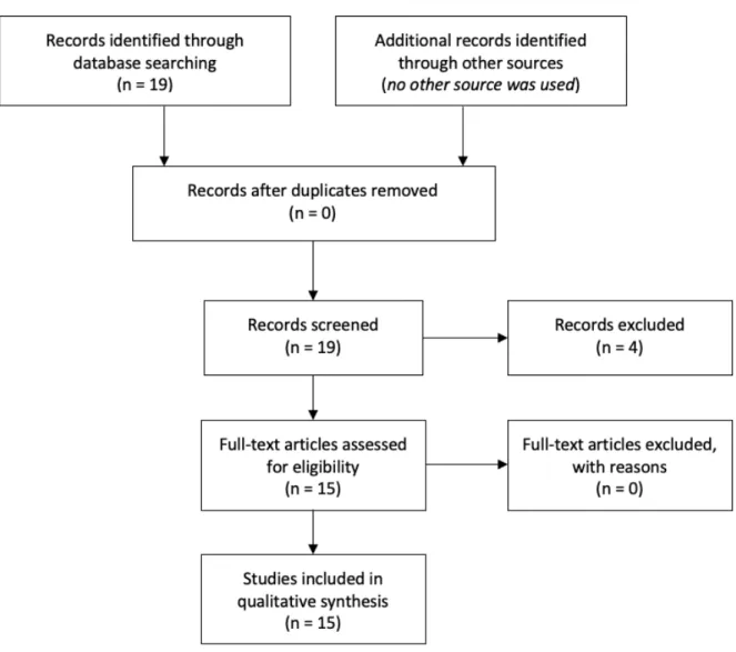
identified through database searching (Fig. 2). After screening, 4 papers were excluded
for 1) not involving patients with anorexia nervosa; 2) not involving monetary
56
Figure 2. PRISMA flow diagram of studies involving “anorexia nervosa” and “money OR monetary reward”.
The 14 remaining papers included (Table 2) reported behavioral, self-report
and neural responses to monetary reward in patients with AN, and the results will be
discussed below.
1.3.1.1. Behavioral responses to monetary rewards in AN
Behavioral data have been obtained by the same tasks used for fMRI research
57
2016; Piccolo, Milos, Bluemel, Schumacher, Muller-Pfeiffer, et al., 2019; Ritschel et al.,
2015; Steinglass et al., 2012; Steinglass et al., 2017; Steward et al., 2017). In general,
research shows that patients with AN prefer larger/later rewards than smaller/sooner
rewards (Decker et al., 2015; Steinglass et al., 2012; Steinglass et al., 2017; Steward et
al., 2017), again, showing greater cognitive control than normal-weight participants.
However, one study found no difference between AN and HC (Ritschel et al., 2015).
The later study also evaluated delay discounting responses to monetary rewards in
recovered patients, and no difference was found.
1.3.1.2. Self-reported responses to monetary rewards in
AN
Self-report measures to reward have been assessed by the Behavioral
Inhibition System/Behavioral Activation System (BIS/BAS) (Carver & White, 1994).
BIS consists of two scales measuring the concern about negative future events, and the
sensitivity to negative past events. BAS measures how rewarding experiences are
found and responses towards goal pursuing.
One study has evaluated how AN responds to reward using BIS/BAS
(DeGuzman, Shott, Yang, Riederer, & Frank, 2017). While AN did not differ from
58
with AN showed higher sensitivity to punishment than HC, both before and after
weight-treatment(de Zwaan, 2001).
1.3.1.3 Neural responses to monetary rewards in AN
Regarding neural responses, six studies used fMRI measures in combination
with a monetary discounting delay task – choosing larger later over smaller sooner
rewards (Bailer et al., 2017; Decker et al., 2015; Ehrlich et al., 2014; King et al., 2016;
Murao et al., 2017; Wierenga et al., 2015). Other tasks, such as card guessing games,
where participants were asked to guess whether a hidden card was higher or smaller
than the number shown, have also been used to measure neural responses to monetary
rewards in AN (Bischoff-Grethe et al., 2013; DeGuzman et al., 2017; Wagner et al.,
2007).
Two studies have shown no difference in striatal activation during monetary
reward anticipation (i.e. when the prospect of a reward is initiated) between patients
with AN and healthy controls (Ehrlich et al., 2014; Murao et al., 2017). For instance,
increasing activation in the striatum, mOFC, and DLPFC were found to increase in
relation to the reward magnitude in both AN and controls (Ehrlich et al., 2014). On the
other hand, Ehrlich et al. (2014) showed stronger activation in the DLPFC in AN
59
Regarding reward delivery (i.e. when the outcome is received), differences
between AN and controls can be found. For instance, Wagner et al. (2007) reported
that, while HC show different activation patterns to winning vs losing in the striatum
(i.e. increased activation to winning vs decreased activation to losing), no difference
between outcomes was seen in AN. Contrarily, Bischoff-Grethe et al. (2013) have
shown stronger activation to winning and decreased activation to losing in NAcc and
60
Study Reason for exclusion
Haynos et al. (2019) Did not involve response to monetary rewards Kekic et al. (2016) Did not involve patients with anorexia nervosa Wierenga et al. (2014) Did not report empirical research
Wagner et al. (2010) Did not involve patients with anorexia nervosa
Table 2. Studies identified through search on PubMed investigating monetary reward processing in anorexia nervosa.
Study Sample Size Measures Instruments Relevant findings
Isobe et al. (2018) AN = 24 HC = 22
Behavioral Ultimatum game
(splitting of money between players)
• AN required more money from the other player than HC to accept an offer.
Steward et al. (2017) AN = 56 (AN-R = 37; AN-BP = 19)
HC = 80
Delay discounting Intertemporal choice task
• AN-BP had greater discounting than HC; • AN-R showed more preference for
delayed reward than AN-BP. Murao et al. (2017) AN = 23 (AN-R = 11;
AN-BP = 12) HC = 20
fMRI Monetary delay task • AN striatum activation during
anticipation was similar to controls; • AN-BP rostral ACC and right posterior
insula activation was greater during loss anticipation than AN-R and HC;
• AN-R ACC and posterior insula activation was similar to HC.
61
DeGuzman et al. (2017) AN = 21 HC = 21
(adolescents before and after weight treatment)
fMRI Self-report
Monetary reward task BIS/BAS
• AN showed greater activation in the caudate body, right caudate head, NAcc, and insula associated with prediction error than HC before treatment;
• AN showed greater activation in the left caudate body during unexpected reward omission before treatment;
• AN showed higher activation than HC in the right dorsal anterior insula during unexpected reward receipt before and after treatment;
• AN showed higher activation than HC in the left dorsal anterior insula and the right ventral anterior insula during unexpected reward receipt only after treatment: • AN showed higher activation than HC in
the right posterior insula during unexpected reward receipt; • AN did not differ from controls in
sensitivity to reward in the acute phase and after treatment;
• AN showed higher sensitivity to
62
activation in NAcc.
Steinglass et al. (2017) AN = 27 HC = 50
Delay discounting Monetary incentive delay task
• AN show higher preference for delayed reward than HC.
Bailer et al. (2017) ANRec = 12 Correlation between fMRI and PET data obtained in previous studies (Bailer et al., 2013; Wagner et al., 2007)
Monetary choice task • ANRec with greater activation in the dorsal caudate in response to winning and losing money had higher middle caudate D2/D3 binding.
King et al. (2016) AN = 31 HC = 31
(predominately adolescents)
fMRI Intertemporal choice
task
• AN delayed discounting did not differ from HC;
• AN reaction times were faster than HC; • AN showed decreased activation in
comparison to HC in the dorsal ACC.
Decker et al. (2015) Delay discounting task AN = 59
Delay discounting fMRI
Monetary delay task • AN showed higher preference for delayed reward than HC only before treatment, with no difference between groups after treatment;
63
fMRI + task AN = 30 HC = 22
(before and after treatment)
in the striatum and dorsal ACC in response to the larger/later compared to the shorter/sooner reward, while HC did not show different between types of reward. This was reversed after treatment.
Ritschel et al. (2015) AN = 34 ANRec = 33 HC = 54 (reevaluation of AN after weight-recovery n = 21)
Delay discounting Intertemporal choice task
• No group differences were found.
Wierenga et al. (2015) ANRec = 23 HC = 17
Delay discounting fMRI
Delay discounting task • ANRec striatal, dorsal caudate and ACC activation, different than HC, did not differ between hunger and satiety


![Calix[6]arènes présentant une chiralité inhérente](data:image/gif;base64,R0lGODlhAQABAIAAAP///wAAACH5BAEAAAAALAAAAAABAAEAAAICRAEAOw==)