HAL Id: hal-03019063
https://hal.inrae.fr/hal-03019063
Submitted on 31 Mar 2021
HAL is a multi-disciplinary open access archive for the deposit and dissemination of sci-entific research documents, whether they are pub-lished or not. The documents may come from teaching and research institutions in France or abroad, or from public or private research centers.
L’archive ouverte pluridisciplinaire HAL, est destinée au dépôt et à la diffusion de documents scientifiques de niveau recherche, publiés ou non, émanant des établissements d’enseignement et de recherche français ou étrangers, des laboratoires publics ou privés.
Distributed under a Creative Commons Attribution| 4.0 International License
mammary cancer model? Limit and uncertainty
Augustin Le Naour, Adrien Rossary, Marie-Paule Vasson
To cite this version:
Augustin Le Naour, Adrien Rossary, Marie-Paule Vasson. EO771, is it a well-characterized cell line for mouse mammary cancer model? Limit and uncertainty. Cancer Medicine, Wiley, 2020, 9 (21), pp.8074-8085. �10.1002/cam4.3295�. �hal-03019063�
8074
|
wileyonlinelibrary.com/journal/cam4 Cancer Medicine. 2020;9:8074–8085.1
|
INTRODUCTION
Breast cancer is the most common cause of cancer deaths among women (522 000 deaths, WHO 2013) and the most frequently diagnosed woman cancer in the world.1 Many
studies are interested in breast cancer (348,598 results for “mammary cancer” and 405,294 results for “breast cancer” on pubmed, 7 April 2020). This cancer is subdivided into sev-eral types, which are well characterized taking into account the cell type of origin, its mutations and its gene expression profile. Before performing clinical trials, experimental mod-els in vitro and in vivo must be used. Thus in preclinical approaches, it is important to choose the right experimental model to know what type of tumor is studied and what type of patient this model could match.
For in vivo experimental approaches, the use of murine models is often done. Among the mice models, the C57BL/6 strain seems to be the most used (about 300 000 publica-tions are found during a pubmed search, 7 April 2020, de-pending on the way it is spelled) in front of the BALB/c strain (about 200 000 publications found on pubmed, 7 April 2020). The C57BL/6J mouse is the most widely used inbred strain and the first to have genome sequenced.2
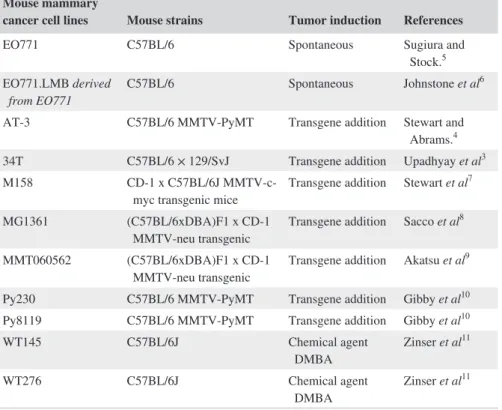
Although this strain is refractory to many tumors, it is a permissive background for maximal expression of most mu-tations. However, despite this massive use, few breast tumor lines result from C57BL/6 genetic background. Indeed, only 10 lines are derived from C57BL/6 mice (Table 1): 34T,3
AT-3,4 EO7715 and its derivative EO771.LMB,6 M158,7
MG1361,8 MMT060562,9 Py230,10 Py8119,10 WT145,11
R E V I E W
EO771, is it a well-characterized cell line for mouse mammary
cancer model? Limit and uncertainty
Augustin Le Naour
1|
Adrien Rossary
1|
Marie-Paule Vasson
1,2This is an open access article under the terms of the Creative Commons Attribution License, which permits use, distribution and reproduction in any medium, provided the original work is properly cited.
© 2020 The Authors. Cancer Medicine published by John Wiley & Sons Ltd. 1UMR 1019 Human Nutrition Unit,
ECREIN team, University of Clermont Auvergne, INRAE, CRNH-Auvergne, Clermont-Ferrand, France
2Department of Nutrition, Gabriel Montpied
University Hospital, Jean Perrin Cancer Centre, Clermont-Ferrand, France
Correspondence
Adrien Rossary, University of Clermont Auvergne, INRAE, UMR 1019 Human Nutrition Unit, ECREIN team, CRNH-Auvergne, 28 place Henri Dunant, 63000 Clermont-Ferrand, France
Email: adrien.rossary@uca.fr
Funding information
This work was supported by funding from the Institut National du Cancer (INCA: MammAdipo project; PLBIO 13-106) and the Comité de l’Allier de la Ligue Contre le Cancer.
Abstract
Among mouse mammary tumor models, syngeneic cell lines present an advantage for the study of immune response. However, few of these models are well character-ized. The tumor line EO771 is derived from spontaneous breast cancer of C57BL/6 mice. These cells are widely used but are referenced under different names: EO771, EO 771, and E0771. The characteristics of the EO771 cells are well described but some data are contradictory. This cell line presents the great interest of developing an immunocompetent neoplastic model using an orthotopic implantation reflecting the mammary tumors encountered in breast cancer patients. This review presents the phenotype characteristics of EO771 and its sensitivity to nutrients and different ther-apies such as radiotherapy, chemotherapy, hormone therapy, and immunotherapy.
K E Y W O R D S
and WT276.11 Of the 10 lines, only the EO771 line and its
derivated line EO771.LMB come from spontaneous tumors, not induced by the addition of a transgene (as 34T, AT-3, M158, MG1361, MMT060562, Py230, and Py8119) or a chemical agent DMBA (7,12-dimethyl-benzanthracene) (as WT145 and WT276).
Therefore, this review is dedicated to the EO771 mam-mary cancer cell line. According to the spelling used to search for this lineage in pubmed, numbers of associated publica-tions found are different. A total of 122 publicapublica-tions are iden-tified following pubmed research on 7 April 2020 (Table 2).
However, publications using these cells but not indicating the lineage in their title or abstract are not identified.12-14 These
cells have been used for many years and the publication of Sugiura and Stock5 from 1952 is frequently presented as the
original publication, but earlier articles using this line exist as Homburger's (1948).15
2
|
EO771 CELL PHENOTYPE
Despite their isolation in the late 1940s, characteriza-tion of hormonal receptors and classificacharacteriza-tion of EO771 line remains controversial. Indeed, its classification di-verges according to the authors, which is considered as triple negative in 10 publications6,12,16-23 and as ERα+
in 19 publications.13,24-41 Some authors prefer to
men-tion the unclear status of this lineage.42,43 Other
publi-cations do not report information on the expression of ERα. However, among the 30 publications considering EO771 cells as triple negative or ERα+, only 3 articles analyzed the expression of ERα.6,40,41 Contreras-Zárate
et al have considered EO771 cells as triple negative and have showed that the proliferation of EO771 cells is in-dependent of the estradiol presence.23 But in the same
time, they showed that estradiol drives the signaling for
Mouse mammary
cancer cell lines Mouse strains Tumor induction References
EO771 C57BL/6 Spontaneous Sugiura and
Stock.5
EO771.LMB derived
from EO771 C57BL/6 Spontaneous Johnstone et al
6
AT-3 C57BL/6 MMTV-PyMT Transgene addition Stewart and
Abrams.4
34T C57BL/6 × 129/SvJ Transgene addition Upadhyay et al3
M158 CD-1 x C57BL/6J
MMTV-c-myc transgenic mice Transgene addition Stewart et al
7
MG1361 (C57BL/6xDBA)F1 x CD-1
MMTV-neu transgenic Transgene addition Sacco et al
8
MMT060562 (C57BL/6xDBA)F1 x CD-1
MMTV-neu transgenic Transgene addition Akatsu et al
9
Py230 C57BL/6 MMTV-PyMT Transgene addition Gibby et al10
Py8119 C57BL/6 MMTV-PyMT Transgene addition Gibby et al10
WT145 C57BL/6J Chemical agent DMBA Zinser et al 11 WT276 C57BL/6J Chemical agent DMBA Zinser et al 11
Note: Breast tumour lines resulted from C57BL/6 genetic background. Among them, only the EO771 line and its derivated line EO771. LMB come from spontaneous tumours. The 34T, AT-3, M158, MG1361, MMT060562, Py230, and Py8119 cell lines were obtained by addition of a transgene. WT145 and WT276 cell lines were obtained by exposure to the chemical agent DMBA (7,12-dimethyl-benzanthracene).
C57BL/6J is the parental substrain; “J” is the laboratory code for The Jackson Laboratory. TABLE 1 C57BL/6 mammary cancer
cell lines
TABLE 2 Various spelling used to identify EO771 mammary cancer cell line in literatur
Mouse mammary cancer cell lines Number of Pubmed results
EO 771 25*
EO771 38
E0771 79
EO771.LMB 1
According to the spelling used the numbers of associated publications found are different. A total of 122 publications are identified following pubmed research on 7 April 2020.
*EO 771:25 results but 5 do not concern the tumor line. Research online 7 April 2020.
brain metastasis of EO771 cells.23 Therefore, Hiraga
et al have observed that the gene encoding ERα is tran-scribed in EO771 cells41 and Gu et al have observed the
protein expression of ERα by western blot.40 However,
they observed that this ERα expression is much weaker than that found in MCF-7 cells (considered as ERα+). In addition, Johnstone et al have observed ERα by immuno-histochemistry in EO771 cells but have considered these cells as ERα- because this receptor is only found in the cytoplasm but not in the nuclear compartment6 which is
the localization found in primary human breast cancers. Thus, based on these publications, the cells could be con-sidered as ERα- because the expression of this receptor is very weak and not at the nuclear level.
Few publications have investigated the status of ERβ, the progesterone receptor and ErbB2 in EO771 cells. The immuno-histochemical analysis performed by Johnstone et al on primary EO771 and EO771.LMB tumors did not detect the expression of ERβ.6 Thus, Hiraga et al and Johnstone et al have not found
gene transcription or protein expression of the progesterone receptor.6,41 Similarly, the ErbB2 status for this line is poorly
described and contradictory. Indeed, Johnstone et al have not found expression of ErbB2 by immunohistochemistry in the tumors formed after injection of EO771 cells6 in vivo whereas
Hiraga et al and Zou et al found respectively a transcription of ErbB2 and a protein expression highlighted by westernblot.44
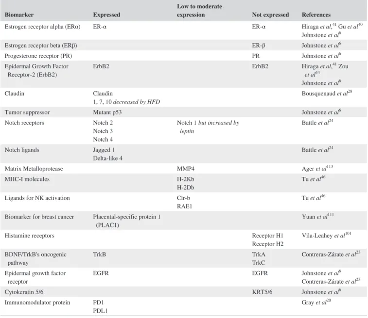
As for hormone receptors, the expression of claudin, a marker for the classification of triple negative cancers,
TABLE 3 Characterisation of hormonal receptors and protein patterns of EO771 line in literature
Biomarker Expressed Low to moderate expression Not expressed References
Estrogen receptor alpha (ERα) ER-α ER-α Hiraga et al,41 Gu et al40
Johnstone et al6
Estrogen receptor beta (ERβ) ER-β Johnstone et al6
Progesterone receptor (PR) PR Johnstone et al6
Epidermal Growth Factor
Receptor-2 (ErbB2) ErbB2 ErbB2 Hiraga et al,
41 Zou et al44 Johnstone et al6 Claudin Claudin 1, 7, 10 decreased by HFD Bousquenaud et al 28
Tumor suppressor Mutant p53 Johnstone et al6
Notch receptors Notch 2
Notch 3 Notch 4
Notch 1 but increased by
leptin Battle et al
24
Notch ligands Jagged 1
Delta-like 4 Battle et al
24
Matrix Metalloprotease MMP4 Ager et al113
MHC-I molecules H-2Kb
H-2Db Tu et al
46
Ligands for NK activation Clr-b
RAE1 Tu et al
46
Biomarker for breast cancer Placental-specific protein 1
(PLAC1) Yuan et al
111
Histamine receptors Receptor H1
Receptor H2 Vila-Leahey et al
101
BDNF/TrkB's oncogenic
pathway TrkB TrkATrkC Contreras-Zárate et al
23
Epidermal growth factor
receptor EGFR EGFR Johnstone et al
6 Contreras-Zárate et al23 Cytokeratin 5/6 KRT5/6 Johnstone et al6 Immunomodulator protein PD1 PDL1 Gray et al 20
aList of publications with partial determination of proteins and hormonal receptors expression. Characterisation of phenotype and the expression of many other markers
from EO771 cells were generally not done. Among the 30 publications considering EO771 cells, only 3 articles analysed the expression of ERα6,40,41. That’s why
is poorly determined. Some authors consider this lineage as claudin-low without checking it.31 On the contrary,
Bousquenaud et al have found an expression of claudin 1, 7, and 10 in EO771 cells.28 However, their expressions
are greatly decreased when the mice are fed with a high-fat diet (HFD), and are associated with a decrease in es-trogen receptor and ErbB2 expression. This suggests that a hyperlipid diet would induce a triple negative phenotype for EO771 cells.28
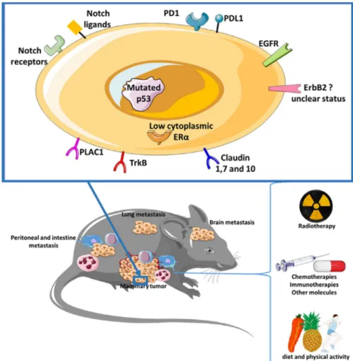
Finally, the expression of many other markers from EO771 cells has been characterized such as p53, Notch re-ceptors and ligand, EGFR, PD1 (Programmed Cell Death 1), PDL1 (Programmed Cell Death Ligand 1), etc (Table 3, Figure 1).
• Modified EO771 cell lines
The EO771 cell line can be genetically modified to follow their growth by fluorescence, for example by ex-pression of green fluorescent protein (GFP),13,14,23,33,45 or
luciferase13 as well as to modify the expression of genes by
the CRISPR/Cas9 technology such as the EO771 line de-void of MHC I or MHC II or expressing the tumor antigen NY-BR-1.46,47
3
|
EO771 MAMMARY TUMOR
MOUSE MODELS
The C57BL/6J mouse is the most widely used inbred strain and the first to have genome sequenced.2 Although this strain
is refractory to many tumors, it is a permissive background for maximal expression of most mutations. It is also susceptible to diet-induced obesity, type 2 diabetes, and atherosclerosis (https://www.jax.org/strai n/000664). Due to the wide use of this mouse strain, many modified strains exist (transgenic, knock out (KO), knock down (KD), overexpression, etc) to evaluate tumor development under various conditions. Indeed, the advantage of having a tumor line from the most used mouse strain, C57BL/6, makes it possible to have a lot of genetically modified models of this strain. Thus, EO771 cell tumor growth can be evaluated i) in KO mice for E2 (estradiol),23 apolipoprotein E and aromatase,42
IFN-γ receptor,48 natural killer lytic-associated molecule
(NKLAM),33 MMP13,49 calcium-independent phospholipase
A2 β,50 IGFBP-3,21,51,52 DUSP1,53 GDF2,54 BMP10,54 and
mKIAA146255 but also ii) in transgenic models such as mice
overexpressing IL-15,56 obese,13,35 NKCKD (exhibiting silenced
Ly49 expression in NK cells),46 GFP-LC3 (LC3 linked to GFP
expressing mice),57 mice transgenically modified to express
the HER2 proto-oncogene (ERBB2)58 or FAT-1 (gene leading
FIGURE 1 Characteristics of the
EO771 cell line and tumor bearing in C57BL/6 mice. Numerous publications have made it possible to refine the phenotype of the EO771 line. This cell line could be considered as a luminal B type mammary cancer. Tumor-bearing mice models are well-known and sensitive to explore numerous therapeutic strategies. After injection of EO771 cells, the major metastatic dissemination sites are in the brain, the lung, the intestine, and the peritoneal cavity
to the endogenous formation of ω3-Polyunsaturated fatty acid from ω6-Polyunsaturated fatty acid).44,59
The use of EO771 cells in mouse models has the advan-tage of having tumor uptake close to 100%, in particular when an orthotopic mammary implantation is used.33 This model
is really close to human breast cancer (Figure 1). In reality, mammary cell line EO771 (5 × 10⁵ cells) in suspension into
Matrigel™ Matrix orthotopically transplanted into the fourth right mammary fat pad leads to large tumors within weeks.38 In
fact, implantation into the mammary gland provides a neoplas-tic model with a complete microenvironment promoting the intercellular dialogue. Syngeneic models have the advantage of being immunocompetent. Thus, communication between different cell types can be explored including the immune response. Therefore, injection of EO771 cells into C57BL/6 mice provides a model of mammary cancer in which the role of host and tumor immunity can be investigated.
• In vivo proliferation and dissemination capacities of
EO771 cells
The tumor proliferation of EO771 cells seems to be similar to other transplantable murine and human tumors, especially in relation to the duration of the S phase.60 The size of the EO771
tumor is correlated with the cell density but also with the propor-tion of necrotic tissue.60 However, the growth curve of EO771
tumors is independent of the number of cells injected but formu-las have been proposed to estimate tumor growth.61 The tumor
growth can also be influenced by the characteristics of its host. Indeed, the tumor growth is linked to the weight of the mouse injected with EO771 cells with a higher initial growth rate fol-lowed by a faster growth deceleration phase in mice with higher body weight compared to mice with lower body weight.62
In addition to their proliferation capacity, the ability of EO771 cells to disseminate has also been studied. In mice, a metastatic spread is found for this cell line, with character-istics similar to the human disease.63 The sites of metastatic
disseminations are multiple from EO771 tumors. Its prefer-ential release seems to be the lungs 6,12,22,26,28,33,36,49,50,54,59,64-68 but other localizations such as the peritoneum,26,36,64,68
bones,6,41 brain,23 diaphragm,36 and intestines36 are observed
(Figure 1). In addition, EO771 cells express the CD73 anti-gen (ecto-5'-nucleotidase), associated with a pro-metastatic phenotype in breast cancer.63
• In vivo cellular interactions in EO771 mammary tumor
mouse model
Innate immune cells, such as macrophages, neutrophils, and dendritic cells, appear to be relatively uninvolved in the growth of the primary tumor but, on the other hand, appear to be important in the metastatic dissemination. Indeed, tumor-associated macrophages and neutrophils and
tumor-infiltrating dendritic cells are less present in the meta-static microenvironment after splenectomy and this decrease was associated with a decrease in the number of mammary cancer lung metastases.67
Similarly, the role of Natural Killer (NK) cells seems im-portant in the control of mammary tumor development and metastatic dissemination. Indeed, the use of an anti-NK1.1, causing NK cell depletion, leads to an increase in tumor growth compared to untreated mice.46 Similarly, the KO of
the NK lytic-associated molecule (NKLAM), which plays an important role in the cytotoxic activity of NK, causes a very large increase of tumor cells in the blood and lungs of mice after orthotopic injection of EO771 cells, compared to wild-type (WT) mice for NKLAM.33 The effect of NK does
not appear to be mediated by Toll-like receptor 3 because its deficiency has a small impact on the tumor growth of EO771 cells.69 Interestingly, a study showing that a prior
immuniza-tion with the injecimmuniza-tion of another type of tumor cell (Colon adenocarcinoma line Colon38) resulted in an absence of EO771 tumor growth. This immunoprevention is mediated mainly by NK1.1 positive cells.70
Finally, the evolution of T lymphocytes and their role during tumor development is also studied in the model of EO771 tumor-bearing mice. Thus, an increase in the CD4 + and CD8 + populations is observed when the size of the tumor increases. However, the CD4/CD8 ratio differs during tumor development with a predominance of CD8 + T lym-phocytes (cytotoxic lymlym-phocytes) in the early stages whereas CD4 + lymphocytes become predominant in late stages. The accumulation of CD8 + T lymphocytes, which may involve IGFBP-3 (Insulin-like Growth Factor Binding Protein-3), is an immune population with a strong antitumor role as shown by the decrease in tumor growth.51 Experiments inducing a
CD8 + T cell depletion exacerbates EO771 tumor growth, emphasizing the importance of CD8 + T cells in controlling tumor growth.71 Within CD4 + cells, the proportions of
pop-ulation subtypes evolve during tumor development with, in the early stages, a predominant Th1 lymphocyte population, known to stimulate antitumor immune responses, and then evolve into subtypes of Treg, associated with a tolerogenic profile, and Th17 cells, in more advanced stages.72
In addition to the applications already described, these cells have also been used with other cell types to study pos-sible interactions. Thus, the interactions of EO771 cells with adipocytes or fibroblasts led to protumoral effects. An increase in EO771 cell proliferation is induced by secre-tions of senescent fibroblasts73 or adipocytes.74 Finally, this
model has also been used to evaluate the effect of the tumor on memory loss,75 muscle damage,76 or tumor development
when associated with a viral infection.77
• Impact of obesity and physical activity in EO771
A sedentary lifestyle and obesity are studied because of their association with breast cancer78 incidence and recurrence.
On the contrary, physical activity is inversely associated with breast cancer risk.78 To study these parameters, the use of the
immunocompetent model is essential because obesity, induc-ing chronic low-grade inflammation, and physical activity are able to modulate the immune system. Therefore, the use of a syngeneic model such as orthotopic injection of EO771 cells in C57BL/6 mice represents a relevant model of breast cancer to study the effect of overweight/obesity and physical activity.
The obesity of the C57BL/6 mice, induced either by taking a HFD28,32,35,36,51,52,79 or by genetic alteration,13,35 leads to a
pro-tumoral effect by increasing tumor growth,13,28,32,52 promoting
tumor angiogenesis,28,32 having an immunomodulatory effect28
as well as activating pro-tumor pathways such as AKT/mTOR.13
Nachat-Kappes et al have used an environmental enrich-ment that leads to spontaneous physical activity in mice.38
Environmental enrichment resulted in a decrease in COX-2, which may suggest a decrease in inflammation, and a decrease in tumor volume and weight associated with a decrease in the prolif-eration index Ki67.38 Physical exercise, whether spontaneous25,38
or forced,31 leads to a slowing down of tumor growth but also
makes it possible to modulate angiogenesis25,31 and to increase
sensitivity to chemotherapy25 compared with controlled mice.
Physical activity also induces changes in the cytokine en-vironment, including adipokines, by increasing the plasma ratio of adiponectin/leptin levels.38 This ratio of adipokines
is decreased in a situation of obesity, which is a major risk of breast cancer.78
• Impact of phytonutrients in EO771 tumor-bearing mice
Various phytonutrients from usual vegetables have been tested in mammary cancer models using EO771 cells. Bioactive compounds such as naringenin (citrus flavonoid),80
[10] -Gingerol (a major phenolic constituent of ginger root),16
secoisolariciresinol diglucoside (polyphenolic plant lig-nan),17 meroxest (synthetic merosesquiterpenes),29,30 EGCG
(Epigallocatechin Gallate, a major green tea catechin)81 and
emodin (a Chinese herb-derived compound)34,65 induced
an-titumor effects against E0771 cells by several ways such as: 1) inducing cell death16,80; 2) inhibiting the cell cycle16,80; 3)
modulating the immune system including macrophages17,34,65
or leukocytes29,34; 4) decreasing pro-angiogenic markers such
as expression of VEGF17,29,81; 5) modulating signaling
path-ways such as NF-κB,17,81 IRF4,34 STAT6,34 and C/EBPβ.34
4
|
SENSITIVITY TO DIFFERENT
THERAPIES
The sensitivity of the EO771 line has been tested against many treatments such as radiotherapy, cytotoxic agents,
antiangiogenics, hormone therapy, immunotherapy, gene therapy, and bioactive compounds.
• Sensitivity to radiotherapy
Breast cancer treatment is multimodal and includes ra-diotherapy.82 Therefore, EO771 cells are exposed to
radio-therapy in animal models.83,84 The EO771 tumor line seems
resistant to irradiation by 30 Gy of Cobalt 60 because, despite a transient effect in tumor cell number, the day after irradi-ation, the effect is limited to a growth delay without tumor regression.84
The radioactivity can also be used as a tracer in EO771 tumor-bearing C57BL/6 mice, such as the use of 125I-labeled
5-iodo-2'-deoxyuridine, which allows tumor progression to be monitored.85,86
• Sensitivity to cytotoxic agents
4.1
|
Inhibitors of topoisomerase II and
alkylating agents
Anthracyclines and topoisomerase II inhibitors, are among the conventional chemotherapies used in breast cancer, whether in the United States where AC regimen (A = dox-orubicin, (anthracycline) and C = cyclophosphamide) is commonly used, or in Europe where FEC100 (combin-ing F = 5-fluorouracil, E = epirubicin (anthracycline) and C = cyclophosphamide) is frequently used.87 Doxorubicin
is tested on EO771 cells in vitro73 and in vivo, in mouse
models to test its antitumor efficacy and its toxicity. Studies on animal models have shown that this line is sen-sitive to doxorubicin and that the activity of the latter could be improved either by conjugating it to nanoparticles27,88
or peptides37,89 or by associating it with other molecules
such as IL-2,64 TNF,90 rapamycin,57 or FTY720 (a
sphingo-sine-1-phosphate receptor functional antagonist).35 These
models also tested the toxicity of doxorubicin in mouse mammary cancer models. This anthracyclin causes skel-etal muscle dysfunction and increases mitochondrial H2O2
production inducing a decrease on the mitochondrial res-piratory complex supported by complex I (pyruvate/gluta-mate/malate) and complex II (succinate) substrates.76 This
toxicity of doxorubicin, in particular at cardiac level, is not modified with the addition of IL-264 but is decreased when
doxorubicin is conjugated with nanoparticles27,88 and
pep-tides64,89 or associated with rapamycin.57
Cyclophosphamide, an alkylating agent, frequently used in breast cancer therapy as in the FEC100 and AC regimen protocols, leads to an initial significant cytotoxic activity but limited in time as the tumor growth restarts.60,91
4.2
|
Other cytotoxic agents
In addition to these commonly used chemotherapies, other cytotoxic drugs can be used in the therapeutic arsenal of breast cancer. Thus, methotrexate, an antifolate, is tested in the mouse model of EO771 mammary cancer showing a sensitivity of this tumor to this drug.92 This animal model
is also tested for the development of a novel antifolate fam-ily as analogues of aminopterin91,93-99 whose antitumor
ac-tivity is superior to methotrexate in EO771 tumor-bearing mice.
Antipyrimidic drugs such as cytosine arabinoside100 and
gemcitabine101 are also active on EO771 tumors. The vinca
alkaloids, such as vinblastine, navelbine, and vindesine in-duced increase in animal survival.98 Similarly, paclitaxel,102
cisplatin,91 and melphalan91 have antitumor activity against
EO771 tumors. In contrast, 5-fluorouracil appears to be of low activity on EO771 tumors.91
Other compounds such as ranitidine,45,101,103 a substituted
3-(5-imidazo[2,1-b] thiazolylmethylene)-2-indolinones104 or
EB-3D (a choline kinase 1 inhibitor)105 have shown
antitu-mor effects in EO771 mammary cancer models.
• Sensitivity to antiangiogenic agents
Angiogenesis plays an important role in tumor progres-sion and metastatic dissemination.106 Thus, the development
of antiangiogenic therapy has generated a great enthusiasm in recent years. The EO771 tumor-bearing mouse model is used to evaluate the role of angiogenesis and to test mole-cules modulating angiogenesis, especially since this cell line expresses vascular endothelial growth factor (VEGF) recep-tors 1 and 2.40 Tumor development seems to be dependent on
angiogenesis. A decrease in neovascularization is found in a mouse model using mKIAA1462-/- mice resulting in a loss of
“junctional protein associated with coronary artery disease,” and is associated with a decrease in tumor volume compared with the control group of EO771 tumor-bearing mice.55
Antiangiogenic agents targeting VEGF or its receptors have been tested in this model. The SU1124839 is a selective
pro-tein kinase inhibitor inducing inhibition of, among others, VEGFR types 1-3 found in human breast cancer. SU11248,39
but also pyrrolidine dithiocarbamate,40 in EO771 tumor
model modulated neoangiogenesis by decreasing intratu-moral microvessel density. This effect is associated with a decrease in tumor weight compared to the control group.39,40
The effect of pyrrolidine dithiocarbamate passed in particu-lar through the inhibition of autocrine and paracrine VEGF effects by decreasing its expression and reducing NFkB acti-vation.40 This molecule pyrrolidine also inhibited the growth
and migration of EO771 cells and had a synergistic effect with the VEGF receptor inhibitor SU5416.40 Lu et al have
showed that EO771 tumor growth is increased by stimulating
VEGF-dependent angiogenesis but that is inhibited by the use of SU5416.107 The antioxidant N-acetylcysteine has also
been studied in models using EO771 cells. However, despite the fact that N-acetylcysteine prevented Hif-1α stabilization under hypoxia in vitro, it did not reduce in vivo the tumor growth or the survival of EO771 tumor-bearing mice but rather, increased the metastatic burden.108 Finally, vascular
endothelial protein tyrosine phosphatase has also been inves-tigated. Inhibition of the latter resulted in a delay in tumor growth during tumor establishment, but had no impact once the tumor is well established.68
• Sensitivity to hormone therapy
Since the status of EO771 cells in hormone receptor ex-pression remains controversial, few studies have evaluated the impact of hormone therapy in this model. Johnstone et al have observed a reduction in the growth of EO771 tu-mors during treatment with tamoxifen,6 which may therefore
suggest an expression of estrogen receptors in these cells.
• Sensitivity to immunotherapy
As previously mentioned, the EO771 tumor-bearing mouse model is immunocompetent and of interest for eval-uating the efficacy of immunotherapies such as the use of cytokines or immune checkpoint inhibitors. Indeed, it rep-resents a model of choice because the EO771 cells express immunomodulatory molecules as PD1 and PDL1 in the basal state.20 Moreover, in the presence of IFNγ, the expression of
PD1 and PDL1 is increased in EO771 cells20 and the
over-expression of mucin 1 in these tumor cells resulted in an in-crease in PDL1 expression.19
Therefore, therapies such as immune checkpoint inhibi-tors have been tested in this model. The use of anticytotoxic T lymphocyte-associated protein 4 (CTLA4) or antipro-grammed cell death 1 (PD1) therapy allowed an increase in the number of circulating CD8 + T cells and IFN-γ leading to T lymphocyte-mediated antitumor response.48 However,
anti-PD1 alone leads to partial antitumor activity in the model of EO771 tumor-bearing mice.22 Thus, it seems
in-teresting to associate them with other treatments, such as surgery, chemotherapy, or even other immunotherapies. Liu et al have showed that neoadjuvant immunotherapy combining anti-PD1 and anti-CD137 enhanced therapeutic efficacy compared to surgery alone or surgery followed by this immunotherapy.12 This increase in therapeutic efficacy
is associated with an increase in CD8 + lymphocytes in the blood, spleen, liver, and lungs.12 However, this
neoad-juvant immunotherapy must be performed shortly before surgery because, after 10 days, the benefits of this treat-ment are lost.18 Anti-PD1 has also been associated with a
tyrosine kinase receptors having immunomodulatory prop-erties. Their association allowed to significantly reduced tumor growth and incidence of lung metastasis, associated with an increase in proinflammatory cytokines and an infil-tration of antitumor effector T cells.22 The use of
phosphati-dylserine-targeting antibodies decreases tumor growth and increases the antitumor efficacy of anti-PD1 in a syngeneic model using EO771 cells by promoting tumor infiltration of T cells and increasing the production of proinflamma-tory cytokines.20
Proinflammatory cytokines, such as interleukin (IL) 2 and tumor necrosis factor (TNF), have also been investigated in models using EO771. Treatment with IL-2 resulted in pro-longed survival of EO771 tumor-bearing mice109,110 and
im-proved the efficacy of doxorubicin chemotherapy.64 This effect
required the action of lymphocytes, especially CD8 +.64,109,110
Similarly, the presence of TNF led to the stimulation of CD8 + cytotoxic T lymphocytes and NK cells, allowing the complete remission of EO771 tumor-bearing mice, when this cytokine was associated with doxorubicin chemotherapy.90
Other immunomodulatory agents acting on other im-mune cells have also been studied in the mouse model of EO771 mammary cancer. For example, the use of a Cxcr2 antagonist decreased tumor growth by acting on different immune populations. In fact, it led in the tumor to a de-crease in immunosuppressive cells such as myeloid-de-rived suppressor cells (MDSC) and regulatory T cells (Treg) and, on the contrary, an increase in antitumor cells such as cytotoxic lymphocytes, NK cells, dendritic cells, and macrophages.111 These latter have an ambivalent role
on tumorigenesis depending of their polarization. Indeed, M1 macrophages have tumoricidal activity, while M2 mac-rophages exhibit low amounts of antigen presentation and suppress antitumor immunity.112 Treatment with an
an-ti-MMP14 inhibitory antibody (DX-2400) increased the tumor-associated macrophage number and polarized them toward an antitumoral M1 phenotype.113
• Sensitivity to gene therapy
Gene therapy has also been tested in the EO771 tu-mor-bearing mouse model. A reduction in tumor growth of the primary tumor but also at the metastatic level is observed when using adenovirus inducing the expression “brain-derived neurotrophic factor”36 or MBP-166 (Myelin Basic Protein 1).
5
|
CONCLUSION
Therefore, the characteristics of the EO771 cells are well described but some data are contradictory. Thus, their mo-lecular classification remains controversial at the present time between the luminal and triple negative subtype, close
to the luminal B phenotype. However, the luminal A phe-notype could be excluded considering the articles showing them as ERα-. More accurate phenotyping of this cell line is needed as well as for other murine cell lines since very few of them are clearly defined in terms of expression of estrogen, progesterone, and ErbB2 receptors. The use of this cell line has been carried out in 2D, 3D and in vivo models. 3D cell culture techniques are now a way of using cancer cell lines. Even if there is currently only one publi-cation on this subject,107 the use of this line in 3D models
is possible. The establishment of 3D models with EO771 cells will provide valuable data for understanding breast cancer.
The EO771 cell line presents the great interest of develop-ing an immunocompetent neoplastic model usdevelop-ing an orthot-opic injection reflecting the mammary tumors encountered in breast cancer patients. This EO771 tumor-bearing mouse model has been well characterized considering its sensitivity to various antineoplastic treatments and even other therapic approaches including nutritional interventions and physical activity (Figure 1).
Despite some uncertainties, all these data lead us to con-sider the EO771 cell line as a very relevant candidate for pro-viding an experimental mammary tumor model very close to human breast cancer.
ACKNOWLEDGMENTS
Not applicable.
CONFLICT OF INTEREST
The authors declare that they have no conflict of interest.
AUTHOR CONTRIBUTIONS
Augustin Le Naour is the major contributor in writing the manuscript and analyzing the literature. Adrien Rossary and Marie-Paule Vasson participated in the scientific discussions for the manuscript writing and during the revision of the manuscript. All the authors approved the final version of the manuscript.
DATA AVAILABILITY STATEMENT
Not applicable.
ORCID
Adrien Rossary https://orcid.org/0000-0001-8311-2419
REFERENCES
1. Winters S, Martin C, Murphy D, Shokar NK. Breast cancer epi-demiology, prevention, and screening. Prog Mol Biol Transl Sci. 2017;151:1-32.
2. Mouse Genome Sequencing Consortium. Initial sequenc-ing and comparative analysis of the mouse genome. Nature. 2002;420(6915):520-562.
3. Upadhyay G, Yin Y, Yuan H, Li X, Derynck R, Glazer RI. Stem cell antigen-1 enhances tumorigenicity by disruption of growth differentiation factor-10 (GDF10)-dependent TGF- signaling.
Proc Natl Acad Sci. 2011;108(19):7820-7825.
4. Stewart TJ, Abrams SI. Altered immune function during long-term host-tumor interactions can be modulated to retard autoch-thonous neoplastic growth. J Immunol. 2007;179(5):2851-2859. 5. Sugiura K, Stock CC. Studies in a tumor spectrum. I. Comparison
of the action of methylbis (2-chloroethyl)amine and 3-bis(2-chlo-roethyl)aminomethyl-4-methoxymethyl -5-hydroxy-6-methylpyr-idine on the growth of a variety of mouse and rat tumors. Cancer. 1952;5(2):382-402.
6. Johnstone CN, Smith YE, Cao Y, et al. Functional and molec-ular characterisation of EO771.LMB tumours, a new C57BL/6-mouse-derived model of spontaneously metastatic mammary cancer. Dis Model Mech. 2015;8(3):237-251.
7. Stewart TA, Pattengale PK, Leder P. Spontaneous mammary ad-enocarcinomas in transgenic mice that carry and express MTV/ myc fusion genes. Cell. 1984;38(3):627-637.
8. Sacco MG, Gribaldo L, Barbieri O, et al. Establishment and char-acterization of a new mammary adenocarcinoma cell line derived from MMTV neu transgenic mice. Breast Cancer Res Treat. 1998;47(2):171-180.
9. Akatsu T, Ono K, Katayama Y, et al. The mouse mammary tumor cell line, MMT060562, produces prostaglandin E2 and leukemia in-hibitory factor and supports osteoclast formation in vitro via a stro-mal cell-dependent pathway. J Bone Miner Res. 1998;13(3):400-408. 10. Gibby K, You W-K, Kadoya K, et al. Early vascular deficits are
correlated with delayed mammary tumorigenesis in the MMTV-PyMT transgenic mouse following genetic ablation of the NG2 proteoglycan. Breast Cancer Res BCR. 2012;14(2):R67.
11. Zinser GM, McEleney K, Welsh J. Characterization of mammary tumor cell lines from wild type and vitamin D3 receptor knockout mice. Mol Cell Endocrinol. 2003;200(1–2):67-80.
12. Liu J, Blake SJ, Yong MCR, et al. Improved efficacy of neoadju-vant compared to adjuneoadju-vant immunotherapy to eradicate metastatic disease. Cancer Discov. 2016;6(12):1382-1399.
13. Fuentes-Mattei E, Velazquez-Torres G, Phan L, et al. Effects of obesity on transcriptomic changes and cancer hallmarks in estrogen receptor-positive breast cancer. J Natl Cancer Inst. 2014;106(7). https://doi.org/10.1093/jnci/dju158
14. Goetz J, Minguet S, Navarro-Lérida I, et al. Biomechanical re-modeling of the microenvironment by stromal caveolin-1 favors tumor invasion and metastasis. Cell. 2011;146(1):148-163. 15. Homburger F. Studies on hypoproteinemia: III. Lymphoid
hy-perplasia and redistribution of nitrogen caused in mice by trans-planted tumors (sarcoma 180 and breast adenocarcinoma EO 771). Science. 1948;107(2790):648-649.
16. Bernard MM, McConnery JR, Hoskin DW. [10]-Gingerol, a major phenolic constituent of ginger root, induces cell cycle ar-rest and apoptosis in triple-negative breast cancer cells. Exp Mol
Pathol. 2017;102(2):370-376.
17. Bowers LW, Lineberger CG, Ford NA, et al. The flaxseed lignan secoisolariciresinol diglucoside decreases local inflammation, suppresses NFκB signaling, and inhibits mammary tumor growth.
Breast Cancer Res Treat. 2019;173(3):545-557.
18. Liu J, O’Donnell JS, Yan J, et al. Timing of neoadjuvant im-munotherapy in relation to surgery is crucial for outcome.
OncoImmunology. 2019;8(5):e1581530.
19. Maeda T, Hiraki M, Jin C, et al. MUC1-C induces PD-L1 and immune evasion in triple-negative breast cancer. Cancer Res. 2018;78(1):205-215.
20. Gray MJ, Gong J, Hatch MMS, et al. Phosphatidylserine-targeting antibodies augment the anti-tumorigenic activity of anti-PD-1 therapy by enhancing immune activation and downregulating pro-oncogenic factors induced by T-cell checkpoint inhibition in murine triple-negative breast cancers. Breast Cancer Res. 2016;18(1):50.
21. Scully T, Scott CD, Firth SM, Pintar JE, Twigg SM, Baxter RC. Contrasting effects of IGF binding protein-3 expression in mam-mary tumor cells and the tumor microenvironment. Exp Cell Res. 2019;374(1):38-45.
22. Kasikara C, Davra V, Calianese D, et al. Pan-TAM tyrosine ki-nase inhibitor BMS-777607 enhances anti–PD-1 mAb efficacy in a murine model of triple-negative breast cancer. Cancer Res. 2019;79(10):2669-2683.
23. Contreras-Zárate MJ, Day NL, Ormond DR, et al. Estradiol in-duces BDNF/TrkB signaling in triple-negative breast cancer to promote brain metastases. Oncogene. 2019;38(24):4685-4699. 24. Battle M, Gillespie C, Quarshie A, et al. Obesity induced a
Notch signaling axis in breast cancer: obesity induced a leptin-Notch signaling axis in BC. Int J Cancer. 2014;134(7):1605-1616. 25. Betof AS, Lascola CD, Weitzel D, et al. Modulation of Murine
Breast Tumor Vascularity, Hypoxia, and Chemotherapeutic Response by Exercise. JNCI. 2015;107(5): https://doi. org/10.1093/jnci/djv040.
26. Ewens A, Mihich E, Ehrke MJ. Distant metastasis from sub-cutaneously grown E0771 medullary breast adenocarcinoma.
Anticancer Res. 2005;25(6B):3905-3915.
27. Prados J, Cabeza L, Ortiz R, et al. Enhanced antitumor activity of doxorubicin in breast cancer through the use of poly(butylcya-noacrylate) nanoparticles. Int J Nanomed. 2015;1291. https://doi. org/10.2147/IJN.S74378.
28. Bousquenaud M, Fico F, Solinas G, Rüegg C, Santamaria-Martínez A. Obesity promotes the expansion of metastasis-initi-ating cells in breast cancer. Breast Cancer Res. 2018;20(1):104. https://doi.org/10.1186/s1305 8-018-1029-4
29. Carrasco E, Garrido JM, Álvarez PJ, et al. Meroxest improves the prognosis of immunocompetent C57BL/6 mice with al-lografts of E0771 mouse breast tumor cells. Arch Med Sci. 2016;5:919-927.
30. Carrasco E, Álvarez PJ, Melguizo C, et al. Novel merosesquiter-pene exerts a potent antitumor activity against breast cancer cells in vitro and in vivo. Eur J Med Chem. 2014;79:1-12.
31. Glass OK, Bowie M, Fuller J, et al. Differential response to exercise in claudin-low breast cancer. Oncotarget. 2017;8(60):100989-101004.
32. Gu J-W, Young E, Patterson SG, et al. Postmenopausal obesity promotes tumor angiogenesis and breast cancer progression in mice. Cancer Biol Ther. 2011;11(10):910-917.
33. Hoover RG, Gullickson G, Kornbluth J. Natural killer lytic-asso-ciated molecule plays a role in controlling tumor dissemination and metastasis. Front Immunol. 2012;3. https://doi.org/10.3389/ fimmu.2012.00393
34. Iwanowycz S, Wang J, Hodge J, Wang Y, Yu F, Fan D. Emodin inhibits breast cancer growth by blocking the tumor-promoting feedforward loop between cancer cells and macrophages. Mol
35. Katsuta E, Yan LI, Nagahashi M, et al. Doxorubicin effect is en-hanced by sphingosine-1-phosphate signaling antagonist in breast cancer. J Surg Res. 2017;219:202-213.
36. Liu X, McMurphy T, Xiao R, Slater A, Huang W, Cao L. Hypothalamic gene transfer of BDNF inhibits breast cancer pro-gression and metastasis in middle age obese mice. Mol Ther. 2014;22(7):1275-1284.
37. Moktan S, Perkins E, Kratz F, Raucher D. Thermal targeting of an acid-sensitive doxorubicin conjugate of elastin-like polypep-tide enhances the therapeutic efficacy compared with the parent compound in vivo. Mol Cancer Ther. 2012;11(7):1547-1556. 38. Nachat-Kappes R, Pinel A, Combe K, et al. Effects of
en-riched environment on COX-2, leptin and eicosanoids in a mouse model of breast cancer. Coleman WB, ed. PLoS One. 2012;7(12):e51525.
39. Young E, Miele L, Tucker KB, Huang M, Wells J, Gu J-W. SU11248, A selective tyrosine kinases inhibitor suppresses breast tumor angiogenesis and growth via targeting both tumor vasculature and breast cancer cells. Cancer Biol Ther. 2010;10(7):703-711.
40. Gu J-W, Young E, Busby B, Covington J, Johnson JW. Oral ad-ministration of Pyrrolidine Dithiocarbamate (PDTC) inhibits VEGF expression, tumor angiogenesis, and growth of breast can-cer in female mice. Cancan-cer Biol Ther. 2009;8(6):514-521. 41. Hiraga T, Ninomiya T. Establishment and characterization of a
C57BL/6 mouse model of bone metastasis of breast cancer. J
Bone Miner Metab. 2019;37(2):235-242.
42. Buss LA, Mandani A, Phillips E, Scott NJA, Currie MJ, Dachs GU. Characterisation of a mouse model of breast cancer with met-abolic syndrome. Vivo. 2018;32(5):1071-1080.
43. Buss LA, Dachs GU. Voluntary exercise slows breast tumor es-tablishment and reduces tumor hypoxia in ApoE −/− mice. J Appl
Physiol. 2018;124(4):938-949.
44. Zou Z, Bellenger S, Massey KA, et al. Inhibition of the HER2 pathway by n-3 polyunsaturated fatty acids prevents breast cancer in fat-1 transgenic mice. J Lipid Res. 2013;54(12):3453-3463. 45. Rogers D, Vila-Leahey A, Pessôa AC, Oldford S, Marignani PA,
Marshall JS. Ranitidine inhibition of breast tumor growth is b cell dependent and associated with an enhanced antitumor antibody response. Front Immunol. 2018;9:1894.
46. Tu MM, Rahim MMA, Sayed C, Mahmoud AB, Makrigiannis AP. Immunosurveillance and immunoediting of breast cancer via class I MHC receptors. Cancer Immunol Res. 2017;5(11):1016- 1028.
47. Das K, Eisel D, Lenkl C, et al. Generation of murine tumor cell lines deficient in MHC molecule surface expression using the CRISPR/ Cas9 system. Fujii H, ed. PLoS One. 2017;12(3):e0174077. 48. Zheng X, Fang Z, Liu X, et al. Increased vessel perfusion
pre-dicts the efficacy of immune checkpoint blockade. J Clin Invest. 2018;128(5):2104-2115.
49. Perry SW, Schueckler JM, Burke K, Arcuri GL, Brown EB. Stromal matrix metalloprotease-13 knockout alters Collagen I structure at the tumor-host interface and increases lung metas-tasis of C57BL/6 syngeneic E0771 mammary tumor cells. BMC
Cancer. 2013;13(1):411.
50. McHowat J, Gullickson G, Hoover RG, Sharma J, Turk J, Kornbluth J. Platelet-activating factor and metastasis: calci-um-independent phospholipase A 2 β deficiency protects against
breast cancer metastasis to the lung. Am J Physiol-Cell Physiol. 2011;300(4):C825-C832.
51. Scully T, Scott CD, Firth SM, et al. Enhancement of mammary tu-mour growth by IGFBP-3 involves impaired T cell accumulation.
Endocr Relat Cancer. 2018;25(2):111-122.
52. Scully T, Firth SM, Scott CD, et al. Insulin-like growth factor binding protein-3 links obesity and breast cancer progression.
Oncotarget. 2016;7(34):55491-55505.
53. Zhang X, Hyer JM, Yu H, D’Silva NJ, Kirkwood KL. DUSP1 phos-phatase regulates the proinflammatory milieu in head and neck squamous cell carcinoma. Cancer Res. 2014;74(24):7191-7197. 54. Ouarné M, Bouvard C, Boneva G, et al. BMP9, but not BMP10,
acts as a quiescence factor on tumor growth, vessel normaliza-tion and metastasis in a mouse model of breast cancer. J Exp Clin
Cancer Res. 2018;37(1):209.
55. Hara T, Monguchi T, Iwamoto N, et al. Targeted disruption of JCAD (Junctional Protein Associated With Coronary Artery Disease)/KIAA1462, a coronary artery disease-associated gene product, inhibits angiogenic processes in vitro and in vivo.
Arterioscler Thromb Vasc Biol. 2017;37(9):1667-1673.
56. Bohlen J, McLaughlin SL, Hazard-Jenkins H, et al. Dysregulation of metabolic-associated pathways in muscle of breast cancer patients: preclinical evaluation of interleukin-15 targeting fa-tigue: skeletal muscle transcriptome of breast cancer patients. J
Cachexia Sarcopenia Muscle. 2018;9(4):701-714.
57. Sishi BJN, Loos B, van Rooyen J, Engelbrecht A-M. Autophagy upregulation promotes survival and attenuates doxorubicin-in-duced cardiotoxicity. Biochem Pharmacol. 2013;85(1):124-134. 58. Wang SH, Lu L, Fan Y, et al. Characterization of a novel
trans-genic mouse tumor model for targeting HER2+ cancer stem cells.
Int J Biol Sci. 2014;10(1):25-32.
59. Yun E-J, Song K-S, Shin S, et al. Docosahexaenoic acid sup-presses breast cancer cell metastasis by targeting matrix-metallo-proteinases. Oncotarget. 2016;7(31):49961-49971.
60. Maurer-Schultze B, Bassukas ID, Loer E. Growth and prolifera-tion of a transplantable mouse tumor and of human tumors grow-ing in nude mice. Acta Histochem Suppl. 1990;39:81-91. 61. Bassukas ID, Maurer-Schultze B. The recursion formula of the
Gompertz function: a simple method for the estimation and comparison of tumor growth curves. Growth Dev Aging GDA. 1988;52(3):113-122.
62. Bassukas ID, Maurer-Schultze B. Relationship between preim-plantation host weight and growth of the mouse adenocarcinoma EO 771. Vivo Athens Greece. 1992;6(1):93-96.
63. Stagg J, Divisekera U, McLaughlin N, et al. Anti-CD73 antibody therapy inhibits breast tumor growth and metastasis. Proc Natl
Acad Sci. 2010;107(4):1547-1552.
64. Ewens A, Luo L, Berleth E, et al. Doxorubicin plus Interleukin-2 chemoimmunotherapy against breast cancer in mice. Cancer Res. 2006;66(10):5419-5426.
65. Jia X, Yu F, Wang J, et al. Emodin suppresses pulmonary metas-tasis of breast cancer accompanied with decreased macrophage recruitment and M2 polarization in the lungs. Breast Cancer Res
Treat. 2014;148(2):291-302.
66. Kanda T, Raychoudhuri A, Steele R, Sagartz JE, West C, Ray RB. MBP-1 inhibits breast cancer growth and metastasis in immuno-competent mice. Cancer Res. 2009;69(24):9354-9359.
67. Stöth M, Freire Valls A, Chen M, et al. Splenectomy reduces lung metastases and tumoral and metastatic niche inflammation. Int J
Cancer. 2019;145(9):2509-2520.
68. Goel S, Gupta N, Walcott BP, et al. Effects of vascular-endo-thelial protein tyrosine phosphatase inhibition on breast
cancer vasculature and metastatic progression. JNCI. 2013;105(16):1188-1201.
69. Guillerey C, Chow MT, Miles K, et al. Toll-like receptor 3 reg-ulates NK cell responses to cytokines and controls experimental metastasis. OncoImmunology. 2015;4(9):e1027468.
70. Sedlacek AL, Gerber SA, Randall TD, van Rooijen N, Frelinger JG, Lord EM. Generation of a dual-functioning an-titumor immune response in the peritoneal cavity. Am J Pathol. 2013;183(4):1318-1328.
71. Karkeni E, Morin SO, Bou Tayeh B, et al. Vitamin D controls tumor growth and CD8+ T cell infiltration in breast cancer. Front
Immunol. 2019;10:1307.
72. Huang YI, Ma C, Zhang Q, et al. CD4+ and CD8+ T cells
have opposing roles in breast cancer progression and outcome.
Oncotarget. 2015;6(19):17462-17478. https://doi.org/10.18632/
oncot arget.3958
73. Fourie C, Davis T, Kriel J, Engelbrecht A-M. The paracrine ef-fects of fibroblasts on doxorubicin-treated breast cancer cells. Exp
Cell Res. 2019;381(2):280-287.
74. Xiong Y, Russell DL, McDonald LT, Cowart LA, LaRue AC. Hematopoietic stem cell-derived adipocytes promote tumor growth and cancer cell migration. Int J Cancer Res Mol Mech. 2017;3(1). https://doi.org/10.16966/ 2381-3318.130
75. Walker AK, Chang A, Ziegler AI, Dhillon HM, Vardy JL, Sloan EK. Low dose aspirin blocks breast cancer-induced cognitive im-pairment in mice. Samant R, ed. PLOS ONE. 2018;13(12). https:// doi.org/10.1371/journ al.pone.0208593
76. Gilliam LAA, Lark DS, Reese LR, et al. Targeted overexpression of mitochondrial catalase protects against cancer chemothera-py-induced skeletal muscle dysfunction. Am J Physiol-Endocrinol
Metab. 2016;311(2):E293-E301.
77. Yang Z, Tang X, Meng G, et al. Latent cytomegalovirus infec-tion in female mice increases breast cancer metastasis. Cancers. 2019;11(4):447.
78. Kerr J, Anderson C, Lippman SM. Physical activity, sedentary be-haviour, diet, and cancer: an update and emerging new evidence.
Lancet Oncol. 2017;18(8):e457-e471.
79. Margolis M, Perez O, Martinez M, et al. Phospholipid makeup of the breast adipose tissue is impacted by obesity and mam-mary cancer in the mouse: results of a pilot study. Biochimie. 2015;108:133-139.
80. Ke J-Y, Banh T, Hsiao Y-H, et al. Citrus flavonoid naringenin reduces mammary tumor cell viability, adipose mass, and adipose inflammation in obese ovariectomized mice. Mol Nutr Food Res. 2017;61(9). https://doi.org/10.1002/mnfr.20160 0934
81. Gu J-W, Makey KL, Tucker KB, et al. EGCG, a major green tea catechin suppresses breast tumor angiogenesis and growth via inhibiting the activation of HIF-1α and NFκB, and VEGF expression. Vasc Cell. 2013;5(1):9. https://doi. org/10.1186/2045-824X-5-9
82. Krug D, Baumann R, Budach W, et al. Current controversies in radiotherapy for breast cancer. Radiat Oncol Lond Engl. 2017;12(1):25.
83. Bornschein R, Porschen R, Porschen W, Mühlensiepen H, Feinendegen LE. Cell loss from viable and necrotic tumor regions after local gamma irradiation measured by 125I-UdR. Strahlenther
Onkol Organ Dtsch Rontgengesellschaft Al. 1987;163(2):114-122.
84. Walter J, Maurer-Schultze B. Regrowth, tumor cell prolifer-ation and morphological alterprolifer-ations of the adenocarcinoma EO 771 following a single dose of 30 Gy 60Co gamma-rays.
Strahlenther Onkol Organ Dtsch Rontgengesellschaft Al.
1987;163(10):687-694.
85. Porschen R, Porschen W, Mühlensiepen H, Feinendegen LE. Evaluation of radio- and chemotoxic effects of 125I-UdR on tumour growth and host survival. Radiat Environ Biophys. 1985;24(3):219-226.
86. Porschen R, Porschen W, Mühlensiepen H, Feinendegen LE. Cell loss from viable and necrotic tumour regions measured by 125I-UdR. Cell Tissue Kinet. 1983;16(6):549-556.
87. Hassan. Chemotherapy for breast cancer (Review). Oncol Rep. 2010;24(5). https://doi.org/10.3892/or_00000963
88. Cabeza L, Ortiz R, Prados J, et al. Improved antitumor activity and reduced toxicity of doxorubicin encapsulated in poly(ε-capro-lactone) nanoparticles in lung and breast cancer treatment: an in vitro and in vivo study. Eur J Pharm Sci. 2017;102:24-34. 89. Walker L, Perkins E, Kratz F, Raucher D. Cell penetrating
pep-tides fused to a thermally targeted biopolymer drug carrier im-prove the delivery and antitumor efficacy of an acid-sensitive doxorubicin derivative. Int J Pharm. 2012;436(1–2):825-832. 90. Mihich E, Ehrke MJ. Anticancer drugs plus cytokines:
immunod-ulation based therapies of mouse tumors. Int J Immunopharmacol. 2000;22(12):1077-1081.
91. Schmid FA, Sirotnak FM, Otter GM, DeGraw JI. Combination che-motherapy with a new folate analog: activity of 10-ethyl-10-dea-za-aminopterin compared to methotrexate with 5-fluorouracil and alkylating agents against advanced metastatic disease in murine tumor models. Cancer Treat Rep. 1987;71(7–8):727-732. 92. Zhao SC, Banerjee D, Mineishi S, Bertino JR. Post-transplant
methotrexate administration leads to improved curability of mice bearing a mammary tumor transplanted with marrow transduced with a mutant human dihydrofolate reductase cDNA. Hum Gene
Ther. 1997;8(8):903-909.
93. Piper JR, Malik ND, Rhee MS, Galivan J, Sirotnak FM. Synthesis and antifolate evaluation of the 10-propargyl derivatives of 5-deazafolic acid, 5-deazaaminopterin, and 5-methyl-5-deazaami-nopterin. J Med Chem. 1992;35(2):332-337.
94. Piper JR, Johnson CA, Maddry JA, et al. Studies on analogues of classical antifolates bearing the naphthoyl group in place of benzoyl in the side chain. J Med Chem. 1993;36(26):4161-4171. 95. Piper JR, Ramamurthy B, Johnson CA, Otter GM, Sirotnak FM.
Analogues of 10-deazaaminopterin and 5-alkyl-5,10-dideazaami-nopterin with the 4-substituted 1-naphthoyl group in the place of 4-substituted benzoyl. J Med Chem. 1996;39(2):614-618. 96. Sirotnak FM, DeGraw JI, Schmid FA, Goutas LJ, Moccio DM.
New folate analogs of the 10-deaza-aminopterin series. Further evidence for markedly increased antitumor efficacy compared with methotrexate in ascitic and solid murine tumor models.
Cancer Chemother Pharmacol. 1984;12(1):26-30.
97. Sirotnak FM, Schmid FA, Samuels LL, DeGraw JI. 10-Ethyl-10-deaza-aminopterin: structural design and biochemical, pharmaco-logic, and antitumor properties. NCI Monogr Publ Natl Cancer
Inst. 1987;5:127-131.
98. Otter GM, Sirotnak FM. Effective combination therapy of met-astatic murine solid tumors with edatrexate and the vinca alka-loids, vinblastine, navelbine and vindesine. Cancer Chemother
Pharmacol. 1994;33(4):286-290.
99. Sirotnak FM, Otter GM, Schmid FA. Markedly improved efficacy of edatrexate compared to methotrexate in a high-dose regimen with leucovorin rescue against metastatic murine solid tumors.
100. Kreis W, Hession C, Soricelli A, Scully K. Combinations of tetra-hydrouridine and cytosine arabinoside in mouse tumors. Cancer
Treat Rep. 1977;61(7):1355-1364.
101. Vila-Leahey A, Oldford SA, Marignani PA, Wang J, Haidl ID, Marshall JS. Ranitidine modifies myeloid cell populations and inhibits breast tumor development and spread in mice.
OncoImmunology. 2016;5(7):e1151591.
102. Bourgeois-Daigneault M-C, St-Germain LE, Roy DG, et al. Combination of Paclitaxel and MG1 oncolytic virus as a suc-cessful strategy for breast cancer treatment. Breast Cancer Res. 2016;18(1):83.
103. Vila-Leahey A, Rogers D, Marshall JS. The impact of ranitidine on monocyte responses in the context of solid tumors. Oncotarget. 2016;7(10):10891-10904.
104. Morigi R, Locatelli A, Leoni A, et al. Synthesis, in vitro and in vivo biological evaluation of substituted 3-(5-imidazo[2,1-b]thi-azolylmethylene)-2-indolinones as new potent anticancer agents.
Eur J Med Chem. 2019;166:514-530.
105. Mariotto E, Viola G, Ronca R, et al. Choline Kinase Alpha in-hibition by EB-3D triggers cellular senescence, reduces tumor growth and metastatic dissemination in breast cancer. Cancers. 2018;10(10):391.
106. Hanahan D, Folkman J. Patterns and emerging mecha-nisms of the angiogenic switch during tumorigenesis. Cell. 1996;86(3):353-364.
107. Lu Y, Ni F, Xu M, et al. Alcohol promotes mammary tumor growth through activation of VEGF-dependent tumor angiogene-sis. Oncol Lett. 2014;8(2):673-678.
108. Sceneay J, Liu MCP, Chen A, Wong CSF, Bowtell DDL, Möller A. The antioxidant N-acetylcysteine prevents HIF-1 stabilization
under hypoxia in vitro but does not affect tumorigenesis in multi-ple breast cancer models in vivo. PLoS One. 2013;8(6):e66388. 109. de Zoeten E, Carr-Brendel V, Markovic D, Taylor-Papadimitriou
J. Cohen EP. Treatment of breast cancer with fibroblasts trans-fected with DNA from breast cancer cells. J Immunol Baltim Md. 1999;162(11):6934-6941.
110. Deshmukh P, Glick RP, Lichtor T, Moser R, Cohen EP. Immunogene therapy with interleukin-2-secreting fibroblasts for intracerebrally metastasizing breast cancer in mice. J Neurosurg. 2001;94(2):287-292.
111. Yuan H, Wang X, Shi C, et al. Plac1 is a key regulator of the inflammatory response and immune tolerance in mammary tum-origenesis. Sci Rep. 2018;8(1):5717.
112. Mantovani A, Sica A, Sozzani S, Allavena P, Vecchi A, Locati M. The chemokine system in diverse forms of macrophage activation and polarization. Trends Immunol. 2004;25(12): 677-686.
113. Ager EI, Kozin SV, Kirkpatrick ND, et al. Blockade of MMP14 ac-tivity in murine breast carcinomas: implications for macrophages, vessels, and radiotherapy. J Natl Cancer Inst. 2015;107(4): https:// doi.org/10.1093/jnci/djv017.
How to cite this article: Le Naour A, Rossary A,
Vasson M-P. EO771, is it a well-characterized cell line for mouse mammary cancer model? Limit and uncertainty. Cancer Med. 2020;9:8074–8085. https:// doi.org/10.1002/cam4.3295