The Transplantation of
v3 PUFA–Altered Gut Microbiota
of fat-1 Mice to Wild-Type Littermates Prevents Obesity
and Associated Metabolic Disorders
Célia Bidu,
1,2,3Quentin Escoula,
1,2,3Sandrine Bellenger,
1,2,3Aymé Spor,
4Maxime Galan,
5Audrey Geissler,
6André Bouchot,
6Dominique Dardevet,
7,8Béatrice Morio,
9Patrice D. Cani,
10Laurent Lagrost,
2,3,11Michel Narce,
1,2,3and Jérôme Bellenger
1,2,3Diabetes 2018;67:1512–1523 | https://doi.org/10.2337/db17-1488
Altering the gut microbiome may be beneficial to the host and recently arose as a promising strategy to manage obesity. Here, we investigated the relative contribution ofv3 polyunsaturated fatty acid (PUFA)–mediated alter-ations in the microbiota to metabolic parameter changes in mice. Four groups were compared: male fat-1 trans-genic mice (with constitutive production ofv3 PUFAs) and male wild-type (WT) littermates fed an obesogenic (high fat/high sucrose [HFHS]) or a control diet. Unlike WT mice, HFHS-fed fat-1 mice were protected against obesity, glucose intolerance, and hepatic steatosis. Unlike WT mice, fat-1 mice maintained a normal bar-rier function, resulting in a significantly lower metabolic endotoxemia. The fat-1 mice displayed greater phylogenic diversity in the cecum, and fecal microbiota transplanta-tion from fat-1 to WT mice was able to reverse weight gain and to normalize glucose tolerance and intestinal permeability. We concluded that thev3 PUFA–mediated alteration of gut microbiota contributed to the prevention
of metabolic syndrome in fat-1 mice. It occurred indepen-dently of changes in the PUFA content of host tissues and may represent a promising strategy to prevent met-abolic disease and preserve a lean phenotype.
The association of obesity and related metabolic disorders with low-grade systemic inflammation is now widely rec-ognized (1,2). A strong correlation has been found be-tween increased fat and energy intake and increased plasma concentrations of bacterial lipopolysaccharides (LPSs), a ma-jor component of the outer wall of Gram-negative bacteria (3,4). LPSs induce fat accumulation in liver and adipose tissue and cause insulin resistance (5). The molecular origin of this inflammation, however, is not fully elucidated (1). LPS detection involves two coreceptors, cluster of differen-tiation 14 (CD14) and Toll-like receptor 4 (TLR4). TLR4– knock-out (KO) mice are resistant to high-fat diet–induced inflammation, obesity, and insulin resistance (6). Similarly,
1University of Bourgogne Franche-Comté, L’Unité de Formation Sciences de la
Vie, de la Terre et de l’Environnement, Lipides Nutrition Cancer UMR1231, Dijon, France
2INSERM, Lipides Nutrition Cancer UMR1231, Dijon, France
3LipSTIC LabEx, Fondation de Coopération Scientifique Bourgogne-Franche
Comté, Dijon, France
4Institut National de la Recherche Agronomique, Unité Mixte de Recherche
1347, Agroécologie, Dijon, France
5Institut National de la Recherche Agronomique, Unité Mixte de Recherche
1062 Centre de Biologie pour la Gestion des Populations (Institut National de la Recherche Agronomique, L’Institut de Recherche pour le Développement, Centre de coopération Internationale en Recherche Agronomique pour le Développement, Montpellier SupAgro), Montferrier-sur-Lez, France
6CellImap–Cellular Imaging Platform, Faculté de Médecine et Pharmacie, Dijon,
France
7Clermont Université, Université d’Auvergne, Unité de Nutrition Humaine,
Clermont-Ferrand, France
8Institut National de la Recherche Agronomique, Unité Mixte de Recherche 1019,
Unité de Nutrition Humaine, Centre de Recherche en Nutrition Humaine Auvergne, Clermont-Ferrand, France
9Institut National de la Recherche Agronomique , Unité Mixte de Recherche 1397,
CarMeN Laboratory, Lyon 1 University, INSERM U1060, Institut National des Sciences Appliquées of Lyon, Rockefeller and Charles Merieux Lyon-Sud Medical Universities, Lyon, France
10Université Catholique de Louvain, Welbio (Walloon Excellence in Life Sciences
and BIOtechnology), Louvain Drug Research Institute, Metabolism and Nutrition Research Group, Brussels, Belgium
11L’Unité de Formation Médecine, Université de Bourgogne, Dijon, France
Corresponding author: Michel Narce, michel.narce@u-bourgogne.fr, or Jérôme Bellenger, jerome.bellenger@u-bourgogne.fr.
Received 8 December 2017 and accepted 21 April 2018. This article contains Supplementary Data online at http://diabetes .diabetesjournals.org/lookup/suppl/doi:10.2337/db17-1488/-/DC1. C.B. and Q.E. participated equally in the study and sharefirst authorship. © 2018 by the American Diabetes Association. Readers may use this article as long as the work is properly cited, the use is educational and not for profit, and the work is not altered. More information is available at http://www.diabetesjournals .org/content/license.
OBESITY
CD14-KO mice are resistant to the development of LPS-induced inflammation compared with wild-type (WT) mice (5).
Endotoxemia has been closely related to intestinal dysbiosis during high-fat feeding in mice (5) and in humans (7). In addition, the modulation of gut microbiota after a high-fat diet increased intestinal permeability by reducing the expression of genes encoding tight junc-tion (TJ) proteins such as zonula-occludens 1 (ZO-1) and occludin (3). Abnormalities in the intestinal barrier re-sult in bacterial translocation, LPS-mediated activation of TLR4 (5), and the release of proinflammatory mediators (1,8). Interestingly, v3 fatty acids, notably eicosapentaenoic (EPA) and docosahexaenoic (DHA) acids, were reported to preserve intestinal barrier integrity and to reduce inflamma-tory cytokines. For example, after intravenous injections of LPS in animal models, dietary DHA supplementation signi fi-cantly increased the expression of intestinal TJ proteins, such as occludin and claudin-1, and decreased intestinal expression of TLR4 (9). However, whereas recent stud-ies suggested that v3 polyunsaturated fatty acid (PUFA) may influence the content of gut microbiota (10,11), other data showed that EPA- and DHA-treated mice were not protected from gut microbiota dysbiosis (12).
Gut microbiome transplantation recently arose as a successful, new, and promising intervention to manage obesity, suggesting that the replacement of a microbial population by a new one that has been manipulated to include healthy factors should confer a beneficial health effect on the host (13). This led us to investigate whether modified gut microbiome by the v3 fatty acid environ-ment might improve metabolic alterations caused by a high fat/high sucrose (HFHS) diet. For that, we used transgenic mice exhibiting endogenously elevated v3 PUFA tissue content and a lower v6-to-v3 PUFA ratio compared with their WT littermates (14) and displaying a reduced endo-toxemia and inflammatory status (15).
In the present work, we took advantage of fat-1 mice to study the effect of gut microbiome alteration versus host tissue fatty acid composition on the metabolic pheno-type in mice fed an HFHS diet. The fat-1 transgenic mice encoding an n-3 fatty acid desaturase are able to catalyze the conversion of v6 to v3 PUFA (14), overcoming the use of dietary manipulation. Compared with conventional dietary intervention (introducing potential confounding factors), this approach is more effective in balancing the v6-to-v3 ratio because it not only elevates tissue con-centrations of v3 PUFA but also decreases the levels of excessive endogenous v6 PUFA (14), which is ideal for identifying the specific roles of v3 PUFA and addressing nutrient-gene interactions. Then, this mouse model would represent a useful in vivo system for giving new insights of the role of the v6-to-v3 fatty acid ratio in the context of obesity and associated metabolic disorders.
We evidenced for thefirst time that a fat-1 microbiome transplanted to WT mice revealed a cause-and-effect re-lationship between microbiome composition and metabolic
changes that highlights the transmissible trait of such a lean phenotype.
RESEARCH DESIGN AND METHODS
Animals
All procedures followed institutional guidelines for the use and care of laboratory animals and were approved by the University of Burgundy Ethics Committee. Male fat-1 homozygous transgenic mice and nontransgenic litter-mate controls (12–16 weeks old) were fed a control (CTL) diet (D12450B; Research Diets) or an HFHS diet (D12451; Research Diets) ad libitum for 18 weeks. Both diets con-tained 20% protein, 10 and 45% fat, and 70 and 35% carbohydrates, respectively. The v6-to-v3 ratios in the diets were 16.5 (CTL) and 13.6 (HFHS).
An oral glucose tolerance test (OGTT) was performed at week 16, as described previously (16). In response to the OGTT, insulinemia was determined using an ultrasensitive ELISA kit (Alpco, Salem, NH) according to the manufac-turer’s protocol. The HOMA of insulin resistance index was calculated based on the following formula: area under the curve (AUC) of glucose3 AUC of insulin 3 1024. Body composition (fat mass and lean mass) was measured in week 1 and 17 by EchoMRI (Echo Medical Systems, Houston, TX). At week 17, intestinal permeability was assessed by force-feeding mice withfluorescein isothiocyanate (FITC)-dextran 4 kDa, collecting blood 4 h after gavage, and measuring plasma fluorescence. At the end of the nutritional study, the fasted mice were killed by lethal intracardiac puncture, and blood was collected for LPS, glucose, and insulin quan-tification. Liver and adipose tissue and the jejunum and cecum contents were collected, snap-frozen in liquid nitro-gen, and stored at280°C until further analysis.
Ex Vivo Muscle Response to Insulin and Glucose Transport
Gastrocnemius skeletal muscles (200 mg) were dissected intact in the fiber axis to assess skeletal muscle glucose transport as previously described by Dardevet et al. (17).
Histological and Immunohistochemical Analysis
Samples were analyzed at the CellImaP core facility (Fac-ulty of Medicine, Dijon, France). Histological analysis was conducted on formalin-fixed, paraffin-embedded liver sections stained with Oil Red O to study neutral lipid con-tent. Immunohistochemistry was performed on formalin-fixed organ sections stained with hematoxylin and eosin. Jejunums and adipose tissue were dehydrated and embed-ded in paraffin. Paraffin blocks were sliced (5-mm-thick slices, two different levels per block), and slices of jejunum and adipose tissue were deposited onto Superfrost Plus slides. ZO-1 and F4/80 (two slides per block at two dif-ferent levels) immunohistofluorescence or immunohisto-chemistry were performed as previously described (18).
Lipidomic Analysis
The fatty acid composition of the different sections of the gastrointestinal tract, liver, and muscle was
determined by gas chromatography as described previously (19).
Real-time Quantitative PCR
Quantitative PCRs were performed using the StepOne Plus Real-Time PCR system (Applied Biosystems, Villebon Sur Yvette, France) (18). Primer sequences for the targeted human and mouse genes are presented in Supplementary Table 1.
Western Blot Analysis
Frozen jejunums were ground to afine powder under liquid nitrogen and transferred into radioimmunoprecipitation assay protein lysis buffer (50 mmol/L Tris [pH 8.0], 150 mmol/L NaCl, 0.1% SDS, 0.5% sodium deoxycholate, and 1% Triton X-100) containing a protease and phos-phatase inhibitor cocktail (Sigma-Aldrich, Saint Quentin Fallavier, France). Proteins expression was performed as described previously (18).
Determination of Intestinal Permeability
Intestinal permeability was assessed using FITC-dextran 4. Briefly, mice fasted for 6 h were force-fed with FITC-dextran 4 (600 mg/kg body weight, 120 mg/mL). After 4 h, each mouse was anesthetized with isoflurane (TEM, Bor-deaux, France), and 120 mL blood was collected from the retro-orbital vein. Plasma was analyzed using afluorescence spectrophotometer to determine FITC-dextran 4 kDa con-centrations. Standard curves were produced by serial di-lution of FITC-dextran 4 kDa in nontreated plasma.
Plasma LPS Quantification
LPS concentrations were assayed in total serum using a recently published method that is based on the direct quantitation of 3-b-hydroxymyristic acid by gas high-performance liquid chromatography/tandem mass spec-trometry, as previously described (20). This new approach is a relevant method to quantitate endotoxin in a sample and evaluate the proinflammatory potential of LPS in vivo.
Cecal Microbiota Analysis
Ceca were recovered quickly after the mice were killed. The contents of each intact cecum were recovered by manual extrusion and snap frozen (280°C) until use. Frozen cecal samples (;100 mg) were used for whole-community DNA extraction in accordance with the guidelines of the QIAamp DNA Stool Mini Kit (Qiagen, Valencia, CA).
DNA samples were sequenced using universal primers targeting the hypervariable region V4 of the 16S rRNA gene (251 base pairs) via Illumina MiSeq (Illumina) sequencing, as previously described (21), to perform PCR amplification, indexing, pooling, multiplexing, and taxonomic identifica-tion using the SILVA SSU Ref NR 119 database (http:// www.arb-silva.de/projects/ssu-ref-nr/) as a reference.
The paired-end sequences obtained were first quality checked and assembled with PEAR (Paired-End Read Merger) software. The assembled sequences were then denoised and processed with QIIME (Quantitative Insights Into Microbial Ecology) software (22). Briefly, reference-based chimera detection was performed using the Greengenes
representative set of 16S rRNA sequences. Sequences were then clustered in operational taxonomic units (OTUs) at 97% similarity using USEARCH (23). Representative sequences for each OTU were then aligned using Python Nearest Alignment Space Termination (PyNAST) (24), and their taxonomy was assigned using the Greengenes database (http://greengenes.lbl.gov/Download/). A phylo-genetic tree was then constructed using FastTree.
Diversity metrics (i.e., Faith’s Phylogenetic Diversity), richness (observed species), and evenness (Simpson’s re-ciprocal index), which describe the structure of microbial communities, were calculated based on rarefied OTU tables. Differences in a-diversity indexes were tested using ANOVAs, followed by Tukey honest significance differ-ence (HSD) tests. Unweighted and weighted UniFrac distance matrices were also computed to detect global variations in the composition of microbial communities. Principal coordinates analyses (PCoAs) were then cal-culated and plotted.
Fecal Microbiota Transplantation
The endogenous microbiota of 12-week-old homozygous fat-1 transgenic and WT mice were depleted by treat-ment with a single dose of streptomycin (Sigma-Aldrich, St. Louis, MO). This process has previously been validated for fecal microbiota transplantation in conventional mice (25). It can transiently reduce by 90% the density of cecal bacteria recovered using anaerobic culture conditions and is known to return to normal levels within 3–4 days (26). Before each fecal transfer treatment, 1 g fecal pellets were collected from eight naïve donor mice, pooled, and placed in 5 mL Dulbecco’s PBS (Life Technologies, Grand Island, NY). The fecal transplantation was performed as described previously (25).
Mice from each group were individually housed from antibiotic treatment to the end of the experiment (fat-1 to WT). Instead of cecal microbiota, fecal microbiota was transplanted for convenience and to minimize the number of animals used as donors. Fecal transplants were initiated 24 h after the oral antibiotic treatment by force-feeding with controlled amounts (200 mL) of the donor fecal matter (n = 6 per group). Control groups of mice (n = 6) were force-fed with 200 mL transfer buffer alone to eliminate the effects of gavage per se. Mice underwent transplantation six times during the following 9 days after streptomycin treatment. To provide additional infor-mation on the incidence of fecal microbiota transplanta-tion itself on the obese phenotype development in the recipient mice, we also transferred WT fecal microbiota to WT recipients (fecal to stomach) and assert that weight gain was comparable to that of WT mice force-fed with buffer alone.
Statistical Analysis
Data are expressed as mean6 SEM for each group. Because the limited sample sizes prevented a proper test of para-metric test assumptions, groups were compared using the nonparametric Kruskal-Wallis test and the Dunn multiple
comparisons test, except for cecal microbiota analysis, where differences in a-diversity indexes were tested using ANOVAs, followed by Tukey HSD tests. When only two groups were present, they were compared using the Mann-Whitney test. All analyses were performed using GraphPad Prism 6.05 software.
RESULTS
fat-1 Transgenic Mice Maintain a Lean Phenotype When Fed an HFHS Diet
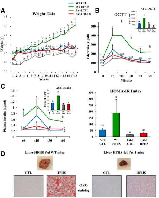
The HFHS diet induced a statistically significant weight gain in WT mice (+20 g relative to controls) (Fig. 1A). Weight gain under the HFHS diet was much lower in fat-1 mice (Fig. 1A and B) and remained comparable to that in their counterparts fed the CTL diet. HFHS-fed fat-1 mice exhibited much less adipose tissue than did WT mice (Supplementary Fig. 1A–C). Moreover, UCP1 mRNA ex-pression in brown adipose tissue was much more elevated in fat-1 mice versus WT (Supplementary Fig. 1D). Food intake did not differ between the two genotypes (Supple-mentary Fig. 6). Thisfinding provides further support to earlier studies that reported that the endogenous production of v3 PUFAs and a reduction in the levels of v6 PUFAs could induce increased energy expenditure, with no effect on energy intake (27).
An HFHS diet is known to have deleterious effects on glucose homeostasis and adiposity (28,29). Our results showed that fat-1 mice were protected against the whole-body glucose intolerance observed in WT mice fed the obesogenic diet (Fig. 1B–D and Supplementary Fig. 3A). In accordance with this remarkable resistance to glucose intolerance, v3 PUFA enrichment of skeletal muscle (Sup-plementary Fig. 4) was associated with improved glucose uptake, independently of the presence of insulin (Supple-mentary Fig. 3C). Moreover, GLUT-1 mRNA expression in skeletal muscle was 2.5-fold higher in fat-1 compared with WT mice (Supplementary Fig. 3B).
Because nonalcoholic fatty liver disease is positively associated with the development of obesity, insulin re-sistance, and ultimately, type 2 diabetes (30,31), we ex-amined different markers of steatosis. Oil Red O staining revealed significantly fewer and smaller lipid droplets in HFHS-fed fat-1 mice livers than in WT mice livers (Fig. 1D). This was confirmed by the unchanged total liver fatty acid mass in fat-1 mice fed the HFHS diet compared with controls, whereas WT mice fed the HFHS diet exhibited a pronounced accumulation of total liver fatty acids (Sup-plementary Fig. 5A) and a twofold increase in the level of hepatic oleic acid (a marker of hepatic steatosis). This level was constant in fat-1 mice, whether fed the HFHS diet or not (Supplementary Fig. 5B). In addition, levels of 20:4 and 22:6 in HFHS-fed WT mice were half those in controls. In fat-1 mice, total liver v3 and v6 PUFA content did not differ whether they were fed the CTL or HFHS diet, but significant increases in 20:4 and 22:6 and decreases in 18:2 and 20:5 levels occurred in fat-1 mice fed the HFHS diet (Supplementary Fig. 5B). mRNA expression of FATCD36
and peroxisome proliferator–activated receptor-g remained comparable to controls in HFHS-fed fat-1 mouse livers and were 2 and 2.5 times higher in HFHS-fed WT mice, re-spectively (Supplementary Fig. 5C).
In addition, F4/80 adipose tissue labeling appeared much higher in HFHS-fed WT mice compared with con-trols but remained remarkably low in fat-1 mice whatever the diet (Supplementary Fig. 2).
fat-1 Mice Are Protected Against Gut Barrier Dysfunction With Consequences on Metabolic Endotoxemia
To bring insights into the beneficial properties of PUFAs in fat-1 mice, intestinal permeability was assessed. As shown in Fig. 2A, a higher flux of 4 kDa FITC-dextran 4 was observed after HFHS feeding in WT mice but not in fat-1 mice. FITC-dextran 4 can be used to assess intestinal permeability but cannot specifically target the lower part of the intes-tine. Measurements of plasma FITC 4 h after gavage mostly corresponded to the lower part of the gut, that is, ileum, cecum, and proximal colon, where most microbes are located (32,33). Even under the CTL diet, fat-1 mice showed lower permeability values than did their WT counterparts (Fig. 2A). ZO-1 immunostaining was 70% higher in fat-1 than in WT mice fed the CTL diet. Even if the HFHS diet induced a decrease of ZO-1 expression in WT (3.6 times) and fat-1 (2 times) mice, it remained close to that of WT controls in fat-1 HFHS-fed mice (Fig. 2B).
Quantitation of LPS mass concentration by liquid chromatography–tandem mass spectrometry revealed that fat-1 mice had low plasma LPS concentrations, which were much lower than in WT mice fed the HFHS diet but com-parable to values measured in WT animals fed the CTL diet (Fig. 2C).
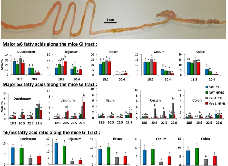
Majorv6 and v3 Fatty Acid Composition and v6-to-v3 Fatty Acid Ratio Along the Gastrointestinal Tract of WT and fat-1 Mice Fed the HFHS and CTL Diets
As shown in Fig. 3, fatty acid composition of total lipids revealed higher levels of v3 fatty acids in fat-1 transgenic animals than in WT mice fed the CTL or HFHS diet, whereas arachidonic acid (20:4 v6) was lower in fat-1 mice than in WT fed the CTL diet. In addition, the ratio of v6 PUFA to the long chain v3 PUFA was markedly decreased compared with the ratio in WT mice, with consistent data from duodenum to colon. The total lipid level did not differ in the two genotypes whatever the regimen (data not shown).
fat-1 and WT Mice Display Different Proportions of Cecal Microbiota Phyla and Different Expression of Intestinal Antimicrobial Peptides When Fed a CTL or HFHS Diet
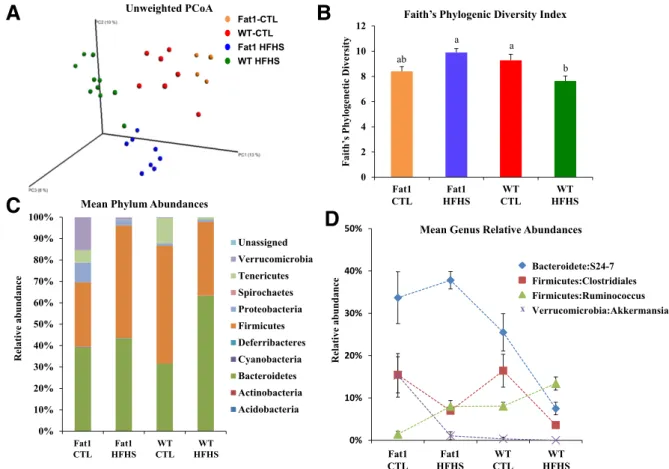
We used 16S rDNA sequencing to investigate changes in the cecal microbiota of fat-1 and WT mice fed the CTL or HFHS diet. A PCoA of the unweighted UniFrac distance matrix showed drastic and significant changes (analysis of similarity, R = 0.78; P , 0.001) in the composition of the cecal microbiota caused by both the mouse genotype and diet (Fig. 4A). Shifts in composition were associated with
significant changes in diversity levels (Faith’s Phylogenetic Diversity) and were also influenced by both the mouse genotype and diet (F1,25= 15.9, P = 0.001). The fat-1 mice
fed the HFHS diet displayed a significantly higher di-versity level than did WT mice fed the same diet (Tukey HSD test, P , 0.05) (Fig. 4B).
At the phylum level (Fig. 4C), differences in OTUs re-vealed decreases in Tenericutes in fat-1 and WT mice phylum when fed the HFHS diet, with a trend of simi-lar magnitude in both genotypes. A more abundant pop-ulation of Verrucomicrobia was observed in fat-1 than in WT mice when fed the CTL diet. Under the HFHS diet, the
Verrucomicrobia phylum was substantially reduced in fat-1 mice and completely disappeared in WT mice.
At the genus level, using a supervised classification ap-proach (nearest shrunken centroid), we identified OTUs that were particularly responsive to the interaction between mouse genotype and diet (Fig. 4D). Indeed, as shown in Fig. 4D, the cecal microbiota of fat-1 mice proved to be enriched in OTUs belonging to the Akkermansia genus, especially when the mice were fed the CTL diet, compared with WT mice. We also found that OTUs belonging to the Rumino-coccus genus were particularly responsive to the HFHS diet for both genotypes, but with significantly lower relative ORO staining e c i m 1 -t a f d e f -S H F H r e v i L e c i m T W d e f -S H F H r e v i L CTL HFHS CTL HFHS
Figure 1—The fat-1 transgenic mice maintain a lean phenotype when fed an HFHS diet. Mice (WT and fat-1 transgenic) were fed a CTL or HFHS diet for 18 weeks.A: Weight gain curves (n = 8 per group). B: OGTT and AUCs for OGTTs. C: Insulin response to OGTT. HOMA-IR, HOMA of insulin resistance.D: Representative photographs of the liver of WT and fat-1 mice fed an HFHS diet and representative Oil Red O (ORO)–stained liver sections (n = 8 per group). Data are expressed as means 6 SEM. Data with different superscript letters are significantly different atP , 0.05, according to the post hoc ANOVA statistical analysis. Similar results were obtained in five independent experiments.
abundance in fat-1 mice than in WT mice. Members of the Clostridials family seemed to be affected by the HFHS diet, with decreasing relative abundance compared with controls for both genotypes. Finally, marked differences were found between WT and fat-1 mice fed the HFHS diet in the relative abundance of members of the S24-7 family. Indeed, al-though they seemed to be unaffected by the HFHS diet in fat-1 mice, the relative abundance of S24-7 members dra-matically dropped in WT mice.
Antimicrobial peptides produced by the host play an important role in maintaining gut microbiota homeostasis and constitute an attractive mechanism for gut ecosystem modulation upon HFHS feeding or upon v3 tissue enrich-ment. Consistent with this hypothesis, we observed that HFHS feeding affected antimicrobial peptide production in the small intestine when v3 tissue enrichment preserved it (Supplementary Fig. 10): A dramatic decrease of Reg3g and a-defensin–Defa expressions was observed in the ileum of WT HFHS compared with WT CTL when it was maintained to the level of WT CTL in fat-1 CTL- and HFHS-fed mice. In the jejunum, Reg3g and phospholipase A2 group II (Pla2g2a) were decreased in WT HFHS com-pared with WT CTL; moreover, Reg3g was increased 2.5-fold in fat-1 mice, and Pla2g2a was at least maintained in fat-1
mice to the levels of WT CTL mice. Therefore, the expression of transcripts for antimicrobial peptides is affected by the HFHS diet in WT mice when it was not in fat-1 mice.
Transplantation of thev3-Modified Fecal Microbiome as a Relevant Way to Preserve a Lean Phenotype in Mice Fed the HFHS Diet
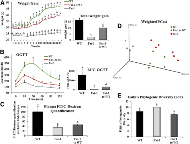
To evaluate the relative contribution of microbiota to the protective effects of v3 PUFAs against diet-induced obesity, WT mice underwent transplantation with fat-1 fecal microbiota and were fed an HFHS diet for 17 weeks. Food intake did not differ between WT, fat-1, and fat-1–to-WT mice (Supplementary Fig. 9). After completion of the dietary intervention, WT mice colonized with fat-1 fecal microbiota exhibited a significantly lower total weight gain (240%) than saline force-fed WT counterparts (10.2 6 1.1 vs. 16.26 2.3 g, respectively) and a significant improvement in glucose tolerance (Fig. 5A and B). WT fecal microbiota transferred to WT recipients had no effect on the ponderal curve or total weight gain of the animals, which remained highly comparable to those observed in saline buffer force-fed control mice (14.56 1.4 vs. 16.2 6 2.3 g, respectively). Intestinal permeability in nontransplanted fat-1 mice was 65% lower than in WT mice (Fig. 5C). A comparable
Figure 2—The fat-1 mice are protected against gut barrier dysfunction with consequences on metabolic endotoxemia. A: Intestinal permeability assay: plasma FITC-dextran 4 (DX, 4,000 molecular weight) oral challenge was measured in WT and fat-1 mice fed the CTL or HFHS diet for 17 weeks.B: Representative immunofluorescence staining for ZO-1 in the jejunum of WT and fat-1 mice fed the CTL or HFHS diet for 18 weeks, and ZO-1 immunostaining quantification of WT and fat-1 mice presented as ZO-1 area–to–DAPI area ratio. C: Direct plasma quantitation of 3-b-hydroxymyristic (OH) acid concentration by gas chromatography-mass spectrometry in WT and fat-1 mice fed the CTL or HFHS diet for 18 weeks (n = 8 per group). Data are means 6 SEM. Groups with different superscript letters are significantly different at P , 0.05, according to the Kruskal-Wallis test and the Dunn multiple comparisons test.
reduction was achieved in WT mice after transplantation with fat-1 fecal microbiota, with values that no longer differed from those in nontransplanted fat-1 mice (Fig. 5C). Finally, we examined changes in the cecal microbiota between the different groups after transplantation. Using PCoA of the weighted UniFrac distance matrix, we detected a strong clustering of mice according to the genotype (Fig. 5D) along the first axis of the PCoA plot that explained 45% of the observed variation in the composition of the cecal microbiota. However, transplanted mice still differed significantly from fat-1 mice in cecal microbial composition (analysis of similarity, R = 0.47; P , 0.001). Again, and as shown in Fig. 4B, analysis of Faith’s Phylogenetic Diversity revealed that fat-1 mice displayed the highest diversity levels (Tukey HSD test, P , 0.05) (Fig. 5E).
DISCUSSION
Early studies investigating the effects of dietary supple-mentation with v3 PUFA enrichment (34,35) and of endogenous v3 PUFA synthesis in fat-1 transgenic mice
(15,27) identified a role for v3 PUFAs in preventing metabolic endotoxemia and low-grade inflammation re-lated to metabolic disorders. The metabolic changes ob-served in the current study—using a specific diet-induced obesity diet containing only traces of cholesterol (0.14%)— are in agreement with those observed by Li et al. (27), who used a diet recommended for atherosclerosis research con-taining 0.2% cholesterol. Nevertheless, extending the work of these authors, we focused here on gut microbiota analysis and transplantation studies. We demonstrated that an alteration in the gut microbiota was an important factor contributing to the v3 beneficial effects: v3 PUFAs in-creased the phylogenic diversity of cecal microbiota and reduced endotoxemia. Most importantly, fecal microbiome transplantation from fat-1 to WT mice was able to prevent the metabolic disorders induced by an obesogenic HFHS diet. In addition, v3 PUFA enrichment in fat-1 mice was able to dampen intestinal epithelial barrier dysfunction in-duced by an HFHS diet, despite no direct contribution of host tissue-embedded PUFAs was evidenced.
Figure 3—Major v6 and v3 fatty acid composition and v6-to-v3 fatty acid ratio along the gastrointestinal (GI) tract of WT and fat-1 mice fed the HFHS and CTL diets. Major fatty acid composition and v6-to-v3 fatty acid ratios of the different sections of the gastrointestinal tract of WT and fat-1 transgenic mice fed the HFHS and CTL diets (mean6 SEM). Groups with different superscript letters are significantly different at P , 0.05, according to the Kruskal-Wallis test and the Dunn multiple comparisons test (n = 5 per group). The v6-to-v3 ratio is given by (18:2 v6 + 20:4 v6 + 22:4 v6 + 22:5 v6)/(18:3 v3 + 20:5 v3 + 22:5 v3 + 22:6 v3).
The rapid and severe 80% increase in body weight observed here was of greater magnitude than the moder-ate increase in body weight of WT mice fed a high v6 PUFA diet as reported by Kaliannan et al. (15). Consistent with earlier investigations (27), our data demonstrated the com-plete prevention of severe diet-induced obesity in fat-1 transgenic mice, an effect that might partly relate to a stim-ulation of energy expenditure (27) and to an increase of UCP1 expression in brown adipose tissue (Supplementary Fig. 1D). The fat-1 transgenic mice were also protected against the metabolic disorders associated with obesity, namely hepatic steatosis, glucose intolerance, and adipose tissue inflammation (Fig. 1 and Supplementary Fig. 2). In the current study, endogenously synthesized v3 PUFAs prevented HFHS-induced fatty liver, thus extending pre-vious observations by Kim et al. (36) and Li et al. (27), who demonstrated that fat-1 gene expression afforded protec-tion against high-fat diet–induced hypercholesterolemia and hepatic steatosis. Furthermore, we showed that homozy-gous fat-1 mice exhibited enhanced muscle glucose uptake, independently of the presence of insulin, and a huge de-crease in the v6-to-v3 ratio compared with WT mice (Supplementary Fig. 4). This protective response might be
attributed to increased docosanoid biosynthesis in mus-cle (37) of fat-1 mice fed a high-fat diet. Indeed, White et al. (38) evidenced that the proresolving lipid mediator, PD1 (10R,17S-dihydroxydocosa-4Z,7Z,11E,13E,15Z,19-Z-hexaenoic acid) was increased by ;176% in muscle tissue of fat-1 mice versus WT mice. Moreover, these authors demonstrated that administration of PD1 to db/db mice substantially improved their insulin sensitivity (38). In the light of these data, we may assume that the high muscle glucose uptake observed in our study could be at least partly due to increased n-3 bioavailability for proresolution me-diator synthesis (as PD1) in the muscle of the fat-1 mice. Chronic low-grade inflammation is a critical factor un-derlying obesity and type 2 diabetes (39), in which significant alterations in the intestinal barrier occur (5,40), resulting in a two-to-threefold increase in serum LPS concentrations, characterizing metabolic endotoxemia (5). As observed in the current study, high-fat diets in mice increase gut permeabil-ity due to reduced expression of TJ proteins (3), resulting in a leaky gut and elevated endotoxin activity (3,40,41).
Whereas high v6 PUFA diets increase metabolic endo-toxemia, v3 PUFAs beneficially affect intestinal permeabil-ity and metabolic endotoxemia (9,35). However, whether
Figure 4—WT and fat-1 mice display different proportions of cecal microbiota phyla when fed a CTL or HFHS diet. A: PCoA of the unweighted UniFrac distance matrix. Red, orange, green, and blue dots represent WT mice fed the CTL diet, fat-1 mice (Fat1) fed the CTL diet, WT mice fed the HFHS diet, and fat-1 mice fed the HFHS diet, respectively.B: Variation between groups of Faith’s Phylogenetic Diversity index. Colors are the same as inA. Groups with different letters are significantly different at P , 0.05, by the Tukey HSD test. C: Mean phylum abundances across the four treatments.D: Mean relative abundances of the most responding OTUs across the four treatments. Data in B, C, and D are means6 SEM.
metabolic endotoxemia is regulated when fat-1 mice are challenged with an obesogenic diet known to dis-rupt intestinal permeability is unknown. Importantly, we found here that endogenous v3 PUFA synthesis in fat-1 mice preserved TJ protein expression, prevented a“leaky” intestinal epithelial barrier, and limited metabolic endotox-emia. The beneficial properties of v3 PUFAs could not only be related to the enrichment of the jejunum in DHA, because our in vitro experiments with Caco-2/TC7 cells did not result in a significant improvement in TJ protein expression and paracellular permeability. Although pre-vious studies (42–44) reported that DHA may directly affect epithelial cells, our results suggest that another mechanism, independently of the enrichment of host cells with v3 PUFAs, has to be considered to account for the potent beneficial effects of v3 PUFAs on gut barrier function. Growing evidence supports the notion that gut micro-biota play a pivotal role in energy homeostasis through the modulation of energy balance (45,46), glucose metabolism, and chronic inflammation associated with obesity (3,5,47).
In addition, gut microbiota-derived LPS has been demon-strated to play a driving role in the onset and progres-sion of inflammation and related metabolic diseases (5). We thus investigated the effect of v3 PUFA enrichment in fat-1 mice on cecal microbiota. Our results show that besides significant differences in cecal microbiota be-tween the two genotypes, fat-1 mice display significantly greater bacterial phylogenic diversity than do WT mice when both are fed the HFHS diet. As shown Fig. 3, cells of the gastrointestinal tract—including the lower part of the intestine where the bacteria are the most abundant—are highly enriched in v3 fatty acids due to the expression of the fat-1 gene (see Supplementary Fig. 8) (v6-to-v3 ratios in fat-1 mice are two- to fourfold lower compared with their WT littermates). So, because the intestinal epithe-lium is continuously renewed, we may speculate that a “cellular” source of long-chain v3 fatty acids brought to the microbiota from the intestinal cell shedding would explain how the microbiota would also be“fed” by v3 fatty acids and would consequently have a positive effect.
Figure 5—Transplantation of the v3-modified microbiome as a relevant way to preserve a lean phenotype in mice fed the HFHS diet. After fecal microbiota transplantation from fat-1 to WT mice, the WT and fat-1 transgenic mice were fed an HFHS diet for 16 weeks.A: Weight gain curves (n = 6 per group). The inset represents the total body weight gain (g, n = 6 per group). B: Mice were fasted for 6 h, and an OGTT was performed after gavage with glucose (2 g/kg body weight). The inset represents the AUC of the three different groups.C: Plasma FITC-dextran 4 oral challenge was measured for the intestinal permeability assay in WT and fat-1 mice fed an HFHS diet for 16 weeks.D: PCoA of the weighted UniFrac distance matrix.E: Variation between groups of Faith’s Phylogenetic Diversity index. Groups with different superscript letters are significantly different at P , 0.05, according to the Kruskal-Wallis test and the Dunn multiple comparisons test.
The signaling pathways by which these changes in flu-ence obesity and associated metabolic parameters remain to be investigated. In this way, this could explain why some genera (in particular Akkermansia and S24-7) previously reported to be protective against weight gain and associated metabolic disorders (46,48) were present at elevated levels in fat-1 mice but not in WT mice. Interestingly, beneficial species such as Akkermansia seem to be crucial for the maintenance of mucus layer integrity (49,50), and the administration of Akkermansia as a beneficial bacteria has been reported to reduce systemic LPS levels in mice fed a high-fat diet, possibly through the preservation of mucus layer thickness and the prevention of leaky gut and endo-toxemia (49). The mechanism by which Akkermansia could protect against obesity is not yet well understood, but recent data indicate that a membrane protein from Akker-mansia muciniphila (Amuc_1100*)—more than the bacteria by itself—could be involved in the protective effects. This hypothesis has been reinforced by the fact that pasteurized bacteria exert similar effects (51).
Because we could not perform microbiota analysis in the upper part of the intestine, due to the paucity of biological material accessible in this specific area, directly linking events occurring in the lower part of the gut with changes observed in the upper part of the gastrointes-tinal tract is indeed difficult. However, it is important to note, as observed in numerous studies and also in the present one, that changing the cecal microbiota composi-tion from the lower part of the gut (measured in the cecal or in the fecal material) can strongly influence the overall phenotype of the mice, even at distance of the colon. For example, seminal papers from Bäckhed et al. (45) showed that transferring the cecal content from mice into germ-free mice strongly affected liver and lipid metabolism. We and others have shown that changing the microbiota of the cecum and colon by using, for instance, prebiotics, may strongly affect the production of gut peptides such as glucagon-like peptide 2, which is known to directly affect the intestinal permeability of the jejunum (40).
Our data also suggest that under the HFHS diet, the host could contribute to the modulation of the microbial community by modulating the production of antimicrobial peptides (Reg3g, Defa, and Pla2g2a) in the small intestine and that v3 tissue enrichment normalized or even in-creased them. We then postulate that the regulation of antimicrobial peptide expression observed in fat-1 CTL- or HFHS-fed mice may represent a potential mechanism by which the host influences the composition of the microbiota. A number of diseases (i.e., atopic diseases, in flamma-tory bowel disease, diabetes, obesity, cancer, and neuro-pathologies) are known to be associated with a significant decrease in microbiota diversity as well as with altered barrier function and increased permeability of the epithe-lium (52). In the current study, and to confirm a cause-and-effect relationship between changes in cecal microbiome and the observed metabolic trait, we transferred fat-1 fecal microbiota to WT mice. We used fecal microbiota for
convenience and to minimize the number of animals used as donors. Moreover, the microbiota of fecal samples were similar to those from the lower gastrointestinal tract. The community membership and structure of fecal samples is indistinguishable from samples derived from the lower gastrointestinal tract (53). The results of our fecal micro-biota transplantation experiments supported the idea that the fat-1 microbiota were the driving force in lowering total weight gain, normalizing glucose tolerance, and restoring gut permeability. The pronounced effects of v3 PUFA– mediated changes in cecal microbiota on body weight and glucose utilization were observable in the present studies in the long-term (after more than 17 weeks of dietary inter-vention). This suggests that the beneficial effect of v3 PUFAs is a transmissible trait that accounts for most of the observed phenotypic differences between fat-1 and WT mice. In parallel, other beneficial effects of v3 PUFAs on host tissue homeostasis occur as already reported (18,19,27,37). As already mentioned in other studies, the transplan-tation process can affect the immune system (54–56). Thus, such effect on the immune system at the very early stage after transplantation could be responsible for met-abolic changes, independently of the implantation of the microbiota. That being said, as mentioned in RESEARCH DESIGN AND METHODS, we showed that WT-to-WT transfer
did not affect the phenotype (data not shown). If any effect on the immune system would have explained the pheno-type observed in WT mice receiving fat-1 microbiota, this should have been seen in WT-to-WT transplantation. We may then assume that all these kinds of any potential changes were not enough to account for the phenotype observed when fat-1 fecal material was used.
In conclusion, the present transplantation study pro-vides evidence that gut microbiota from fat-1 mice account for the protective effects observed in diet-induced obesity and associated metabolic disorders. This main finding provides strong evidence that modulation of the micro-biota by v3 PUFAs may improve the metabolic profile in obese individuals and that the microbiome would be a transmissible trait of a lean phenotype. Although future studies are required to decipher the molecular mechanisms involved, fecal microbiota profiling may become a relevant and usable means to evaluate the positive effect of v3 PUFAs in individualized medicine.
Acknowledgments.The authors thank Amandine Bataille (University of Bourgogne Franche-Comté), Amandine Chlémaire (University of Bourgogne Franche-Comté), Elise Lalarme (Institut National de la Recherche Agronomique, Dijon), Loïc Roblet (University of Bourgogne Franche-Comté), and Chad Stroud (University of Bourgogne Franche-Comté) for technical assistance. The authors thank Dr. John Seubert (University of Alberta, Edmonton, Alberta, Canada) and Philip Bastable (University Hospital of Dijon, Dijon, France) for manuscript editing and Stéphane Garnier (University of Bourgogne Franche-Comté) for his expertise in statistics analysis.
Funding.This work was supported by INSERM, the Regional Council of Bourgogne, the European Regional Development Fund, the University of Bour-gogne, the Fondation de France, a CIFRE grant from Valorex (Combourtillié,
France), and a French government grant managed by the French National Research Agency (ANR) under the program “Investissements d’Avenir” with reference ANR-11-LABX-0021-01-LipSTIC LabEx. P.D.C. is research associate at FRS-FNRS (Fonds de la Recherche Scientifique) and supported by the FRFS-WELBIO under grant WELBIO-CGR-2017-C-02 funds Baillet Latour (grant for Medical Re-search 2015). P.D.C. is a recipient of an ERC Starting grant.
Duality of Interest.No potential conflicts of interest relevant to this article were reported.
Author Contributions.C.B. and Q.E. performed experiments. S.B. and L.L. analyzed data and wrote the manuscript. A.S. analyzed microbiota data and wrote the manuscript. M.G., A.G., A.B., D.D., and B.M. performed experiments. P.D.C. advised on the experimental design for intestinal studies. M.N. designed research, analyzed data, and wrote the manuscript. J.B. designed research, researched and analyzed data, and wrote the manuscript. C.B., M.N., and J.B. are the guarantors of this work and, as such, had full access to all the data in the study and take responsibility for the integrity of the data and the accuracy of the data analysis.
References
1. Hotamisligil GS. Inflammation and metabolic disorders. Nature 2006;444: 860–867
2. Horng T, Hotamisligil GS. Linking the inflammasome to obesity-related disease. Nat Med 2011;17:164–165
3. Cani PD, Bibiloni R, Knauf C, et al. Changes in gut microbiota control metabolic endotoxemia-induced inflammation in high-fat diet-induced obesity and diabetes in mice. Diabetes 2008;57:1470–1481
4. Creely SJ, McTernan PG, Kusminski CM, et al. Lipopolysaccharide activates an innate immune system response in human adipose tissue in obesity and type 2 diabetes. Am J Physiol Endocrinol Metab 2007;292:E740–E747
5. Cani PD, Amar J, Iglesias MA, et al. Metabolic endotoxemia initiates obesity and insulin resistance. Diabetes 2007;56:1761–1772
6. Tsukumo DM, Carvalho-Filho MA, Carvalheira JB, et al. Loss-of-function mutation in Toll-like receptor 4 prevents diet-induced obesity and insulin re-sistance [retracted in: Diabetes 2016;65:1126–1127]. Diabetes 2007;56:1986– 1998
7. Amar J, Burcelin R, Ruidavets JB, et al. Energy intake is associated with endotoxemia in apparently healthy men. Am J Clin Nutr 2008;87:1219–1223
8. Weisberg SP, McCann D, Desai M, Rosenbaum M, Leibel RL, Ferrante AW Jr. Obesity is associated with macrophage accumulation in adipose tissue. J Clin Invest 2003;112:1796–1808
9. Liu Y, Chen F, Odle J, et al. Fish oil enhances intestinal integrity and inhi-bits TLR4 and NOD2 signaling pathways in weaned pigs after LPS challenge. J Nutr 2012;142:2017–2024
10. Ghosh S, DeCoffe D, Brown K, et al. Fish oil attenuates omega-6 poly-unsaturated fatty acid-induced dysbiosis and infectious colitis but impairs LPS dephosphorylation activity causing sepsis. PLoS One 2013;8:e55468 11. Yu HN, Zhu J, Pan WS, Shen SR, Shan WG, Das UN. Effects offish oil with a high content of n-3 polyunsaturated fatty acids on mouse gut microbiota. Arch Med Res 2014;45:195–202
12. Mujico JR, Baccan GC, Gheorghe A, Díaz LE, Marcos A. Changes in gut microbiota due to supplemented fatty acids in diet-induced obese mice. Br J Nutr 2013;110:711–720
13. Jayasinghe TN, Chiavaroli V, Holland DJ, Cutfield WS, O’Sullivan JM. The new era of treatment for obesity and metabolic disorders: evidence and expectations for gut microbiome transplantation. Front Cell Infect Microbiol 2016;6:15 14. Kang JX, Wang J, Wu L, Kang ZB. Transgenic mice: fat-1 mice convert n-6 to n-3 fatty acids. Nature 2004;427:504
15. Kaliannan K, Wang B, Li XY, Kim KJ, Kang JX. A host-microbiome inter-action mediates the opposing effects of omega-6 and omega-3 fatty acids on metabolic endotoxemia. Sci Rep 2015;5:11276
16. Fèvre C, Bellenger S, Pierre AS, et al. The metabolic cascade leading to eicosanoid precursors–desaturases, elongases, and phospholipases A2–is altered in Zucker fatty rats. Biochim Biophys Acta 2011;1811:409–417
17. Dardevet D, Sornet C, Attaix D, Baracos VE, Grizard J. Insulin-like growth factor-1 and insulin resistance in skeletal muscles of adult and old rats. Endo-crinology 1994;134:1475–1484
18. Zou Z, Bellenger S, Massey KA, et al. Inhibition of the HER2 pathway by n-3 polyunsaturated fatty acids prevents breast cancer in fat-1 transgenic mice. J Lipid Res 2013;54:3453–3463
19. Bellenger J, Bellenger S, Bataille A, et al. High pancreatic n-3 fatty acids prevent STZ-induced diabetes in fat-1 mice: inflammatory pathway inhibition. Diabetes 2011;60:1090–1099
20. Pais de Barros JP, Gautier T, Sali W, et al. Quantitative lipopolysaccharide analysis using HPLC/MS/MS and its combination with the limulus amebocyte lysate assay. J Lipid Res 2015;56:1363–1369
21. Kozich JJ, Westcott SL, Baxter NT, Highlander SK, Schloss PD. Development of a dual-index sequencing strategy and curation pipeline for analyzing amplicon sequence data on the MiSeq Illumina sequencing platform. Appl Environ Microbiol 2013;79:5112–5120
22. Caporaso JG, Kuczynski J, Stombaugh J, et al. QIIME allows analysis of high-throughput community sequencing data. Nat Methods 2010;7:335–336 23. Edgar RC. Search and clustering orders of magnitude faster than BLAST. Bioinformatics 2010;26:2460–2461
24. Caporaso JG, Bittinger K, Bushman FD, DeSantis TZ, Andersen GL, Knight R. PyNAST: a flexible tool for aligning sequences to a template alignment. Bioinformatics 2010;26:266–267
25. Myers-Morales T, Bussell KM, D’Orazio SE. Fecal transplantation does not transfer either susceptibility or resistance to food borne listeriosis in C57BL/6 and BALB/c/By mice. F1000 Res 2013;2:177
26. Stecher B, Robbiani R, Walker AW, et al. Salmonella enterica serovar typhimurium exploits inflammation to compete with the intestinal microbiota. PLoS Biol 2007;5:2177–2189
27. Li J, Li FR, Wei D, et al. Endogenousv-3 polyunsaturated fatty acid pro-duction confers resistance to obesity, dyslipidemia, and diabetes in mice. Mol Endocrinol 2014;28:1316–1328
28. Eckel RH, Grundy SM, Zimmet PZ. The metabolic syndrome. Lancet 2005; 365:1415–1428
29. Ogden CL, Yanovski SZ, Carroll MD, Flegal KM. The epidemiology of obesity. Gastroenterology 2007;132:2087–2102
30. Jacobs M, van Greevenbroek MM, van der Kallen CJ, et al. The association between the metabolic syndrome and alanine amino transferase is mediated by insulin resistance via related metabolic intermediates (the Cohort on Diabetes and Atherosclerosis Maastricht [CODAM] study). Metabolism 2011;60:969–975 31. Polyzos SA, Kountouras J, Zavos C. Nonalcoholic fatty liver disease: the pathogenetic roles of insulin resistance and adipocytokines. Curr Mol Med 2009;9:299–314
32. Wang Q, Fang CH, Hasselgren PO. Intestinal permeability is reduced and IL-10 levels are increased in septic IL-6 knockout mice. Am J Physiol Regul Integr Comp Physiol 2001;281:R1013–R1023
33. Padmanabhan P, Grosse J, Asad AB, Radda GK, Golay X. Gastrointestinal transit measurements in mice with99mTc-DTPA-labeled activated charcoal using NanoSPECT-CT. EJNMMI Res 2013;3:60
34. Wall R, Ross RP, Fitzgerald GF, Stanton C. Fatty acids fromfish: the anti-inflammatory potential of long-chain omega-3 fatty acids. Nutr Rev 2010;68:280– 289
35. Mani V, Hollis JH, Gabler NK. Dietary oil composition differentially modulates intestinal endotoxin transport and postprandial endotoxemia. Nutr Metab (Lond) 2013;10:6
36. Kim EH, Bae JS, Hahm KB, Cha JY. Endogenously synthesized n-3 poly-unsaturated fatty acids in fat-1 mice ameliorate high-fat diet-induced non-alcoholic fatty liver disease. Biochem Pharmacol 2012;84:1359–1365 37. White PJ, Arita M, Taguchi R, Kang JX, Marette A. Transgenic restoration of long-chain n-3 fatty acids in insulin target tissues improves resolution capacity and alleviates obesity-linked inflammation and insulin resistance in high-fat-fed mice. Diabetes 2010;59:3066–3073
38. White PJ, St-Pierre P, Charbonneau A, et al. Protectin DX alleviates insulin resistance by activating a myokine-liver glucoregulatory axis. Nat Med 2014;20: 664–669
39. Wellen KE, Hotamisligil GS. Inflammation, stress, and diabetes. J Clin Invest 2005;115:1111–1119
40. Cani PD, Possemiers S, Van de Wiele T, et al. Changes in gut microbiota control inflammation in obese mice through a mechanism involving GLP-2-driven improvement of gut permeability. Gut 2009;58:1091–1103
41. Brun P, Castagliuolo I, Di Leo V, et al. Increased intestinal permeability in obese mice: new evidence in the pathogenesis of nonalcoholic steatohepatitis. Am J Physiol Gastrointest Liver Physiol 2007;292:G518–G525
42. Li Q, Zhang Q, Wang M, Zhao S, Xu G, Li J. n-3 polyunsaturated fatty acids prevent disruption of epithelial barrier function induced by proinflammatory cy-tokines. Mol Immunol 2008;45:1356–1365
43. Willemsen LE, Koetsier MA, Balvers M, Beermann C, Stahl B, van Tol EA. Polyunsaturated fatty acids support epithelial barrier integrity and reduce IL-4 mediated permeability in vitro. Eur J Nutr 2008;47:183–191
44. Roig-Pérez S, Guardiola F, Moretó M, Ferrer R. Lipid peroxidation induced by DHA enrichment modifies paracellular permeability in Caco-2 cells: protective role of taurine. J Lipid Res 2004;45:1418–1428
45. Bäckhed F, Ding H, Wang T, et al. The gut microbiota as an environmental factor that regulates fat storage. Proc Natl Acad Sci U S A 2004;101:15718–15723 46. Turnbaugh PJ, Ley RE, Mahowald MA, Magrini V, Mardis ER, Gordon JI. An obesity-associated gut microbiome with increased capacity for energy harvest. Nature 2006;444:1027–1031
47. Cani PD, Neyrinck AM, Maton N, Delzenne NM. Oligofructose promotes satiety in rats fed a high-fat diet: involvement of glucagon-like peptide-1. Obes Res 2005;13:1000–1007
48. Dao MC, Everard A, Aron-Wisnewsky J, et al.; MICRO-Obes Consortium. Akkermansia muciniphila and improved metabolic health during a dietary in-tervention in obesity: relationship with gut microbiome richness and ecology. Gut 2016;65:426–436
49. Everard A, Belzer C, Geurts L, et al. Cross-talk between Akkermansia muciniphila and intestinal epithelium controls diet-induced obesity. Proc Natl Acad Sci U S A 2013;110:9066–9071
50. Belzer C, de Vos WM. Microbes inside–from diversity to function: the case of Akkermansia. ISME J 2012;6:1449–1458
51. Plovier H, Everard A, Druart C, et al. A purified membrane protein from Akkermansia muciniphila or the pasteurized bacterium improves metabolism in obese and diabetic mice. Nat Med 2017;23:107–113
52. Bischoff SC, Barbara G, Buurman W, et al. Intestinal permeability–a new target for disease prevention and therapy. BMC Gastroenterol 2014;14:189 53. Suzuki TA, Nachman MW. Spatial heterogeneity of gut microbial composition along the gastrointestinal tract in natural populations of house mice. PLoS One 2016;11:e0163720
54. Ekmekciu I, von Klitzing E, Fiebiger U, et al. Immune responses to broad-spectrum antibiotic treatment and fecal microbiota transplantation in mice. Front Immunol 2017;8:397
55. Ekmekciu I, von Klitzing E, Neumann C, et al. Fecal microbiota trans-plantation, commensal Escherichia coli and Lactobacillus johnsonii strains dif-ferentially restore intestinal and systemic adaptive immune cell populations following broad-spectrum antibiotic treatment. Front Microbiol 2017;8:2430
56. Burrello C, Garavaglia F, Cribiù FM, et al. Short-term oral antibiotics treatment promotes inflammatory activation of colonic invariant natural killer T and conventional CD4+T cells. Front Med (Lausanne) 2018;5:21