Abstract Introduction: For routine staging of patients with primary breast cancer, clinical practice guidelines of many medical societies recommend chest X-ray, liver ultrasound and bone scan. With respect to expanding health care costs and patients’ psychological distress it has been supposed, that there might be a group of breast cancer patients, who do not need these imaging studies. Methods: Four hundred and eighty-eight consecutive pa-tients with primary breast cancer who had primary sur-gery at our institution and complete work-up for distant metastases including chest X-ray, liver ultrasound, and bone scan were studied. Results: We found distant me-tastases at the time of primary diagnosis in 19 patients (3.9%). Bone metastases were found in 2.7%, liver me-tastases in 1.0%, and pulmonary meme-tastases in 0.4%. However, in breast tumors smaller than 1 cm, no meta-static lesions were found, whereas 18.2% of the patients with pT4 tumors had metastases. In 2.4% of screening imaging studies, metastases were ruled out by additional imaging. Conclusion: Based on our data and a review of the literature, we suggest that chest X-ray, liver ultra-sound and bone scan can be omitted in the staging of asymptomatic patients with pT1a or pT1b disease. Keywords Primary breast cancer · Metastases · Chest radiography · Liver ultrasound · Bone scan
Introduction
Screening of breast cancer patients for distant metastasis at the time of presentation is required for staging and
subsequent management of these patients. In response to increasing demands for effectiveness of costs from government and private third-party payers, health care providers are asked to re-examine the current staging strategies and to optimize utilization of resources. On the other side as their patient’s advocate, they have to do their best to exclude or detect metastatic lesions at initial presentation in order to avoid over- and undertreatment.
In breast cancer patients, metastases are found most frequently in bones, lung and liver, and therefore, the staging methods recommended are bone scan, chest X-ray and liver ultrasound. In case of unclear findings, further imaging studies such as computer tomography (CT) or magnetic resonance imaging (MRI) will be per-formed to confirm or rule out the presence of metastases. The aim of this study was to determine the frequency and distribution pattern of metastases at the time of pri-mary surgery, and to identify a group of breast cancer patients in whom imaging studies for staging can be omitted.
Patients and methods
From January 1996 to June 2001, 497 consecutive patients with primary breast cancer had surgery and subsequent therapy at the Division of Gynecology of the University Hospital of Zurich, Switzerland. Of these patients, 9 had incomplete staging due to si-multaneous life limiting diseases or due to refusal, leaving 488 pa-tients for further analysis. The mean age of these 488 papa-tients was 60.9 years, ranging from 20 to 94 years. One hundred and forty (28.7%) patients were premenopausal and 348 (71.3%) postmeno-pausal. The patients were routinely staged with chest X-ray, liver ultrasound and bone scan. The detection rate of metastases was correlated with the size of the primary tumor as well as the pres-ence of positive axillary lymph nodes. Staging was performed ac-cording to the American Joint Committee on Cancer (AJCC) clas-sification system [7].
Results
Metastases were found with routine imaging studies in 19 (3.9%) patients at the time of primary surgery. The C. Schneider · M.K. Fehr · D. Hagen · U. Haller · D. Fink (
✉
)Department of Obstetrics and Gynecology,
Division of Gynecology, University Hospital of Zurich, 8091 Zurich, Switzerland
e-mail: [email protected]
Tel.: +41-1-2555327, Fax: +41-1-2554553 R.A. Steiner
Women’s Hospital Fontana, Chur, Switzerland Arch Gynecol Obstet (2003) 269:9–12 DOI 10.1007/s00404-002-0445-x
O R I G I N A L A R T I C L E
Christoph Schneider · Mathias K. Fehr Rolf A. Steiner · Daniela Hagen · Urs Haller Daniel Fink
Frequency and distribution pattern of distant metastases
in breast cancer patients at the time of primary presentation
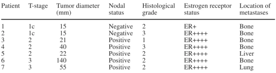
Received: 6 August 2002 / Accepted: 24 September 2002 / Published online: 15 November 2002most frequent location of metastasis was bone (2.7%), followed by liver (1.0%) and lung (0.4%) (Table 1). Patients with pT1 tumor rarely had metastatic lesions. Only 2 of 106 (1.3%) patients with pT1c tumors had dis-tant metastases, all of which were bone metastases. Moreover, both patients were node negative. In larger primary tumors, metastatic lesions occurred in lymph node positive breast cancer patients only. The highest percentage of distant metastases had pT4d tumors with 30%. Patient and tumor characteristics of the 7 patients with pT1–3 tumor and distant metastases are shown in Table 2.
Axillary lymph node dissection was routinely per-formed in level I and II. In 268 (54.9%) patients the axil-lary lymph nodes were negative at primary therapy. Only 2 (0.7%) of these had distant metastases (bone metastas-es), detected by bone scan and confirmed with directed X-ray studies. No lung or liver metastases were found in node negative breast cancer patients at the time of prima-ry surgeprima-ry. Due to the suspicion of metastases in routine staging, 19 (7.1%) node negative patients had 21 addi-tional radiologic studies performed: directed X-rays of bones in 10 cases, CT scan and/or MRI of the abdomen in 8 cases and CT scans of the chest in 3 cases. Metasta-tic lesions in all these 19 patients have been finally ruled out.
Positive axillary lymph nodes were found in 216 (44.3%) patients. In 4 patients the nodal status was not
evaluable, mostly due to patients’ refusal to axillary sur-gery or renunciation due to otherwise reduced life ex-pectancy. None of these 4 patients had distant metastas-es.
In the 216 patients with node positive breast cancer, the diagnosis of distant metastases could be made by bone scan, liver ultrasound or chest X-ray in 9 patients (4.2%). Additional imaging studies were required in 22 of 216 patients (10.2%): 14 directed X-rays of bones, 5 CT scans and/or MRI of the abdomen, 5 CT scans of the chest and 1 CT scan of the bone. Only in 8 patients, metastases were confirmed. Hence, 17 (7.9%) of the node positive patients had distant metastases; one of these patients had both bone and liver metastases. Four patients with metastases had less then 4 positive axillary lymph nodes (23.5%). Of 28 patients with axillary mi-crometastasis (pN1a), one patient (3.6%) had detectable bone metastases. The number of false positive imaging studies routinely performed was high in both, node nega-tive and node posinega-tive patients: In 4.3% of suspicious bone scans, 1.0% of suspicious chest X-ray and 1.8% of suspicious liver ultrasound, metastases were ruled out by additional imaging studies.
Table 3 shows the correlation of the metastases and stage according to AJCC classification. Of the 5 patients with liver metastases, 3 had normal blood chemistry. The other 2 have had either elevated transaminases or elevat-ed alkaline phosphatase.
10
Table 1 Frequency and
distri-bution pattern of metastases ac-cording to the tumor size and nodal status (n=484)
pT Nodal Number of Distant Bone Lung Liver
status patients metastases
1a + 2 0 – 18 0 1b + 3 0 – 35 0 1c + 45 0 – 106 2 (1.9%) 2 (1.9%) 0 0 2 + 93 3 (3.2%) 2 (2.2%) 0 1 (1.1%) – 84 0 3 + 23 2 (8.7%) 1 (4.3%) 1 (4.3%) 0 – 10 0 4a–c + 40 9 (22.5%) 6 (15%) 1 (2.5%) 3 (7.5%) – 11 0 4d + 10 3 (30%) 2 (20%) 0 1 (10%) – 4 0 Table 2 Characteristics of
breast cancer patients with pT1–3 tumors and metastases
Patient T-stage Tumor diameter Nodal Histological Estrogen receptor Location of
(mm) status grade status metastases
1 1c 15 Negative 2 ER+ Bone
2 1c 15 Negative 3 ER++++ Bone
3 2 21 Positive 1 ER++++ Bone
4 2 40 Positive 3 ER++++ Bone
5 2 22 Positive 2 ER++++ Liver
6 3 140 Positive 2 ER++++ Bone
Discussion
Traditionally, chest X-ray, liver ultrasound and bone scan are performed in all patients with breast cancer at the time of first presentation to rule out lung, liver and bone metastases. This work-up is recommended by many clin-ical guidelines, e.g., the National Comprehensive Cancer Network [2]. Only few studies have been focusing on the necessity of these examinations in primary breast cancer, despite expanding health care costs. It has been hypothe-sized that staging examinations might be individualized according to tumor size, nodal status and stage.
Bone is the most common location of distant metas-tases in breast cancer. In a review of nine studies, the rate of bone metastases at the time of primary surgery was 3.1% [8]. In our study, we found an overall rate of 2.7%. However, no patient with pT1a or pT1b disease was found to have distant metastases. Bone metastases were found in 1.3% of our patients with pT1 pN0 dis-ease, which is in good agreement with the rates reported by Ciatto et al. [4] and Cox et al. [5]. In patients with presumable stage II disease, 1.3% of our patients had de-tectable distant metastases, which is similar to other re-ports [4, 5]. While 9.7% of our patients with presumable stage III disease had a skeletal involvement, Ciatto et al. [4] detected bone metastases in 1.2% and Cox et al. [5] in 16.2%.
Pulmonary metastases were present in only 2 of 488 patients (1.0%). Both patients had presumable stage III disease. This is similar to the rate of 0.3% reported by Ciatto et al. [4]. Chen et al. [3] reported an detection rate of 0.099% for asymptomatic lung metastases in 1003 pa-tients. Although many anesthesiologists traditionally re-quire chest X-ray before general anesthesia, there is no strong medical indication for routine preoperative chest X-ray in asymptomatic, otherwise healthy breast cancer patients [13]. Furthermore, chest X-ray has never been shown to improve outcome in the care of patients with clinical stage I and II breast cancer [3]. Our data support the view that chest X-ray may be eliminated in the routine staging for asymptomatic patients with stage I or II breast cancer.
Liver metastases at the time of primary therapy are described in 0.3 to 0.5% [1, 4]. In our study, we found liver metastases in 5 patients (1.0%). Accordant to the results of Ciatto et al. [4], we never found liver metastas-es in patients with stage I breast cancer. In prmetastas-esumable stage II disease, we detected liver involvement in 1 pa-tient (0.4%). In agreement with other studies [5, 6], 4.3% of our patients with presumable stage III disease had liver metastases at the time of primary surgery. Myers et al. [8] and Ravaioli et al. [10, 11] conclude that liver ultrasound can be omitted in the work-up of stage I and II breast cancer unless symptoms or blood chemistry suggest liver metastases. With this strategy, we would have missed one patient with stage II disease with asymptomatic liver metastases and normal blood chemis-try.
Based on our results and a review of the literature, it is reasonable and safe to reduce costs by omitting routine staging imaging studies of asymptomatic patients with pT1a and pT1b tumors. In patients with pT1c tumors routine staging yielded distant metastases in 1.3% of patients. Therefore, we believe that routine work-up for distant metastases should not be omitted in pT1c patients. Calculations on cost savings were made by Norum and Andreassen [9]. They suggested to screen patients in stage I and stage II breast cancer with less than five affected axillary lymph nodes by chest X-ray and blood tests only. According to their calculation, this strategy resulted in significant cost savings without in-fluencing survival. By omitting imaging studies for asymptomatic patients with pT1a and pT1b disease, we recommend a more conservative strategy than others, since these publications accept a rate of missed metastat-ic disease up to 3% [8, 11, 12]. Using our approach, im-aging studies for stim-aging would have been omitted in 58 patients (11.9%) without missing distant metastases and two of 35 false-positive imaging studies would have been avoided (5.7%).
11
Table 3 Frequency of distant
metastases according to the stage (AJCC classification)
Study Stage Distant metastases
Bone Lung Liver
Present study I 1.3% (2/159) 0 (0/159) 0 (0/159) II 0.8% (2/233) 0 (0/233) 0.4% (1/233) III 9.7% (9/93) 2.2% (2/93) 4.3% (4/93) [4] I 0.2% (1/550) 0.1% (1/873) 0 (0/132) II 1.1% (14/1317) 0.2% (3/1943) 0.2% (1/462) III 1.2% (6/508) 1.0% (7/682) 0.5% (1/194) [5] I 0.8% (1/122) 0 (0/127) II 1.1% (2/180) 0 (0/182) III 16.2% (6/37) 5.4% (2/37) [6] I 0 (0/64) 0 (0/54) II 0.8% (2/240) 1.8% (3/167) III 7.3% (6/82) 4.0% (2/50)
References
1. Bruneton JN, Balu-Maestro C, Raffaelli C, Mourou MY, Cambon P, Granon C (1996) Indications for hepatic ultraso-nography in breast cancer staging and follow-up. Breast Can-cer Res Treat 37:115–121
2. Carlson RW, Goldstein LJ, Gradishar WJ, Lichter AS, McCormick B, Moe RE, Theriault RL (1996) NCCN breast cancer guidelines: the National Comprehensive Cancer Net-work. Oncology 10:47–75
3. Chen EA, Carlson GA, Coughlin BF, Reed WP Jr, Garb JL, Frank JL (2000) Routine chest roentgenography is unneces-sary in the work-up of stage I and II breast cancer. J Clin Oncol 18:3503–3506
4. Ciatto S, Pacini P, Azzini V, Neri A, Jannini A, Gosso P, Molino A, Capelli C, Di Costanzo F, Pucciatti MA, Andreoli C, Santoro G, Farante G, Ciurli M, Costa A, Brignone G, Ravaioli A, Scarpellini M, Rosetti P, De Leo G, Punzo C, Oliva V (1988) Preoperative staging of primary breast cancer: a mul-ticentric study. Cancer 61:1038–1040
5. Cox MR, Gilliland R, Odling-Smee GW, Spence RA (1992) An evaluation of radionuclide bone scanning and liver ultraso-nography for staging breast cancer. Aust N Z J Surg 62:550– 555
6. Glynne-Jones R, Young T, Ahmed A, Ell PJ, Berry RJ (1991) How far investigations for occult metastases in breast cancer aid the clinician. Clin Oncol 3:65–72
7. Harris JR (2000) Staging of breast cancer. In: Harris JR, Lippman ME, Marrow M, Osborne CK (eds) Diseases of the breast, 2nd edn. Lippincott, Williams and Wilkins, Philadel-phia, pp 403–406
8. Myers RE, Johnston M, Pritchard K, Levine M, Oliver T (2001) Baseline staging tests in primary breast cancer: a prac-tice guideline. CMAJ 164:1439–1444
9. Norum J, Andreassen T (2000) Screening for metastatic dis-ease in newly diagnosed breast cancer patients. What is cost-effective? Anticancer Res 20:2193–2196
10. Ravaioli A, Tassinari D (2000) Staging of breast cancer: recommended standards. Ann Oncol 11:3–6
11. Ravaioli A, Tassinari D, Pasini G, Polselli A, Papi M, Fattori PP, Pasquini E, Masi A, Alessandrini F, Canuti D, Panzini I, Drudi G (1998) Staging of breast cancer: what standards should be used in research and clinical practice? Ann Oncol 9:1173–1177
12. Samant R, Ganguly P (1999) Staging investigations in patients with breast cancer. Arch Surg 134:551–553
13. Tape TG, Mushlin AI (1988) How useful are routine chest x-rays of preoperative patients at risk for postoperative chest disease? J Gen Intern Med 3:15–20