Annals of Oncology 24: 2985–2989, 2013 doi:10.1093/annonc/mdt359 Published online 7 September 2013
Phase I trial combining temozolomide plus lapatinib
for the treatment of brain metastases in patients with
HER2-positive metastatic breast cancer: the LAPTEM
trial
E. de Azambuja
1*,
†, D. Zardavas
1,†, M. Lemort
1, J. Rossari
2, C. Moulin
3, A. Buttice
1, V. D
’Hondt
1,
F. Lebrun
1, Y. Lalami
1, F. Cardoso
4, C. Sotiriou
1, T. Gil
1, D. Devriendt
1, M. Paesmans
1,
M. Piccart-Gebhart
1& A. Awada
11
Breast Unit, Institut Jules Bordet, Université Libre de Bruxelles, Brussels, Belgium;2
Department of Oncology, Hospital Moinhos de Vento, Porto Alegre, Brazil;
3
Department of Oncology, Centre Hospitalier Universitaire Vaudois, Lausanne, Switzerland;4
Breast Cancer Unit, Champalimaud Cancer Center, Lisbon, Portugal
Received 18 June 2013; revised 16 July 2013; accepted 22 July 2013
Background:
Brain metastases (BMs) pose a clinical challenge in breast cancer (BC). Lapatinib or temozolomide showed activity in BM. Our study assessed the combination of both drugs as treatment for patients with HER2-positive BC and BM.Methods:
Eighteen patients were enrolled, with sixteen of them having recurrent or progressive BM. Any type of previous therapy was allowed, and disease was assessed by gadolinium (Gd)-enhanced magnetic resonance imaging (MRI). The primary end points were the evaluation of the dose-limiting toxicities (DLTs) and the determination of the maximum-tolerated dose (MTD). The secondary end points included objective response rate, clinical benefit and duration of response.Results:
The lapatinib–temozolomide regimen showed a favorable toxicity profile because the MTD could not be reached. The most common adverse events (AEs) were fatigue, diarrhea and constipation. Disease stabilization was achieved in 10 out of 15 assessable patients. The estimated median survival time for the 16 patients with BM reached 10.94 months (95% CI: 1.09–20.79), whereas the median progression-free survival time was 2.60 months [95% confidence interval (CI): 1.82–3.37].Conclusions:
The lapatinib–temozolomide combination is well tolerated. Preliminary evidence of clinical activity was observed in a heavily pretreated population, as indicated by the volumetric reductions occurring in brain lesions. Key words: breast cancer, brain metastases, HER2, lapatinib, temozolomideintroduction
HER-2 overexpression is reported in
∼20% of breast cancer
(BC) patients, conferring aggressive biological [
1
,
2
]. The
treatment of patients with HER2-positive BC has been
revolutionized by trastuzumab and lapatinib [
3
].
Brain metastatic dissemination remains an unresolved medical
need, with BC being the second most common cause of brain
metastases (BM) [
4
]. HER-2 positivity is a risk factor for BM in
patients diagnosed with BC [
5
], with evidence supporting that
trastuzumab-treated HER2 overexpressing metastatic BC bear an
increased risk for BM [
6
]. The reasons for this are debatable:
trastuzumab does not fully penetrate the intact blood–brain
barrier (BBB), thus sparring cancer cells in the central nervous
system (CNS) [
7
]. Alternatively, its high ef
ficacy in controlling
extracranial disease prolongs overall survival, turning the brain
into a
‘sanctuary’ for metastatic disease [
8
].
Standard therapeutic options for BM include local
approaches such as neurosurgical resection, stereotactic
radiosurgery (SRS) and whole brain radiation therapy (WBRT),
with the role of systemic therapy being unclear.
Lapatinib as a small-molecule inhibitor can penetrate more
ef
ficiently the BBB [
9
]. Moreover, the lack of cross-resistance
with trastuzumab promises ef
ficacy in trastuzumab-pretreated
patients [
10
–
13
].
Temozolomide is an alkylating agent, which has a lipophilic
structure enabling BBB penetration. It has been tested in BM
from different tumors either alone or in combination with
capecitabine showing some activity [
14
–
16
].
†EDA and DZ contributed equally to this work.
*Correspondence to: Dr Evandro de Azambuja, Br.E.A.S.T. Data Centre, Jules Bordet Institute, Blvd de Waterloo, 121 (7th Floor), 1000 Brussels, Belgium. Tel: +32-02-541-7244; Fax: +32-02-541-3477; E-mail: [email protected]
© The Author 2013. Published by Oxford University Press on behalf of the European Society for Medical Oncology. All rights reserved. For permissions, please email: [email protected].
In our trial (NCT00614978), we studied the combination of
lapatinib and temozolomide in patients with HER2-positive BC
presenting BM.
materials and methods
eligibility
This study was conducted at the Jules Bordet Institute (IJB), Brussels, Belgium. All patients enrolled received lapatinib combined with temozolomide. Eligibility criteria are published online only.
study design
This was an open-label phase I study. The primary end point was to evaluate the dose-limiting toxicities (DLTs) and determine the maximum-tolerated dose (MTD) of lapatinib plus temozolomide (supplementary Table S1, available at Annals of Oncology online). The secondary end point was to evaluate their clinical activity.
Three to six subjects were enrolled infive dose cohorts (supplementary Table S2, available at Annals of Oncology online), with dose escalation following the classical‘3 + 3’ phase I trial design.
treatment planning
Temozolomide was given orally, once a day, at three dose levels: 100, 150 and 200 mg/m2/day, days 1–5. Lapatinib was given orally, once a day at three dose levels: 1000, 1250, 1500 mg/day. Both agents were given until progression of the disease, intolerable toxicity or a maximum of six cycles, whichever camefirst. A cycle was defined as 28 days of therapy.
response assessments
Brain magnetic resonance imagings (MRIs) were obtained at study entry and every 8 weeks thereafter. They included volumetric, 3D-gradient echo T1-weighted images, before and 10–15 min after intravenous administration of Gd at a dose of 0.1 mmol/Kg body weight. The image assessments, carried out at IJB, were reviewed by one expert radiologist (ML) using both 2D measurement of the longest axial diameters and 3D volumetric
measurement of the lesions using 3D segmentation software (VOXAR 3D® from Toshiba Medical Systems). A CNS objective response was defined as either a complete response (CR: disappearance of all target lesions) or a partial response (PR: at least a 30% decrease in the sum of the longest diameter of the target lesions). Progressive disease (PD) was defined as the occurrence of either a new lesion or a >20% increase in the sum of the longest diameter of the target lesions. Stable disease (SD) was defined as neither sufficient shrinkage to qualify for PR nor sufficient increase to qualify for PD, taking as a reference point the smallest sum of the longest diameter since the treatment started.
Treatment response outside of the CNS was evaluated by the investigator according to the Response Evaluation Criteria in Solid Tumors (RECIST, Version 1.0) guidelines, but was not mandatory as per protocol and was carried out by the investigator as part of the patient’s standard of care.
safety assessments
In the present study, adverse events (AEs) and/or adverse drug reactions were recorded according to the National Cancer Institute Common Terminology Criteria for Adverse Events version 3.0 (NCI CTCAE v3.0). Cardiac monitoring was carried out, with an LVEF assessment using either ECHO or MUGA scan at baseline and every 12 weeks thereafter.
statistical analysis
The study’s sample size was determined according to the well-established current methodology used to design dose-finding studies in oncology (‘3 + 3’ design). The‘safety analyses set’ of subjects consisted of all patients who received at least one dose of lapatinib and temozolomide. AEs were listed by cohort/dose-level and evaluation time point using descriptive statistics. The ‘efficacy analysis set’ of participants included all eligible patients who had received at least 28 days of therapy and who had at least one post-therapeutic assessment of disease status by MRI. The efficacy end points are listed per dose level and cycle using descriptive statistics.
results
patients
Eighteen HER2-positive metastatic BC patients were enrolled
between January 2008 and May 2011. Demographic and clinical
characteristics are displayed in Table
1
. Sixteen patients showed
at least one BM but, because of slow study accrual, two patients
with no BM and no standard therapeutic options available were
enrolled.
The median age of all patients was 50 years (range, 34
–77
years). This was a heavily pretreated population, with a median
of four prior regimens (range, 1–7).
Regarding CNS disease, all but three patients had received
prior CNS irradiation, and one had received additionally SRS.
Sixteen patients had multiple BM, whereas one patient started
the study treatment before SRS. The distribution of patients
according to the treatment dose levels is shown in
Supplementary Table S2, available at Annals of Oncology online.
The median treatment duration reached 64 days (range, 23
–169
days), with a median of two cycles (range, one to six cycles). The
study drugs were stopped because of disease progression
(n = 14), AEs (n = 3) and death (n = 1).
side effects
The non-hematologic toxicity was mainly grade 2 or higher
(Table
2
), with the most common AEs being fatigue, diarrhea
and constipation. The following grade 3 AEs were observed:
diarrhea in one patient at dose level I resulting in treatment
discontinuation; fatigue in three patients at dose levels I, III and
V, respectively; an infection ( port-a-cath associated bacteremia)
in one patient at dose level IV resulting in study withdrawal;
bacterial pneumonia in one patient at dose level V; nausea in
one patient at dose level V, which was managed with
medication.
Few hematologic AEs were reported (Table
2
), with no
neutropenic fever. Per protocol definition, the MTD was not
reached and both agents can be administered combined fully
dosed. One patient developed grade 3 thrombocytopenia while
on her
first cycle at dose level V, with no major bleeding event.
However, the treatment was delayed for 2 weeks, which
constituted the only DLT observed. After the
first cycle, this
patient developed combined grade 4 leukopenia/neutropenia,
not constituting a DLT (duration <7 days).
In terms of cardiac safety, no major toxicity events were
observed. Nine patients underwent a second cardiac monitoring
at 12 weeks. None of them showed an LVEF <50% or an
absolute LVEF drop >15%.
tumor response
Evaluation of response using volumetric brain MRI was a
secondary end point. Two patients did not have BM at baseline;
a third patient received one cycle of treatment and stopped
because of clinical PD. For the 15 remaining assessable patients,
SD was achieved in 10 patients (67%) and PD in 5 patients
(33%). The best response was in each case achieved after two or
three cycles (one patient with SD was not assessed beyond two
cycles).
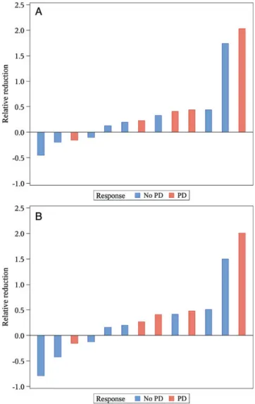
The analysis of volumetric changes from the largest brain
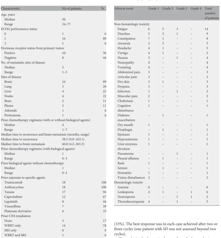
lesion was carried out in 13 patients (Figure
1
A). Five patients
out of the 18 were non-assessable for the purposes of this
analysis: two had no BM at baseline, one had no reported
volume at baseline and two had no measurements after baseline.
For the same cohort of 13 patients, a separate volumetric
changes analysis was undertaken using the sum of the
volumetric measures of all lesions as calculated at baseline and
at the follow-up brain MRI (Figure
1
B). The relative evolution
from baseline was calculated and the largest reduction was
selected.
Table 1. Patient characteristics
Characteristic No of patients %
Age, years
Median 50
Range 34–77
ECOG performance status
0 1 6
1 16 89
2 1 6
Hormone receptor status from primary tumor
Positive 10 56
Negative 8 44
No. of metastatic sites of disease
Median 2 Range 1–3 Sites of disease Brain 16 89 Lung 5 28 Liver 4 22 Nodes 4 22 Bone 2 11 Pleura 2 11 Adrenals 1 6 Peritoneum 1 6
Prior chemotherapy regimens (with or without biological agents)
Median 4
Range 1–7
Median time to recurrence and brain metastasis (months, range)
Median time to recurrence 38.5 (0.0–163.1)
Median time to brain metastasis 60.8 (4.2–265.3) Prior chemotherapy regimens (with biological agents)
Median 1
Range 0–3
Prior biological agents without chemotherapy
Median 1
Range 0–2
Prior exposure to specific agents
Trastuzumab 18 100 Anthracycline 18 100 Taxane 17 94 Capecitabine 12 67 Lapatinib 8 44 Vinorelbine 7 39 Platinum derivative 6 33 Prior CNS irradiation None 3 17 WBRT only 14 78 SRS only 0 0 WBRT and SRS 1 6
ECOG, Eastern Cooperative Oncology Group; SRS, stereotactic radiosurgery; WBRT, whole brain radiotherapy.
Table 2. Toxicity by frequency
Adverse event Grade 1 Grade 2 Grade 3 Grade 4 Total
number of patients Non-hematologic toxicity Fatigue 6 5 3 – 14 Diarrhea 5 3 1 – 9 Constipation 7 1 – – 8 Anorexia 2 3 – – 5 Headache 4 1 – – 5 Vertigo 4 1 – – 5 Nausea 3 – 1 – 4 Neuropathy 4 – – – 4 Vomiting 4 – – – 4 Abdominal pain 3 – – – 3 Articular pain 3 – – – 3 Dry skin 2 1 – – 3 Dyspnea 3 – – – 3 Infection 1 – 1 – 2 Muscular pain 2 – – – 2 Cholestasis 1 – – – 1 Cognitive disturbance 1 – – – 1 Diabetes exacerbation – 1 – – 1 Dry mouth 1 – – – 1 Dysphagia – 1 – – 1 Epistaxis 1 – – – 1 Hyponatremia 1 – – – 1 Liver enzymes elevation 1 – – – 1 Pneumonia – – 1 – 1 Pleural effusion – 1 – – 1 Rash 1 – – – 1 Seizure – 1 – – 1 Stomatitis 1 – – – 1 Vision disturbances 2 – – – 2 Hematologic toxicity Anemia 6 – – – 6 Leukopenia 4 1 – 1 6 Neutropenia – – 1 1 2 Thrombocytopenia 4 – 1 – 5
Overall, as of 31 May 2012, all 18 patients experienced tumor
progression. The estimated median progression-free survival
was 2.60 months [95% con
fidence interval (CI): 1.78–3.42] for
all 18 patients. It was also 2.60 months (95% CI: 1.82
–3.37) for
the 16 patients with BM. Death occurred in 14 patients, which
corresponded to an estimated median survival time of 10.9
months (95% CI: 2.5
–19.3) for all 18 patients, and 10.94 months
(95% CI: 1.09
–20.79) for the 16 patients with BM.
In terms of extracranial disease assessment, three patients
showed SD of the brain but experienced extracranial PD after
two, three and six cycles of treatment, respectively. Regarding
the two patients without BM at study entry, one received six
cycles and the other one cycle of the study treatment.
Unfortunately, both the patients experienced systemic PD.
However, the former of these latter two patients initially showed
a metabolic PR of intrahepatic metastases on a PET scan.
discussion
This phase I study evaluated the safety of lapatinib combined
with temozolomide in patients with HER2-positive BC and BM.
This combination was found to be well tolerable.
Fatigue, diarrhea and constipation were the most frequent
AEs. Hematologic toxicity was not frequent, and no cases of
neutropenic fever or need for transfusions were reported. No
cardiac toxicity was observed in assessable patients.
Preliminary ef
ficacy data were generated in our study, with 10
out of 15 assessable patients achieving a SD (67%). The
volumetric analyses carried out either for the sum of all BM or
for the largest lesion indicated reductions in size, suggesting
clinical bene
fit. With heavily pretreated patients in this study,
including lapatinib pretreatment, it is conceivable that this
element masked the efficacy of our regimen. Furthermore, all
but one patient with BM had undergone local treatment
consisting of WBRT and/or SRS.
Putting our trial in context with other relevant studies, we
noted that in a retrospective analysis of patients treated with
lapatinib combined with capecitabine, seven PRs were reported
for BM, with three of these PRs achieved in the absence of prior
local treatment [
17
]. Building on that concept, the
LANDSCAPE trial assessed the use of lapatinib plus
capecitabine before WBRT and found this to be an alternative to
WBRT as front-line treatment [
18
]. Such an approach could
substantially improve the quality of life of patients with BM by
postponing the WBRT-induced cognitive de
ficit [
19
]. The
results of the randomized phase III CEREBEL study, comparing
the incidence of BM in patients with HER2-positive metastatic
BC treated with lapatinib plus capecitabine versus trastuzumab
plus capecitabine, have been presented [
20
]. CEREBEL did not
show a decrease of the incidence of BM as site of
first relapse for
patients treated with the lapatinib
–capecitabine compared with
trastuzumab–capecitabine (8 versus 12 cases, P = 0.360). These
results must be interpreted with caution, because the incidence
of BM as the
first site of progression in both arms was low [
21
].
WBRT combined with systemic treatment could be another
approach, analogous to a trial in which WBRT was administered
with a protracted low dose of oral vinorelbine and
temozolomide in BC patients with newly diagnosed BM [
22
].
Out of 36 patients, 3 CRs and 16 PRs were reported (ORR of
52%), with a favorable toxicity profile. The advantage here is
mainly that WBRT disrupts the BBB, thus potentiating a higher
penetration of the systemic treatment in the brain [
23
]. Closely
monitoring the dosing of the systemic agents to avoid excessive
toxicity is essential.
Our study has some limitations such as the absence of
neurocognitive function evaluation. However, our patient
population was almost uniformly exposed to WBRT, and thus
Figure 1. (A) Best relative volumetric change in the largest brain metastasis when compared with baseline. Each bar represents a patient having received a baseline and at least an 8-week volumetric brain magnetic resonance imaging (MRI) evaluation. Patients in red are those with progression as best response. The vertical y-axis represents a relative reduction in the largest brain metastasis (vol after treatment– Vol at baseline)/vol at baseline, with negativity corresponding to a reduction and positivity corresponding to an increase in the volume of the lesion. (B) Best relative volumetric change in the sum of the volumetric measures of all brain lesions. Each bar represents a patient having received a baseline and at least an 8-week volumetric brain MRI evaluation. Patients in red are those with progression as best response. The vertical y-axis represents a relative reduction in the sum of the volumetric measures of all brain lesions (sum vol after treatment– sum vol at baseline)/sum vol at baseline, with negativity corresponding to a reduction and positivity corresponding to an increase in the sum of the volumetric lesions.
the chance that the patients had already developed
neurocognitive de
ficits is high. The volumetric changes analysis
we undertook should be interpreted as exploratory, since no
clinically meaningful thresholds for tumor reduction exist.
However, in other trials volumetric reduction of CNS lesions
has been associated with clinical bene
fit [
11
,
24
].
In summary, this study proves the feasibility of treating
patients with BM originating from HER2-positive BC with the
combination of lapatinib and temozolomide at their
single-agent recommended doses. Volumetric reductions in BM were
also achieved. However, the lack of objective responses suggests
limited antitumor activity of the regimen in this heavily
pretreated population.
funding
The Jules Bordet Institute was the sponsor of this clinical trial
and received research grants from GlaxoSmithKline and MSD
(formerly Schering-Plough). Lapatinib was provided by
GlaxoSmithKline and temozolomide was provided by MSD.
Neither company had influence on the study database or data
analyses, which were all carried out at the Jules Bordet Institute.
acknowledgements
We thank Isabelle Mayne, GSK employee, for reviewing the
final
version of the manuscript.
disclosure
EDA: Research Grant (GSK) and Travel Grant (GSK). FC:
Honorarium Consultant (GSK) and Speakers Bureau (GSK). All
the remaining authors have declared no con
flicts of interest.
references
1. Slamon DJ, Clark GM, Wong SG et al. Human breast cancer: correlation of relapse and survival with amplification of the HER-2/neu oncogene. Science 1987; 235 (4785): 177–182.
2. Eccles SA. The epidermal growth factor receptor/Erb-B/HER family in normal and malignant breast biology. Int J Dev Biol 2011; 55(7–9): 685–696.
3. Arteaga CL, Sliwkowski MX, Osborne CK et al. Treatment of HER2-positive breast cancer: current status and future perspectives. Nat Rev Clin Oncol 2011; 9(1): 16–32. 4. Nussbaum ES, Djalilian HR, Cho KH et al. Brain metastases. Histology, multiplicity,
surgery, and survival. Cancer 1996; 78(8): 1781–1788.
5. Pestalozzi BC, Zahrieh D, Price KN et al. Identifying breast cancer patients at risk for Central Nervous System (CNS) metastases in trials of the International Breast Cancer Study Group (IBCSG). Ann Oncol 2006; 17(6): 935–944.
6. Pinder MC, Chang H, Broglio KR et al. Trastuzumab treatment and the risk of central nervous system (CNS) metastases. J Clin Oncol 2007; 25: 36s (suppl 18; abstr 1018).
7. Stemmler H-J, Schmitt M, Willems A et al. Ratio of trastuzumab levels in serum and cerebrospinalfluid is altered in HER2-positive breast cancer patients with
brain metastases and impairment of blood–brain barrier. Anticancer Drugs 2007; 18(1): 23–28.
8. Steeg PS. Brain Metastasis: a Unique Microenvironment and a Sanctuary from Chemotherapy. AACR 101st Annual Meeting. Educational Book 2010, p.91–93. 9. Taskar KS, Rudraraju V, Mittapalli RK et al. Lapatinib distribution in HER2
overexpressing experimental brain metastases of breast cancer. Pharm Res 2012; 29(3): 770–781.
10. Nahta R, Yuan LXH, Du Y et al. Lapatinib induces apoptosis in trastuzumab-resistant breast cancer cells: effects on insulin-like growth factor I signaling. Mol Cancer Ther 2007; 6(2): 667–674.
11. Lin NU, Carey LA, Liu MC et al. Phase II trial of lapatinib for brain metastases in patients with human epidermal growth factor receptor 2-positive breast cancer. J Clin Oncol 2008; 26(12): 1993–1999.
12. Geyer CE, Forster J, Lindquist D et al. Lapatinib plus capecitabine for HER2-positive advanced breast cancer. N Engl J Med 2006; 355(26): 2733–2743. 13. Lin NU, Dieras V, Paul D et al. EGF105084 Study Group. EGF105084, a phase II
study of lapatinib for brain metastases in patients ( pts) with HER2+ breast cancer following trastuzumab (H) based systemic therapy and cranial radiotherapy (RT). J Clin Oncol 2007 ASCO Annual Meeting Proceedings Part I. 25 (18S) (June 20 Supplement), 2007: 1012.
14. Addeo R, Caraglia M. Combining temozolomide with other antitumor drugs and target-based agents in the treatment of brain metastases: an unending quest or chasing a chimera? Expert Opin Investig Drugs. 2011; 20(7): 881–895. 15. Lin NU. CNS metastases in breast cancer. J Clin Oncol 2004; 22(17):
3608–3617.
16. Rivera E, Meyers C, Groves M et al. Phase I study of capecitabine in combination with temozolomide in the treatment of patients with brain metastases from breast carcinoma. Cancer 2006; 107(6): 1348–1354.
17. Metro G, Foglietta J, Russillo M et al. Clinical outcome of patients with brain metastases from HER2-positive breast cancer treated with lapatinib and capecitabine. Ann Oncol 2011; 22(3): 625–630.
18. Bachelot T, Romieu G, Campone M et al. LANDSCAPE: an FNCLCC phase II study with lapatinib (L) and capecitabine (C) in patients with brain metastases (BM) from HER2-positive (+) metastatic breast cancer (MBC) before whole-brain radiotherapy (WBR). 2011 ASCO Annual Meeting J Clin Oncol 2011; 9 (suppl; abstr 509).
19. Platta CS, Khuntia D, Mehta MP et al. Current treatment strategies for brain metastasis and complications from therapeutic techniques: a review of current literature. Am J Clin Oncol 2010; 33(4): 398–407.
20. Pivot X, Semiglazov V, Zurawski B et al. CEREBEL (EGF111438): An open label randomized Phase III study comparing the incidence of CNS metastases in patients ( pts) with HER2+ metastatic breast cancer (MBC), treated with lapatinib plus capecitabine (LC) versus trastuzumab plus capecitabine (TC). ESMO 2012 Congress, Ann Oncol 2012; 23 (suppl 9; LBA LBA11).
21. Addeo R, Caraglia M. The oral tyrosine kinase inhibitors lapatinib and sunitinib: new opportunities for the treatment of brain metastases from breast cancer? Expert Rev Anticancer Ther 2011; 11(2): 139–142.
22. Addeo R, Sperlongano P, Montella L et al. Protracted low dose of oral vinorelbine and temozolomide with whole-brain radiotherapy in the treatment for breast cancer patients with brain metastases. Cancer Chemother Pharmacol. [Internet]. 2012. http://www.ncbi.nlm.nih.gov/pubmed/22890892(21 September 2012, date last accessed).
23. Rubin P, Gash DM, Hansen JT et al. Disruption of the blood–brain barrier as the primary effect of CNS irradiation. Radiother Oncol 1994; 31(1): 51–60. 24. Lin NU, Diéras V, Paul D et al. Multicenter phase II study of lapatinib in patients
with brain metastases from HER2-positive breast cancer. Clin Cancer Res 2009; 15(4): 1452–1459.