Persistent Candida albicans colonization and molecular mechanisms
of azole resistance in autoimmune polyendocrinopathy –candidiasis–
ectodermal dystrophy (APECED) patients
Emilia Siikala
1–3*, Riina Rautemaa
1,3,4, Malcolm Richardson
5, Harri Saxen
6, Paul Bowyer
4and Dominique Sanglard
21
Department of Bacteriology and Immunology, Haartman Institute, University of Helsinki, Haartmaninkatu 3, FI-00014 Helsinki, Finland;
2Institute of Microbiology, University of Lausanne and University Hospital Center, Lausanne, Rue du Bugnon 48, CH-1011 Lausanne,
Switzerland;3Department of Oral and Maxillofacial Diseases, Helsinki University Central Hospital, Helsinki, Kasarmikatu 11-13, FI-00029
HUS, Finland;4The University of Manchester, Manchester Academic Health Science Centre, Translational Research Facility, School of
Translational Medicine, and University Hospital of South Manchester, Wythenshawe Hospital, Southmoor Road, Manchester M23 9LT, UK;
5Mycology Reference Centre, University Hospital of South Manchester, Wythenshawe Hospital and The University of Manchester,
Manchester Academic Health Science Centre, Translational Research Facility, School of Translational Medicine, Southmoor Road,
Manchester M23 9LT, UK;6Hospital for Children and Adolescents, Helsinki University Central Hospital and University of Helsinki,
Stenba¨ckinkatu 11, FI-00029 HUS, Finland
*Corresponding author. Department of Bacteriology and Immunology, Haartman Institute, University of Helsinki, Haartmaninkatu 3, PO Box 21,
FI-00014 Helsinki, Finland. Tel:+358-919126377; Fax: +358-919126382; E-mail: [email protected]
Received 18 March 2010; returned 10 May 2010; revised 3 August 2010; accepted 24 August 2010 Objectives: Patients with autoimmune polyendocrinopathy– candidiasis –ectodermal dystrophy (APECED, APS-I) suffer from chronic candidosis caused mainly by Candida albicans, and repeated courses of azole antifun-gals have led to the development of resistance in the APECED patient population in Finland. The aim of our study was to address whether the patients are persistently colonized with the same or genetically closely related strains, whether epidemic strains are present and which molecular mechanisms account for azole resistance.
Methods: Sets of C. albicans (n¼ 19) isolates from nine APECED patients reported with decreased susceptibility to fluconazole isolated up to 9 years apart were included. The strains were typed by multilocus sequence typing. CDR1/2, MDR1 and ERG11 mRNA expression was analysed by northern blotting and Cdr1, Cdr2 and Mdr1 protein expression by western blotting, and TAC1 and ERG11 genes were sequenced.
Results: All seven patients with multiple C. albicans isolates analysed were persistently colonized with the same or a genetically closely related strain for a mean of 5 years. All patients were colonized with different strains and no epidemic strains were found. The major molecular mechanisms behind the azole resistance were mutations in TAC1 contributing to overexpression of CDR1 and CDR2. Six new TAC1 mutations were found, one of which (N740S) is likely to be a gain-of-function mutation. Most isolates were found to have gained multiple TAC1 and ERG11 point mutations.
Conclusions: Despite clinically successful treatment leading to relief of symptoms, colonization by C. albicans strains is persistent within APECED patients. Microevolution and point mutations occur within strains, leading to the development of azole-resistant isolates.
Keywords: chronic oral candidosis, candidosis, CMC, fluconazole, resistance, TAC1, CDR1, CDR2
Introduction
Autoimmune polyendocrine syndrome type I (APECED, APS-I) is an autosomal recessive disease caused by loss-of-function mutations of the autoimmune regulator gene (Aire) on
chromo-some 21 q22.3.1,2Lack of expression of the Aire protein allows
autoreactive T cells to escape negative selection in the thymus.
This leads to an autoimmune reaction mainly against various endocrine glands as well as to the development of high-titre
auto-antibodies against interferon-v and -a.2Although the
syn-drome is rare, cases have been reported in several geographical
areas.3It has a relatively high prevalence in Finland (1 : 25 000).
The first symptom of the disease in early childhood is often a
superficial Candida infection of the oral mucosa.3Most patients
#The Author 2010. Published by Oxford University Press on behalf of the British Society for Antimicrobial Chemotherapy. All rights reserved. For Permissions, please e-mail: [email protected]
develop high titres of Candida-specific antibodies but fail to respond to Candida antigens and develop chronic
mucocuta-neous candidosis (CMC).4–7 Consequently, patients receive
repeated courses of antifungals from childhood. In the Finnish APECED population, liberal use of azole antifungals has resulted in clinical and microbiological resistance to azoles of the
coloniz-ing Candida albicans strains.8–10Emergence of cross-resistance
between the azoles that the patients had been treated with and newer azole compounds that they had not been exposed
to has also been reported.10In HIV patients, long-term use of
azoles has similarly been reported to cause loss of susceptibility of C. albicans to fluconazole as well as the development of
azole cross-resistance.11 The resistance mechanisms operating
in C. albicans from APECED patients have not yet been investigated.
Several mechanisms are known to result in azole resistance in C. albicans. Azoles, including fluconazole, target lanosterol
14a-demethylase, which is a product of the ERG11 gene.12
Point mutations and upregulation of ERG11 can occur and have
been identified in resistant clinical isolates.12–14The majority of
the point mutations associated with resistance alter the
binding of azoles to Erg11p.15Another mechanism involved in
azole resistance is the failure to accumulate azoles in yeast cells, which may be caused either by impaired drug import or
increased drug efflux.16Multidrug efflux transporters of the
ATP-binding cassette (ABC) superfamily and of the major facilitator (MF) class have a key role in this low-level azole accumulation. CDR1 and CDR2, genes for ABC transporters, and MDR1, a gene for MF transporters, have been shown to be upregulated in
resist-ant strains, leading to enhanced drug efflux.12,13,17This in turn
leads to a reduced inhibition of their target encoded by the ERG11 gene. MDR1 is often not detectable in azole-susceptible strains, whereas the transcription of CDR1 may be detectable also in azole-susceptible strains and is elevated in strains with decreased susceptibility. Deletion of CDR1 and CDR2 results in
hypersusceptible isolates.13,18CDR2 upregulation is usually
com-bined with CDR1 upregulation due to a mutual activator, the
transcriptional activator of CDR genes (TAC1).19TAC1 is located
close to the mating-type locus (MTL), and previous studies have shown a correlation between homozygosity (a/a or a/a) at the MTL and azole resistance.14,20,21
It has been demonstrated that TAC1 mutations, so-called gain-of-function (GOF) mutations, are associated with hyperac-tivity of the encoded protein, which is itself responsible for enhanced transcription of TAC1 target genes, including CDR1
and CDR2.19 It is therefore expected that isolates with high
CDR1 and CDR2 expression will exhibit GOF mutations. Mrr1p has been shown to control MDR1 expression in C. albicans and GOF mutations in the MRR1 gene result in upregulation of
MDR1.22
Using sets of multiple isolates, our study aimed firstly to analyse whether APECED patients with persistent oral candidosis were colonized with the same C. albicans strain over several years or whether they had become re-infected with new strains after antifungal treatment. Secondly, we asked whether epidemic strains existed within the APECED population in Finland. Thirdly, we analysed the molecular mechanisms behind azole resistance in these isolates and looked for signs of microvariation in the persisting C. albicans strains developing resistance.
Patients and methods
C. albicans isolates
The 19 C. albicans isolates used in this study had been isolated from 1995 to 2007 from the oral cavities of nine APECED patients suffering from CMC (1 – 3 isolates/patient). Patients with at least one isolate with decreased susceptibility to fluconazole or signs of clinical resist-ance were included. From these patients multiple isolates with various susceptibilities over many years were selected when available. The C. albicans isolates had been identified from patient samples using con-ventional culture and identification methods at the Clinical Microbiology Laboratory of Helsinki University Central Hospital. The identification of
C. albicans was based on colony morphology on CHROMagarwCandida
medium (CHROMagar, Paris, France) and a negative Bichro-Dubliw
latex co-agglutination test result for Candida dubliniensis (Fumouze Diagnostics, Levallois Perret, France). The strains had been stored in milk – glycerin at – 708C.
Growth media
The C. albicans strains were subcultured in complete YEPD broth (1% Bacto peptone; Difco Laboratories, Basel, Switzerland), 0.5% yeast extract (Difco) and 2% glucose (Fluka, Buchs, Switzerland) and sub-sequently on YEPD agar.
Susceptibility testing
The susceptibility profiles of the isolates to fluconazole and itraconazole were re-analysed by Etest (AB Biodisk, Sweden) according to the
manu-facturer’s instructions and as described previously.23Quality control for
susceptibility testing was performed using C. albicans strains ATCC 90028 and ATCC 24433 and Candida glabrata strain ATCC 90030.
Strain typing
Multilocus sequence typing (MLST) was used to type the C. albicans iso-lates. Sequences of bases in the gene fragments from seven housekeep-ing genes (AAT1a, ACC1, ADP1, MPI1b, SYA1, VPS13 and ZWF1b) were determined and the isolates were tested by MLST as described earlier
using an ABI Prism 3700 genetic analyser (PE Applied Biosystems).24,25
In brief, isolation of genomic DNA was performed as described earlier
from an overnight culture.26 DNA polymerase Expand High Fidelity
(Roche, Basel) was used for the amplification of DNA by PCR. Sequences
of primers used are listed in Table1. Sequence data were analysed for
polymorphisms using Contig Express software (Invitrogen, Basel). Sequence data were entered into the MLST database (http://test1.mlst. net) to provide a diploid sequence type (DST) for each strain. According to sequence types, strains were analysed to determine clonal clusters by the eBURST program and each strain was given a clonal cluster number. Clonal clusters were defined as groups of isolates with six of
the seven genotype sequences being identical.27The unweighted
pair-group method with arithmetic averages (UPGMA) was used to display strains as a dendrogram.
Analysis of MTL status
The MTL type/status was determined by PCR by amplifying the MTLa and
MTLagenes as described previously.28Sequences of primers used are
listed in Table1. The MTL was assessed as heterozygous (a/a),
homozy-gous (a/a) or homozyhomozy-gous (a/a). The correlations between the MTL type and MIC and between MTL type and aggregated years of exposure to azoles were analysed. Aggregated years on azoles signifies the number of years the patient had been on one or more azoles
Siikala et al.
aggregatively before isolation of the strain. The years of concomitant exposure to multiple azoles have been multiplied by the number of pre-scribed azoles.
Northern blotting
Small-scale isolation of total RNA was performed from liquid cultures in
logarithmic growth phase as described earlier.26Northern blotting was
performed to determine the mRNA expression for CDR1, CDR2, ERG11
and MDR1 as described previously.13 RNA samples were separated by
agarose gel electrophoresis and transferred to a nitrocellulose membrane using the Vacuum Blotting System (Hoefer Scientific Instruments,
San Francisco, CA, USA). Probes were labelled with [a-32P]dATP with
random priming using the MegaPrime DNA Labeling System dNTP Kit (GE Healthcare) according to the manufacturer’s instructions. Radioactive signals were revealed by exposure to Kodak BioMax MR film (GE Health-care). Signals obtained in blotted membranes were quantified by count-ing radioactivity (Typhoon Trio; GE Healthcare). CDR1, CDR2, MDR, ERG11 and ACT1 probes were prepared by PCR using primers CB and CYP-NS2, ACT-RT– PCR-F and -R. Primers used in this study are listed in
Table1. As a control for the evaluation of expression levels of CDR1/2,
MDR1 and ERG11, the membranes were hybridized with ACT1 and the amount of RNA was normalized according to the expression of ACT1. The azole-susceptible strain isolated in 1995 from Patient 2 (fluconazole MIC 1 mg/L) was chosen as a baseline control strain (0-level) and the expression levels of the other isolates were quantified relative to the sus-ceptible strain.
Immunoblotting
Western blotting was performed to evaluate Cdr1, Cdr2 and Mdr1 protein expression. A C. albicans strain (DSY3849) known to overexpress Cdr1p and Cdr2p was used as a positive control. A positive control for Mdr1p was established by exposing the azole-susceptible strain isolated in
1995 from Patient 2 to benomyl for 30 min as described previously.29
Cell extracts were prepared as described by Sanglard et al.30Ten
microli-tres of solubilized yeast protein was separated by 10% SDS/PAGE and transferred by western blot onto a nitrocellulose membrane (Bio-Rad Membrane). Immunodetection of Cdr1, Cdr2 and Mdr1 was performed
as described previously.19 A Super Signal West Pico Chemiluminescent
Kit (Thermo Scientific) and Amersham Hyperfilm (GE Healthcare) were used according to the manufacturer’s instructions. The protein signals visualized on the exposed films were interpreted as positive or negative.
Sequence analysis of ERG11 and TAC1
Genomic DNA for sequencing for TAC1 and ERG11 mutations was
extracted as described earlier.26 Sequences were amplified from
genomic DNAs by standard protocols as described previously for TAC131
Table 1. Primers used in this study
Primer Sequence MLST AAT1a-F 5′-CAGCAACATGATTAGCCC-3′ AAT1a-R 5′-ACTCAAGCTAGATTTTTGGC-3′ ACC1-F 5′-GCAAGAGAAATTTTAATTCAATG-3′ ACC1-R 5′-TTCATCAACATCATCCAAGTG-3′ ADP1-F 5′-GAGCCAAGTATGAATGATTTG-3′ ADP1-R 5′-TTGATCAACAAACCCGATAAT-3′ MPIb-F 5′-ACCAGAAATGGCCATTGC-3′ MPIb-R 5′-GCAGCCATGCATTCAATTAT-3′ SYA1-F 5′-AGAAGAATTGTTGCTGTTACTG-3′ SYA1-R 5′-GTTACCTTTACCACCAGCTTT-3′ VPS13-F 5′-TCGTTGAGAGATATTCGACTT-3′ VPS13-R 5′-ACGGATGGATCTCCAGTCC-3′ ZWF1b-F 5′-GTTTCATTTGATCCTGAAGC-3′ ZWF1b-R 5′-GCCATTGATAAGTACCTGGAT-3′ MTL status MTLaplha 1-F 5′-TACATTCTGGTCGCGATGCTC-3′ MTLaplha 1-R 5′-GTAATCCAAAGCCTCGCATAA-3′ Northern blotting CDR1-F 5′-AGTTGAGATCTACCCTTTAAGATATT-3′ CDR1-R 5′-TAATGACCAATTGAATCCCGATTCAG-3′ CDR2-F 5′-TGGTATATAAACTGGACAACATATCTG-3′ CDR2-R 5′-TAATGAACCAATTGAATCCCGATTCAG-3′ ERG11N 5′-CGGGATCCATGGGTGGTCAACATACTTCT-3′ ERG11C 5′-CGGAATTCCCTGCTGGTTCAGTAGGTAAAAC-3′ BENRN 5′-AAAAGCTTATGCATTACAGATTTTTAAGAG-3′ BENRC 5′-AAAAGCTTCTAATTAGCATACTTAGATCTT-3′ ACT1-RT-PCR-F 5′-GTTCCCAGGTATTGCTGAAC-3′ ACT1-RT-PCR-R 5′-CAATGGATGGACCAGATTCG-3′ TAC1 sequencing Amplification Zinc2-604 5′-ATAAGAGTGGCATGTGATA-3′ Zinc2-1123 5′’-GATGCCAACGAATTATTGA-3′ Zinc2-1708 5′-CAGAATTCGTTGGAGAATA-3′ Zinc2-2242 5′-GCCTTGTTACAATCAAGAA-3′ Zinc2-2683 5′-GCAGCATATCTTGTATTAG-3′ Zinc2-3224 5′-ATGCTCAGTCACCAAGTTA-3′ Zinc2-3087c 5′-GGTGTTCCTGCTACCACAA-3′ Zinc2-1789c 5′-ACATCAACAATGCCTTCTAC-3′ Zinc2-1247c 5′-TCTTCACCGTATGAACCTA-3′ Zinc2-778c 5′-CGTTGCTATTGGCGGTTGA-3′ Zinc2-1169 5′-TGTTGGTACTCATTCAATT-3′ Zinc2-1722 5′-TTGGAGAATAGTGCCATTT-3′ Zinc2-1510 5′-GCTACCAAGCGAAGGAGAT-3′ Zinc2-2465c 5′-TCTCTCGCCTAATTGACGT-3′ Sequencing ZCF36SEQ1 5′-ATTACAATGTGTCCCACACAGG-3′ ZCF36SEQ2 5′-CAGTTTACTTTATCCATTTATGCC-3′ ERG11 sequencing Amplification ERG11-xho 5′-GCGCAACTCGAGCTCATATGAACAAGGTTGGGTAGTAA-3′ Continued Table 1. Continued Primer Sequence ERG11-Kpn 5′-GAGCATGGGTACCGGCGCGCGATTGTACGT GG-3′ Sequencing ERG11-3B 5′-CCCATTAAGAATCCCTGAA-3′ ERG11-5B 5′-CAGGGATTCTTAATGGGT-3′ ERG11-4 5′-CTGCTGGTTCAGTAGGTA-3′ ERG11-6 5′-GAGCAAATGAACGGTCAA-3′
MLST, multilocus sequence typing; MTL, mating-type locus.
and ERG11.31The sequences of primers used are listed in Table1. The alleles were sequenced using an AB3730XL DNA Analyzer (Applied Biosys-tems). The resulting data were analysed using the Mutation Surveyor software (Soft Genetics).
Statistical analysis
Data were analysed by using Graph Pad Prism version 5.00 (GraphPad Inc., San Diego, CA, USA). Geometric means and range were used for the analyses of MICs. The two-tailed Mann–Whitney t-test was used
for the comparisons of groups, and Spearman’s rho (rS) was used for
the analyses of correlations. Correlations are presented with a 95%
confidence interval and P value. P values of less than 0.05 were con-sidered statistically significant.
Results
MLST typing
All seven patients with multiple C. albicans isolates analysed were persistently colonized with the same strain for a mean of 5.0
years (range 1–9) (Table2). Five of these seven patients (2, 4, 5, 6
and 7) had a pair of isolates identical by MLST. The mean time
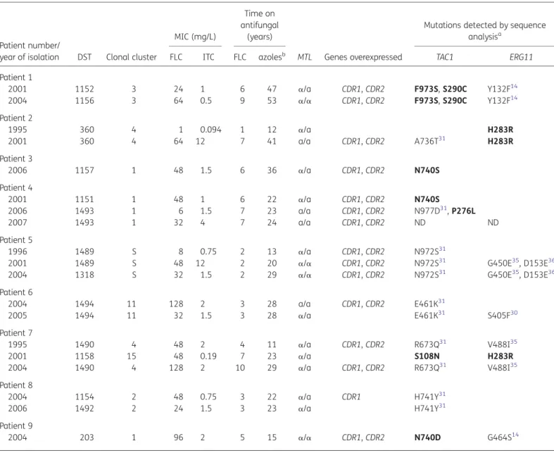
Table 2. Characteristics of C. albicans isolates used in this study
Patient number/
year of isolation DST Clonal cluster
MIC (mg/L)
Time on antifungal
(years)
MTL Genes overexpressed
Mutations detected by sequence
analysisa
FLC ITC FLC azolesb TAC1 ERG11
Patient 1 2001 1152 3 24 1 6 47 a/a CDR1, CDR2 F973S, S290C Y132F14 2004 1156 3 64 0.5 9 53 a/a CDR1, CDR2 F973S, S290C Y132F14 Patient 2 1995 360 4 1 0.094 1 12 a/a H283R 2001 360 4 64 12 7 41 a/a CDR1, CDR2 A736T31 H283R Patient 3 2006 1157 1 48 1.5 6 36 a/a CDR1, CDR2 N740S Patient 4 2001 1151 1 48 1 6 22 a/a CDR1, CDR2 N740S 2006 1493 1 6 1.5 7 23 a/a CDR1, CDR2 N977D31, P276L 2007 1493 1 32 4 7 24 a/a CDR1, CDR2 ND ND Patient 5 1996 1489 S 8 0.75 2 13 a/a CDR1, CDR2 N972S31
2001 1489 S 48 12 2 20 a/a CDR1, CDR2 N972S31 G450E35, D153E36
2004 1318 S 32 1.5 2 29 a/a CDR1, CDR2 N972S31 G450E35, D153E36
Patient 6 2004 1494 11 128 2 3 28 a/a CDR1, CDR2 E461K31 2005 1494 11 32 1.5 3 28 a/a E461K31 S405F30 Patient 7 1995 1490 4 48 2 4 11 a/a CDR1, CDR2 R673Q31 V488I35 2001 1158 15 48 0.19 7 23 a/a S108N H283R 2004 1490 4 128 2 10 29 a/a CDR1, CDR2 R673Q31 V488I35 Patient 8 2004 1154 2 48 0.75 3 22 a/a CDR1 H741Y31 2006 1492 2 24 1.5 3 23 a/a H741Y31 Patient 9 2004 203 1 96 2 5 15 a/a CDR1, CDR2 N740D G464S14
MIC, minimum inhibitory concentration; DST, diploid sequence type; FLC, fluconazole; ITC, itraconazole; MTL, mating-type locus; ND, sequence not determined; S, singleton.
a
Mutations A736T, N977D, N972S, E461K, R673Q and H741Y found in TAC1 alleles have previously been described as gain-of-function (GOF) mutations. Mutations S108N, P276L, S290C, F973S, H283R, N740D and N740S (in bold) have not been previously described.
b
Aggregated years on azoles before isolation of the strain.
Siikala et al.
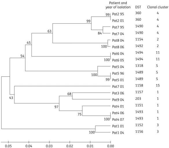
elapsed between the isolation of these strains was 4.4 years (range 1–9). The isolates from the remaining two patients with multiple isolates (Patients 1 and 8) differed in one or two genotypes and were designated by different DST numbers but assigned to the same clonal cluster. These differences mainly resulted from a loss of heterozygosity at the sequenced loci. In one patient (Patient 7) two strains belonging to different clonal clusters were identified. Isolates from three patients (Patients 3, 4 and 9) belonged to clonal cluster 1, and isolates from two patients (Patients 2 and 7) belonged to clonal cluster 4. All other sets of isolates belonged to different clonal clusters (2, 3 and 11). Isolates from Patient 5 were not assignable to a clonal cluster and were designated as sin-gletons. The UPGMA dendrogram of the isolates is shown in
Figure1. Isolates that belonged to clonal cluster 1 but were from
different patients did not co-cluster with very high levels of simi-larity, whereas those that belonged to clonal cluster 4 formed a more compact cluster. However, all patients were colonized with different strains and no epidemic strains were found.
Susceptibility profiles of C. albicans isolates
The MIC of fluconazole for three isolates from three patients
(2/1995, 4/2006 and 5/1996) was ≤8 mg/L (Table 2). The
remaining 16 isolates had MICs≥16 mg/L, of which five had MICs
≥64 mg/L. One patient (Patient 2) had one isolate (isolated in
1995) with an MIC of≤0.125 mg/L of itraconazole and the
fluco-nazole MIC for this isolate was 1 mg/L. All other isolates had MICs
of≥0.25 mg/L of itraconazole, and 14 of these isolates had MICs
of≥1 mg/L. There was an increase in the fluconazole MICs for the
sets of isolates from four (Patients 2, 4, 5 and 7) of the remaining five patients, with two isolates identical by MLST. An increase in the itraconazole MICs was seen in the sets of isolates from three (Patients 2, 4 and 5) of these five patients.
One patient set (Patient 1) showed an increase in the MIC of fluconazole only. Four of the five identical DST pairs showed an increase in fluconazole and itraconazole MICs. In all of these cases systemic and/or topical azoles had been used between
the isolation of the strains (Table2). A decrease in the MICs of
both azoles could be seen in one set (Patient 6) where azoles had not been used between the isolation of the pair of strains. In another patient set (Patient 8), where only miconazole had been used between the isolation of the strains, a decrease in the MIC of fluconazole but not itraconazole was seen. In neither of these cases, however, did the MICs decrease signifi-cantly or fall below the susceptible breakpoints according to
the CLSI standard.32
99 99 63 45 54 43 97 68 99 100 100 0.05 0.04 0.03 0.02 0.01 0.00 100 75 84 100 100 Patient and year of isolation DST Pat2 95 Pat2 01 Pat7 95 Pat7 04 Pat8 04 Pat8 06 Pat6 04 Pat6 05 Pat5 04 Pat5 96 Pat5 01 Pat7 01 Pat3 06 Pat9 04 Pat4 01 Pat4 06 Pat4 07 Pat1 01 Pat1 04 4 4 4 4 2 2 11 11 S S S 15 1 1 1 1 1 3 3 1493 1151 203 1157 1158 1489 1489 1318 1494 1494 1492 1154 1490 1490 360 360 1493 1152 1156 Clonal cluster
Figure 1. Unweighted pair-group method with arithmetic mean (UPGMA) dendrogram of the isolates typed by multilocus sequence typing (MLST). The scale bar shows genetic distance between the isolates. DST, diploid sequence type; S, singleton.
MTL status
Of the 19 strains, 8 were found to be MTL homozygous (a/a or
a/a) and 11 were MTL heterozygous (a/a). The mean MICs for
the homozygous isolates (a/a or a/a) of fluconazole and itraco-nazole were 45.4 (range 6 –128) and 4.7 mg/L (range 0.5 –12), respectively. The mean MIC for the a/a type isolates (n¼ 4) was 35.1 mg/L (range 6 –128) of fluconazole and 4.6 mg/L (range 2 –12) of itraconazole. The mean MIC for the a/a type isolates (n¼ 4) was 60.0 mg/L of fluconazole and 7.0 mg/L of itracona-zole. The heterozygous (a/a) isolates had a mean MIC of 26.7 mg/L (range 1 –128) of fluconazole and 1.4 mg/L (range 0.094– 2) of itraconazole. The difference between the MICs for homozygous and heterozygous isolates was statistically signifi-cant for itraconazole (P¼ 0.0327) but not for fluconazole. In the two isolate pairs with identical DSTs where loss of MTL het-erozygosity could be observed (2/1995 and 2/2001; 5/1996 and 5/2001), the MICs of fluconazole increased. In one patient (Patient 6), who had two strains with identical DSTs isolated 1 year apart, a decrease in the fluconazole MIC was seen conco-mitantly with a shift from homozygosity to heterozygosity in MTL status. MTL status did not correlate with years of exposure to azoles. The correlation of MTL status with the aggregative exposure to all azoles was also analysed since APECED patients are often prescribed different azole antifungal agents and
formu-lations concomitantly (Table2). The mean aggregated exposure
of the homozygous isolates (a/a and a/a) to azoles was 29.1 years and to fluconazole it was 5.3 years. For the heterozy-gous isolates, the mean aggregated exposure to azoles was 24.2 years and to fluconazole it was 4.6 years.
CDR1/2, MDR1 and ERG11 mRNA expression
Fifteen (79%) of the 19 isolates showed overexpression of CDR1 and 14 (74%) showed overexpression of CDR2 by a mean of 9.8-fold (range 3.5 –19.4) and 20.4-fold (range 7.8–65.1),
respect-ively, when compared with the control isolate (Figure2). Thirteen
(87%) of the isolates overexpressing CDR1 and 12 (86%)
overex-pressing CDR2 had fluconazole MICs≥16 mg/L. Fourteen isolates
overexpressed both CDR1 and CDR2 and 12 (86%) of these had
flu-conazole MICs of≥16 mg/L. All isolates overexpressing CDR1 or
CDR2 had itraconazole MICs≥0.25 mg/L. In the four sets of
iso-lates identical by MLST with an increase in fluconazole MICs, the relative expression level of CDR1 increased by a mean of 3.3-fold (range 0.5 –4.4) and that of CDR2 by a mean of 4.0-fold (range 0.5 –11.2). The one pair of isolates identical by MLST (Patient 6), where a decrease in the MIC of fluconazole was seen and the rela-tive expression level of CDR1 and CDR2 decreased to the level of the control isolate in 1 year, the patient had not been exposed to any azoles between the isolation of the isolates.
Only two isolates (2006 isolate from Patient 3 and 2004 from Patient 8) showed a slight (3.2- and 4.9-fold, respectively) increase in the relative expression of MDR1 compared with the control strain. The isolates had an MIC of 48 mg/L of fluconazole and 0.75 and 1.5 mg/L of itraconazole. However, these isolates also showed increased CDR1 mRNA levels. The expression of ERG11 was low in our set of isolates (relative expression ranging from 0.2 to 1.3). Fourteen isolates expressed ERG11 at lower levels than the control, illustrating that azole resistance does not necessarily arise from overexpression of ERG11.
Protein expression levels of Cdr1, Cdr2 and Mdr1
All isolates showing elevated Cdr1 and Cdr2 protein levels over-expressed CDR1 and CDR2 mRNA. One isolate showed slightly elevated CDR1 mRNA levels (3.9-fold when compared with the
control isolate) but no Cdr1 could be detected (Figure2). Isolates
expressing Cdr1 also expressed Cdr2. Of the isolates that expressed Cdr1 and Cdr2, the mean MIC of fluconazole was 44.5 mg/L and that of itraconazole was 1.8 mg/L, whereas the isolates that did not express Cdr1p and Cdr2p had a mean MIC of 6.9 mg/L of fluconazole and 0.3 mg/L of itraconazole. Mdr1p expression was not detected.
TAC1 and ERG11 sequence analysis
TAC1 mutations were detected in all but one isolate (isolate 1995
from patient 2) (Table 2). Twelve different mutations (A736T,
N977D, N972S, E461K, R673Q, H741Y, S108N, P276L, S290C, F973S, N740S and N740D) were found in total. Six of these mutations (S108N, P276L, S290C, F973S, N740S and N740D) have not been described previously. Of these, all but P276L were found in isolates with elevated azole MICs, and N740S occurred in the absence of any other TAC1 or ERG11 mutations. N740S was found in two unrelated strains isolated from different patients. The mean relative expression level of CDR1 and CDR2 in these two isolates was 11.1-fold (CDR1) and 14.6-fold (CDR2) compared with the control isolate and their MIC of fluconazole were 48 mg/L.
The sequence analysis of ERG11 revealed seven point mutations (Y132F, H283R, G450E, D153E, S405F, V488I and G464S) in 11 isolates. Two isolates (2001 and 2004) from Patient 5 had two mutations (G450E and D153E). One mutation (H283R) in ERG11 had not been previously reported. It was detected in three isolates from two patients (Patients 2 and 7), one of which was susceptible to azoles. In one pair of isolates identical by MLST (Patient 6), acquisition of an S405F mutation was observed in association with a decrease in azole MICs. Pairs of isolates identical by MLST from three patients (Patients 1, 5 and 6) gained additional TAC1 or ERG11 mutations as well as maintaining the pre-existing ones.
Discussion
Despite clinically successful antifungal treatment courses, the APECED patients investigated here were found to be persistently colonized with the same C. albicans strain over many years. All patients were colonized with different strains and no epidemic strains were found. Our findings are in accordance with those
of Li et al.,11who found three HIV-infected patients to be
persist-ently colonized with closely related C. albicans strains despite antifungal treatment. Microvariation was evidenced as small differences between MLST types, resulting in most instances from a loss of heterozygosity at one or more of the sequenced loci. In the present study isolates from APECED patients exhibited similar microvariation.
We have previously shown that the incidence of azole
resist-ance is high in C. albicans isolated from APECED patients.8 In
this study patients carrying C. albicans with decreased flucona-zole susceptibility were included and multiple isolates with various susceptibilities isolated over many years were analysed.
Of the 19 isolates, 5 were found to be resistant, 11 were susceptible-dose-dependent, and 3 were susceptible to
flucona-zole.32Upregulation of CDR1 and CDR2 was significantly
associ-ated with the development of resistance. Fourteen (74%) of the isolates showed increased expression of both genes and one isolate only of CDR1. The Cdr1 and Cdr2 protein levels corre-lated with the mRNA levels of CDR1 and CDR2. Upregulation of CDR1 and CDR2 was reported to be the main molecular mechan-ism of resistance in HIV patients with prolonged colonization by
C. albicans in a study by Perea et al.33In their study, however, the
effect of exposure to antifungal agents was not analysed. We have previously reported that the length of exposure to azoles
correlated with azole MICs in C. albicans isolated from APECED
patients.34In the present study, six patients had been exposed
to azoles in the intervening years. The relative expression levels of CDR1 and CDR2 in closely related pairs of isolates increased on average by 2.1-fold (CDR1) and 2.7-fold (CDR2). The mean aggregated exposure to azoles between these isolates was 8.0 years.
Several groups have reported a positive correlation between loss of heterozygosity at the MTL locus and an increase in azole MICs in C. albicans.14,20,21In our set of isolates a similar
trend was observed, since the MIC of fluconazole increased in both isolate pairs identical by MLST with a loss of heterozygosity Pat1 (a) (b) (c) 2001 2004 1995 2001 2006 2001 2006 2007 1996 2001 2004 2004 2005 1995 2001 2004 2004 2006 2004 2001 2004 1995 2001 2006 2001 2006 2007 1996 2001 2004 2004 2005 1995 2001 2004 2004 2006 2004 24 64 1 64 48 48 8 32 8 48 32 128 32 48 48 128 48 24 96 MIC CDR1 ACT1 CDR2 ERG11 ACT1 70 R elativ e expr ession (f old) R elativ e expr ession (f old) 60 50 40 30 20 10 0 7 6 5 4 3 2 1 0 1
Pat2 Pat3 Pat4 Pat5 Pat6 Pat7 Pat8 Pat9
Pat1 Pat2 Pat3 Pat4 Pat5 Pat6 Pat7 Pat8 Pat9
MDR ERG11 CDR1 CDR2
2001 2004 1995 2001 2006 2001 2006 2007 1996 2001 2004 2004 2005 1995 2001 2004 2004 2006 2004
Pat1 Pat2 Pat3 Pat4 Pat5 Pat6 Pat7 Pat8 Pat9
Figure 2. RNA expression levels of CDR1, CDR2, MDR1, ERG11 and ACT1. (a) View of northern blot membranes and MICs (mg/L) of fluconazole. (b) Relative expression of CDR1 and CDR2. (c) Relative expression of MDR1 and ERG11. Expression was quantified by comparison with ACT1 and then relative expression levels of CDR1, CDR2, MDR1 and ERG11 were calculated for each isolate by comparison with the cognate levels of these genes from the azole-susceptible isolate from Patient 2, isolated in 1995 (fluconazole MIC 1 mg/L). Dotted lines in (b) and (c) represent control isolate 1-fold expression levels for reference.
at the MTL locus, i.e. there was a correlation between loss of het-erozygosity and increased MICs for both fluconazole and itraco-nazole. A change from homozygosity to heterozygosity at the MTL locus was seen in one pair of MLST-identical strains (Patient 6). It is possible that the patient was persistently colo-nized with a number of closely related strains sharing a parent strain that had undergone microvariation, and the chronology of the isolation of the strains disagrees with the chronology of genetic changes.
It has been demonstrated that GOF mutations in TAC1 are associated with hyperactivity of the encoded protein, which is itself responsible for enhanced transcription of TAC1 target
genes, including CDR1 and CDR2.19,35In this study, TAC1 mutations
were detected in all isolates with decreased susceptibility to azole antifungals. Six new TAC1 mutations were found in addition to six
that had previously been described as GOF mutations.31 Of the
previously unidentified TAC1 mutations, N740S is a probable GOF mutation since it is the only mutation present in two independent isolates with raised azole MICs. The mutations S108N, S290C, F973S and N740D are to be considered as potential GOF mutations but their role remains unclear because they were detected in isolates that contained other mutations. Six previously described ERG11 mutations contributing to azole resistance were also identified.14,16,36–38 In addition, a previously unidentified mutation (H283R) was detected in ERG11, but it was found in both azole-resistant and azole-susceptible isolates and is there-fore unlikely to be a GOF mutation.
Point mutations in TAC1 and ERG11 seemed to remain stable during the period (years) between isolates. Isolates (2001 and 2004) from Patient 5 had acquired two point mutations in ERG11 after the isolation of the 1996 strain. The mutations were accompanied by loss of heterozygosity at the MTL locus, which was also likely to contribute to the increase in the MICs in the presence of a rather surprising 2.2-fold decrease in the expression of both CDR1 and CDR2. However, both CDR1 and CDR2 were upregulated in all isolates relative to the susceptible control strain. The TAC1 mutation remained identical in all these isolates. The patient had not been exposed to fluconazole during the intervening years but had been administered other azoles. Patient 2 had gained a GOF mutation in TAC1. This patient had been exposed to fluconazole for 6 years between the isolation timepoints and the MIC of fluconazole had increased from 1 to 64 mg/L. The DST had remained the same. In one pair of isolates identical by MLST (Patient 6), acquisition of a point mutation in ERG11 (S405F) was detected in association with minor increases in relative ERG11 and MDR1 expression and decreases in CDR1 and CDR2 expression, resulting in a decrease in azole MICs. In another pair of isolates identical by MLST (1995 and 2004, Patient 7) with identical TAC1 and ERG11 mutations and the same MTL status, the fluconazole MIC increased (from 48 to 128 mg/L) in association with a 4.3-fold increase in CDR1 and 1.5-fold increase in CDR2 expression levels.
In conclusion, our results confirm that APECED patients become persistently colonized with unique C. albicans strains undergoing microvariation over time. Clinically successful treat-ment has not led to eradication of the colonizing Candida popu-lation and re-infection with another strain. The major molecular mechanisms mediating azole resistance were GOF mutation in TAC1, contributing to overexpression of CDR1 and CDR2, and point mutations in ERG11. Six new TAC1 mutations were
detected, one of which (N740S) is likely to be a GOF mutation. Most isolates were found to have gained multiple TAC1 and ERG11 point mutations. However, the role of individual mutations in azole resistance in C. albicans needs further analysis.
Acknowledgements
We acknowledge the expertise and kind help of Professor Frank Odds for providing the unweighted pair group method with arithmetic mean dendrogram of the isolates typed with MLST.
Funding
The work was supported by grants from Helsinki University Central Hospi-tal (Grants TLE82M0033 and T1020V0015 EVO to R. R.), the Finnish Dental Association Apollonia (grant number 017008 to E. S.), the Yrjo¨ Jahnsson Foundation (grant number 5952 to E. S.); and the University of Helsinki (grant number 370056/65703217 to H. S.).
Transparency declarations
M. R. has received grant support from Gilead Sciences, Pfizer and MSD, and acts as a consultant and speaker for Gilead Sciences, Pfizer, Astellas, and Schering-Plough. All other authors, none to declare.
References
1 Aaltonen J, Bjo¨rses P, Sandkuijl L et al. An autosomal locus causing autoimmune polyglandular disease type I assigned to chromosome 21. Nat Genet 1994; 8: 83–7.
2 Zuklys S, Balciunaite G, Agarwal A et al. Normal thymic architecture and negative selection are associated with Aire expression, the gene defective in the autoimmune-polyendocrinopathy-candidiasis-ectodermal dystrophy (APECED). J Immunol 2000; 165: 1976–83.
3 Husebye ES, Perheentupa J, Rautemaa R et al. Clinical manifestations
and management of patients with autoimmune polyendocrine
syndrome type I. J Intern Med 2009; 265: 514–29.
4 Lilic D, Calvert JE, Cant AJ et al. Chronic mucocutaneous candidiasis. II. Class and subclass of specific antibody responses in vivo and in vitro. Clin Exp Immunol 1996; 105: 213–9.
5 Peterson P, Perheentupa J, Krohn KJE. Detection of candidal antigens in autoimmune polyglandular syndrome type I. Clin Diagn Lab Immunol 1996; 3: 290–4.
6 Bjo¨rses P, Aaltonen J, Horelli-Kuitunen N et al. Gene defect behind APECED: a new clue to autoimmunity. Hum Mol Genet 1998; 7: 1547–53. 7 Keka¨la¨inen E, Tuovinen H, Joensuu J et al. A defect of regulatory T cells in patients with autoimmune polyendocrinpathy-candidiasis-ectodermal dystrophy. J Immunol 2007; 15: 1208–15.
8 Rautemaa R, Richardson M, Pfaller M et al. Decreased susceptibility of Candida albicans to azole antifungals: a complication of long-term treatment in autoimmune polyendocrinopathy-candidiasis-ectodermal dystrophy (APECED) patients. J Antimicrob Chemother 2007; 60: 889–92. 9 Rautemaa R, Richardson M, Pfaller M et al. Activity of amphotericin B, anidulafungin, caspofungin, micafungin, posaconazole, and voriconazole against Candida albicans with decreased susceptibility to fluconazole from APECED patients on long-term azole treatment of chronic mucocutaneous candidiasis. Diagn Microbiol Infect Dis 2008; 62: 182– 5. 10 Siikala E, Richardson M, Pfaller MA et al. Candida albicans isolates from APECED patients show decreased susceptibility to miconazole. Int J Antimicrob Agents 2009; 34: 607–9.
11 Li SY, Yang YL, Chen KW et al. Molecular epidemiology of long-term colonization of Candida albicans strains from HIV-infected patients. Epidemiol Infect 2006; 134: 265–9.
12 White TC, Holleman S, Dy F et al. Resistance mechanisms in clinical isolates of Candida albicans. Antimicrob Agents Chemother 2002; 46: 1704– 13.
13 Sanglard D, Kuchler K, Ischer F et al. Mechanisms of resistance to azole antifungal agents in Candida albicans isolates from AIDS patients involve specific multidrug transporters. Antimicrob Agents Chemother 1995; 39: 2378– 86.
14 Goldman GH, da Silva Ferreira ME, dos Reis Marques E et al. Evaluation of fluconazole resistance mechanisms in Candida albicans clinical isolates from HIV-infected patients in Brazil. Diagn Microbiol Infect Dis 2004; 50: 25– 32.
15 Kelly SL, Lamb DC, Loeffler J et al. The G464S amino acid substitution in Candida albicans sterol 14alpha-demethylase causes fluconazole resistance in the clinic through reduced affinity. Biochem Biophys Res Commun 1999; 262: 174–9.
16 Sanglard D, Ischer F, Calabrese D et al. Multiple resistance mechanisms to azole antifungals in yeast clinical isolates. Drug Resist Updat 1998; 1: 255–65.
17 Morschha¨user J, Barker KS, Liu TT et al. The transcription factor Mrr1p controls expression of the MDR1 efflux pump and mediates multidrug resistance in Candida albicans. PLoS Pathog 2007; 3: e164.
18 Tsao S, Rahkhoodaee F, Raymond M. Relative contributions of the Candida albicans ABC transporters Cdr1p and Cdr2p to clinical azole resistance. Antimicrob Agents Chemother 2009; 53: 1344– 52.
19 Coste AT, Karababa M, Ischer F et al. TAC1, transcriptional activator of CDR genes, is a new transcription factor involved in the regulation of Candida albicans ABC transporters CDR1 and CDR2. Eukaryot Cell 2004; 3: 1639– 52.
20 Rustad TR, Stevens DA, Pfaller MA et al. Homozygosity at the Candida albicans MTL locus associated with azole resistance. Microbiology 2002; 148: 1061– 72.
21 Basma R, Barada G, Ojaimi N et al. Susceptibility of Candida albicans to common and novel antifungal drugs, and relationship between the mating type locus and resistance, in Lebanese hospital isolates. Mycoses 2009; 52: 141– 8.
22 Dunkel N, Blass J, Rogers PD et al. Mutations in the multi-drug resistance regulator MRR1, followed by loss of heterozygosity, are the main cause of MDR1 overexpression in fluconazole resistant Candida albicans strains. Mol Microbiol 2008; 6: 827–40.
23 Arendrup M, Lundgren B, Jensen IM et al. Comparison of Etest and a tablet diffusion test with the NCCLS broth microdilution method for fluconazole and amphotericin B susceptibility testing of Candida isolates. J Antimicrob Chemother 2001; 47: 521–6.
24 Bougnoux ME, Morand S, d’Enfert C. Usefulness of multilocus sequence typing for characterization of clinical isolates of Candida albicans. J Clin Microbiol 2002; 40: 1290– 7.
25 Bougnoux ME, Tavanti A, Bouchier C et al. Collaborative consensus for optimized multilocus sequence typing of Candida albicans. J Clin Microbiol 2003; 41: 5265– 6.
26 Sanglard D, Ischer F, Calabrese D et al. The ATP binding cassette transporter gene CgCDR1 from Candida glabrata is involved in the resistance of clinical isolates to azole antifungal agents. Antimicrob Agents Chemother 1999; 43: 2753–65.
27 Odds FC, Davidson AD, Jacobsen MD et al. Candida albicans strain maintenance, replacement, and microvariation demonstrated by multilocus sequence typing. J Clin Microbiol 2006; 44: 3647–58. 28 Coste A, Selmecki A, Forche A et al. Genotypic evolution of azole resistance mechanisms in sequential Candida albicans isolates. Eukaryot Cell 2007; 6: 1889–904.
29 Rognon B, Kozovska Z, Coste AT et al. Identification of promoter elements responsible for the regulation of MDR1 from Candida albicans, a major facilitator transporter involved in azole resistance. Microbiology 2006; 152: 3701– 22.
30 Sanglard D, Ischer F, Koymans L et al. Amino acid substitutions in the cytochrome P-450 lanosterol 14a-demethylase (CYP51A1) from azole resistant Candida albicans clinical isolates contribute to resistance to azole antifungal agents. Antimicrob Agents Chemother 1998; 42: 241–53.
31 Coste AT, Crittin J, Bauser C et al. Functional analysis of cis- and trans-acting elements of the Candida albicans CDR2 promoter with a novel promoter reporter system. Eukaryot Cell 2009; 8: 1250– 67. 32 Clinical and Laboratory Standards Institute. Reference Method For Broth Dilution Antifungal Susceptibility Testing of Yeasts—Second Edition. Approved Standard M27-A2. CLSI, Wayne, PA, 2002.
33 Perea S, Lo´pez-Ribot JL, Kirkpatrick WR et al. Prevalence of molecular mechanisms of resistance to azole antifungal agents in Candida albicans strains displaying high-level fluconazole resistance isolated from human immunodeficiency virus-infected patients. Antimicrob Agents Chemother 2001; 45: 2676– 84.
34 Rautemaa R, Richardson M, Pfaller M et al. Reduction of fluconazole susceptibility of Candida albicans in APECED patients due to long-term use of ketoconazole and miconazole. Scand J Infect Dis 2008; 40: 904– 7. 35 Morschha¨user J. Regulation of multidrug resistance in pathogenic fungi. Fungal Genet Biol 2010; 47: 94 –106.
36 Lo¨ffler J, Kelly SL, Hebart H et al. Molecular analysis of cyp51 from fluconazole-resistant Candida albicans strains. FEMS Microbiol Lett 1997; 15: 1263– 8.
37 Marichal P, Koymans L, Willemsens S et al. Contribution of mutations in the cytochrome P450 14alpha-demethylase (Erg11p, Cyp51p) to azole resistance in Candida albicans. Microbiology 1999; 145: 2701– 13.
38 Park S, Perlin DS. Establishing surrogate markers for fluconazole resistance in Candida albicans. Microb Drug Resist 2005; 11: 232– 8.