HAL Id: hal-01942455
https://hal.archives-ouvertes.fr/hal-01942455v2
Submitted on 10 Jan 2019
HAL is a multi-disciplinary open access
archive for the deposit and dissemination of
sci-entific research documents, whether they are
pub-lished or not. The documents may come from
teaching and research institutions in France or
abroad, or from public or private research centers.
L’archive ouverte pluridisciplinaire HAL, est
destinée au dépôt et à la diffusion de documents
scientifiques de niveau recherche, publiés ou non,
émanant des établissements d’enseignement et de
recherche français ou étrangers, des laboratoires
publics ou privés.
Purinergic signaling in epilepsy
François Rassendren, Etienne Audinat
To cite this version:
François Rassendren, Etienne Audinat. Purinergic signaling in epilepsy. Journal of Neuroscience
Research, Wiley, 2016, 94 (9), pp.781–93. �10.1002/jnr.23770�. �hal-01942455v2�
Purinergic Signaling in Epilepsy
François Rassendren
1,2,3,4and Etienne Audinat
5,6*
1CNRS, UMR 5203, Institut de Genomique Fonctionnelle,Montpellier, France 2INSERM, U1191, Montpellier, France
3Universite de Montpellier, UMR5203, Montpellier, France 4Labex ICST, Montpellier, France
5INSERM, U1128, Paris, France
6Laboratory of Neurophysiology and New Microscopies, Paris Descartes University,Paris, France
Until recently, analysis of the mechanisms underlying epi-lepsy was centered on neuron dysfunctions. Accordingly, most of the available pharmacological treatments aim at reducing neuronal excitation or at potentiating neuronal inhibition. These therapeutic options can lead to obvious
secondary effects, and, moreover, seizures cannot be controlled by any known medication in one-third of the patients. A purely neurocentric view of brain functions and dysfunctions has been seriously questioned during
the past 2 decades because of the accumulation of experimental data showing the functional importance of reciprocal interactions between glial cells and neurons. In the case of epilepsy, our current knowledge of the human disease and analysis of animal models clearly favor the involvement of astrocytes and microglial cells during the progression of the disease, including at very early stages, opening the way to the identification of new therapeutic targets. Purinergic signaling is a fundamental feature of neuron–glia interactions, and increasing evidence indi-cates that modifications of this pathway contribute to the
functional remodeling of the epileptic brain. This Review discusses the recent experimental results indicating the roles of astrocytic and microglial P2X and P2Y receptors in epilepsy.
Key words: ATP; P2X; P2Y; microglia; astrocyte; status epilepticus; seizure
Epilepsy is a condition of the brain characterized by the unpredictable occurrence of seizures that result from the abnormal synchronization of neuronal networks in the forebrain. Seizures, therefore, correspond to tran-sient disturbances of neuronal brain functions that can lead to motor behavior abnormalities and loss of con-sciousness, with devastating psychosocial consequences for patients. Roughly 50 million people worldwide suffer
from epilepsy (Hesdorffer et al., 2011). Epilepsy can be controlled but cannot be cured by medication, and about
one-third of patients are refractory to pharmacological treatments. For decades, analyses of the mechanisms underlying epilepsy as well as therapeutic approaches have
been dominated by a neurocentric view seeking to
under-stand dysfunctions of synaptic and intrinsic membrane properties of neurons. However, it is now clear that reciprocal interactions between neurons and glial cells
dynamically modulate synaptic transmission and neuronal network excitability through a large number of signaling pathways (Perea et al., 2009; Kettenmann et al., 2011; Araque et al., 2014; Bergles and Richardson, 2015; Mal-donado and Angulo, 2015). Accordingly, increasing evi-dence suggests that neuron–glia interactions are critical
actors in determining the pathophysiology of various
neu-rological disorders, including epilepsy (Steinhauser and
SIGNIFICANCE
Increasing evidence indicatesthat interactions between neurons and glial cells are fundamental components of brain functions and dys-functions. Epilepsy haslong been studied from a neurocentric point of view, but we now know that astrocytes and microglia are recruited at different phases ofthe disease,including at very early stages.Puri-neric signaling is central in neuron–glia interactions, and purinergic receptors represent promising therapeutic targets. This Review dis-cusses the experimentalresultson the roles of purinergic signaling in epilepsy, with an emphasis on P2X and P2Y receptors expressed by astrocytes and microglialcells.
Contract grant sponsor:Fondation pour la Recherche Medicale; Contract grant number: DEQ20140329488; Contract grant sponsor: European Union ERA-NET Neuron BrIE; Contract grant sponsor: Universite Sor-bonne Paris Cite RIPPTSA; Contract grant sponsor: Paris School of Neuroscience; Contract grant sponsor: Program Investissementd’Avenir (LabEx Ion Channel Science and Therapeutics);Contract grant sponsor: Agence Nationale de la Recherche Optogating; Contract grant number: ANR-14-CE11-0004-02
*Correspondence to: Etienne Audinat, INSERM U1128,Paris Descartes University, 45 rue des Saints Pe`res, 75006 Paris, France. E-mail: etienne. [email protected]
Boison, 2012; Devinsky et al., 2013). Therefore, the role of glia in seizures and epileptogenesis has been put for-ward as a research priority (Baulac and Pitkanen, 2008; Noebels et al., 2012).
Modifications of glial cell phenotypes are important determinants of the inflammatory reaction that contrib-utes to the remodeling of neuronal networks in epilepsy. These modifications are usually regrouped under the generic terms of gliosis, glial activation, or reactive glia. They actually represent a variety of cell behaviors that are dis-ease and context specific, can be graded and reversible, and may not be uniform or homogeneous within a glial cell population for a given disease (Hanisch and Kettenmann, 2007; Ransohoff and Perry, 2009; Pekny et al., 2016). Reactive astrocytes and microglial cells, the resident macrophages of the CNS, have been observed in specimens of human patients with different forms of epi-lepsy as well as in animal models of the disease (Vezzani et al., 2011; Devinsky et al., 2013; Marchi et al., 2014). Reactive gliosis has been mostly studied in animal models of temporal lobe epilepsy (TLE). In the case of astrocytes this includes up-regulation of GFAP, modification of the astrocyte morphology, alterations in expression, localiza-tion and funclocaliza-tion of connexins, potassium and water channels, impaired gliotransmission, glutamate uptake and functions of the glutamate- and of the adenosine-convert-ing enzymes, glutamine synthetase and adenosine kinase, respectively (Steinhauser et al., 2015). In the case of microglia, seizure-induced reactivity (or activation) includes morphological changes, proliferation, upregula-tion of potassium channels and of purinergic receptors, upregulation and release of inflammatory mediators, and modification of process motility (Avignone et al., 2008, 2015; Devinsky et al., 2013). Microglial cells probably react very rapidly to seizure initiation, and the first signs of activation (i.e., production of inflammatory mediators and increased number of processes) are readily detected 30–45 minutes after the start of a status epilepticus (SE; De Simoni et al., 2000; Eyo et al., 2014), even though some other changes require several days to occur (Avignone et al., 2008). It is generally assumed that microglia activation initiates the inflammatory reaction and drives the astrocyte activation. However, functional changes in astrocytes can develop much faster (within a few hours) than upregulation of GFAP, for instance (Bed-ner et al., 2015), and reciprocal interactions between these two cell types are probably important in setting up the determinants of seizure-induced inflammation. Clearly, our knowledge on the actual phenotypic changes of astro-cytes and microglia at different steps of the disease is still fragmented, and there is a crucial requirement for longitu-dinal and combinatorial studies that would better describe the dynamics of glial cell adaptation to the epileptic condition.
Purinergic signaling is one of the key signaling path-ways regulating neuron–glia interactions. In addition to its classical role as a transmitter released by neuronal vesicles at specific synapses, ATP is also released by glial cells, which, in addition, express purinergic receptors that
sustain key functions of these cells (Butt, 2011; Matute and Cavaliere, 2011; Rodrigues et al., 2015). The roles of adenosine and its receptors in epilepsy have been exten-sively studied (for review see Boison, 2015). Adenosine has anticonvulsant effects in both in vitro and in vivo models of epilepsy (Huber et al., 2001; Avsar and Empson, 2004; Etherington and Frenguelli, 2004; Vianna et al., 2005). This action is mediated through the activa-tion of A1 receptors that hyperpolarize neurons and decrease the probability of glutamate release at many exci-tatory synapses, whereas A2 and A3 receptors have oppo-site convulsive effects (Matute and Cavaliere, 2011; Rodrigues et al., 2015). One key function of astrocytes in regulating the anticonvulsant action of adenosine relies on the activity of the astrocytic enzyme ADK. The expres-sion of ADK is upregulated during epilepsy, leading to a deficiency of adenosine concentration and, consequently, of A1 receptor activation (Boison, 2015). In contrast, the roles of P2 receptors in epilepsy remain more controver-sial. In the following paragraphs, we review the roles of P2X and P2Y receptors expressed by astrocytes and microglia in epilepsy, focusing mostly on TLE.
MECHANISMS OF ATP RELEASE
Extracellular signaling properties of ATP were identified during the first half of 20th century, yet it took almost 50 years to establish clearly that ATP fulfills almost all criteria defining a neurotransmitter and 20 additional years to elu-cidate the molecular nature of purinergic receptors (Burnstock, 2006a). Even now, numerous questions with respect to extracellular ATP remain unanswered or con-troversial, among which are questions pertaining to the mechanisms of ATP release.
Unlike other signaling molecules, ATP can be released in the extracellular space through different mech-anisms. In neurons, following the work of Holton (1959), studies using synaptosomes showed that synaptic vesicles contain high concentrations of ATP (White, 1978). Later, corelease of ATP with other neurotransmitters, such as acetylcholine, GABA, or glutamate, was demonstrated (Fields, 2011a). It has been only recently that the nucleo-tide transporter (VNUT), concentrating ATP into vesicles, was identified, further supporting a classical vesicular release of ATP by neurons that underlies fast synaptic purinergic transmission (Sawada et al., 2008). VNUT is also expressed in nonneuronal cells, in which it is localized in vesicles of the regulatory secretory pathway, underlying the possibility for numerous cell types to release ATP in a calcium-dependent manner (Lazarowski, 2012). Such mechanisms are well described for T lymphocytes or myeloid cells, including microglia (Imura et al., 2013). VNUT is also associated with vesicles of the lysosomal exocytosis pathways. In astrocytes, several stud-ies have demonstrated that vesicular ATP is released by lysosomal exocytosis and sustains the propagation of inter-cellular calcium waves in vitro (Bowser and Khakh, 2007; Zhang et al., 2007; Hamilton et al., 2008; Oya et al., 2013).
In addition to these mechanisms of vesicular release, cytosolic ATP can be released through so-called conduc-tive pathways (Lazarowski, 2012). The best characterized pathway is through hemichannel proteins. Hemichannels are hexameric proteins that form a pore through the plasma membrane. Two families of proteins can form hemichannels, the connexins and the pannexins. Although these two families of protein do not share sig-nificant amino acid sequence homology, they have similar membrane organization, function, and pharmacology (Orellana and Stehberg, 2014). Unlike gap junction, hemichannels are not involved in cellular coupling but allow direct exchange between the cytoplasmic compart-ment and the extracellular space. Most connexin and pan-nexin hemichannels exhibit ATP permeability, although with various efficacy and cell specificity (Lohman and Isakson, 2014). Experimental conditions leading to ATP efflux through connexin pores require low external cal-cium and strong depolarization that are outside physiolog-ical range, suggesting that connexin-evoked ATP release may be related mostly to pathological conditions. Among the three pannexin proteins, pannexin-1 (PANX1) is the best characterized isoform. Many studies have demon-strated that, under physiological conditions, PANX1 can release ATP in response to the activation of different sig-naling pathways (Bao et al., 2004). Because PANX1 is widely expressed and activated for membrane potential in the physiological range, it has been considered as the major hemichannel allowing cellular ATP efflux. PANX1 activity can be triggered by purinergic receptor activation (Pelegrin and Surprenant, 2006). This positive feedback loop constituted by ATP-induced ATP release may sup-port the role of ATP as a danger signal under pathological conditions (Rodrigues et al., 2015). Although results obtained from connexin- or pannexin-deficient mice are somewhat prone to controversy (Hanstein et al., 2013), data obtained from these mice support the view that these two hemichannel families contribute to the release of ATP into the extracellular space, particularly under path-ological conditions.
Other conductive pathways have also been docu-mented. Cellular swelling and mechanical cell deforma-tion lead to a surge in extracellular ATP. Although PANX1 appears to be mechanosensitive (Beckel et al., 2014), ATP release evoked by cell swelling is more likely related to activation of the large-conductance volume-regulated anionic channel (VRAC) and maxianion channels. Although ATP efflux through VRAC is still controversial, there is compelling evidence that maxian-ion channels contribute to ATP release (Islam et al., 2012). For neurons, these channels have been proposed to be activated by local variations of osmolarity along the axon that occur during action potential propagation (Fields, 2011b), leading to an activity-dependent ATP release from axons. Similarly, based on pharmacological sensitivity to gadolinium, massive ATP release from astro-cytes evoked by oxygen–glucose deprivation likely is due to the activity of maxianion channels (Liu et al., 2008).
In conclusion, ATP release involves multiple mech-anisms that can be differentially triggered by physiological or pathological stimuli. In the nervous system, extracellu-lar ATP has important functions in neuron–glia
commu-nication through the activation of purinergic P2 receptors.
PURINERGIC RECEPTORS
Extracellular tri- and dinucleotides act on two main classes of membrane receptors, P2Y and P2X receptors, that are G-protein-coupled receptors and ATP-gated channels, respectively (Burnstock, 2006b). In mammals, height P2Y receptors are known (P2Y1, 2, 4, 6, 11, 12, 13, and 14), although P2Y11 is not present in rat and mouse genomes. They exhibit differential sensitivity to either adenine or uracil nucleotides (von K€ ugelgen and Hoffmann, 2015); most P2Y receptors have a lower affin-ity for ATP than for other purines such as ADP, UTP, UDP, or UDP-glucose. Pharmacology of P2Y receptors is well defined. There are specific antagonists for all of them except for P2Y2 and P2Y14; however, these two receptors can be activated by specific agonists. All eight P2Y receptors are expressed in the brain, particularly in glial cells.
There are seven cloned P2X subunits. These subu-nits can assemble to form homo- or heterotrimeric chan-nels, potentially encoding an important number of receptor complexes. However, only limited numbers of heterotrimeric P2X receptors have been unambiguously characterized thus far. Unlike P2Y receptors, P2X recep-tors are activated only by ATP or its derivatives, such abmeATP, ATP-g-S, or BzATP. They are not sensitive to dinucleotides or uracil nucleotides. Only a few specific antagonists for P2X receptors are available. These mole-cules target, with high specificity, P2X1, P2X2, P2X3, P2X2/3, or P2X7 channels. Specific and well-characterized antagonists for P2X4 receptors are still lack-ing, despite some recent improvement (Khakh and North, 2012). P2X5 and P2X6 have received little atten-tion because they are either nonfunctional as a homo-meric receptor (P2X6) or inappropriately spliced in humans (P2X5; Collo et al., 1996; Bo et al., 2003).
In the nervous system, all cell types express P2X receptors, yet each cell type expresses only a subset of subunits. P2X2, P2X3, and P2X6 are found only in neu-rons, whereas P2X4 has been characterized in neuneu-rons, microglia, and oligodendrocytes. The pattern of expres-sion of P2X7 is more problematic. Although different groups have reported the expression of P2X7 in neurons and astrocytes, these findings, often based on poorly selec-tive pharmacological tools, are still controversial; unam-biguous expression of P2X7 has been demonstrated in microglia (Khakh and North, 2012). A subset of cortical astrocytes expresses functional heteromeric P2X1/P2X5 receptors (Lalo et al., 2008). It is important to keep in mind that expression of P2X receptors is highly dynamic and can dramatically change under pathological condi-tions. For example, P2X4 is absent from resting microglia
but is strongly upregulated in activated cells following nerve injury or induction of an SE (Tsuda et al., 2003; Ulmann et al., 2008, 2013).
PURINERGIC SIGNALING IN THE CNS P2X Receptors
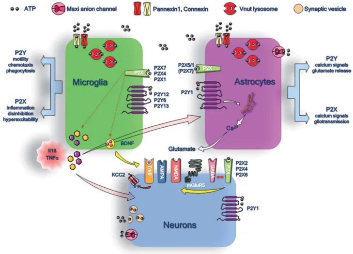
Figure 1 summarizes the main purinergic signaling pathways, and their functions, between neurons, astro-cytes, and microglial cells. Just as with other ligand-gated channels, P2X receptors could contribute to fast synaptic transmission. Consistent with this idea, several studies have demonstrated that P2X receptors can be activated at synapses of different brain regions. However, these excita-tory postsynaptic currents are small and rare and occur in few cells within given brain nuclei/areas (Pankratov et al., 2007; Khakh and North, 2012). Our current understand-ing of P2X signalunderstand-ing in neurons is that these receptors
have neuromodulatory rather than true excitatory proper-ties. Supporting this view, P2X receptors can alter NMDA-dependent synaptic functions either by regulating the membrane insertion of the GluN2B subunit (Baxter et al., 2011) or by regulating NMDA receptor inactiva-tion (Pankratov et al., 2002). Similarly, a recent study showed that P2X receptor activation triggers AMPA receptor internalization and thus can regulate glutamater-gic fast synaptic transmission in the hippocampus (Poug-net et al., 2014). By direct interaction with GABA A receptors, P2X4 receptors can negatively control the gen-eration of inhibitory postsynaptic currents, promoting, although indirectly, network excitability (Jo et al., 2011).
The function of P2X receptors in astrocytes is poorly documented. In the hippocampus, where astro-cytes have been studied the most, fast applications of P2X receptor agonists during whole-cell recordings of astro-cytes in the mouse, rat, and human CA1 region failed to Fig. 1. Purinergic signaling pathways between neurons, astrocytes, and microglialcells. The different
release mechanisms ofATP and the expression of P2X and P2Y receptors are indicated for micro-glia (green), astrocytes (violet), and neurons (blue). The involvement of micromicro-glial P2X receptors in the production of the IL-1b, TNF-a, and BDNF and that of astrocytic P2Y1 receptors in the release of glutamate are highlighted. The putative functions of glial P2X and P2Y receptors are also indicated at right and at left of the microglial cell and of the astrocyte, respectively.
trigger any detectable membrane current that could sup-port the existence of functional P2X receptors in these cells (Jabs et al., 2007), yet, as stated above, heteromeric P2X1/P2X5 receptors have been functionally character-ized in a subset of cortical astrocytes (Lalo et al., 2008); however, the physiological role of this heteromeric recep-tor remains speculative. It is possible that P2X1/5 activa-tion can contribute to astroglial calcium signaling and may represent a fast neuron-to-glia signaling mechanism at the synaptic level. Alternatively, P2X-evoked sodium influx in astrocytes may interfere with the glutamate buf-fering capacity of astrocytes.
The expression of P2X7 in astrocytes is highly con-troversial. Indeed most studies demonstrating functional P2X7-like receptors in astrocytes in vitro or ex vivo rely on the use of pharmacological tools with little specificity. Moreover, a recent study demonstrated that heteromeric P2X2/5 receptors have pharmacological and functional properties close to those of P2X7, including activation by micromolar BzATP concentration, pore dilatation charac-teristics, and induction of pseudoapoptosis, supporting the view that other P2X receptors present the same hallmark of activation as P2X7 (Compan et al., 2012). Neverthe-less, several studies have provided convincing results showing the expression of P2X7 in radial glial cells, such as Muller glia in the retina (Pannicke et al., 2000) or Bergmann cells in the cerebellum (Habbas et al., 2011). Moreover, it has recently been shown in vivo that the induction of P2X7 expression in activated cortical astro-cytes contributes to ischemic tolerance induced by a pre-conditioning sublethal insult (Hirayama et al., 2015). P2X receptor expression in cultured oligodendrocyte precursor cells has been reported; however, these observations have not been reproduced in mature oligodendrocytes in situ (Matute, 2008). P2X7 is potentially expressed in a subset of oligodendroglia (Domercq et al., 2010), yet these stud-ies suffer from the same limitation as described above.
Expression of P2X receptors in microglia is well documented. Microglial cells are brain-resident macro-phages known for their involvement in neuroinflamma-tion (Hanisch and Kettenmann, 2007; Ransohoff and Perry, 2009). Increasing evidence indicates that microglial cells also have numerous homeostatic functions in healthy brain or during development (Kettenmann et al., 2013; Wu et al., 2013; Shemer et al., 2015; Arnoux and Audi-nat, 2015). Microglia behaviors are shaped by the activity of nearby networks, including neurons and astrocytes under both healthy and pathological conditions (Kettenmann et al., 2013). Microglial cells have long and branched processes that present remarkable and unique motility properties, allowing them to scan their environ-ment and establish transient contact with synapses. These processes can also rapidly extend toward a local lesion, and several studies have demonstrated that purinergic sig-naling, particularly through the P2Y12 receptor, is a cen-tral regulator of microglial process motility (Parkhurst and Gan, 2010). Purinergic receptors shape many other func-tions of microglial cells, particularly their ability to inter-act with other brain cell types, and, from this point of
view, they share many characteristics with peripheral macrophages, which are known to express both P2X and P2Y receptors.
In microglia, as in macrophages, the expression of P2X1, P2X4, and P2X7 receptors is clearly established. P2X7 receptors, which are highly expressed in all myeloid cells, display unusual biophysical, pharmacological, and functional properties compared with other P2X receptors (but see Compan et al., 2012); they have a very low sensi-tivity for ATP (EC50 above 1 mM), and their activation
opens a permeation pathway for high-molecular-weight molecules. The main functional characteristic of P2X7 is its ability to activate the inflammasome, leading to procaspase-1 cleavage and interleukin (IL)-1b processing and release. Mice deficient for the P2X7 gene are unable to process and secrete mature IL-1b or IL-18 in response to an inflammatory challenge and display a severe altera-tion of the expression of other proinflammatory cyto-kines, including tumor necrosis factor-a (TNF-a; Solle et al., 2001). P2X7-deficient mice have normal pain sen-sation but do not show any hypersensitivity to mechanical stimuli in models of pathological pain (Chessell et al., 2005). This phenotype likely is due to an alteration of microglial proinflammatory functions in the spinal cord. It is surprising that only a few studies have investigated the cellular mechanisms underlying the contribution of microglial P2X7 receptors in brain pathologies and inflammation. P2X7 receptors are also expressed by microglia under physiological conditions (Avignone et al., 2008), suggesting that these receptors contribute to microglia homeostatic functions.
Unlike P2X7, P2X4 receptors are not expressed in quiescent microglia but are strongly upregulated in acti-vated cells under diverse pathological conditions. After sciatic nerve ligation, a model of neuropathic pain, P2X4 is expressed de novo in activated spinal microglia in which this receptor contributes to a calcium-dependent brain-derived neurotrophic factor (BDNF) release. The latter will, in turn, promote network excitability through downregulation of the chloride–potassium cotransporter KCC2 and GABAergic disinhibition (Beggs et al., 2012). P2X4 has a strong permeability to calcium, equivalent to that of NMDA receptors (Egan and Khakh, 2004). In myeloid cells, P2X4-evoked calcium entry activates phos-pholipase A2 and stimulates the production of arachidonic acid and prostaglandin 2, the latter inducing sensitization of sensory neurons (Ulmann et al., 2010). P2X4 is likely to have a similar function in brain microglia, potentially contributing to neuronal excitability within the CNS. Other microglial functions are regulated by P2X4-evoked calcium influx, as suggested by the fact that microglial cells in P2X4-deficient mice show attenuated activation hallmarks in disease models (Ulmann et al., 2008, 2013).
The role of P2X1 in microglia has not been investi-gated. In macrophage, P2X1-deficient mice have altered neutrophil extravasation and reduced plasma levels of key cytokines and chemokines in an endotoxemia model (Maitre et al., 2015). This phenotype suggests that P2X1
could also contribute to microglial proinflammatory functions.
P2Y Receptors
The functions of P2Y receptors in the CNS are still poorly characterized. As for most other G-protein-coupled receptors, P2Y receptors in neurons appear to have neuromodulatory functions, yet there is only sparse evidence that P2Y receptors endorse important neuronal functions (but see Bowser and Khakh, 2004). By contrast, P2Y receptors are major determinants of glial cell activity. Astrocytes are thought to express several types of P2Y1, P2Y2, and P2Y4 receptors in situ (Verkhratsky et al., 2009), among which P2Y1 has been the most extensively studied. Activation of P2Y1 triggers increases in cytosolic calcium concentration through the phospholipase C path-way, mobilizing calcium from intracellular stores. Varia-tion of cytosolic calcium is an important feature of astrocyte activity through which these cells integrate and respond to external stimuli (Zorec et al., 2012; Bazargani and Attwell, 2016). For instance, P2Y1-mediated calcium signaling in astrocytes leads to changes in the extracellular concentration of transmitters by modulating either the activity of astrocyte transporters (Jacob et al., 2014) or the release of astrocyte transmitters or gliotransmitters, includ-ing glutamate and ATP (Araque et al., 2014). Under pathological conditions, P2Y1 expression is upregulated, and calcium signaling in activated astrocytes is usually increased (Franke et al., 2012). The best-described exam-ple is that of animal models of Alzheimer’s disease. In vivo two-photon imaging showed that intracellular con-centration of calcium and the frequency of calcium transi-ents are increased in astrocytes of cortical areas including b-amyloid plaques (Kuchibhotla et al., 2009) and that inhibiting P2Y1 receptors normalizes calcium signaling (Delekate et al., 2014). These studies of Alzheimer’s mice provide the only reports of intercellular propagation of calcium waves in the astrocyte syncytium in vivo. Cal-cium waves in astrocytes have been observed and studied extensively in cell cultures and acute slice preparations but never in vivo under physiological conditions. P2Y1 plays a pivotal role in initiating or propagating these waves (Giaume et al., 2010).
Several P2Y receptors are expressed by microglial cells (P2Y6, P2Y12, and P2Y13), some at very high levels in resting cells (P2Y12, P2Y13). The expression of these receptors is highly dependent on the activation state of microglia. Under some conditions of activation, the expression of P2Y12 and P2Y13 receptors is strongly downregulated, and that of P2Y6 is increased (Haynes et al., 2006; Koizumi et al., 2007), whereas the expression of all these receptors is upregulated under other patholog-ical conditions (Avignone et al., 2008; Kobayashi et al., 2012). The lack of specific pharmacological tools has pre-cluded the analysis of the functional roles of P2Y13 receptors, and there are few data on its neuronal expres-sion. Its close homolog, P2Y12, however, has received much more attention. In the brain, P2Y12 is expressed
almost exclusively in microglia. Early studies using CX3CR1 1/eGFPreporter mice have demonstrated that P2Y12 is a key regulator of the dynamics of microglial process and chemotaxis (Haynes et al., 2006). P2Y12 appears to be a highly sensitive sensor of extracellular ATP, underlying response of microglial response to neu-ronal excitability or acute pathogenic event. For example, it has been proposed that NMDA receptor activity can lead to a local production of ATP that induces a P2Y12-dependent microglial process outgrowth toward the active neuron (Li et al., 2012; Dissing-Olesen et al., 2014; Eyo et al., 2014).
UDP-sensitive P2Y6 receptors have been shown to regulate the phagocytic activity of microglia (Koizumi et al., 2007); this receptor can also induce the expression of the chemokines CCL2 and CCL3, although the down-stream signaling pathway is still unclear (B. Kim et al., 2011; Morioka et al., 2013). Altogether, recent data dem-onstrate that astroglial and microglial purinergic receptors are central to brain functions under homeostatic and path-ological conditions, yet the role of purinergic receptors expressed by other brain cells should not be neglected.
PURINERGIC SIGNALING IN EPILEPSY
The involvement of purinergic signaling in pathological situations has received much attention. This is due mainly to the common belief that extracellular ATP concentra-tions drastically increase under many pathological condi-tions, including trauma, ischemia, inflammation, periods of intense neuronal activity, and different chronic or neu-rodegenerative pathologies (Franke et al., 2012; Rodrigues et al., 2015). The consequences of this surge in extracellular ATP in pathological states are not fully appreciated. There are several hurdles limiting the charac-terization of the involvement of purinergic signaling in pathological states. First, the different mechanisms leading to ATP release are still poorly characterized. Second, ATP is rapidly metabolized in ADP and adenosine, which in turn activates their specific receptors. This dynamic process of ATP conversion is difficult to apprehend experimentally. Finally, the number of pharmacologically active molecules that can be used in vivo or ex vivo to target specifically the diverse purinergic receptors remains limited, despite some recent improvement (Bhattacharya et al., 2013). The use of genetically modified mice has provided convincing results on the involvement of puri-nergic receptors in diverse pathological models, indicating that some purinergic receptors could represent interesting therapeutic targets.
The involvement of purinergic signaling has been particularly investigated in the field of neuropathic pain and seizure or SE. It turns out that these two models share unexpected similarities and that microglial purinergic receptors play an active role in the development of these pathologies. The mechanisms underlying purinergic con-tribution to chronic pain have been reviewed elsewhere (Beggs et al., 2012; Tsuda et al., 2013).
Increased levels of purines during epileptic seizure have been described in humans, yet this observation has been restricted to adenosine (During and Spencer, 1992). The use of electrode biosensor detecting adenosine in the CA1 area of the hippocampus has revealed a rise in aden-osine levels following brief, electrically evoked electro-graphic seizures. However, there is still no evidence for an increase of ATP levels following seizures in this struc-ture (Lopatar et al., 2011). In the CA3 area, dihydroxy-phenylglycine, a metabotropic glutamate agonist, induces epileptiform activities and concomitant ATP release (Lopatar et al., 2015). In the two hippocampal regions, there is no correlation between the levels of extracellular ATP and adenosine, suggesting that ATP degradation by ectonucleotidase does not significantly contribute to extracellular adenosine concentration. Still, the ATP release pattern might depend on the type of stimulation eliciting epileptic activity. For example, astrocytic release of ATP can be triggered by K 1 -evoked depolarization in brain slices but not by electrical stimulation, which further complicates the interpretation of these studies. If electrode biosensors allow for a direct and dynamic analysis of purine release, this technique has limitations. Indeed, even if the spatial resolution of the measurement is improved compared with the bulk analysis of ATP released in supernatant, it still provides information that may be unrelated to the concentration of ATP in the syn-aptic cleft or at the immediate vicinity of cells. In addition these biosensors rely on the enzymatic conversion of purine that generates peroxide. The kinetics of this con-version may be slower than the actual degradation of ATP by membrane-bound ecto-ATPase. Finally, these biosensors have been used mainly in ex vivo brain slice preparations, in which extracellular ATP levels may have been perturbed during tissue processing.
Independently of whether the release of ATP is enhanced in the epileptic brain, there is general agree-ment that purines contribute to the pathology of epilepsy. Direct application of ATP analogs in the brain is known to evoke neuronal hyperexcitability and to promote sei-zure activity (Knutsen and Murray, 1997). Moreover, ATP released from astrocytes contributes to ictogenesis and epileptogenesis. On the one hand, genetic impair-ment of vesicular ATP release by expressing a dominant-negative soluble NSF attachment protein receptor in astrocytes delays the onset of recurrent seizure, decreases the frequency of these seizures, and reduces hippocampal damage after a pilocarpine-induced SE (Clasadonte et al., 2013). This effect is probably mediated through the con-trol of NMDA receptor expression that is regulated by adenosine A1 receptor activation following ATP release from astrocytes (Fellin et al., 2009; Deng et al., 2011). On the other hand, it has been suggested that ATP release through PANX1 contributes to neuronal hyperexcitabil-ity in seizures because mice with PANX1 deficiency or silencing are less prone to kainate- or pilocarpin-evoked SE (Santiago et al., 2011). Finally, there is a profound remodeling of the expression of hippocampal P2 receptors following SE in rodents (Avignone et al., 2008; Engel
et al., 2016); this will be the focus of the following paragraphs.
P2X Receptors in Epilepsy
Because of their excitatory properties, their activa-tion during episode of high neuronal activity, and their expression in microglial cells, the involvement of iono-tropic P2X receptors in tissue remodeling after the induc-tion of SE has received special atteninduc-tion. After SE, a local inflammatory reaction occurs in brain areas that have been the most active during the seizure episode, and this inflammation persists for days or weeks. There is now strong evidence that this inflammatory response promotes neuronal damage and contributes to long-lasting alteration of local network properties (Devinsky et al., 2013; Marchi et al., 2014). IL-1b and TNF-a are key proinflammatory cytokines, which, in the CNS, can sustain neuronal hyperexcitability, and their involvement in the initiation of seizure and epileptogenesis was proposed several years ago (Vezzani et al., 2008). The fact that P2X7 activity is the main trigger of IL-1b processing and contributes to TNF-a production led to investigating the role of these receptors in epilepsy. In human specimens from patients suffering with focal cortical dysplasia (FCD), P2X7 mRNA and protein expressions were increased vs. con-trol samples. This was accompanied by an increased immunoreactivity against IL-1b in microglia and also in dysmorphic neurons and balloon cells, which are charac-teristic cells of FCD (Wei et al., 2015). The roles of P2X7 in epilepsy have been investigated in different animal models in which the modulation of the receptor activity was performed with different approaches, either pharma-cological or genetic. Opposite results were reported when SE was induced through systemic injection of proconvul-sive agents or through direct intra-amygdala injection of kainate. In the pilocarpine-evoked SE model, P2X7 appeared to have protective properties. Indeed, inhibition of P2X7 activity with either specific antagonists or P2X7-deficient mice increased seizure susceptibility after pilo-carpine injection. P2X7-anticonvulsive effects were linked to PANX1 because siRNA-mediated downregula-tion of PANX1 also reduced the threshold of seizure apparition (Kim and Kang, 2011). It has been suggested that the protective effect of P2X7 receptor in pilocarpine-induced SE could be mediated through TNF-a pathways, at least in the CA3 area (J.E. Kim et al., 2011). In a region-specific manner, P2X7 receptors control the hip-pocampal nonapoptotic astrocyte death after pilocarpine-induced SE (Kim et al., 2015).
On the other hand, P2X7 activation appears to have proconvulsive effects when SE is triggered by an intra-amygdala injection of KA. In this model, P2X7-deficient mice showed reduced electrographic seizure during SE, and pretreatment with P2X7 antagonists (brilliant blue G or A-438079) reduced behavioral seizures, neuronal cell death in the hippocampus, and increase of IL-1b triggered by SE. Treatment with a combination of a classical anti-convulsivant (lorazepam) and A-438079 during the SE
refractory period, a period during which SE cannot be blocked by anticonvulsants alone, efficiently stopped the SE (for review see Jimenez-Mateos et al., 2015). The reg-ulation of P2X7 expression in this model is controlled by micro-RNA-22, which has anticonvulsive effects by inhibiting P2X7 expression (Jimenez-Mateos et al., 2015). Whether this regulation holds true also in humans has not been documented.
The reason for the discrepancies between the two models is not clear. The same antagonists and the same strain of P2X7-deficient mice were used in both cases; however, these two experimental models of SE are very different, and it is possible that, in the pilocarpine model, the peripheral mode of administration involves a P2X7-dependent intermediary step that is not activated when SE is induced by direct intra-amygdala injection of kai-nate. It is worth noting that adhesion of circulating leuko-cytes on endothelial cells of cerebral blood vessels is probably an initial and mandatory step of SE induction in the pilocarpine model (Fabene et al., 2008). A recent study in rats showed that, 3 months after systemic kainate injections, treatment with a new P2X7 antagonist that crosses the blood–brain barrier decreased the severity of spontaneous recurrent seizures (Amhaoul et al., 2016). Although the seizure frequency remained unaltered by this new P2X7 antagonist, this study further strengthens the idea that blocking P2X7 might have beneficial effects in regulating seizure during not only the acute but also the chronic phases of the disease.
P2X7 may thus appear as a potential therapeutic tar-get to reduce the burden of seizures. However, several technical issues with respect to the results discussed in this Review should be kept in mind. First, the antagonists used in these studies are specific for P2X7 receptors com-pared with other P2X receptors or other receptors and channels but may show off-target activity. Such a case was reported recently for a P2X1 antagonist that also inhibits the activity of CCR5 and CXCR4 (Giroud et al., 2015). Second, the tissue and cellular distribution of P2X7 is still unclear; antibodies and mouse reporter gene lack specificity or can reasonably be questioned. With respect to the Gensat P2X7-GFP reporter mouse (http:// www.gensat.org/imagenavigator.jsp?imageID=36838), there are strong discrepancies between the pattern of expression of GFP in this mouse strain and mRNA tissue distribution of the receptor that can be visualized in the Allen brain atlas in situ hybridization database (http:// mouse.brain-map.org/gene/show/18206). Moreover, it is surprising that no expression of GFP in microglial cells can be observed in this mouse because there are clear demonstrations that P2X7 is functionally expressed in microglial cells under physiological conditions (see above). Finally, whether P2X7 is efficiently deleted in the two commonly used P2X7-deficient mice is still debated (Taylor et al., 2009). For all these reasons, further work is required to establish unambiguously whether P2X7 is a viable therapeutic target in epilepsy. Particularly, investi-gating the involvement of P2X7 in seizure activity and chronic epilepsy in conditional knockout mice will be an
essential step. There is a common agreement that the expression of P2X7 is enhanced following SE in the hip-pocampus. Whether this expression is restricted to micro-glia, as functionally demonstrated (Avignone et al., 2008),
or is also present in neurons and astrocytes requires a clear examination. Finally, it is important to note that all
clini-cal trials testing the effectiveness of P2X7 antagonists have failed to demonstrate any beneficial effect in rheumatoid arthritis (Keystone et al., 2012), despite the clear demon-stration of involvement of this receptor in animal models of the disease.
P2X4 is the only other P2X receptor that has been studied for its potential involvement in epilepsy. As for P2X7, there are important discrepancies in the literature, mainly regarding whether P2X4 expression is up- or downregulated in the hippocampus following SE. In mice, two studies showed no change or a decrease of P2X4 immunostaining within 24 hr post-SE, evoked either by systemic pilocarpine or by intra-amygdala kai-nate injection (Dona et al., 2009; Engel et al., 2012). A similar finding was also reported in seizure-sensitive com-pared with seizure-resistant gerbil hippocampus (Kang et al., 2003). However, no specificity for the P2X4 anti-bodies used in any of these studies was provided. Another study using intraperitoneal injection of kainate to elicit SE demonstrated an increase of the transcription and protein expression in the hippocampus 24 and 48 hr post-SE (Ulmann et al., 2013). Particularly, upregulated P2X4 receptors were localized in activated microglial cells in the stratum radiatum. In this study, the specificity of P2X4 antibody staining was confirmed in P2X4-deficient mice. Furthermore, Western blot experiments performed on purified hippocampal microglia clearly demonstrated a de novo expression of P2X4 in microglia 48 hr after the induction of SE. Involvement of P2X4 in epileptogenesis was also investigated with the P2X4-deficient mice. The data show a reduction of distinct hallmarks of microglial activation after the induction of SE in the P2X4-deficient mice, including a reduction of cell density and morpho-logical changes. Analysis of microglia electrophysiomorpho-logical properties in acute hippocampal slices revealed that the upregulation of Kv1.3-mediated outward K 1 currents normally occurring in activated microglia of wild-type mice was reduced in epileptic P2X4-deficient mice, yet the functional upregulation of P2 receptors (i.e., P2X7, P2Y12, and P2Y6) was not altered by the deletion of P2X4. In addition, a reduction in the expression of differ-ent proinflammatory genes and a lower neuronal cell death in the CAI area were also reported. Although SE was not analyzed by EEG recordings in P2X4-deficient mice, no difference of susceptibility to kainate-induced SE assessed by scoring behavioral seizures was observed between wild-type and P2X4-deficient mice. Whether P2X4-deficient mice can develop spontaneous recurrent seizure has not been determined. Thus, even though both microglial activation and neuronal damage are signifi-cantly attenuated by the inactivation of this receptor, the role of P2X4 must be further substantiated by EEG recordings during acute and chronic phases of epilepsy. In
addition, P2X4 conditional allele deletion to inactivate P2X4 in either neurons or microglia should also be per-formed. Indeed, P2X4 is expressed in hippocampal neu-rons, where its activation is triggered by episodes of high neuronal activity (Sim et al., 2006) and could lead to hyperexcitability, which in turn could promote microglial activation or neuronal death. On the other hand, as docu-mented in the neuropathic pain model (Beggs et al., 2012), de novo expression of P2X4 in microglia after SE could control the release of BDNF and KCC2 downreg-ulation, thereby inducing local hyperexcitability and pro-moting epileptogenesis. Clearly, there is a crucial requirement for specific P2X4 antagonists and for more targeted genetic approaches to clarify the actual roles of P2X4 in epilepsy.
P2Y Receptors in Epilepsy
The role of P2Y receptors in epilepsy has received little attention. From a purely neuronal point of view, there is little evidence supporting a role for these recep-tors in mediating hyperexcitability or epileptogenesis. However, functional excitatory P2Y1 receptors are expressed by subpopulations of inhibitory interneurons in the hippocampus (Bowser and Khakh, 2004). Some of these interneurons degenerate after SE (Dinocourt et al., 2003), and this could lead to a modification of the P2Y1 regulation of the excitation/inhibition balance in the hip-pocampal neuronal network.
The expression of P2Y receptors in glia and their remodeling under pathological conditions suggest that these receptors may contribute to phenotypic changes taking place after seizures. Supporting this idea, P2Y12 receptors are functionally upregulated in microglia after SE, and this leads to increased process motility toward a source of ATP (Avignone et al., 2008). Other aspects of microglia process motility (e.g., basal motility or process extension toward a laser-induced lesion) are not affected in this model (Avignone et al., 2015). These studies were performed with either local application of agonists or laser-evoked microlesion in acute slices after SE induction in vivo. Eyo and coworkers (2014) also demonstrated, in vivo, that P2Y12 receptor-mediated elongation of micro-glia processes toward active dendrites are enhanced after SE. This phenomenon was impaired in P2Y12-deficient mice, and, remarkably, these mice showed a worsening of kainate-induced seizures, suggesting that microglial P2Y12 signaling has a beneficial effect on the outcome of the disease.
Microglial P2Y6 receptors are also upregulated after SE (Avignone et al., 2008), but the consequences of this upregulation on epilepto- and ictogenesis have not been evaluated. These receptors control the phagocytic func-tion of microglial cells (Koizumi et al., 2007), so it is important to test whether P2Y6 contributes to the phago-cytosis of hippocampal neurons after SE as well as to the regulation of adult neurogenesis in the dentate gyrus (Sierra et al., 2010), which is also affected after seizures (Sierra et al., 2015).
Astrocytic purinergic signaling in epilepsy has received little attention despite the fact that P2Y1
recep-tors control calcium signaling in astrocytes and contribute to glutamate-mediated gliotransmission (see above). In
the hippocampus, the astrocytic release of glutamate fol-lowing P2Y1 activation is controlled by TNF-a and pros-taglandins (Domercq et al., 2006). Accordingly, this form of gliotransmission is enhanced in the inflammatory con-text of a mouse model of multiple sclerosis and leads to alterations of synaptic functions and impairment of con-textual learning and memory (Habbas et al., 2015).
TNF-a TNF-and prostTNF-aglTNF-andin signTNF-aling TNF-are rTNF-apidly upregulTNF-ated TNF-after SE (Avignone et al., 2008), so it is very likely that altered gliotransmission will contribute to the remodeling of the hippocampus during epileptogenesis and to the generation of seizures. In an in vitro model of focal seizures, calcium elevation in astrocytes, partially dependent on P2 recep-tors, contributes to drive neurons to the threshold of ictal but not interictal events (Gomez-Gonzalo et al., 2010).
Moreover, calcium signaling is enhanced in cortical astro-cytes in vivo during and up to 3 days after pilocarpine-induced SE (Ding et al., 2007). Unfortunately, despite the fact that this enhanced calcium signaling favors the release of astrocytic glutamate and SE-induced neuronal death, the contribution of P2Y receptors has not been tested. A more recent study on a model of kindling in the rat also indicated enhanced calcium signaling in astrocytes that depends on P2Y1 receptor activation (Alvarez-Ferradas et al., 2015). In agreement with the pilocarpine model, processes downstream of the kindling-evoked calcium signaling include mGluR5-dependent increase of presyn-aptic glutamate release probability and NMDA receptor-dependent enhanced neuronal excitability. ATP released by microglia in hippocampal slices of na€ıve animalscan rapidly mobilize this signaling cascade through the activa-tion of astrocyte P2Y1 receptors, the release of astrocyte glutamate, and the activation of mGluR5 receptors on presynaptic glutamatergic terminals, leading to an enhancement of excitability and favoring the appearance of epileptiform activities (Pascual et al., 2012). These results support the hypothesis of an active role of astro-cytic P2Y1 receptors in epileptogenesis. However, spe-cific inhibition or invalidation at specific stages of the disease is still required to establish more clearly the actual impact of this enhanced P2Y1 signaling. Moreover, a careful examination of the involvement of other P2Y receptors that may be upregulated in reactive astrocytes is required.
CONCLUSIONS AND PERSPECTIVES
Increasing evidence indicates that this glial reactivity is actively involved in remodeling of the epileptic brain (Matute and Cavaliere, 2011; Steinhauser and Boison, 2012; Devinsky et al., 2013; Heuser et al., 2014) and that ATP is a danger signal in many disorders, including epi-lepsy (Rodrigues et al., 2015). Here, we have reviewed several modifications of purinergic signaling pathways that have been observed in animal models of epilepsy, and we
believe that, as fundamental components of neuron–glia interactions, purinergic receptors represent promising therapeutic targets to cure or to control the disease better.
Unlike glutamate and GABA receptors, purinergic
recep-tors are not expressed by all neurons of the brain. Some of these receptors are expressed almost exclusively by astrocytes or microglia and control several key functions of these glial cells. Moreover, some of these receptors are
selectively upregulated in glial cells after these cells are activated under pathological conditions. This is the case, for instance, for P2X4 in microglia after SE (Ulmann et al., 2013). However, the repertoire of purinergic receptors expressed by astrocytes and by microglia at
dif-ferent stages of the disease is still largely unknown and should be carefully determined. Functional studies to establish the roles of these receptors will certainly be an important step, but the lack of specific pharmacological tools will be an obstacle. An initial transcriptome analysis to identify the set of genes up- or downregulated in
astro-cytes and in microglia at different stages of the disease appears to be essential before designing genetic and func-tional strategies to understand better the specific roles of purinergic receptors during the time course of reactive gliosis occurring in epilepsy.
ACKNOWLEDGMENTS
The authors thank all their team members who have con-tributed to the study of purinergic signaling pathways in microglia and astrocytes over the past 10 years.
CONFLICT OF INTEREST STATEMENT The authors have no conflicts of interest to declare.
ROLE OF AUTHORS
FR and EA contributed equally to the writing of this Review.
REFERENCES
Alvarez-Ferradas C, Morales JC, Wellmann M, Nualart F, Roncagliolo M, Fuenzalida M, Bonansco C. 2015. Enhanced astroglial Ca21 signal-ing increases excitatory synaptic strength in the epileptic brain. Glia 63: 1507–1521.
Amhaoul H, Ali I, Mola M, Van EA, Szewczyk K, Missault S, Bielen K, Kumar-Singh S, Rech J, Lord B, Ceusters M, Bhattacharya A, Dedeurwaerdere S. 2016. P2X7 receptor antagonism reduces the sever-ity of spontaneous seizures in a chronic model of temporal lobe epi-lepsy. Neuropharmacology 105:175–185.
Araque A, Carmignoto G, Haydon PG, Oliet SH, Robitaille R, Volterra A. 2014. Gliotransmitters travel in time and space. Neuron 81:728–739. Arnoux I, Audinat E. 2015. Fractalkine signaling and microglia functions
in the developing brain. Neural Plast 2015:689404.
Avignone E, Ulmann L, Levavasseur F, Rassendren F, Audinat E. 2008. Status epilepticus induces a particular microglial activation state charac-terized by enhanced purinergic signaling. J Neurosci 28:9133–9144. Avignone E, Lepleux M, Angibaud J, Nagerl UV. 2015. Altered
mor-phological dynamics of activated microglia after induction of status epi-lepticus. J Neuroinflammation 12:202.
Avsar E, Empson RM. 2004. Adenosine acting via A1 receptors controls the transition to status epilepticus-like behaviour in an in vitro model of epilepsy. Neuropharmacology 47:427–437.
Bao L, Locovei S, Dahl G. 2004. Pannexin membrane channels are mechanosensitive conduits for ATP. FEBS Lett 572:65–68.
Baulac M, Pitkanen A. 2008. Research priorities in epilepsy for the next decade: a representative view of the European scientific community. Epilepsia 50:571–578.
Baxter AW, Choi SJ,Sim JA, North RA. 2011. Role of P2X4 receptors in synaptic strengthening in mouse CA1 hippocampal neurons. Eur J Neurosci 34:213–220.
Bazargani N, Attwell D. 2016. Astrocyte calcium signaling: the third wave. Nat Neurosci 19:182–189.
Beckel JM, Argall AJ, Lim JC, Xia J, Lu W, Coffey EE, Macarak EJ, Shahidullah M, Delamere NA, Zode GS, Sheffield VC, Shestopalov VI, Laties AM, Mitchell CH. 2014. Mechanosensitive release ofadeno-sine 50-triphosphate through pannexin channels and mechanosensitive upregulation of pannexin channels in optic nerve head astrocytes: a mechanism for purinergic involvement in chronic strain. Glia 62:1486– 1501.
Bedner P, Dupper A, Huttmann K, Muller J, Herde MK, Dublin P, Deshpande T, Schramm J, Haussler U, Haas CA, Henneberger C, Theis M, Steinhauser C. 2015. Astrocyte uncoupling as a cause of human temporal lobe epilepsy. Brain 138:1208–1222.
BeggsS, Trang T, Salter MW. 2012. P2X4R 1 microglia drive neuro-pathic pain. Nat Neurosci 15:1068–1073.
Bergles DE, Richardson WD. 2015. Oligodendrocyte development and plasticity. Cold Spring Harbor Perspect Biol 8.
Bhattacharya A,Wang Q, Ao H, Shoblock JR, Lord B, Aluisio L, Fraser I, Nepomuceno D, Neff RA, Welty N, Lovenberg TW, Bonaventure P, Wickenden AD, Letavic MA. 2013. Pharmacological characterization of a novel centrally permeable P2X7 receptor antagonist: JNJ-47965567. Br J Pharmacol170:624–640.
Bo X, Jiang LH, Wilson HL, Kim M, Burnstock G, Surprenant A, North RA. 2003. Pharmacological and biophysical properties of the human P2X5 receptor. Mol Pharmacol63:1407–1416.
Boison D. 2015. Adenosinergic signaling in epilepsy. Neuropharmacology [E-pub ahead of print] doi: 10.1016/j.neuropharm.2015.08.046 Bowser DN, Khakh BS. 2004. ATP excites interneurons and astrocytes
to increase synaptic inhibition in neuronal networks. J Neurosci 24: 8606–8620.
Bowser DN, Khakh BS. 2007. Vesicular ATP is the predominant cause of intercellular calcium waves in astrocytes. J Gen Physiol129:485–491. Burnstock G. 2006a. Historical review: ATP as a neurotransmitter.
Trends PharmacolSci 27:166–176.
Burnstock G. 2006b. Pathophysiology and therapeutic potential of puri-nergic signaling. PharmacolRev 58:58–86.
Butt AM. 2011. ATP: a ubiquitous gliotransmitter integrating neuron– glial networks. Semin Cell Dev Biol 22:205–213.
ChessellIP, Hatcher JP, Bountra C, Michel AD, Hughes JP, Green P, Egerton J, Murfin M, Richardson J, Peck WL, GrahamesCB, Casula MA, Yiangou Y, Birch R, Anand P, Buell GN. 2005. Disruption of the P2X7 purinoceptor gene abolishes chronic inflammatory and neu-ropathic pain. Pain 114:386–396.
Clasadonte J,Dong J, Hines DJ, Haydon PG. 2013. Astrocyte control of synaptic NMDA receptors contributes to the progressive development of temporal lobe epilepsy. Proc Natl Acad Sci U S A 110:17540– 17545.
Collo G, North RA, Kawashima E, Merlo-Pich E, Neidhart S, Surprenant A, Buell G. 1996. Cloning OF P2X5 and P2X6 receptors and the distribution and properties of an extended family of ATP-gated ion channels. J Neurosci 16:2495–2507.
Compan V, Ulmann L, Stelmashenko O, Chemin J, Chaumont S, Rassendren F. 2012. P2X2 and P2X5 subunits define a new hetero-meric receptor with P2X7-like properties. J Neurosci 32:4284–4296. De Simoni MG, Perego C, Ravizza T, Moneta D, Conti M, Marchesi
and related genes are induced in the rat hippocampus by limbic status epilepticus. Eur J Neurosci 12:2623–2633.
Delekate A, Fuchtemeier M, Schumacher T, Ulbrich C, Foddis M, Petzold GC. 2014. Metabotropic P2Y1 receptor signalling mediates astrocytic hyperactivity in vivo in an Alzheimer’s disease mouse model. Nat Commun 5:5422.
Deng Q, Terunuma M, Fellin T, Moss SJ, Haydon PG. 2011. Astrocytic activation of A1 receptors regulates the surface expression of NMDA receptors through a Src kinase dependent pathway. Glia 59:1084–1093. Devinsky O, Vezzani A, Najjar S, De Lanerolle NC, Rogawski MA.
2013. Glia and epilepsy: excitability and inflammation. Trends Neurosci 36:174–184.
Ding S, Fellin T, Zhu Y, Lee SY, Auberson YP, Meaney DF, Coulter DA, Carmignoto G, Haydon PG. 2007. Enhanced astrocytic Ca 21 sig-nals contribute to neuronal excitotoxicity after status epilepticus. J Neurosci 27:10674–10684.
Dinocourt C, Petanjek Z, Freund TF, Ben Ari Y, Esclapez M. 2003. Loss of interneurons innervating pyramidal cell dendrites and axon ini-tial segments in the CA1 region of the hippocampus following pilocarpine-induced seizures. J Comp Neurol 459:407–425.
Dissing-Olesen L, LeDue JM, Rungta RL, Hefendehl JK, Choi HB, MacVicar BA. 2014. Activation of neuronal NMDA receptors triggers transient ATP-mediated microglial process outgrowth. J Neurosci 34: 10511–10527.
Domercq M, Brambilla L, Pilati E, Marchaland J, Volterra A, Bezzi P. 2006. P2Y1 receptor-evoked glutamate exocytosis from astrocytes: con-trol by tumor necrosis factor-alpha and prostaglandins. J Biol Chem 281:30684–30696.
Domercq M, Perez-Samartin A, Aparicio D, Alberdi E, Pampliega O, Matute C. 2010. P2X7 receptors mediate ischemic damage to oligoden-drocytes. Glia 58:730–740.
Dona F, Ulrich H, Persike DS, Conceicao IM, Blini JP, Cavalheiro EA, Fernandes MJ. 2009. Alteration of purinergic P2X4 and P2X7 receptor expression in rats with temporal-lobe epilepsy induced by pilocarpine. Epilepsy Res 83:157–167.
During MJ, Spencer DD. 1992. Adenosine: a potential mediator of sei-zure arrest and postictal refractoriness. Ann Neurol 32:618–624. Egan TM, Khakh BS. 2004. Contribution of calcium ions to P2X
chan-nel responses. J Neurosci 24:3413–3420.
Engel T, Jimenez-Pacheco A, Miras-Portugal MT, Diaz-Hernandez M, Henshall DC. 2012. P2X7 receptor in epilepsy; role in pathophysiology and potential targeting for seizure control. Int J Physiol Pathophysiol Pharmacol 4:174–187.
Engel T, Alves M, Sheedy C, Henshall DC. 2016. ATPergic signalling during seizures and epilepsy. Neuropharmacology. 104:140–153. Etherington LA, Frenguelli BG. 2004. Endogenous adenosine modulates
epileptiform activity in rat hippocampus in a receptor subtype-dependent manner. Eur J Neurosci 19:2539–2550.
Eyo UB, Peng J, Swiatkowski P, Mukherjee A, Bispo A, Wu LJ. 2014. Neuronal hyperactivity recruits microglial processes via neuronal NMDA receptors and microglial P2Y12 receptors after status epilepti-cus. J Neurosci 34:10528–10540.
Fabene PF, Navarro MG, Martinello M, Rossi B, Merigo F, Ottoboni L, Bach S, Angiari S, Benati D, Chakir A, Zanetti L, Schio F, Osculati A, Marzola P, Nicolato E, Homeister JW, Xia L, Lowe JB, McEver RP, Osculati F, Sbarbati A, Butcher EC, Constantin G. 2008. A role for leukocyte-endothelial adhesion mechanisms in epilepsy. Nat Med 14: 1377–1383.
Fellin T, Halassa MM, Terunuma M, Succol F, Takano H, Frank M, Moss SJ, Haydon PG. 2009. Endogenous nonneuronal modulators of synaptic transmission control cortical slow oscillations in vivo. Proc Natl Acad Sci U S A 106:15037–15042.
Fields RD. 2011a. Nonsynaptic and nonvesicular ATP release from neu-rons and relevance to neuron–glia signaling. Semin Cell Dev Biol 22: 214–219.
Fields RD. 2011b. Signaling by neuronal swelling. Sci Signal 4:tr1. Franke H, Verkhratsky A, Burnstock G, Illes P. 2012. Pathophysiology
of astroglial purinergic signalling. Purinergic Signal 8:629–657.
Giaume C, Koulakoff A, Roux L, Holcman D, Rouach N. 2010. Astro-glial networks: a step further in neuroAstro-glial and gliovascular interactions. Nat Rev Neurosci 11:87–99.
Giroud C, Marin M, Hammonds J, Spearman P, Melikyan GB. 2015. P2X1 receptor antagonists inhibit HIV-1 fusion by blocking virus–core-ceptor interactions. J Virol 89:9368–9382.
Gomez-Gonzalo M, Losi G, Chiavegato A, Zonta M, Cammarota M, Brondi M, Vetri F, Uva L, Pozzan T, de CM, Ratto GM, Carmignoto G. 2010. An excitatory loop with astrocytes contributes to drive neu-rons to seizure threshold. PLoS Biol 8:e1000352.
Habbas S, Ango F, Daniel H, Galante M. 2011. Purinergic signaling in the cerebellum: Bergmann glial cells express functional ionotropic P2X7 receptors. Glia 59:1800–1812.
Habbas S, Santello M, Becker D, Stubbe H, Zappia G, Liaudet N, Klaus FR, Kollias G, Fontana A, Pryce CR, Suter T, Volterra A. 2015. Neu-roinflammatory TNFa impairs memory via astrocyte signaling. Cell 163:1730–1741.
Hamilton N, Vayro S, Kirchhoff F, Verkhratsky A, Robbins J, Gorecki DC, Butt AM. 2008. Mechanisms of ATP- and glutamate-mediated calcium signaling in white matter astrocytes. Glia 56:734–749.
Hanisch UK, Kettenmann H. 2007. Microglia: active sensor and versatile effec-tor cells in the normal and pathologic brain. Nat Neurosci 10:1387–1394. Hanstein R, Negoro H, Patel NK, Charollais A, Meda P, Spray DC,
Suadicani SO, Scemes E. 2013. Promises and pitfalls of a Pannexin1 transgenic mouse line. Front Pharmacol 4:61.
Haynes SE, Hollopeter G, Yang G, Kurpius D, Dailey ME, Gan WB, Julius D. 2006. The P2Y12 receptor regulates microglial activation by extracellular nucleotides. Nat Neurosci 9:1512–1519.
Hesdorffer DC, Logroscino G, Benn EK, Katri N, Cascino G, Hauser WA. 2011. Estimating risk for developing epilepsy: a population-based study in Rochester, Minnesota. Neurology 76:23–27.
Heuser K, Szokol K, Tauboll E. 2014. [The role of glial cells in epi-lepsy]. Tidsskr Nor Laegeforen 134:37–41.
Hirayama Y, Ikeda-Matsuo Y, Notomi S, Enaida H, Kinouchi H, Koizumi S. 2015. Astrocyte-mediated ischemic tolerance. J Neurosci 35:3794–3805.
Holton P. 1959. The liberation of adenosine triphosphate on antidromic stimulation of sensory nerves. J Physiol 145:494–504.
Huber A, Padrun V, Deglon N, Aebischer P, Mohler H, Boison D. 2001. Grafts of adenosine-releasing cells suppress seizures in kindling epilepsy. Proc Natl Acad Sci U S A 98:7611–7616.
Imura Y, Morizawa Y, Komatsu R, Shibata K, Shinozaki Y, Kasai H, Moriishi K, Moriyama Y, Koizumi S. 2013. Microglia release ATP by exocytosis. Glia 61:1320–1330.
Islam MR, Uramoto H, Okada T, Sabirov RZ, Okada Y. 2012. Maxian-ion channel and pannexin 1 hemichannel constitute separate pathways for swelling-induced ATP release in murine L929 fibrosarcoma cells. Am J Physiol Cell Physiol 303:C924–C935.
Jabs R, Matthias K, Grote A, Grauer M, Seifert G, Steinhauser C. 2007. Lack of P2X receptor mediated currents in astrocytes and GluR type glial cells of the hippocampal CA1 region. Glia 55:1648–1655.
Jacob PF, Vaz SH, Ribeiro JA, Sebastiao AM. 2014. P2Y1 receptor inhibits GABA transport through a calcium signalling-dependent mech-anism in rat cortical astrocytes. Glia 62:1211–1226.
Jimenez-Mateos EM, Arribas-Blazquez M, Sanz-Rodriguez A, Concannon C, Olivos-Ore LA, Reschke CR, Mooney CM, Mooney C, Lugara E, Morgan J, Langa E, Jimenez-Pacheco A, Silva LF, Mesuret G, Boison D, Miras-Portugal MT, Letavic M, Artalejo AR,
Bhattacharya A, Diaz-Hernandez M, Henshall DC, Engel T. 2015. microRNA targeting of the P2X7 purinoceptor opposes a contralateral epileptogenic focus in the hippocampus. Sci Rep 5:17486.
Jo YH, Donier E, Martinez A, Garret M, Toulme E, Boue-Grabot E. 2011. Cross-talk between P2X4 and gamma-aminobutyric acid type A receptors determines synaptic efficacy at a central synapse. J Biol Chem 286:19993–20004.
Kang TC, An SJ, Park SK, Hwang IK, Won MH. 2003. P2X2 and P2X4 receptor expression is regulated by a GABA A receptor-mediated mechanism in the gerbil hippocampus. Brain Res Mol Brain Res 116: 168–175.
Kettenmann H, Hanisch UK, Noda M, Verkhratsky A. 2011. Physiology of microglia. Physiol Rev 91:461–553.
Kettenmann H, Kirchhoff F, Verkhratsky A. 2013. Microglia: new roles for the synaptic stripper. Neuron 77:10–18.
Keystone EC, Wang MM, Layton M, Hollis S, McInnes IB. 2012. Clini-cal evaluation of the efficacy of the P2X7 purinergic receptor antagonist AZD9056 on the signs and symptoms of rheumatoid arthritis in patients with active disease despite treatment with methotrexate or sulphasala-zine. Ann Rheum Dis 71:1630–1635.
Khakh BS, North RA. 2012. Neuromodulation by extracellular ATP and P2X receptors in the CNS. Neuron 76:51–69.
Kim B, Jeong HK, Kim JH, Lee SY, Jou I, Joe EH. 2011. Uridine 5 0-diphosphate induces chemokine expression in microglia and astrocytes through activation of the P2Y6 receptor. J Immunol 186:3701–3709. Kim JE, Kang TC. 2011. The P2X7 receptor-pannexin-1 complex
decreases muscarinic acetylcholine receptor-mediated seizure susceptibil-ity in mice. J Clin Invest 121:2037–2047.
Kim JE, Ryu HJ, Kang TC. 2011. P2X7 receptor activation ameliorates CA3 neuronal damage via a tumor necrosis factor-alpha-mediated path-way in the rat hippocampus following status epilepticus. J Neuroinflammation 8:62.
Kim JY, Ko AR, Kim JE. 2015. P2X7 receptor-mediated PARP1 activ-ity regulates astroglial death in the rat hippocampus following status epi-lepticus. Front Cell Neurosci 9:352.
Knutsen LJS, Murray TF. 1997. Adenosine and ATP in epilepsy. In: Jacobson KA, Jarvis MF (eds) Purinergic appraoches in experimental therapeutics. Wiley-Lyss, New York, pp 423–447.
Kobayashi K, Yamanaka H, Yanamoto F, Okubo M, Noguchi K. 2012. Multiple P2Y subtypes in spinal microglia are involved in neuropathic pain after peripheral nerve injury. Glia 60:1529–1539.
Koizumi S, Shigemoto-Mogami Y, Nasu-Tada K, Shinozaki Y, Ohsawa K, Tsuda M, Joshi BV, Jacobson KA, Kohsaka S, Inoue K. 2007. UDP acting at P2Y6 receptors is a mediator of microglial phagocytosis. Nature 446:1091–1095.
Kuchibhotla KV, Lattarulo CR, Hyman BT, Bacskai BJ. 2009. Synchro-nous hyperactivity and intercellular calcium waves in astrocytes in Alz-heimer mice. Science 323:1211–1215.
Lalo U, Pankratov Y, Wichert SP, Rossner MJ, North RA, Kirchhoff F, Verkhratsky A. 2008. P2X1 and P2X5 subunits form the functional P2X receptor in mouse cortical astrocytes. J Neurosci 28:5473–5480. Lazarowski ER. 2012. Vesicular and conductive mechanisms of
nucleo-tide release. Purinergic Signal 8:359–373.
Li Y, Du XF, Liu CS, Wen ZL, Du JL. 2012. Reciprocal regulation between resting microglial dynamics and neuronal activity in vivo. Dev Cell 23:1189–1202.
Liu HT, Toychiev AH, Takahashi N, Sabirov RZ, Okada Y. 2008. Maxianion channel as a candidate pathway for osmosensitive ATP release from mouse astrocytes in primary culture. Cell Res 18:558–565. Lohman AW, Isakson BE. 2014. Differentiating connexin hemichannels
and pannexin channels in cellular ATP release. FEBS Lett 588:1379– 1388.
Lopatar J, Dale N, Frenguelli BG. 2011. Minor contribution of ATP P2 receptors to electrically-evoked electrographic seizure activity in
hippo-campal slices: evidence from purine biosensors and P2 receptor agonists and antagonists. Neuropharmacology 61:25–34.
Lopatar J, Dale N, Frenguelli BG. 2015. Pannexin-1-mediated ATP release from area CA3 drives mGlu5-dependent neuronal oscillations. Neuropharmacology 93:219–228.
Maitre B, Magnenat S, Heim V, Ravanat C, Evans RJ, de la SH, Gachet C, Hechler B. 2015. The P2X1 receptor is required for neutrophil extravasation during lipopolysaccharide-induced lethal endotoxemia in mice. J Immunol 194:739–749.
Maldonado PP, Angulo MC. 2015. Multiple modes of communication between neurons and oligodendrocyte precursor cells. Neuroscientist 21:266–276.
Marchi N, Granata T, Janigro D. 2014. Inflammatory pathways of seizure disorders. Trends Neurosci 37:55–65.
Matute C. 2008. P2X7 receptors in oligodendrocytes: a novel target for neuroprotection. Mol Neurobiol 38:123–128.
Matute C, Cavaliere F. 2011. Neuroglial interactions mediated by puri-nergic signalling in the pathophysiology of CNS disorders. Semin Cell Dev Biol 22:252–259.
Morioka N, Tokuhara M, Harano S, Nakamura Y, Hisaoka-Nakashima K, Nakata Y. 2013. The activation of P2Y6 receptor in cultured spinal microglia induces the production of CCL2 through the MAP kinases-NF-kappaB pathway. Neuropharmacology 75:116–125.
Noebels JL, Rogawski M, Olsen RW, Delgado-Escueta AV. 2012. The next decade of research in the basic mechanisms of the epilepsies. In: Noebels JL, Avoli M, Rogawski M, Olsen RW, Delgado-Escueta AV, editors. Jasper’s basic mechanisms of the epilepsies. Bethesda, MD: Oxford University Press. p 3–11.
Orellana JA, Stehberg J. 2014. Hemichannels: new roles in astroglial function. Front Physiol 5:193.
Oya M, Kitaguchi T, Yanagihara Y, Numano R, Kakeyama M, Ikematsu K, Tsuboi T. 2013. Vesicular nucleotide transporter is involved in ATP storage of secretory lysosomes in astrocytes. Biochem Biophys Res Commun 438:145–151.
Pankratov YV, Lalo UV, Krishtal OA. 2002. Role for P2X receptors in long-term potentiation. J Neurosci 22:8363–8369.
Pankratov Y, Lalo U, Verkhratsky A, North RA. 2007. Quantal release of ATP in mouse cortex. J Gen Physiol 129:257–265.
Pannicke T, Fischer W, Biedermann B, Schadlich H, Grosche J, Faude F, Wiedemann P, Allgaier C, Illes P, Burnstock G, Reichenbach A. 2000. P2X7 receptors in Muller glial cells from the human retina. J Neurosci 20:5965–5972.
Parkhurst CN, Gan WB. 2010. Microglia dynamics and function in the CNS. Curr Opin Neurobiol 20:595–600.
Pascual O, Ben AS, Rostaing P, Triller A, Bessis A. 2012. Microglia acti-vation triggers astrocyte-mediated modulation of excitatory neurotrans-mission. Proc Natl Acad Sci U S A 109:E197–E205.
Pekny M, Pekna M, Messing A, Steinhauser C, Lee JM, Parpura V, Hol EM, Sofroniew MV, Verkhratsky A. 2016. Astrocytes: a central ele-ment in neurological diseases. Acta Neuropathol 131:323–345.
Pelegrin P, Surprenant A. 2006. Pannexin-1 mediates large pore forma-tion and interleukin-1beta release by the ATP-gated P2X7 receptor. EMBO J 25:5071–5082.
Perea G, Navarrete M, Araque A. 2009. Tripartite synapses: astrocytes process and control synaptic information. Trends Neurosci 32:421–431. Pougnet JT, Toulme E, Martinez A, Choquet D, Hosy E, Boue-Grabot
E. 2014. ATP P2X receptors downregulate AMPA receptor trafficking and postsynaptic efficacy in hippocampal neurons. Neuron 83:417–430. Ransohoff RM, Perry VH. 2009. Microglial physiology: unique stimuli,
specialized responses. Annu Rev Immunol 27:119–145.
Rodrigues RJ, Tome AR, Cunha RA. 2015. ATP as a multitarget dan-ger signal in the brain. Front Neurosci 9:148.