Trajectory of coronary motion and its significance
in robotic motion cancellation
q,qq
Philippe Cattin
a,*
,1, Hitendu Dave
b,1, Ju¨rg Gru¨nenfelder
b,
Gabor Szekely
a, Marko Turina
b, Gregor Zu¨nd
baComputer Vision Laboratory, Swiss Federal Institute of Technology, ETH-Zurich, Gloriastrasse 35, 8092 Zurich, Switzerland b
Clinic Cardiovascular Surgery, University Hospital Zurich, Raemistrasse 100, 8091 Zurich, Switzerland Received 10 October 2003; received in revised form 5 January 2004; accepted 7 January 2004
Abstract
Objectives: To characterize remaining coronary artery motion of beating pig hearts after stabilization with an ‘Octopus’ using an optical remote analysis technique. Methods: Three pigs (40, 60 and 65 kg) underwent full sternotomy after receiving general anesthesia. An 8-bit high speed black and white video camera (50 frames/s) coupled with a laser sensor (60 mm resolution) were used to capture heart wall motion in all three dimensions. Dopamine infusion was used to deliberately modulate cardiac contractility. Synchronized ECG, blood pressure, airway pressure and video data of the region around the first branching point of the left anterior descending (LAD) coronary artery after Octopus stabilization were captured for stretches of 8 s each. Several sequences of the same region were captured over a period of several minutes. Computerized off-line analysis allowed us to perform minute characterization of the heart wall motion. Results: The movement of the points of interest on the LAD ranged from 0.22 to 0.81 mm in the lateral plane (x=y-axis) and 0.5–2.6 mm out of the plane (z-axis). Fast excursions (. 50 mm/s in the lateral plane) occurred corresponding to the QRS complex and the T wave; while slow excursion phases (, 50 mm/s in the lateral plane) were observed during the P wave and the ST segment. The trajectories of the points of interest during consecutive cardiac cycles as well as during cardiac cycles minutes apart remained comparable (the differences were negligible), provided the hemodynamics remained stable. Inotrope-induced changes in cardiac contractility influenced not only the maximum excursion, but also the shape of the trajectory. Normal positive pressure ventilation displacing the heart in the thoracic cage was evident by the displacement of the reference point of the trajectory. Conclusions: The movement of the coronary artery after stabilization appears to be still significant. Minute characterization of the trajectory of motion could provide the substrate for achieving motion cancellation for existing robotic systems. Velocity plots could also help improve gated cardiac imaging.
q2004 Elsevier B.V. All rights reserved.
Keywords: Off-pump; Coronary artery bypass; Robotics
1. Introduction
Recent developments in robotic technology have enhanced surgical precision while operating through less invasive approaches in various surgical subspecialties. However, it is not surprising that use of robotics for performing coronary artery bypass surgery has been slow
because of the additional challenge of perpetual cardiac motion, and the precision demanded for a graft to coronary artery anastomosis. It is here that adding further intelligence to robotic control could probably help. The remaining motion after coronary stabilization forces the surgeon to adapt to the movement of the heart; and this could be responsible for the inferior quality of anastomosis and increased operative time[1].
This study was aimed to precisely characterize all aspects of remaining coronary artery motion at a point of interest after ‘Octopus’ stabilization on pig beating hearts, to understand its significance with regard to surgical precision during off-pump coronary artery bypass surgery (OPCAB) and to explore the possibilities of using it for mechanical motion cancellation.
www.elsevier.com/locate/ejcts
1010-7940/$ - see front matter q 2004 Elsevier B.V. All rights reserved. doi:10.1016/j.ejcts.2004.01.019
q
Supplementary data associated with this article can be found, in the online version, at doi:10.1016/j.ejcts.2004.01.019.
Presented at the joint 17th Annual Meeting of the European Association for Cardio-thoracic Surgery and the 11th Annual Meeting of the European Society of Thoracic Surgeons, Vienna, Austria, October 12 – 15, 2003.
1 Both these authors contributed equally towards this paper.
* Corresponding author. Tel.: þ41-1-632-25-29; fax: þ41-1-632-11-99. E-mail address: cattin@vision.ee.ethz.ch (Ph. Cattin).
2. Materials and methods
From November 2002 to February 2003, three pigs weighing 55 ^ 3 kg (40, 60 and 65 kg) underwent full sternotomy after receiving general anesthesia.2All animals received humane care in accordance with the ‘Guide for the Care and Use of Laboratory Animals’ published by the National Institutes of Health (NIH publication 85-23, revised 1985) and in accordance with the European Convention on Animal Care. The study was approved by the institutional ethics committee. A pericardial well was created to optimize exposure. The first branching point of the left anterior descending coronary artery (LAD) was stabilized using an Octopus III stabilizer in a standard fashion (Medtronic Inc., Minneapolis, MN, USA). The two-dimensional lateral cardiac surface motion (x=y-axis) of the afore-mentioned point of interest on the beating heart was captured using a fast progressive scan 8-bit (992 £ 1003 pixels; frame rate of 50/s) black and white CCD video camera (Model A1000m, Adimec Inc., Eindhoven, The Netherlands). The out-of-plane (z-axis) motion was acquired using a laser triangulation sensor (Baumer electric, Frauenfeld, Switzerland, range 30 – 130 mm; spatial resol-ution of 60 mm) specifically coupled with the camera so as to have convergent focus at the point of interest. The above-mentioned system was suspended above the operative field using a standard multijoint arm attached to the operating table (Fig. 1). The absolute reference point for all measurements was therefore the operating table.
Dopamine infusions were used to temporarily modu-late the cardiac contractility. ECG, blood pressure (BP) and airway pressure (AP) synchronized heart wall motion of the first branching point on the LAD after Octopus stabilization, as well as multiple points on the stabilizer suction bars were captured for stretches of 8 s each, over a period of several minutes.
The remaining heart wall and stabilizer motion was then analyzed by a computer program using a proprietary template matching algorithm as described by Berger [2]. This software allowed us to track the movements of multiple points of interest in the lateral space with an accuracy of approximately 10 mm. The ECG, BP, AP, z-axis excursion and x=y-axis video signals were captured in a known time delay relation, so that one could exactly pinpoint the state of the heart at any given point in time.
3. Results
3.1. Efficacy of stabilization with an Octopus stabilizer Motion analysis of a LAD branching point, showed a significant reduction of maximal excursion, speed of
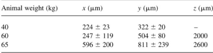
excursion and motion trajectory excursions after Octopus stabilization. An unstabilized point on the LAD moved with an excursion of 7.2 ^ 0.3 mm, which was reduced to 300 ^ 6.4 mm after Octopus stabilization. The average maximum excursions in x-, y- and z-directions were as shown in Table 1.
Fig. 2 depicts motion trajectories of various points of interest on the heart wall as well as on the stabilizer suction bars of the Octopus.
Fig. 3 shows on the left side, the state of a heart, i.e. ECG, BP, AP, z-excursion, and lateral speed, whereas the right side depicts the corresponding trajectory of proximal point on the LAD (see video online). For improved understanding the trajectory has been labeled with correlating landmarks on the ECG.
The stroke width of the trajectory vector corresponds with the excursion velocity. Fast excursions were found corresponding with the QRS complex and the T wave, while slow excursions were observed during the P wave and ST segment, lasting for about 60 ms each (heart rate 61/min). 3.2. Influence of inotropes
Dopamine-induced change in hemodynamics not only increased the maximum excursion by about 1 mm for every 1 mmHg rise in mean BP (Table 2), but also led to a change in the shape of trajectory of cardiac motion (Fig. 4). This could underline the role of temporary b blockade during OPCAB.
3.3. Variability over time
During a phase of stable hemodynamics, the variability of maximum excursion in the lateral plane, between consecutive cardiac cycles (10 cardiac cycles over 8 s)
Fig. 1. Scheme of the measurement setup used.
Table 1
Average maximum excursion in x-, y- and z-directions after octopus stabilization
Animal weight (kg) x (mm) y (mm) z (mm) 40 224 ^ 23 322 ^ 20 – 60 247 ^ 119 504 ^ 80 2000 65 596 ^ 200 811 ^ 239 2600
2 This research was sponsored by the NCCR CO-ME of the Swiss
was in the range of 8.8 ^ 7 mm; while the maximum variability between cycles minutes apart (10 cycles over 8 s) under the same conditions was 18 ^ 12 mm.
Following the same trend, trajectories of a point of interest during consecutive cardiac cycles, as well as those captured minutes apart showed no significant change in shape, provided the hemodynamics remained stable (Fig. 5).
This understanding could play an important role in providing mechanical motion cancellation.
3.4. Influence of positive pressure ventilation on absolute coronary artery motion
Synchronized AP plotting could help correlate a significant influence of lung ventilation on the remaining motion of a point of interest. Fig. 6 shows a distinct correlation between the AP and motion in the direction of the z-axis. The amplitude of the motion component attributed to ventilation in the x=y-plane was in the range of 100 mm and in the z-direction in the range of 500 mm.
However, high frequency low tidal volume ventilation should ameliorate the influence of positive pressure ventilation on the remaining cardiac wall motion after stabilization.
4. Discussion
With the increasing age of patients being subjected to coronary bypass surgery, increasing attention must be paid to the adverse effects of heart lung machine and the effect of coexisting diseases on the overall outcome. OPCAB has gradually started becoming acceptable thanks to the gadgets like stabilizers, mist blowers, intraoperative doppler flow measurement devices, etc. In addition to avoiding the adverse effects of heart lung machine, cardiac surgeons have sought to reduce the surgical trauma by using port access approaches.
Totally endoscopic OPCAB surgery is seen as the logical culmination of all port access coronary bypass procedures.
Fig. 2. Cardiac surface motion in the x=y-plane for various points of interest on the heart surface as well as on the Octopus stabilizer.
Table 2
Change in excursions on a proximal branch on the LAD (x=y-plane) with modulation of cardiac contractility
Excursion (M ^ SD) (mm) Mean BP (mmHg) Heart rate (per min)
300 ^ 6.4 55 82
318 ^ 8.4 78 86
330 ^ 8 88 116
Fig. 3. (a) Time synchronous plot of the ECG, BP, airway pressure, z-excursion and velocity in the x=y-plane. (b) Trajectory of a point of interest correlated with landmark points on the ECG. The thickness of trajectory vector corresponds with excursion velocity.
Fig. 4. Trajectories of the same bifurcation point on the LAD at mean pressures of 55 (left) and 88 (right) mmHg.
The rigid design of conventional endoscopic instruments which were tried earlier had their inherent limitations for application in closed chest cardiac procedures[3].
The introduction of surgical telemanipulators with their fulcrum effect not only improved the maneuverability of the end effectors, but with their motion scaling, tremor filters and three-dimensional vision helped improve surgical precision. This enabled construction of a reliable quality of anastomosis on a static heart in an acceptable duration of time after a brief learning curve[3].
However, the same group reported suboptimal anasto-mosis and prolonged ischaemic – anastomotic times required for performing a telemanipulator-assisted anastomosis on a beating heart[4].
Inadequate stabilization and lack of reliable occlusion of the target coronary artery appear to be the basic impedi-ments to the widespread acceptability of telemanipulator assistance for coronary bypass procedures.
In this study we try to characterize the coronary artery motion in multiple dimensions, to assess its significance with regards to the quality of OPCAB anastomosis, and explore the possibilities of using this knowledge for some form of robotic motion cancellation.
Our observations show that the unstabilized anterior heart wall excursions can be up to a mean of 7.2 mm. Octopus stabilization reduces this motion significantly. However, remnant coronary artery excursion after stabili-zation of about 300 – 330 mm in the x=y-plane and of about 2 – 2.6 mm in the z-plane could still be significant in certain situations, resulting in a loss of anastomotic caliber. According to Falk [5], the geometric accuracy to aim a needle at the desired target (arterial wall) under ideal conditions (nonmoving target or arrested heart) is in the range of 0.1 – 0.2 mm. This level of accuracy in ‘relative positioning’ would amount to a deviation of about 20% for a small coronary artery (1 mm diameter); and this would be even more if the target were moving as in the case of OPCAB. The complexity of performing a stitch on a stabilized coronary artery corresponds to the complexity of the trajectory of motion (even after stabilization) as well as surgical factors like wall irregularities (necessitating precise targeting at an optimal part of the coronary wall). These factors may result in prolonged duration and inferior caliber. We believe that some form of motion cancellation, in addition to state of the art stabilization, could help in improving the acceptability of telemanipulator assistance and OPCAB procedures in general.
The trajectory of coronary motion appears to be comparable over a prolonged time phase provided the hemodynamics remains stable (Fig. 5). This postulation could open the possibility of teaching the robot to replicate a trajectory of motion based on its immediate preceding observation. On the other hand, depiction of the trajectory parameters could be more readily amenable to virtual cancellation of motion. Of how much usefulness such a virtual motion compensation could be is not clear. However, if mechanical cancellation can be put into practice, it appears to have the potential to significantly improve the accuracy of anastomosis, even while accounting for minor variations in the trajectory of motion between successive cardiac cycles. In our approach, the heart wall motion can be synchronized with the ECG, thus enabling compensation for the movements even if a surgical tool occludes the view to the anastomotic site. This would be an advantage over methods of motion compensation based on online visual tools as suggested by Ortmaier et al. [6], which could be interrupted by obstructing the view.
The instantaneous velocity plots of coronary motion (which according to our observations were up to 6 mm/s in the lateral plane and up to 20 mm/s in the z-axis direction), would be useful to explore the practicability of coupling mechanical motion compensation to the existing telemani-pulator systems.
Fig. 5. The motion trajectory remains stable even for cardiac cycle minutes apart (6 min) provided hemodynamics remain stable. However, the reference point of the trajectory is shifted in this example due to positive pressure ventilation.
Fig. 6. The ventilation pattern is clearly visible in the z-axis motion of the heart wall.
The information about the slowest excursion phase of the coronary motion in relation to the ECG could optimize gated imaging of the heart using CT and MRI by reducing motion blur.
Though, it is well known, we could verify during our study, that positive pressure ventilation causes significant baseline shift of the heart in the thoracic cage in the range of 100 mm in the lateral plane and 500 mm in the z-direction. However, this adverse effect could be minimized using high frequency low tidal volume ventilation. Furthermore, we could observe that dopamine-induced changes in hemo-dynamics not only increased the maximum excursion observed, but also led to a change in the shape of trajectory of cardiac motion. This could underline the role of temporary b blockage during OPCAB. Further studies would be needed to establish clinical protocols for ventilation, heart rate and BP control so as to harness their advantage vis-a-vis the quality of beating heart coronary bypass anastomosis.
While using different orientations of the arm to stabilize the same point of interest on the beating heart, we noticed significant differences in stabilizing effect. Although our study did not give us the scope to investigate it in detail, we noticed that the stabilizer setup has a significant influence on stabilizing efficacy. Our setup could therefore be used to standardize the orientation of the stabilizer arm for various parts of the coronary artery tree so as to obtain maximum stabilization, as also to provide vital clues for mechanical improvements in the stabilizing arm.
5. Conclusion
This study characterizes the motion of a point of interest on the coronary artery in multiple dimensions with respect to time and ECG. There is significant remnant coronary artery motion after state of the art stabilization, which can affect the quality of anastomosis in certain unfavorable situations. We believe that variability of excursions and the change in trajectory of motion between consecutive cardiac cycles is negligible. This could help formulating motion compen-sation strategies for the surgical robot or active stabilizer, which in turn could lead to better results. Additionally, velocity plots constructed in our study, are likely to help in gated cardiac imaging. In conclusion we believe that future robots, in addition to compensating for the surgeons tremor, could help more intelligently by compensating for the remaining motion of the operation field.
Acknowledgements
This work has been supported by the CO-ME/NCCR research network of the Swiss National Science Foundation (http://co-me.ch). We are also grateful to Dr med. E. Hiltbrand, Dr med. Berenger, Dr med. F. Terrier
and their staff of the University Hospital in Geneva, Switzerland.
References
[1] Diodato LH, Scarborough JE, Domkowski PW, Smith ML, Biswas SS, Schwartz T, Landolfo KP. Robotically assisted versus conventional freehand technique during beating heart anastomosis of left internal thoracic artery to left anterior descending artery. Ann Thorac Surg 2002;73:825– 9.
[2] Berger M. Deformable area-based template matching with application to low contrast imagery. Selected readings in vision and graphics; 1999. ISSN 1439-5053.
[3] Falk V, Gummerta JF, Walthera T, Hayaseb M, Berryc GJ, Mohra FW. Quality of computer enhanced totally endoscopic coronary bypass graft anastomosis—comparison to conventional technique. Eur J Cardi-othorac Surg 1999;15:260 – 5.
[4] Falk V, Fann JI, Grunenfelder J, Daunt D, Burdon TA. Endoscopic computer-enhanced beating heart coronary artery bypass grafting. Ann Thorac Surg 2000;70:2029– 33.
[5] Falk V. Manual control and tracking—a human factor analysis relevant for beating heart surgery. Ann Thorac Surg 2002;74:624 – 8. [6] Ortmaier T, Groeger M, Kotzor D. Bewegungsschaetzung in der
minimal invasiven Herzchirurgie. Automatisierungstechnik 2002;6.
Appendix A. Conference discussion
Dr A. Wechsler (Philadelphia, USA): This is obviously a precursor to the development of any kind of haptic system or some means of interacting directly with the coronary. Just for you to speculate a little bit, if you had a choice of using these data to create the optical illusion that there was no motion, or to accept motion and alter the robotic arm and create a mechanical fix, what would your choice be? How would you do this?
Dr Cattin: I would definitely take a mechanical solution first. And if the stabilization effect is not enough, then you can maybe add a controller to compensate the remaining motion afterwards. But definitely a mechanical solution first. The advantage of this two-stage approach is that, the speeds and accelerations required by the robot to compensate the remaining motion are a lot less than for a pure robotic solution.
Dr J. Vaage (Oslo, Norway): As far as I know, without being an expert, a lot of the technology of removing the movements is already there, for instance, in the war industry. I think in a warship at sea, they can actually fix their guns at a point and compensate for the movement of the waves. And when I ask this to the manufacturers of the robotic systems, why this is not included, their answer is that this is far too difficult, it will increase the cost tremendously, etc. How do you look at the future prospect of this?
Dr Cattin: Solving this problem for heart surgery of course involves additional difficulties. It is still not clear what combination of sensors is best for the task but the sensor system has to be very small for minimally invasive surgery. If the already available endoscopic cameras were used as part of the sensor system the occlusion problem has to be solved.
Additionally, when we started our research it was unclear what speeds and accelerations we were facing. This information is crucial when designing a robot to compensate remaining heart wall motion.
Dr M. Turina (Zurich, Switzerland): If I may answer Mr Chairman’s question. The motion cancellation, even if it is performed, would severely limit the surgeon’s ability to react to the extrasystole or to the larger excursion, because you will not allowed to do it, because the motion cancellation system does not let you react like your normal brain very easily does if an extrasystole comes when you are suturing the anastomosis. So there are drawbacks of motion cancellation which the manufacturers are very well aware of.