Images in cardio-thoracic surgery
Caught in the act
A. Delabays, P. Ruchat*, L.K. von Segesser, L. Kappenberger
Department of Cardiology and Cardio-thoracic Surgery, University Hospital, 1011 Lausanne, SwitzerlandReceived 5 August 1998; revised version received 7 August 1998; accepted 8 September 1998
A 59-year-old woman presented with acute pulmonary
embolism. A routine echocardiography showed pulmonary
hypertension, right ventricular dysfunction and a mass
float-ing in the left atrium (Fig. 1).
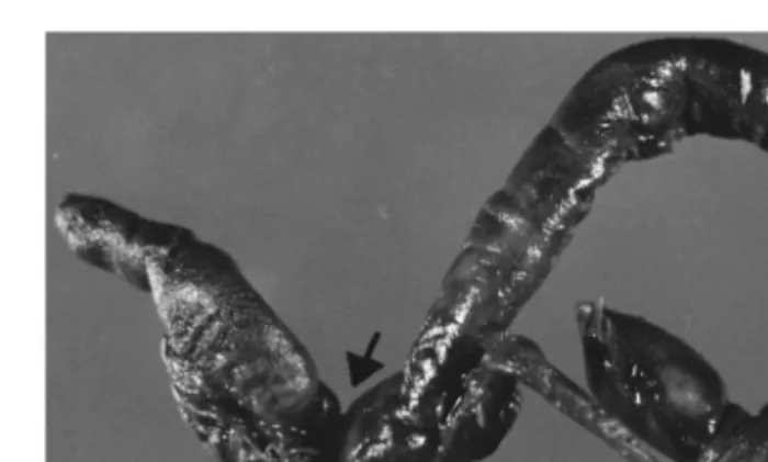
Numerous clots could also be removed from the left
pul-monary artery (Fig. 2). An inferior vena cava filter was
inserted post-operatively and the patient recovered
unevent-fully. At the 6 months follow-up visit, she was
asympto-matic.
European Journal of Cardio-thoracic Surgery 14 (1998) 516
1010-7940/98/$19.00 1998 Elsevier Science B.V. All rights reserved
P I I S 1 0 1 0 - 7 9 4 0 ( 9 8 ) 0 0 2 3 7 - 1
* Corresponding author. Tel.: +41 21 3142280; fax: +41 21 3142278; e-mail: [email protected]
Fig. 1. Transesophageal view of an elongated thrombus crossing the intera-trial septum with its major portion in the left atrium (LA, left atrium; RA, right atrium; AO, aorta).
Fig. 2. Macroscopic view of the clot removed under cardio-pulmonary by-pass. Arrows indicate inprint of the foramen ovale.