© The Author 2015. Published by Oxford University Press on behalf of the European Orthodontic Society. All rights reserved. For permissions, please email: journals.permissions@oup.com
Original article
Non-invasive removal of sandblasted and
acid-etched titanium palatal implants, a
retrospective study
Mirjam Kuhn
*
, Peter Göllner
**
, Marc Schätzle
*
and Michael P. Hänggi
***
*Clinic of Orthodontics and Paediatric Dentistry, Centre of Dental Medicine, University of Zurich, **Private Practice, Bern, Switzerland, ***Private Practice, Basel, Switzerland
Correspondence to: Michael P. Hänggi, Private Practitioner, Birsigstrasse 105, CH-4054 Basel, Switzerland. E-mail: info@ praxishaenggi.ch
Summary
Background: Short, rough-surfaced palatal implants are an established and reliable anchor for orthodontic treatment. Until recently, removal was only possible surgically using a hollow cylinder trephine. This standard method retrieves the implant combined with a larger bone volume and is therefore considered invasive and has known complications. Lately, an explantation tool which allows a sufficient force application to break the bone-implant-connection and unscrew the palatal implant was developed and, since its introduction, has been used as the method of choice in several orthodontic offices.
Objectives: The aim of this study was to assess the complications caused by removing rough-surfaced palatal implants simply by unscrewing them with an explantation tool in contrast to standard protocol by surgical removal with a trephine.
Material and methods: The removal of 73 palatal implants using a customized explantation tool has been evaluated retrospectively and was compared to an existing sample of 44 conventional surgical explantations.
Results: The new clinical procedure resulted in successful removal of 71 (97.3 per cent) palatal implants. In two cases, the new method failed but removal with the established surgical method was still possible with no further complications. The non-invasive palatal implant removal with a customized explantation tool had less medical complications compared to an existing sample of surgical explantations.
Conclusions: User’s opinion was that the new method is more easily executed, less invasive, and also applicable without local anaesthesia. Therefore, it is considered to be beneficial for patients and the treatment approach of choice. However, further research is needed for verification.
Introduction
In orthodontic treatment, reliable anchorage is required in various treatment approaches to achieve a satisfactory result. Traditionally, the most common appliance for anchorage was a headgear which is predominantly dependent on patient cooperation (1, 2). More than a decade ago, temporary anchorage devices (TAD) were introduced (3–8). TADs offer reliable and predictable skeletal anchorage for
orthodontic treatment, independent of patient cooperation, and are well accepted by patients (9). Comparing different TADs, it has been shown that rough-surfaced palatal implants and miniplates have a statistical significantly higher survival rate than miniscrews (10). However, this might partially be influenced by the location, as recent studies showed an excellent survival rate for miniscrews placed in the palate (11).
doi:10.1093/ejo/cju099 Advance Access publication 9 February 2015
Palatal implants might be positioned in the midsagittal of the hard palate (3, 12–14) or at a paramedian location (15, 16). For paramedian palatal implants, a 4.8 per cent risk of failure was found which took place during the healing period. No palatal implant was lost under orthodontic loading (17). In a different study, a failure rate of 6.7 per cent during the healing period was described for the midsagittal location (18). A survival rate of 92 per cent was shown for palatal implants placed midsagittally used for orthodontic treat-ment (19). Another study described a success rate of 91 per cent independent of median or paramedian placement site (20).
Until recently, one of the disadvantages of rough-surfaced palatal implants was the invasive surgical removal by trephine after treat-ment (3, 4, 21). The hollow cylinder of the trephine has a larger diameter than the implant itself (Figures 1 and 2). Besides causing a larger wound through explantation, the procedure is accompa-nied by risks of disturbed wound healing, perforation of nasal floor, injury of the nasopalatal nerve or the roots of neighbouring teeth, devitalization of incisors, secondary bleeding, and implant fracture. Furthermore, the water-cooling of the trephine hardly reaches the tip of the explantation bur, possibly leading to overheating or oste-onecrosis of bone. To avoid this invasive procedure, it was suggested that the palatal implant be left permanently in the bone (22). Neither surgical explantation nor leaving the implant permanently in place is a satisfactory solution (23).
It has been shown that osseointegrated micro-implants of a dif-ferent type (Exacta MS) might be removed at the end of treatment by simple atraumatic unscrewing (23). The desire to be able to do the same for the more widely used second-generation Orthosystem palatal implant (Institute Straumann AG, Basel, Switzerland) arose. Therefore, to simplify the explantation process and reduce complica-tions, one of the authors (MPH) developed an explantation tool to unscrew palatal implants. The tool precisely grasps the 1 mm high triangular abutment connection of the implant, allowing sufficient force application to unscrew the palatal implant without primary drilling.
The aim of this study was to assess retrospectively the advantages and disadvantages of removing rough-surfaced palatal implants sim-ply by unscrewing them with an explantation tool in contrast to standard protocol by surgical removal with a trephine (3–5, 21).
Material and methods
Selection of the sample
The retrospectively evaluated data for this study were derived from 73 patients (44 female, 29 male; mean age at the time of explanta-tion: 23.8 years; range: 12.0–57.2; SD ± 11.9 years) from two private practices in Switzerland (MPH, Basel, n = 32; PG, Bern, n = 26) and from the Clinic of Orthodontics and Paediatric Dentistry, Centre of Dental Medicine, University of Zurich, Zurich, Switzerland (n = 15). All patients previously received a second-generation Orthosystem palatal implant as an anchorage device for their orthodontic treat-ment. The palatal implants were made of pure titanium and had a diameter of 4.1 mm, an endosteal part with SLA (sandblasted with large grits of 0.25–0.5 mm and acid etched with HCl/H2SO4) surface of 4.2 mm length, a smooth neck of 1.8 mm height, and an almost tri-angular-shaped abutment connection of 1 mm height (Orthosystem; Institute Straumann AG). After treatment was finished, the implants had to be removed. Since the introduction of the explantation tool in 2013, all three involved clinics only removed the palatal implants with the explantation tool. Therefore, from the moment the tool was available all patients of the three named clinics who needed removal of an osseointegrated palatal implant were included.
As the surgical removal by trephine was subjectively considered to be more invasive and bore a higher risk of complications, it is no longer considered as the procedure of choice and is only indicated in specific clinical situations. Therefore, a randomized clinical trial was considered problematic for an ethical approval in Switzerland and consequently no current control group was available. Instead, a group of 44 patients with surgical explantations in the time period 1999–2010 from a previous study (21) was used for comparison. Those patients were all treated at the Clinic of Orthodontics and Paediatric Dentistry, Centre of Dental Medicine, University of Zurich, Zurich, Switzerland. In this earlier study, the surgical explan-tations were performed with a 5.5 mm trephine drill and a guiding cylinder, local anaesthesia was used in all 44 surgical explantations.
Removal procedure with the customized explantation tool
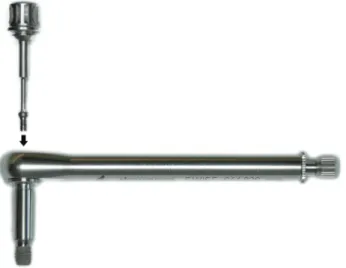
The following material was used in order to unscrew an Orthosystem palatal implant: explantation tool (Bussmann Orthodontie-Labor AG, Luzern, Switzerland), ratchet (Art. No. 046.119, Institute Straumann, Basel, Switzerland), SCS (Screw Carrying System) screw driver length 27 mm (Art. No. 046.402, Institute Straumann), and occlusal screw (Bussmann Orthodontie-Labor AG) made of hard-ened steel, length 4.4 mm (Figures 3 and 4).
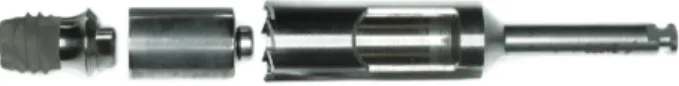
Figure 1. Conventional surgical method of explantation; left: Orthosystem
palatal implant with a 4 mm rough surface and a smooth neck; middle: conduct-cylinder which is mounted on top of the implant to guide the trephine parallel to the implant while drilling; right: trephine hollow cylinder. After drilling down along the implant with the trephine, the implant’s bottom side is still firmly attached to the bone and a strong force with an extraction plier is needed for the final removal of the implant.
Figure 2. Palatal implant within the hollow cylinder at the end of drilling with
the conventional surgical method. Note that a larger bone volume has to be removed and that external cooling by water is very limited.
Subjective experience reported by patients showed that the procedure is possible without local anaesthesia as patients feel only momentary pressure during the initial loosening. This was not entirely unexpected as the removal of miniscrews is also per-formed without local anaesthesia because bone has no innervation. However, this has not been confirmed by a VAS (visual analogue scale) as there was no direct comparison possible to a control group with surgical removal. Nonetheless, local anaesthesia obviously still is recommended for implants that are covered by gingiva.
The explantation tool is placed on the 1 mm high triangular abut-ment connection of the implant and secured by an occlusal screw made of hardened steel, similar to the fixation of a normal abutment. Only after a tight and gapless fit has been ensured, the ratchet is installed on the explantation tool. The ratchet is slightly tilted in all directions for some preloosening and then the implant is unscrewed by turning the ratchet counter-clockwise (24).
Data collection
The data of 73 consecutive patients was collected from two pri-vate practices in Switzerland (MPH and PG) and from the Clinic of Orthodontics and Paediatric Dentistry, Centre of Dental Medicine, University of Zurich, Zurich, Switzerland. The investigator received all data irreversibly anonymized and randomized.
Statistical method
For statistical analysis, the Statistical Package for the Social Sciences (SPSS) version 22 for Windows was used (IBM Corporation, Armonk, New York, USA). Discrete variables were evaluated by chi-square tests (Pearson chi-chi-square test or Fisher’s Exact Test). The level of statistical significance was set at P <0.05.
Results
The explantation of 73 (29 male and 44 female) successfully osseoin-tegrated palatal implants was assessed. The average interval between implantation and explantation was 2.7 years (range: 0.9–6.4; SD ± 1.2 years). In some cases, the interval was very long since the implant was left in place even though the intended orthodontic treatment was long finished but patients were hesitant about the surgical inter-vention. A total of 27 palatal implants had been used for active tooth movement, 44 for passive anchorage of teeth, and 2 had not been loaded due to change of treatment plan. Totally, 36 (49.3 per cent) explantations were performed without any anaesthesia, 13 (17.8 per cent) with topical anaesthesia, and 24 (32.9 per cent) with local anaesthesia.
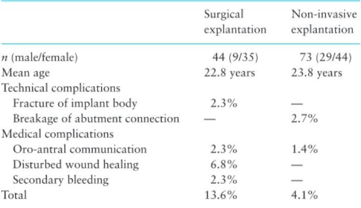
Implant removal with the new non-invasive method was com-pletely successful in 70 (95.9 per cent) patients. Three cases (4.1 per cent) showed minor complications: in one case, an opening of the nasal floor due to a deeply inserted implant in a particularly thin palatal bone was detected. Nevertheless, this explantation was considered successful and the opening of the nasal floor seemed unavoidable. In another case, there was an overloading of the implant head due to accidental clockwise turning instead of coun-ter-clockwise rotation. Another complication was a fracture of the almost triangular-shaped abutment connection (1 mm) of the pala-tal implant, leaving the remaining part of the implant still in situ. Since the implant body was unharmed, subsequent removal with the traditional method by trephine was still possible without prob-lems. A comparison of the 44 surgical removals and the 73 non-invasive explantations is given in Table 1. For statistical analysis, the observed complications were divided into two groups of either medical or technical complications. Significantly less medical com-plications were found for the non-invasive removal compared to the surgical removal (χ2 = 5.656, P = 0.018). No significant differences were found for technical complications (χ2 = 0.281, P = 0.596).
Discussion
After an initial healing period, rough-surfaced palatal implants have proven to be extremely reliable during passive or active orthodontic force application (17, 19). This is not unexpected, as they are basi-cally just a smaller version of typical prosthetic implants, which are commonly used in general dentistry. Even short prosthetic implants are able to successfully withstand very high intermitted biting forces for more than a decade (25). Although the rough surface of palatal implants is unquestionably responsible for its greatest advantage, which is the ability to withstand high forces and its high success rate, it is also responsible for the greatest disadvantage: complicating
Figure 3. Custom made explantation tool. The tool precisely grasps the
1 mm high triangular abutment connection of the implant and represents the negative form of the implant’s head. The tool contains a passage for vertical fixation with an occlusal screw on the implant. Force can be applied to the explantation tool by a ratchet that fits on the top.
Figure 4. Custom made explantation tool mounted on the palatal implant
with ratchet in position. Above: screwdriver and fixation screw ready to be inserted through the hollow passage.
implant removal after an orthodontic treatment. Clinicians trying to remove such a short palatal implant for the first time might be surprised by the force necessary. Therefore, conventional surgi-cal removal after losurgi-cal anaesthesia with a hollow cylinder trephine and subsequent extraction with a plier is quite difficult and requires much more force than could be expected. In addition to the difficulty of the surgical task, a variety of complications are known with this invasive removal method, partly because of insufficient water-cool-ing of the hollow cylinder and partly because of the adjacent bone volume that has to be removed (Figure 2).
In an existing sample of 44 surgical explantations conducted at the Clinic of Orthodontics and Paediatric Dentistry, Centre of Dental Medicine, University of Zurich, Zurich, Switzerland, the following complications were found: disturbed wound healing: 3 (6.8 per cent), perforation of nasal floor: 1 (2.3 per cent), secondary bleeding: 1 (2.3 per cent), and fracture of implant: 1 (2.3 per cent) (21). Damage to the nerve-vessel thread of an upper incisor lead-ing to grey tooth discoloration and temporary reduced sensibility as well as permanent sensory loss of the anterior palatal region have been reported (26, 21). Furthermore, damage to the roots of upper incisors and tooth devitalisation caused by surgical explantation has been described (27). Also perforation of the nasal cavity is a known risk during surgical explantation (21).
This new method with a customized explantation tool had signif-icantly less medical complications (Table 1). Specifically, there were no cases with disturbed wound healing or secondary bleeding. This is most likely due to the fact that no larger wound is created and the tissue does not overheat during the removal procedure. This is illustrated by the observation that the surfaces of the explanted pala-tal implants are not covered with bone, except in their apical anti-rotational notches (Figure 5). Subjectively, all three operators (MPH, PG, and MS) had the impression that the new explantation method was technically easier and better tolerated by patients. Contributing factors could be that the surgical removal by trephine is very loud for patients and a local anaesthesia is mandatory. The non-invasive palatal implant removal seems to generate much less noise and pain. With this method it was even possible to remove the palatal implants completely without any anaesthesia or just with topical anaesthesia.
Nevertheless, local anaesthesia was used in 24 cases. This was partly because in the first couple of cases, no one had thought that local anaesthesia might not be needed. On the other hand, local anaesthesia was also used in situations where the implant was very deeply inserted. A third reason for local anaesthesia was in cases were gingiva covered the implant, particularly in those cases in which the implant was left for a longer time after treatment was finished
and no abutment was mounted to cover the implant. Generally, it is safe to say that under normal circumstances, no local anaesthesia is necessary for the explantation but for anxious or sensitive patients, topical anaesthesia is recommended and might be beneficial.
Of the three reported complications with the new removal method, two can easily be explained. One was due to operator fail-ure (unintentional rotation in wrong direction) and one was due to a pre-existing perforation of the nasal floor by the implant, resulting in an oro-antral communication after removal. The only incident that was not clearly comprehensible was a case where the top triangular part (1 mm) of the implant came off, leaving the rest of the implant still in place. The reasons for this breakage could be 1. insufficient placement of the removal tool, 2. not enough preloosening, con-centrating all the force on the abutment connection, 3. denser bone with a higher removal torque value, and 4. material weakness of the implant. However, as only the top 1 mm part of the implant was separated, subsequent removal with the traditional method by tre-phine was still possible without problems. In no case was a fracture of the endosteal implant part detected.
Our findings are supported by a similar study for Exacta MS osseointegrated palatal implants which showed that a non-invasive implant removal by unscrewing with a special implant key is easy and atraumatic with no complications during the particular pro-cedure or the subsequent healing period (23). Generally, the above mentioned way of explantation is considered to have the follow-ing advantages: easier practicability, less invasive, less painful, fewer medical complications, and faster healing due to a smaller bone defect. However, the non-invasive implant removal is slightly tech-nique-sensitive and requires some experience. A secure position of the removal tool is mandatory and it is recommended to not just unscrew the implant directly in counter-clockwise rotation, but to do some preloosening first.
Conclusion
The non-invasive palatal implant removal with a customized explan-tation tool yielded less medical complications compared to an exist-ing sample of surgical explantations. User’s opinion was that the new method is more easily executed, less invasive, and also applicable without local anaesthesia. Therefore, it is considered to be beneficial for patients and the treatment approach of choice. However, further research is needed to verify this.
Acknowledgement
MPH developed the explantation tool, but for this study, no financial support was received.
Table 1. Complications associated with surgical and non-invasive
explantation. Surgical explantation Non-invasive explantation n (male/female) 44 (9/35) 73 (29/44)
Mean age 22.8 years 23.8 years Technical complications
Fracture of implant body 2.3% — Breakage of abutment connection — 2.7% Medical complications
Oro-antral communication 2.3% 1.4% Disturbed wound healing 6.8% — Secondary bleeding 2.3% —
Total 13.6% 4.1%
Figure 5. Palatal implant directly after removal with the new method. Note
that barely any bone remained on the implant except in the apical anti-rotational notches.
References
1. Jambi, S., Thiruvenkatachari, B., O’Brien, K.D. and Walsh, T. (2013) Orthodontic treatment for distalising upper first molars in children and adolescents. The Cochrane Database of Systematic Reviews, 10, CD008375.
2. Nanda, R.S. and Kierl, M.J. (1992) Prediction of cooperation in orthodon-tic treatment. American Journal of Orthodonorthodon-tics and Dentofacial Ortho-pedics, 102, 15–21.
3. Triaca, A., Antonini, M. and Wintermantel, E. (1992) Ein neues Titan-Flachschrauben-Implantat zur orthodontischen Verankerung am anteri-oren Gaumen. Informationen aus Orthodontie und Kieferorthopädie, 24, 251–257.
4. Wehrbein, H., Merz, B.R., Diedrich, P. and Glatzmaier, J. (1996) The use of palatal implants for orthodontic anchorage. Design and clinical applica-tion of the orthosystem. Clinical Oral Implants Research, 7, 410–416. 5. Wehrbein, H., Glatzmaier, J., Mundwiller, U. and Diedrich, P. (1996) The
Orthosystem–a new implant system for orthodontic anchorage in the pal-ate. Journal of Orofacial Orthopedics, 57, 142–153.
6. Glatzmaier, J., Wehrbein, H. and Diedrich, P. (1995) The development of a resorbable implant system for orthodontic anchorage. The BIOS implant system. Bioresorbable implant anchor for orthodontic systems. Fortschritte der Kieferorthopädie, 56, 175–181.
7. Kanomi, R. (1997) Mini-implant for orthodontic anchorage. Journal of Clinical Orthodontics: JCO, 31, 763–767.
8. Costa, A., Raffainl, M. and Melsen, B. (1998) Miniscrews as orthodon-tic anchorage: a preliminary report. The International Journal of Adult Orthodontics and Orthognathic Surgery, 13, 201–209.
9. Gündüz, E., Schneider-Del Savio, T.T., Kucher, G., Schneider, B. and Bant-leon, H.P. (2004) Acceptance rate of palatal implants: a questionnaire study. American Journal of Orthodontics and Dentofacial Orthopedics, 126, 623–626.
10. Schätzle, M., Männchen, R., Zwahlen, M. and Lang, N.P. (2009) Survival and failure rates of orthodontic temporary anchorage devices: a systematic review. Clinical Oral Implants Research, 20, 1351–1359.
11. Karagkiolidou, A., Ludwig, B., Pazera, P., Gkantidis, N., Pandis, N. and Katsaros, C. (2013) Survival of palatal miniscrews used for orthodontic appliance anchorage: a retrospective cohort study. American Journal of Orthodontics and Dentofacial Orthopedics, 143, 767–772.
12. Wehrbein, H., Merz, B.R. and Diedrich, P. (1999) Palatal bone support for orthodontic implant anchorage–a clinical and radiological study. Euro-pean Journal of Orthodontics, 21, 65–70.
13. Wehrbein, H. (2009) Bone quality in the midpalate for temporary anchor-age devices. Clinical Oral Implants Research, 20, 45–49.
14. Stockmann, P., Schlegel, K.A., Srour, S., Neukam, F.W., Fenner, M. and Felszeghy, E. (2009) Which region of the median palate is a suitable loca-tion of temporary orthodontic anchorage devices? A
histomorphomet-ric study on human cadavers aged 15-20 years. Clinical Oral Implants Research, 20, 306–312.
15. Bernhart, T., Vollgruber, A., Gahleitner, A., Dörtbudak, O. and Haas, R. (2000) Alternative to the median region of the palate for placement of an orthodontic implant. Clinical Oral Implants Research, 11, 595–601. 16. Bernhart, T., Freudenthaler, J., Dörtbudak, O., Bantleon, H.P. and Watzek,
G. (2001) Short epithetic implants for orthodontic anchorage in the paramedian region of the palate. A clinical study. Clinical Oral Implants Research, 12, 624–631.
17. Züger, J., Pandis, N., Wallkamm, B., Grossen, J. and Katsaros, C. (2014) Success rate of paramedian palatal implants in adolescent and adult ortho-dontic patients: a retrospective cohort study. European Journal of Ortho-dontics, 36, 22–25.
18. Jung, B.A., Kunkel, M., Göllner, P., Liechti, T. and Wehrbein, H. (2009) Success rate of second-generation palatal implants. The Angle Orthodon-tist, 79, 85–90.
19. Männchen, R. and Schätzle, M. (2008) Success rate of palatal orthodon-tic implants: a prospective longitudinal study. Clinical Oral Implants Research, 19, 665–669.
20. Asscherickx, K., Vannet, B.V., Bottenberg, P., Wehrbein, H. and Sabzevar, M.M. (2010) Clinical observations and success rates of palatal implants. American Journal of Orthodontics and Dentofacial Orthopedics, 137, 114–122.
21. Fäh, R. and Schätzle, M. (2014) Complications and adverse patient reactions associated with the surgical insertion and removal of palatal implants: a retrospective study. Clinical Oral Implants Research, 25, 653– 658.
22. Mura, P., Maino, B.G. and Paoletto, E. (2000) Midplant: l’ancoraggio assoluto in ortodonzia. Ortodonzia Tecnica, 3, 7–11.
23. Favero, L.G., Pisoni, A. and Paganelli, C. (2007) Removal torque of osse-ointegrated mini-implants: an in vivo evaluation. European Journal of Orthodontics, 29, 443–448.
24. Hänggi, M., Kuhn, M., Göllner, P. and Schätzle, M. (2014) Noninva-sive palatal implant removal. Clinical Oral Implants Research, 1–3. doi: 10.1111/clr.12501
25. Buser, D., Janner, S.F., Wittneben, J.G., Brägger, U., Ramseier, C.A. and Salvi, G.E. (2012) 10-year survival and success rates of 511 titanium implants with a sandblasted and acid-etched surface: a retrospective study in 303 partially edentulous patients. Clinical Implant Dentistry and Related Research, 14, 839–851.
26. Schätzle, M., Männchen, R., Balbach, U., Hämmerle, C.H., Toutenburg, H. and Jung, R.E. (2009) Stability change of chemically modified sand-blasted/acid-etched titanium palatal implants. A randomized-controlled clinical trial. Clinical Oral Implants Research, 20, 489–495.
27. Nicolas, G. and Bart, V.V. (2008) Aspects in post-orthodontic removal of Orthosystem implants. Clinical Oral Implants Research, 19, 1290–1294.