Complex mechanics of the heterogeneous
extracellular matrix in cancer
The MIT Faculty has made this article openly available. Please share
how this access benefits you. Your story matters.
Citation
Malandrino, Andrea, Michael Mak, Roger D. Kamm, and Emad
Moeendarbary. “Complex Mechanics of the Heterogeneous
Extracellular Matrix in Cancer.” Extreme Mechanics Letters 21 (May
2018): 25–34. © 2018 The Authors
As Published
http://dx.doi.org/10.1016/J.EML.2018.02.003
Publisher
Elsevier BV
Version
Final published version
Citable link
http://hdl.handle.net/1721.1/119642
Terms of Use
Creative Commons Attribution 4.0 International License
Extreme Mechanics Letters 21 (2018) 25–34
Contents lists available atScienceDirect
Extreme Mechanics Letters
journal homepage:www.elsevier.com/locate/emlComplex mechanics of the heterogeneous extracellular matrix in
cancer
Andrea Malandrino
a,b,*
,1, Michael Mak
c,*
,1, Roger D. Kamm
d,*
,1,
Emad Moeendarbary
d,e,*
,1aInstitute for Bioengineering of Catalonia, Barcelona, Spain bEuropean Molecular Biology Laboratory, Barcelona, Spain
cDepartment of Biomedical Engineering, Yale University, New Haven, CT, USA
dDepartments of Biological Engineering and Mechanical Engineering, Massachusetts Institute of Technology, Cambridge, MA, USA eDepartment of Mechanical Engineering, University College London, London, UK
a r t i c l e i n f o Article history:
Received 16 October 2017
Received in revised form 7 February 2018 Accepted 20 February 2018
Available online 28 February 2018
a b s t r a c t
The extracellular matrix (ECM) performs many critical functions, one of which is to provide structural and mechanical integrity, and many of the constituent proteins have clear mechanical roles. The composition and structural characteristics of the ECM are widely variable among different tissues, suiting diverse functional needs. In diseased tissues, particularly solid tumors, the ECM is complex and influences disease progression. Cancer and stromal cells can significantly influence the matrix composition and structure and thus the mechanical properties of the tumor microenvironment (TME). In this review, we describe the interactions that give rise to the structural heterogeneity of the ECM and present the techniques that are widely employed to measure ECM properties and remodeling dynamics. Furthermore, we review the tools for measuring the distinct nature of cell–ECM interactions within the TME.
© 2018 The Authors. Published by Elsevier Ltd. This is an open access article under the CC BY license (http://creativecommons.org/licenses/by/4.0/).
Contents
1. General overview... 25
2. The ECM... 26
3. Modes of interaction between cells and the ECM... 26
3.1. Local ECM tension, degradation, and production... 26
3.2. Cell motility and ECM... 27
3.3. Cell populations and ECM... 28
4. Measuring ECM physical properties in cancer... 28
4.1. Optics-based techniques... 28
4.2. Mechanics-based measurements... 28
5. Techniques for measuring cell–ECM interactions... 29
5.1. 3D traction force techniques... 29
5.2. Particle tracking methods... 30
5.3. Laser ablation... 30
5.4. Analysis of proteolytic tracks... 30
5.5. Examples of cell–ECM interaction dynamics distinct in cancer... 31
6. Future directions and concluding remarks... 31
Acknowledgments... 31
References... 31
*
Corresponding authors.E-mail addresses:andrea.malandrino@embl.es(A. Malandrino),
michael.mak@yale.edu(M. Mak),rdkamm@mit.edu(R.D. Kamm),
e.moeendarbary@ucl.ac.uk(E. Moeendarbary).
1Equal contribution.
1. General overview
We review the specific roles of ECM mechanics in tumor pro-gression, with emphasis on mechanobiological phenomena arising from the complex interactions between the heterogeneous ECM
https://doi.org/10.1016/j.eml.2018.02.003
microenvironment and tumor and stromal cells. While comple-menting other reviews (see [1–9]) we highlight (i) the heterogene-ity of ECM mechanical properties as a result of dynamic cellular in-teractions and remodeling processes and (ii) the current advances in measuring these properties. Here we focus on solid tumors, in which the ECM, acting as a scaffolding medium, has proven to impact cell mechanical responses, such as migration, contractile forces, mechanotransduction, and mechanosensing, which in turn influence the degree of tumor malignancy and metastatic poten-tial. In the following sections, we cover ECM functions, cell–ECM interactions, and advances in biophysical techniques for corre-sponding measurements.
2. The ECM
Tissues are typically comprised of ECM, cells, blood-filled vas-cular space, in addition to a collection of other proteins used for signaling between cells, but the proportions differ drastically among anatomical locations. Some, such as cartilage or the cornea, show low cellularity (and lack a vascular supply), so are primarily comprised of ECM, having unique mechanical, and in the case of the cornea, optical properties. Others, such as the heart or pancreas, for example, are dominated by their cellular content, both in terms of their function and their mechanical stiffness. In tissues, structure generally follows function [10].
The ECM is comprised of approximately 300 proteins, and they serve a variety of functions. Some cross-link to form into long filaments that in turn bundle into fibers and serve largely a struc-tural role: collagen, elastin and fibronectin are common [1]. But even at this level, there are fundamental mechanical differences— e.g., elastin exhibits linear, entropic elastic behavior and can sus-tain high levels of strain without fracture, whereas collagen is highly non-linear, much stiffer, and strains very little before frac-ture [11,12]. As with most filaments, both collagen and elastin tend to be stiff under tension, but buckle under compressive stress. Other constituents serve different functions, such as the proteogly-cans (PGs), which are glycoproteins decorated with highly charged, space-filling glycosaminoglycans (GAGs). Due to their high nega-tive charge density, they primarily resist compressive stress, and are especially important in cartilaginous tissues [13].
Fiber arrangement can also be an important determinant of ECM material properties. Collagen and elastin in particular can align into cylindrical chords such as tendons and ligaments, or sheet-like structures, stiff in the plane of the sheet, but compliant perpendicular to it [14–16]. Tissues like the cornea or the interver-tebral disk, are especially interesting examples in which the col-lagen is arranged in layers, alternating in fiber orientation [13,17]. Non-linearity can arise from a variety of sources, but in collagen-rich tissues, it often results from a progressively increasing fraction of the filaments becoming taut with a concomitant increase in stiffness as the tissue is strained [12,18].
One of the unique features of biological tissues that help to distinguish them from abiotic ones, is their ability to remodel in re-sponse to various factors, an effect largely mediated by the cellular content and the ability of the cells to sense and respond to mechan-ical stimuli. Cells can alter the ECM by synthesizing new matrix proteins, altering the extent of crosslinking, or secreting enzymes that selectively break down matrix elements [10]. Cross-linking occurs via several mechanisms, but disulfide bonding is common, occurring in many collagens and laminins. Matrix degradation is mediated again by a variety of proteins, including matrix met-alloproteases (MMPs), ADAMTS proteases, elastaces, and cathep-sins [19,20]. Most have specific sequence targets enabling the cells to fine-tune the mechanical properties of their environment. In order to respond to stress, the cells need to sense it, and this is done via several families of cell–matrix adhesion molecules, but
predominantly proteins in the integrin family. These are promi-nent transmembrane proteins that link the ECM to the intracellular structural members such as the actin cytoskeleton [21], and are again, highly selective in terms of their specific binding partners. For example, laminin binds to dimers consisting of the beta-3 integrin coupled with alpha 3, 6, or 7, and is largely responsible for tethering the basement membrane associated with blood vessels to the vascular endothelium [22,23]. Finally, although they do not participate in the load-bearing function of tissues, the ECM contains a plethora of signaling molecules, and these often bind to specific ECM proteins. Thus, the ECM serves as an effective reser-voir of factors that can, in turn, regulate numerous cell functions, such as growth, migration, and protein synthesis and secretion.
As the architecture of the ECM, composed of many constituents linked together into a complex network, can contribute to func-tional roles and guide cell behavior, various imaging methods have been applied to directly visualize local and global organizational patterns. Electron microscopy provides high resolution imaging of individual fibers, revealing their fine structures [24]. To im-age ECMs along with cells under various physiologically relevant conditions, optical imaging enables non-destructive visualization, which can also be performed with live cells. Common methods for optical imaging of the ECM, particularly for common matrix proteins collagen I and fibrin, include fluorescence excitation and confocal reflectance [25–27]. For collagen I, second harmonic gen-eration microscopy is also applicable and has been used to visualize collagen in tissues with various diseases, including cancer, fibrosis, and atherosclerosis [28]. Imaging studies have shown that many common ECMs are organized into a complex network of intercon-nected fibers. In stromal tissues, cells are typically encapsulated inside this 3-dimensional fiber matrix, which provides a physically and geometrically distinct environment compared to traditional cell culture conditions on flat (2D) substrates [29].
Imaging of the ECM in cancer specimens, from preclinical and clinical biopsies and in vitro samples, demonstrate distinctive fea-tures, including increased collagen density and matrix alignment in the vicinity of tumors [30,31]. These ECM signatures are corre-lated with disease progression and poor prognosis [30]. Tumor tis-sue environments, particularly from aggressive tumors, have also been shown to be stiffer [30,32], potentially due to a combination of increased ECM concentration, higher matrix crosslinking, and nonlinear stiffening of the ECM fiber network under cancer cell generated tension [33–35], discussed more later.
3. Modes of interaction between cells and the ECM
3.1. Local ECM tension, degradation, and production
Through integrins, cells engage the ECM utilizing cytoskeletal contractile forces generated by molecular motors (myosins) walk-ing on actin filaments. Contractile forces are transmitted to the ECM network, leading to matrix stiffening [35]. Stiffer substrates induce increased cell-generated tension [36,37], generating a pos-itive mechanical feedback. In addition to pulling on matrix fibers, cells can synthesize and degrade the ECM through different types of MMPs specialized in degrading different ECM proteins. MMP-1, for instance, cleaves fibrillar collagen I, while MMP-2 and MMP-9 degrades the basement membrane [38], consisting largely of col-lagen IV and non-colcol-lagenous components such as laminin [2,39]. Production of new ECM occurs as a highly integrated process in which ECM molecular components are synthesized and pack-aged inside the cell, secreted, and self-assemble into the existing
matrix [2]. Depending on the microenvironment, highly ordered
fiber networks (e.g. in some connective tissues) or amorphous gels (e.g. in the brain) can be generated. The molecular content and network architecture of the ECM determine mechanical and functional properties and cell–matrix interactions [3,4,40–44].
A. Malandrino et al. / Extreme Mechanics Letters 21 (2018) 25–34 27
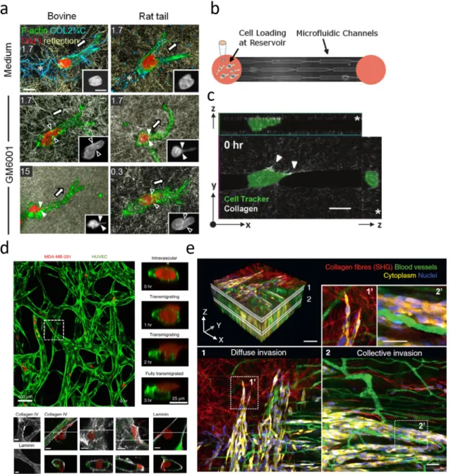
Fig. 1. Capturing the interactions between cancer cells and their microenvironment at varying levels of complexity. (a) A cancer cell navigates through a reconstituted 3D
collagen ECM with or without inhibition of MMPs via GM6001 [45]. (b,c) Microfluidic (b) and micropatterning (c) techniques can reproduce and isolate key features of the TME, such as confinement or ECM tracks [47,48]. (d) In vitro co-culture systems can capture specific cell–cell and cell–ECM interactions, such as a cancer cell extravasating from a microvascular network (top) or breaching the basement membrane during extravasation (bottom) [49]. (e) Intravital imaging in mice captures complex TMEs with multiple cell types and in vivo ECMs [50].
3.2. Cell motility and ECM
The surrounding ECM influences cell behavior, including growth and migration. Collagen density determines the pore size of a collagen matrix. Reduced pore sizes, especially when smaller than the cell nucleus, restrict the mechanical motion of cells, requiring motile cells to undergo substantial deformations and utilize MMPs [45,46]. Inhibition of MMPs reduces cell migration rates in dense matrices but less so in sparse matrices [45] (Fig. 1a). MMPs therefore may be expendable in sparse matrices.
Although ECMs are often quantified by bulk metrics such as average pore size or average fiber length, they have a high degree of heterogeneity due to the intrinsic disorder of the fiber network. Micro-patterning and manipulation methods have been applied to accentuate certain local features and to determine their impact on cell behavior. Microchannels of variable dimensions and bi-furcating paths revealed that migratory decision making depends
on both the dimensionality and directionality of the path [51]. Aligned paths with larger dimensions are favored. Paths with cross-sections smaller than the cell nucleus require additional time for cell transmigration as the nucleus deforms under persistent force generation [47,52] (Fig. 1b). Aligning collagen matrices by controlling flow and temperature during gelation leads to cells that preferentially extend and migrate in the same direction [41,53]. Cells themselves can also induce ECM alignment by applying ten-sion [54,55]. Furthermore, cells have been shown to migrate along gradients of substrate stiffness and ligand density [56–58]. Cell migratory patterns are therefore biased by local ECM properties. Importantly, these features (dimensionality, alignment, stiffness) are all inducible by cells themselves via force generation, matrix degradation and synthesis, and secretion of crosslinking factors.
In TMEs, enhanced force generation by aggressive cells [37], along with secretion of collagen crosslinking factors [33], can lead to local stiffening of the matrix and alignment of matrix fibers,
generating features conducive toward invasion. Additionally, MMP activity by tumor cells can generate cell-scaled tracks along migra-tory paths [59]. Tumors (and stromal cells) therefore act as local sources of ECM remodeling, resulting in heterogeneous spatial and temporal profiles of the ECM network. These profiles can then in-fluence the migration of surrounding cells. In micropatterned col-lagen tracks that mimic tube-like paths cleared by MMP-mediated degradation, cancer cells have been shown to migrate with
in-creased speeds in an MMP-independent manner [48] (Fig. 1c),
as cells in these paths do not need to squeeze through or clear constrictive mechanical barriers.
3.3. Cell populations and ECM
The tumor stroma encapsulates many other cell types in addi-tion to the cancer cells. Fibroblasts, immune cells, and endothe-lial cells have all been shown to interact with cancer cells and influence invasion and metastasis. Macrophages, which secrete
TNF
α
and TGFβ
, stimulate MMT1-MMP and MMP1 in cancer cells,leading to increased migratory speed and persistence in collagen matrices [60]. Cancer-associated fibroblasts (CAFs) influence the TME in a number of ways. They can use transmembrane proteins to pull on cancer cells and lead them to disseminate away from the local bulk tumor and into the ECM [61]. This may facilitate the invasion of tumors that tend to stay localized. CAFs also re-lease pro-inflammatory factors, which promote the recruitment of
macrophages, MMP activity, and angiogenesis [62]. To spread to
distant sites, cancer cells need to transmigrate across endothelial barriers in order to access and exit from the vascular system. Trans-migration involves both squeezing through endothelial junctions and penetrating through the basement membrane, a relatively thin matrix produced by endothelial cells that separate the endothe-lium from the surrounding connective tissue (Fig. 1d). Different MMPs are required to degrade the basement membrane and in-terstitial matrices. Additionally, integrin
β
1 appears to be critical for tumor cells to penetrate through the basement membrane after migrating through endothelial junctions, as cells with integrinβ
1 knocked down appear to be able to transverse endothelial junc-tions but not the basement membrane [23,49].The TME therefore hosts many diverse mechanical and bio-chemical interactions during cancer progression and metastasis. Some of these interactions are being targeted actively in ther-apeutic development, such as angiogenesis, MMPs, and chronic inflammation [5,63], whereas other factors such as mechanical interactions and force generation, which are also important in normal tissue maintenance and function, may require novel strate-gies. Various methods, from 3D mono- and co-culture systems to micropatterning and microfluidics to in vivo imaging [50] (Fig. 1e), can address the different degrees of complexity between cells and their surrounding environment (Fig. 1).
4. Measuring ECM physical properties in cancer
Abnormal ECM composition, architecture and stiffness have been identified to play integral roles in cancer progression at all steps of metastasis [6,40]. It is crucial to measure and quan-tify the changes in ECM properties since they regulate tumor growth, transformation to malignancy, and invasion [7]. Depend-ing on in vivo, ex vivo, and in vitro conditions and the associ-ated technological limitations, tumor tissues and cancer associassoci-ated ECMs have been mechanically characterized from macro to micro and nano scales. Elastography techniques based on
ultrasonogra-phy [64,65], optical coherence tomography [66–68], and
mag-netic resonance imaging [69,70] (Fig. 2a) are among the widely-employed techniques for non-invasive measurement of in vivo me-chanical properties of tumors in patients and animals. Elastogra-phy measurements revealed significant stiffening of tumor tissues
in vivo, particularly for malignant tumors, compared to normal tissues [71,72]. While these in vivo mechanical measurements can identify the presence of abnormal changes in the stiffness of the tumor bulk and can be considered as a diagnostic approach alter-native to conventional palpation methods, they lack the resolution to dissect the contribution of various tissue elements, such as cells and ECMs, and the role of intra-tumor stresses. Fundamentally, the increased stiffness of tumors in vivo can result from the combined effects from the alterations in cellular and extracellular compo-sitions and structures, such as excessive proliferation of cancer cells, causing ECM remodeling and a build-up of growth induced solid stress within the tumor [73], and changes in stromal cells and vascular architecture, causing unnatural interaction of blood flow within and surrounding the tumor and build-up of interstitial fluid pressure [8]. A myriad of other techniques, mostly based on ex vivo and in vitro conditions, have been employed to provide high resolution physical characterization of the local tumor microenvi-ronment at microscales and the capacity to dissect the contribution of cells and ECM physical properties, discussed below and inFig. 2.
4.1. Optics-based techniques
Optical techniques have been widely applied to quantify changes in ECM composition and remodeling in ex vivo slices of tumors or in vitro assays. Confocal microscopy in reflectance or fluorescence modes has been applied to reveal the images of ECM structures mainly in thin tissue slices, due to limited optical
penetration depth [79]. While reflectance confocal microscopy
is the most straightforward label-free method of characterizing the remodeling of pre-existing ECM in simple in vitro assays, immunofluorescence in combination with fluorescence confocal
microscopy provides the ability to probe remodeling and
depo-sition of multiple types of tumor associated ECMs, particularly in ex vivo tumor slices, with submicron resolution [79]. Providing high penetration depth (up to 1 mm) and contrast in addition to submicron resolution, multiphoton microscopy has been an extremely useful optical tool for capturing high resolution images of ECM alignment particularly in live tumor specimens [80,81]. By taking advantage of the large penetration depth and high sensitiv-ity of second harmonic generation (SHG) for label-free imaging of collagen structures (Fig. 2b–c), it has been possible to perform live imaging of the organization of the collagen matrix and its interac-tions with other fluorescently labeled ECM proteins, cancer cells, and stromal cells [74,82,83]. Another set of emerging optical tools involve the extraction of mechanical properties based on unique interactions between photons and phonons and the changes in the behavior of optically generated acoustic waves upon experienc-ing different material properties [79]; Brillouin microscopy is an opto-mechanical characterization method that has been recently integrated with confocal microscopy, allowing non-contact extrac-tion of high resoluextrac-tion stiffness maps of biological samples [84,85] and potentially tumor tissues [86].
4.2. Mechanics-based measurements
Since direct application of forces typically requires the contact between a mechanical probe and the sample, mechanics based techniques to measure tumor stiffness are mostly performed in ex vivo or in vitro conditions. Conventional engineering methods such as compression and shear tests have been applied to quantify the stiffness of ex vivo tumors [71,87]. However, these bulk measure-ments do not have sufficient accuracy and sensitivity to capture local heterogeneous mechanical properties of tumors. Indentation is a robust mechanical characterization method of soft materials. In particular, by tuning the size of the indenter and the sensitivity
A. Malandrino et al. / Extreme Mechanics Letters 21 (2018) 25–34 29
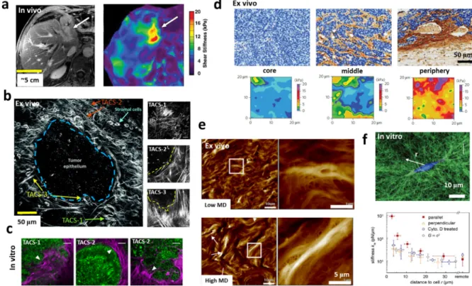
Fig. 2. Characterization of physical properties of tumor tissue and associated ECM via different tools. (a) Significant stiffening of the tumor (from an average of 4.3 kPa in
liver parenchyma to an average of 15.5 kPa at the tumor site) is probed via magnetic resonance elastography (right image) of cholangiocarcinoma invaded surrounding left lobe of human liver (arrow in the left T1-weighted magnetic resonance image) [70] (b) Second harmonic generation image of an ex vivo mouse mammary tumor indicates three tumor associated collagen signatures (TACS): The first signature (TACS-1) is related to a wavy collagen similar to a normal mammary gland but with increased density at regions near tumor. The second (TACS-2) and third (TACS-3) signatures can be characterized by straightened and aligned collagen fibers oriented parallel or perpendicular to the tumor edge respectively [74]. A clearer representation of different TACS is shown in the right panels [31]. (c) In vitro model of CT26 tumor spheroid embedded in 3D collagen I exhibited similar TACS [75]. (d) The stiffness maps (bottom panels) of mouse mammary tumors extracted via AFM indentations show extreme tissue stiffening (∼5-fold) in peripheral regions compared to the tumor core which can be correlated to significant changes in collagen density, structure, and morphology as well as cell density as indicated in immunohistochemistry images in top panels [76]. (e) Topographic maps of human breast tissue measured via AFM show more bundled and aligned collagen fibers in patients with high mammographic density (MD) compared to patients with low MD [77]. (f) Quantifying mechanics of collagen I under the influence of single cancer cells. Confocal reflectance microscopy (top panel) shows remodeling of the collagen network around an MDA-MB-231 breast cancer cell. Quantification of the network stiffness via optical tweezers indicates stiffening of the collagen network at long distances (∼>20µm) away from the cancer cell [78].
of the mechanical apparatus, indentation can offer high resolution micro/nano scale quantification [88,89].
Indentation and topography tests via atomic force microscopy, a very high resolution versatile tool for studying biological sam-ples, have been pivotal in the field of cancer biomechanics and revealed a myriad of mechanical information about the TME at molecular, cellular, and tissue levels [90,91]. Nanomechanical in-dentation tests and topography measurements, performed via AFM on ex vivo tissue slices, revealed a high degree of heterogeneity in the stiffness and collagen architecture of tumors [92]. Interest-ingly, at the core of a tumor, where cancer cells are abundant, the tumor exhibits a soft mechanical signature while the adja-cent peripheral regions, where collagen alignment is apparent, are stiffened [76,93] (Fig. 2d). Based on recent AFM measurements, it has been suggested that the remodeling of ECM microarchitecture, particularly in collagen, leads to tumor stroma stiffening which triggers the epithelial to mesenchymal transition, invasion of tu-mor cells, and metastasis [77,94,95] (Fig. 2e). Moreover, it has been concluded that in addition to the ECM, the tumor epithelium and the tumor-associated vasculature contribute to the stiffening of the tumor stroma, as quantified via AFM [96].
In addition to quantifying physical properties of tumors in vivo and ex vivo conditions, numerous in vitro assays have been used to study the effects of cancer cells and tumor associated stromal cells, such as fibroblasts, on 3D remodeling of naturally derived ECMs [97]. Interestingly, mechanical quantification via AFM of gels embedded with fibroblasts, revealed that activation of YAP mechano-signaling in cancer-associated fibroblasts induces
extreme ECM remodeling and stiffening, more than 8-fold com-pared to stiffening by normal fibroblasts, which contributes to the tumor bulk stiffening observed in vivo [98]. Magnetic and optical
tweezers are also among promising high resolution mechanical
techniques that have been recently employed to characterize ECM properties [99–101]. Interestingly, measurements of cancer cell-induced ECM contractions at the single cell level using optical tweezers revealed long-ranged stress stiffening of the ECM cor-related with ECM remodeling and non-linear elasticity as well as inelastic behavior of collagen networks [78,102] (Fig. 2f).
5. Techniques for measuring cell–ECM interactions
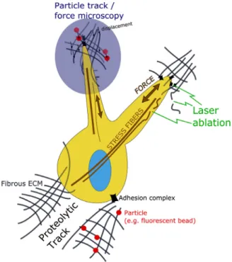
To measure the interactions between cells and their surround-ing matrices, probes and methodologies are required at the cellular and subcellular resolutions (Fig. 3) and over biologically relevant time scales. The readouts of interest of many advanced techniques, which we review here, are often forces and matrix architecture.
5.1. 3D traction force techniques
The techniques that back-calculate forces that cells apply on planar substrates – mainly as a 2D geometrical problem – have seen rapid development and adoption and reached good accuracy (see for instance [103]). Most of these methods use elastic sub-strates, such as polyacrylamide gels with embedded tracer parti-cles, and computational algorithms to extract cell-generated forces based on substrate deformations, captured in microscopy images
Fig. 3. Schematic of cell–ECM interactions that can be measured in 3D by utilizing
the reviewed methodologies. Some examples are illustrated for (i) measuring ECM displacements resulting from contractile forces from tracking of fluorescent parti-cles or cross-correlations of ECM images before and after cellular force application (ii) measuring ECM or stress fiber relaxation after laser ablation and (iii) imaging and assessing cellular proteolytic processes in the ECM.
of tracer displacement profiles. Also, for such force inference, mi-cropatterned substrates (e.g. micropillars of known stiffness) that can be elastically deformed and followed over time have proved useful and accurate to measure forces [104,105]. These methods have been extended to cells surrounded by a 3D ECM and have shown presumed feasibility when the ECM itself can be treated as an elastic continuum whose stiffness is used for back-calculation of forces from the tracer bead displacement data [106–108]. How-ever, when cells alter this stiffness spatiotemporally, many 3D traction force microscopy techniques lose accuracy [109]. Stiffness alteration by cell activity includes local proteolytic degradation, inelastic remodeling (such as matrix densification), and any non-linearity of stiffness values.
Algorithms for 3D determination of displacement fields from the tracking of fluorescent beads or cross-correlation of 3D volu-metric image data must ensure that the resulting distribution of all forces applied from cell processes (such as filopodial dynam-ics) are mechanically self-equilibrated. This is often accomplished with inverse optimization procedures and can be a non-trivial task when multicellular entities are under examination. Powerful algorithms have been recently developed for 3D traction
measure-ments from fluorescence microscopy [108–110]. When combined
with experimental techniques such as matrix fluorescent labeling or reflectance imaging (e.g. collagen or fibrin gel fibers, [110,111]), these algorithms have provided reliable results at the subcellular resolution.
5.2. Particle tracking methods
In 3D traction microscopy of cells in fibrous biopolymer ECMs, fiber bundles or exogenous beads covalently attached to the fiber lattice can be spatiotemporally tracked. Generalizing this concept, single molecules or macromolecular assemblies are also parti-cles that can be tracked using computational approaches. Particle
tracking has thus vast applicability. It has been used for the study of the dynamics of cytoskeletal microtubules ends, showing how alternating periods of growth and shrinkage modulate cell archi-tecture and cytoskeletal forces [112]. Particle tracking is suitable to extract relevant data from highly dynamical processes, such as the assembly and disassembly of focal adhesion complexes. In one study of this kind, several features – e.g. geometry, fluorescence intensity, and position – of Paxillin and FAK were tracked with available tools and have helped determine adhesion lifetimes and turnover rates [113].
Moreover, by using video microscopy and recording the time-dependence of average quantities such as the mean square dis-placement of moving particles, tracking can inform on the modes of motion of molecular entities. In turn, these modes relate to diffusion processes [114]. For instance, membrane dynamics stud-ies could detect both Brownian and non-Brownian motions and transition phases among modes of motions, revealing spatiotem-poral phenomena such as the partition of molecules into different subspecies or the transition to active motion modes, such as the binding to a motor protein [115,116]. Also, passive microrheology – measuring rheological properties from the Brownian motion of ECM-embedded particles – can provide important data to un-derstand the dynamics of cell–ECM interaction. In a recent paper, Schultz and coworkers functionalized a cell-laden hydrogel and tracked microenvironmental changes at multiple time and size scales. The authors could correlate cell-mediated initial proteolytic changes in the ECM farther away from the cells to cytoskeletal tension across the material. On longer timescales, particle track-ing provided evidence for a transition of the pericellular ECM-mimicking hydrogel from an elastic gel to a viscous liquid, medi-ated by degradation processes [117].
5.3. Laser ablation
To probe mechanical stresses at the subcellular level one can ar-tificially relax the tension built by a molecular assembly. The laser ablation technique can be used for this purpose. The technique con-sists of sublimating or evaporating a portion of the molecular as-sembly using the energy of a focused laser beam. Laser ablation acts as a nano scale scissor that results in expansion (or shrinkage) of tissues, revealing the tension (or compression) that kept the tissue together before ablation. Laser ablation can be used in combination with bead tracking and/or knowledge of material properties: the tracking of movements of the surrounding ECM when laser ablat-ing acto-myosin assemblies is used to back-calculate the mechan-ical stress. Laser ablation studies have elucidated the dynamics of multicellular cooperation mediated by the ECM, resulting in a rapid force transmission to the ECM when single stress fibers are disrupted [118,119]. Viscoelastic effects must be expected in cell mechanics applications. Therefore ablation and related time-scales of the expansions/shrinkages can reveal both viscous and elastic constants [118,120]. Laser ablation shares with all of the cell– ECM tools introduced so far an important limitation that concerns the proteolytic- and/or remodeling-driven stiffness modifications, which undermine the accuracy of the back-calculated values of tension. Yet, laser ablation is a powerful technique for selectively probing cell–ECM-related structures such as cytoskeletal cables, cell–matrix adhesion proteins, and ECM components.
5.4. Analysis of proteolytic tracks
Cell migration, especially during invasive spreading through fibrous 3D ECMs, often entails proteolytic activity for fiber break-down. Proteolytic remodeling, via upregulation of MMPs, ser-ine and cysteser-ine proteases, results in cells forming tracks in the ECM. This proteolytic cleavage occurs at the subcellular, cell–ECM
A. Malandrino et al. / Extreme Mechanics Letters 21 (2018) 25–34 31
interface, and is often co-localized with the cell adhesion
molec-ular machinery [121]. Imaging of the ECM structure (through
confocal reflectance or fluorescence microscopy) is often used to study these processes. Also, fluorophore dequenching highlighting proteolytic activity, combined with analyses of cell deformation and migration speed has delivered relevant insights. With these combined tools, proteolytic activity can be measured on cancer cells seeded within 3D collagen lattices containing labeled type I collagen monomers [122,123]. Furthermore, several techniques for in vivo research have been developed to image the effect of MMPs in TMEs, such as optical imaging, positron emission tomography, single photon emission computed tomography, and magnetic res-onance imaging. These target cancer progression-mediated ECM changes through the use of contrast agents linked to MMP in-hibitors or to engineered substrates amplified during enzymatic processes [124].
5.5. Examples of cell–ECM interaction dynamics distinct in cancer
The techniques covered so far have been applied to study the mechanical interactions between the cell types and the complex landscape of ECM proteins characterizing the TME. As for cell-generated forces, these techniques have reported differences be-tween non-cancerous and cancerous cells. For instance, traction force microscopy has measured larger contractile forces generated by more malignant cells on different substrates, compared to non-metastatic cells [37]. Another example related to mechanical sig-naling is the possibility of studying integrins as mechanoreceptors and their distinct features when interacting with the TME. Based on traction force measurements, it has been shown that the ability to exert Rho GTPase-dependent cytoskeletal tensions is functionally linked to the ECM stiffness. This has provided evidence of an important mechanism by which cells may use ligands to feel the crosslinking of exogenous ECMs [125].
Although mostly focused on cells seeded on 2D substrates, laser ablation has also been used to study force propagation in the ECM. Recently, laser ablation was used to measure the me-chanical tension within a collagen gel 48h after seeding of cancer spheroids [75]. It was shown that contractile forces rapidly de-form the surrounding ECM in a centripetal fashion. Interestingly, selectively ablating the 3D collagen lattice reduced spheroid cell spreading, suggesting a prestress-dependent mechanotransduc-tion regulamechanotransduc-tion of cancer invasion.
Finally, particle tracking of intracellular beads has been used to show further linking of intracellular regulation and stiffening to cell motility and perturbed mechanotransduction in breast cancer, which further confirms that the adaptation of intracellular con-tractility and stiffness are ECM stiffness-dependent [126]. Matrix-embedded beads can also be tracked in combination with prote-olytic tracks analyses [127]. In this study, beads were tethered to collagen I fibers near migrating fibrosarcoma cells in the absence and presence of proteolytic inhibitors and acto-myosin contrac-tile forces. Taking the axis of cell migration as reference during forward cell motion, ECM release due to proteolytic activity near the trailing edge was measured. ECM degradation was asymmetric to the axis and produced inelastic deformation, while symmetry was observed at the ECM deformations near the leading edge, with these deformations being elastically recoverable.
Beyond the reductionist approach employed in many tumor biology studies, one important effort would be to channel these methodologies to study mechanical signaling when multiple cell types and macromolecular assemblies that characterize cancer complexity are integrated [19,128]. Many cell type-specific pro-cesses contribute toward furbishing the TME, such as those of immune and endothelial cells, mesenchymal stem cells, as well
as pericytes and cancer-associated fibroblasts. More realistic ex-perimental models should further include 3D tumor-driven angio-genesis and cell spreading, as well as additional ECM-type specific entities interacting with cells (e.g. basement membrane proteins, such as laminin and collagen IV).
6. Future directions and concluding remarks
A profound body of evidence indicates that aberrations in the mechanics of the ECM significantly contribute to tumor progres-sion and metastasis. Therefore, there is an increasing need for new techniques to resolve spatiotemporal changes in ECM mechanics and its underpinning biology. However, inherent limitations asso-ciated with optical and mechanical tools impose challenges toward capturing high resolution spatiotemporal changes of ECM mechan-ics in vivo. In vitro methodologies based on the combination of 3D cultures with microfluidic techniques are ideal platforms that can realistically and efficiently recapitulate various bio-mechanical el-ements of the TME at specific progression points while monitoring dynamic cell–cell and cell–ECM interactions with high resolution. Since the ECM plays such a prominent role in cancer pro-gression, modulating ECM mechanics offers the potential for new approaches to cancer therapy. New methods are being actively pursued in several laboratories (see, e.g., [129]) to limit the spread of tumor by the use of drugs that alter TME mechanical prop-erties or their spatial gradients. Related studies are addressing the underlying mechanisms that give rise to matrix remodeling. Further work is needed, however, before we can fully characterize the mechanical complexity of the TME, understand the processes that contribute to it, and finally, how it might be regulated for therapeutic benefit.
Acknowledgments
A.M. received funding from the People Programme (Marie Curie Actions) of the European Union’s Seventh Framework Programme FP7/2007–2013/under REA (Grant 625500). Funding from the U.S. National Cancer Institute (U01 CA202177-01) to R.K. is gratefully acknowledged. This work was supported by Cancer Research UK Multidisciplinary Award [C57744/A22057] and CRUK-UCL Cen-tre Award [C416/A25145] to EM. E.M. was the recipient of a Wellcome Trust-Massachusetts Institute of Technology Fellowship (WT103883). M.M. acknowledges support from Yale University.
References
[1] R.O. Hynes, A. Naba, Overview of the matrisome-An inventory of extracellular matrix constituents and functions, Cold Spring Harb. Perspect. Biol. 4 (2012).
http://dx.doi.org/10.1101/cshperspect.a004903. a004903–a004903. [2] J.K. Mouw, G. Ou, V.M. Weaver, Extracellular matrix assembly: A multiscale
deconstruction, Nat. Rev. Mol. Cell Biol. 15 (2014) 771–785.http://dx.doi.org/ 10.1038/nrm3902.
[3] S.R. Peyton, C.M. Ghajar, C.B. Khatiwala, A.J. Putnam, The emergence of ECM mechanics and cytoskeletal tension as important regulators of cell func-tion, Cell Biochem. Biophys. 47 (2007) 300–320.http://dx.doi.org/10.1007/ s12013-007-0004-y.
[4] D.H. Kim, P.P. Provenzano, C.L. Smith, A. Levchenko, Matrix nanotopography as a regulator of cell function, J. Cell Biol. 197 (2012) 351–360.http://dx.doi. org/10.1083/jcb.201108062.
[5] M. Egeblad, Z. Werb, New functions for the matrix metalloproteinases in cancer progression, Nat. Rev. Cancer. 2 (2002) 161–174.http://dx.doi.org/10. 1038/nrc745.
[6] T.R. Cox, J.T. Erler, Remodeling and homeostasis of the extracellular matrix: implications for fibrotic diseases and cancer, Dis. Model. Mech. 4 (2011) 165– 178.http://dx.doi.org/10.1242/dmm.004077.
[7] M.W. Pickup, J.K. Mouw, V.M. Weaver, The extracellular matrix modulates the hallmarks of cancer, EMBO Rep. 15 (2014) 1243–1253.http://dx.doi.org/ 10.15252/embr.201439246.
[8] R.K. Jain, J.D. Martin, T. Stylianopoulos, The role of mechanical forces in tumor growth and therapy, Annu. Rev. Biomed. Eng. 16 (2014) 321–346.
[9] M.J. Mitchell, R.K. Jain, R. Langer, Engineering and physical sciences in on-cology: Challenges and opportunities, Nat. Rev. Cancer. 17 (2017) 659–675.
http://dx.doi.org/10.1038/nrc.2017.83.
[10] B. Alberts, A. Johnson, J. Lewis, Molecular Biology of the Cell, Garland Science, 2007.
[11] C.A.J. Hoeve, P.J. Flory, The elastic properties of elastin, Biopolymers. 13 (1974) 677–686.http://dx.doi.org/10.1002/bip.1974.360130404.
[12] D. Vader, A. Kabla, D. Weitz, L. Mahadevan, Strain-induced alignment in collagen gels, PLoS One 4 (2009) e5902.http://dx.doi.org/10.1371/journal. pone.0005902.
[13] A. Malandrino, A.R. Jackson, J.M. Huyghe, J. Noailly, Poroelastic modeling of the intervertebral disc: A path toward integrated studies of tissue biophysics and organ degeneration, MRS Bull. 40 (2015) 324–332.http://dx.doi.org/10. 1557/mrs.2015.68.
[14] L. Rossetti, L.A. Kuntz, E. Kunold, J. Schock, K.W. Müller, H. Grabmayr, J. Stolberg-Stolberg, F. Pfeiffer, S.A. Sieber, R. Burgkart, A.R. Bausch, The microstructure and micromechanics of the tendon–bone insertion, Nature Mater. 16 (2017) 664–670.http://dx.doi.org/10.1038/nmat4863.
[15] J.E. Scott, C.R. Orford, E.W. Hughes, Proteoglycan-collagen arrangements in developing rat tail tendon: An electron microscopical and biochemical in-vestigation, Biochem. J. 195 (1981) 573–581.
[16] M. Benjamin, J.R. Ralphs, Tendons and ligaments - An overview, Histol. Histopathol. 12 (1997) 1135–1144. http://dx.doi.org/10.1016/B978-0-323-09138-1.00007-3.
[17] K.M. Meek, C. Knupp, Corneal structure and transparency, Prog. Retin. Eye Res. 49 (2015) 1–16.http://dx.doi.org/10.1016/j.preteyeres.2015.07.001. [18] S. Nam, K.H. Hu, M.J. Butte, O. Chaudhuri, Strain-enhanced stress relaxation
impacts nonlinear elasticity in collagen gels, Proc. Natl. Acad. Sci. 113 (2016) 5492–5497.http://dx.doi.org/10.1073/pnas.1523906113.
[19] R. Zent, A. Pozzi, Cell-extracellular matrix interactions in cancer, 2010,http: //dx.doi.org/10.1007/978-1-4419-0814-8.
[20] K. Kessenbrock, V. Plaks, Z. Werb, Matrix metalloproteinases: Regulators of the tumor microenvironment, Cell 141 (2010) 52–67.http://dx.doi.org/10. 1016/j.cell.2010.03.015.
[21] L.B. Case, C.M. Waterman, Integration of actin dynamics and cell adhesion by a three-dimensional, mechanosensitive molecular clutch, Nat. Cell Biol. 17 (2015) 955–963.http://dx.doi.org/10.1038/ncb3191.
[22] T. Tennenbaum, L. Li, A.J. Belanger, L.M. De Luca, S.H. Yuspa, Selective changes in laminin adhesion and alpha 6 beta 4 integrin regulation are associated with the initial steps in keratinocyte maturation, Cell Growth Differ. 7 (1996) 615– 628.
[23] M.B. Chen, J.M. Lamar, R. Li, R.O. Hynes, R.D. Kamm, Elucidation of the roles of tumor integrin 1 in the extravasation stage of the metastasis cascade, Cancer Res. 76 (2016) 2513–2524. http://dx.doi.org/10.1158/0008-5472.CAN-15-1325.
[24] D.E. Birk, J.M. Fitch, J.P. Babiarz, K.J. Doane, T.F. Linsenmayer, Collagen fibrillo-genesis in vitro: Interaction of types I and V collagen regulates fibril diameter, J. Cell Sci. (1990) 649–658.
[25] A.O. Brightman, B.P. Rajwa, J.E. Sturgis, M.E. McCallister, J.P. Robinson, S.L. Voytik-Harbin, Time-lapse confocal reflection microscopy of collagen fibril-logenesis and extracellular matrix assembly in vitro, Biopolymers. 54 (2000) 222–234. http://dx.doi.org/10.1002/1097-0282(200009)54:3. < 222::AID-BIP80>3.0.CO;2-K..
[26] Y.L. Yang, L.J. Kaufman, Rheology and confocal reflectance microscopy as probes of mechanical properties and structure during collagen and colla-gen/hyaluronan self-assembly, Biophys. J. 96 (2009) 1566–1585.http://dx. doi.org/10.1016/j.bpj.2008.10.063.
[27] S. Geraldo, A. Simon, N. Elkhatib, D. Louvard, L. Fetler, D.M. Vignjevic, Do cancer cells have distinct adhesions in 3D collagen matrices and in vivo? Eur. J. Cell Biol. 91 (2012) 930–937.http://dx.doi.org/10.1016/j.ejcb.2012.07.005. [28] X. Chen, O. Nadiarynkh, S. Plotnikov, P.J. Campagnola, Second harmonic generation microscopy for quantitative analysis of collagen fibrillar structure, Nat. Protoc. 7 (2012) 654–669.http://dx.doi.org/10.1038/nprot.2012.009. [29] A.D. Doyle, R.J. Petrie, M.L. Kutys, K.M. Yamada, Dimensions in cell migration,
Curr. Opin. Cell Biol. 25 (2013) 642–649. http://dx.doi.org/10.1016/j.ceb. 2013.06.004.
[30] M.W. Conklin, J.C. Eickhoff, K.M. Riching, C.A. Pehlke, K.W. Eliceiri, P.P. Provenzano, A. Friedl, P.J. Keely, Aligned collagen is a prognostic signature for survival in human breast carcinoma, Am. J. Pathol. 178 (2011) 1221–1232.
http://dx.doi.org/10.1016/j.ajpath.2010.11.076.
[31] P.P. Provenzano, K.W. Eliceiri, J.M. Campbell, D.R. Inman, J.G. White, P.J. Keely, Collagen reorganization at the tumor-stromal interface facilitates local invasion, BMC Med. 4 (2006) 38.http://dx.doi.org/10.1186/1741-7015-4-38. [32] A. Evans, P. Whelehan, K. Thomson, D. McLean, K. Brauer, C. Purdie, L. Baker, L. Jordan, P. Rauchhaus, A. Thompson, Invasive breast cancer: Relationship between shear-wave elastographic findings and histologic prognostic factors, Radiology 263 (2012) 673–677.http://dx.doi.org/10.1148/radiol.12111317. [33] K.R. Levental, H. Yu, L. Kass, J.N. Lakins, M. Egeblad, J.T. Erler, S.F.T. Fong, K.
Csiszar, A. Giaccia, W. Weninger, M. Yamauchi, D.L. Gasser, V.M. Weaver, Matrix crosslinking forces tumor progression by enhancing integrin signal-ing, Cell 139 (2009) 891–906.http://dx.doi.org/10.1016/j.cell.2009.10.027.
[34] S. Münster, L.M. Jawerth, B.A. Leslie, J.I. Weitz, B. Fabry, D.A. Weitz, Strain history dependence of the nonlinear stress response of fibrin and collagen networks, Proc. Natl. Acad. Sci. USA 110 (2013) 12197–12202.http://dx.doi. org/10.1073/pnas.1222787110.
[35] C. Storm, J.J. Pastore, F. MacKintosh, T. Lubensky, P.A. Jamney, Nonlinear elasticity in biological gels, Nature 435 (2005) 191–194.http://dx.doi.org/10. 1038/nature03497.1.
[36] D. Mitrossilis, J. Fouchard, A. Guiroy, N. Desprat, N. Rodriguez, B. Fabry, A. Asnacios, Single-cell response to stiffness exhibits muscle-like behavior, Proc. Natl. Acad. Sci. USA 106 (2009) 18243–18248.http://dx.doi.org/10.1073/ pnas.0903994106.
[37] C.M. Kraning-Rush, J.P. Califano, C.A. Reinhart-King, Cellular traction stresses increase with increasing metastatic potential, PLoS One 7 (2012) e32572.
http://dx.doi.org/10.1371/journal.pone.0032572.
[38] H.K. Kleinman, G.R. Martin, Matrigel: Basement membrane matrix with bio-logical activity, Semin. Cancer Biol. 15 (2005) 378–386.http://dx.doi.org/10. 1016/j.semcancer.2005.05.004.
[39] R. Timpl, H. Rohde, P.G. Robey, S.I. Rennard, J.M. Foidart, G.R. Martin, Laminin– a glycoprotein from basement membranes, J. Biol. Chem. 254 (1979) 9933– 9937.
[40] P. Lu, V.M. Weaver, Z. Werb, The extracellular matrix: A dynamic niche in cancer progression, J. Cell Biol. 196 (2012) 395–406.http://dx.doi.org/10. 1083/jcb.201102147.
[41] P.P. Provenzano, D.R. Inman, K.W. Eliceiri, S.M. Trier, P.J. Keely, Contact guid-ance mediated three-dimensional cell migration is regulated by Rho/ROCK-dependent matrix reorganization, Biophys. J. 95 (2008) 5374–5384.http: //dx.doi.org/10.1529/biophysj.108.133116.
[42] A.L. Willis, F. Sabeh, X.Y. Li, S.J. Weiss, Extracellular matrix determinants and the regulation of cancer cell invasion stratagems, J. Microsc. 251 (2013) 250– 260.http://dx.doi.org/10.1111/jmi.12064.
[43] B. Katz, E. Zamir, A. Bershadsky, Z. Kam, K. Yamada, B. Geiger, Physical state of the extracellular matrix regulates the structure and molecular composition of cell-matrix adhesions, Mol. Biol. Cell. (2000).http://dx.doi.org/10.1091/mbc. 11.3.1047.
[44] D.E. Discher, P. Janmey, Y.L. Wang, Tissue cells feel and respond to the stiffness of their substrate, Science 310 (2005) 1139–1143.http://dx.doi.org/ 10.1126/science.1116995.
[45] K. Wolf, M. te Lindert, M. Krause, S. Alexander, J. te Riet, A.L. Willis, R.M. Hoffman, C.G. Figdor, S.J. Weiss, P. Friedl, Physical limits of cell migration: Control by ECM space and nuclear deformation and tuning by proteolysis and traction force, J. Cell Biol. 201 (2013) 1069–1084.http://dx.doi.org/10.1083/ jcb.201210152.
[46] S.I. Fraley, P. Wu, L. He, Y. Feng, R. Krisnamurthy, G.D. Longmore, D. Wirtz, Three-dimensional matrix fiber alignment modulates cell migration and MT1-MMP utility by spatially and temporally directing protrusions, Sci. Rep. 5 (2015) 14580.http://dx.doi.org/10.1038/srep14580.
[47] M. Mak, C.A. Reinhart-King, D. Erickson, Elucidating mechanical transition effects of invading cancer cells with a subnucleus-scaled microfluidic serial dimensional modulation device, Lab Chip. 13 (2013) 340–348.http://dx.doi. org/10.1039/C2LC41117B.
[48] C.M. Kraning-Rush, S.P. Carey, M.C. Lampi, C.a. Reinhart-King, Microfabricated collagen tracks facilitate single cell metastatic invasion in 3D, Integr Biol. 5 (2013) 606–616.http://dx.doi.org/10.1039/c3ib20196a.
[49] M.B. Chen, J.A. Whisler, J. Fröse, C. Yu, Y. Shin, R.D. Kamm, On-chip human microvasculature assay for visualization and quantification of tumor cell extravasation dynamics, Nat. Protoc. 12 (2017) 865–880.http://dx.doi.org/ 10.1038/nprot.2017.018.
[50] S. Alexander, B. Weigelin, F. Winkler, P. Friedl, Preclinical intravital mi-croscopy of the tumour-stroma interface: Invasion, metastasis, and therapy response, Curr. Opin. Cell Biol. 25 (2013) 659–671.http://dx.doi.org/10.1016/ j.ceb.2013.07.001.
[51] M. Mak, D. Erickson, Mechanical decision trees for investigating and mod-ulating single-cell cancer invasion dynamics, Lab Chip. 14 (2014) 964–971.
http://dx.doi.org/10.1039/C3LC51173A.
[52] M. Mak, D. Erickson, A serial micropipette microfluidic device with applica-tions to cancer cell repeated deformation studies, Integr. Biol. 5 (2013) 1374.
http://dx.doi.org/10.1039/c3ib40128f.
[53] K.E. Sung, G. Su, C. Pehlke, S.M. Trier, K.W. Eliceiri, P.J. Keely, A. Friedl, D.J. Beebe, Control of 3-dimensional collagen matrix polymerization for reproducible human mammary fibroblast cell culture in microfluidic devices, Biomaterials 30 (2009) 4833–4841.http://dx.doi.org/10.1016/j.biomaterials. 2009.05.043.
[54] J.J. Tomasek, C.J. Haaksma, R.J. Eddy, M.B. Vaughan, Fibroblast contraction occurs on release of tension in attached collagen lattices: Dependency on an organized actin cytoskeleton and serum, Anat. Rec. 232 (1992) 359–368.
http://dx.doi.org/10.1002/ar.1092320305.
[55] H. Wang, A.S. Abhilash, C.S. Chen, R.G. Wells, V.B. Shenoy, Long-range force transmission in fibrous matrices enabled by tension-driven alignment of fibers, Biophys. J. 107 (2014) 2592–2603.http://dx.doi.org/10.1016/j.bpj. 2014.09.044.
A. Malandrino et al. / Extreme Mechanics Letters 21 (2018) 25–34 33 [56] C.M. Lo, H.B. Wang, M. Dembo, Y.L. Wang, Cell movement is guided by the
rigidity of the substrate, Biophys. J. 79 (2000) 144–152.http://dx.doi.org/10. 1016/S0006-3495(00)76279-5.
[57] S. Aznavoorian, M.L. Stracke, H. Krutzsch, E. Schiffmann, L.A. Liotta, Signal transduction for chemotaxis and haptotaxis by matrix molecules in tumor cells, J. Cell Biol. 110 (1990) 1427–1438.http://dx.doi.org/10.1083/jcb.110.4. 1427.
[58] J.T. Smith, J.T. Elkin, W.M. Reichert, Directed cell migration on fibronectin gradients: Effect of gradient slope, Exp. Cell Res. 312 (2006) 2424–2432.
http://dx.doi.org/10.1016/j.yexcr.2006.04.005.
[59] K. Wolf, P. Friedl, Mapping proteolytic cancer cell-extracellular matrix in-terfaces, Clin. Exp. Metastasis. 26 (2009) 289–298.http://dx.doi.org/10.1007/ s10585-008-9190-2.
[60] R. Li, J.D. Hebert, T.A. Lee, H. Xing, A. Boussommier-Calleja, R.O. Hynes, D.A. Lauffenburger, R.D. Kamm, Macrophage-secreted TNFαand TGFβ1 influence migration speed and persistence of cancer cells in 3D tissue culture via independent pathways, Cancer Res. 77 (2017) 279–290.http://dx.doi.org/10. 1158/0008-5472.CAN-16-0442.
[61] A. Labernadie, T. Kato, A. Brugués, X. Serra-Picamal, S. Derzsi, E. Arwert, A. Weston, V. González-Tarragó, A. Elosegui-Artola, L. Albertazzi, J. Alcaraz, P. Roca-Cusachs, E. Sahai, X. Trepat, A mechanically active heterotypic E-cadherin/N-cadherin adhesion enables fibroblasts to drive cancer cell inva-sion, Nat. Cell Biol. 19 (2017) 224–237.http://dx.doi.org/10.1038/ncb3478. [62] N. Erez, M. Truitt, P. Olson, D. Hanahan, Cancer-associated fibroblasts are
activated in incipient neoplasia to orchestrate tumor-promoting inflamma-tion in an NF-κB-dependent manner, Cancer Cell. 17 (2010) 135–147.http: //dx.doi.org/10.1016/j.ccr.2009.12.041.
[63] P. Carmeliet, R.K. Jain, Angiogenesis in cancer and other diseases, Nature 407 (2000) 249–257.http://dx.doi.org/10.1038/35025220.
[64] P.N.T. Wells, H.-D. Liang, Medical ultrasound: Imaging of soft tissue strain and elasticity, J. R. Soc. Interface. 8 (2011) 1521–1549.http://dx.doi.org/10.1098/ rsif.2011.0054.
[65] M.F. Insana, C. Pellot-Barakat, M. Sridhar, K.K. Lindfors, Viscoelastic imaging of breast tumor microenvironment with ultrasound, J. Mammary Gland Biol. Neoplasia 9 (2004) 393–404.http://dx.doi.org/10.1007/s10911-004-1409-5. [66] B.F. Kennedy, R.A. McLaughlin, K.M. Kennedy, L. Chin, A. Curatolo, A. Tien, B. Latham, C.M. Saunders, D.D. Sampson, Optical coherence micro-elastography: Mechanical-contrast imaging of tissue microstructure, Biomed. Opt. Express. 5 (2014) 2113.http://dx.doi.org/10.1364/BOE.5.002113. [67] K.M. Kennedy, L. Chin, R.A. McLaughlin, B. Latham, C.M. Saunders, D.D.
Sampson, B.F. Kennedy, Quantitative micro-elastography: Imaging of tissue elasticity using compression optical coherence elastography, Sci. Rep. 5 (2015) 15538.http://dx.doi.org/10.1038/srep15538.
[68] A. Srivastava, Y. Verma, K.D. Rao, P.K. Gupta, Determination of elastic prop-erties of resected human breast tissue samples using optical coherence to-mographic elastography, Strain 47 (2011) 75–87.http://dx.doi.org/10.1111/ j.1475-1305.2009.00627.x.
[69]D.B. Plewes, J. Bishop, A. Samani, J. Sciarretta, Visualization and quantification of breast cancer biomechanical properties with magnetic resonance elastog-raphy, Phys. Med. Biol. 45 (2000) 1591–1610.
[70] S.K. Venkatesh, M. Yin, J.F. Glockner, N. Takahashi, P.A. Araoz, J.A. Talwalkar, R.L. Ehman, MR elastography of liver tumors: Preliminary results, Am. J. Roentgenol. 190 (2008) 1534–1540.http://dx.doi.org/10.2214/AJR.07.3123. [71] N.G. Ramião, P.S. Martins, R. Rynkevic, A.A. Fernandes, M. Barroso, D.C.
Santos, Biomechanical properties of breast tissue, a state-of-the-art review, Biomech. Model. Mechanobiol. 15 (2016) 1307–1323.http://dx.doi.org/10. 1007/s10237-016-0763-8.
[72] E. Cukierman, D.E. Bassi, Physico-mechanical aspects of extracellular matrix influences on tumorigenic behaviors, Semin. Cancer Biol. 20 (2010) 139–145.
http://dx.doi.org/10.1016/j.semcancer.2010.04.004.
[73] H.T. Nia, H. Liu, G. Seano, M. Datta, D. Jones, N. Rahbari, J. Incio, V.P. Chauhan, K. Jung, J.D. Martin, V. Askoxylakis, T.P. Padera, D. Fukumura, Y. Boucher, F.J. Hornicek, A.J. Grodzinsky, J.W. Baish, L.L. Munn, R.K. Jain, Solid stress and elastic energy as measures of tumour mechanopathology, Nat. Biomed. Eng. 1 (2017) 1–11.http://dx.doi.org/10.1038/s41551-016-0004.
[74] M.W. Conklin, P.J. Keely, Why the stroma matters in breast cancer: Insights into breast cancer patient outcomes through the examination of stromal biomarkers, Cell Adhes. Migr. 6 (2012) 249–260.http://dx.doi.org/10.4161/ cam.20567.
[75] K.S. Kopanska, Y. Alcheikh, R. Staneva, D. Vignjevic, T. Betz, Tensile forces originating from cancer spheroids facilitate tumor invasion, PLoS One 11 (2016) e0156442.http://dx.doi.org/10.1371/journal.pone.0156442. [76] M. Plodinec, M. Loparic, C.A. Monnier, E.C. Obermann, R. Zanetti-Dallenbach,
P. Oertle, J.T. Hyotyla, U. Aebi, M. Bentires-Alj, R.Y.H. Lim, C.-A. Schoenenberger, The nanomechanical signature of breast cancer, Nat. Nan-otechnol. 7 (2012) 757–765.http://dx.doi.org/10.1038/nnano.2012.167. [77] J.C. McConnell, O.V. O’Connell, K. Brennan, L. Weiping, M. Howe, L. Joseph, D.
Knight, R. O’Cualain, Y. Lim, A. Leek, R. Waddington, J. Rogan, S.M. Astley, A. Gandhi, C.C. Kirwan, M.J. Sherratt, C.H. Streuli, Increased peri-ductal collagen
micro-organization may contribute to raised mammographic density, Breast Cancer Res. 18 (2016) 5.http://dx.doi.org/10.1186/s13058-015-0664-2. [78] Y.L. Han, P. Ronceray, G. Xu, A. Malandrino, R. Kamm, M. Lenz, C.P. Broedersz,
M. Guo, Cell contraction induces long-ranged stress stiffening in the extra-cellular matrix, arXiv Prepr.arXiv:1709.00793, 2017.
[79] W. Goth, J. Lesicko, M.S. Sacks, J.W. Tunnell, Optical-based analysis of soft tissue structures, Annu. Rev. Biomed. Eng. 18 (2016) 357–385.http://dx.doi. org/10.1146/annurev-bioeng-071114-040625.
[80] P.-C. Wu, T.-Y. Hsieh, Z.-U. Tsai, T.-M. Liu, In vivo quantification of the struc-tural changes of collagens in a melanoma microenvironment with second and third harmonic generation microscopy, Sci. Rep. 5 (2015) 8879.http: //dx.doi.org/10.1038/srep08879.
[81] P.P. Provenzano, D.R. Inman, K.W. Eliceiri, J.G. Knittel, L. Yan, C.T. Rueden, J.G. White, P.J. Keely, Collagen density promotes mammary tumor initiation and progression, BMC Med. 6 (2008) 11. http://dx.doi.org/10.1186/1741-7015-6-11.
[82] E. Güç, M. Fankhauser, A.W. Lund, M.A. Swartz, W.W. Kilarski, Long-term intravital immunofluorescence imaging of tissue matrix components with epifluorescence and two-photon microscopy, J. Vis. Exp. (2014).http://dx.doi. org/10.3791/51388.
[83] A. Keikhosravi, J.S. Bredfeldt, A.K. Sagar, K.W. Eliceiri, Second-harmonic gen-eration imaging of cancer, in: Methods Cell Biol. 2014, pp. 531–546http: //dx.doi.org/10.1016/B978-0-12-420138-5.00028-8.
[84] G. Scarcelli, S.H. Yun, Brillouin confocal microscopy for three-dimensional mechanical imaging, Nat. Photon. 2 (2008) 39–43.
[85] G. Scarcelli, W.J. Polacheck, H.T. Nia, K. Patel, A.J. Grodzinsky, R.D. Kamm, S.H. Yun, Noncontact three-dimensional mapping of intracellular hydromechan-ical properties by brillouin microscopy, Nat. Methods. 12 (2015) 1132–1134.
http://dx.doi.org/10.1038/nmeth.3616.
[86] M. Troyanova-Wood, Z. Meng, V.V. Yakovlev, Elasticity-based identification of tumor margins using Brillouin spectroscopy, International Society for Op-tics and Photonics, 2016, p. 97190Phttp://dx.doi.org/10.1117/12.2213268. [87] C. Madsen, T. Cox, Relative stiffness measurements of tumour tissues by shear
rheology, Bio-Protocol 7 (2017).http://dx.doi.org/10.21769/BioProtoc.2265. [88] H.O.B. Gautier, A.J. Thompson, S. Achouri, D.E. Koser, K. Holtzmann, E.
Moeendarbary, K. Franze, Chapter 12 –Atomic force microscopy-based force measurements on animal cells and tissues, Methods Cell Biol. 125 (2015) 211–235.http://dx.doi.org/10.1016/bs.mcb.2014.10.005.
[89] M.C. Díaz de la Loza, A. Díaz-Torres, F. Zurita, A.E. Rosales-Nieves, E. Moeendarbary, K. Franze, M.D. Martín-Bermudo, A. González-Reyes, Laminin levels regulate tissue migration and anterior-posterior polarity during egg morphogenesis in drosophila, Cell Rep. 20 (2017) 211–223.http://dx.doi.org/ 10.1016/j.celrep.2017.06.031.
[90] R. Kaul-Ghanekar, S. Singh, H. Mamgain, A. Jalota-Badhwar, K.M. Paknikar, S. Chattopadhyay, Tumor suppressor protein SMAR1 modulates the roughness of cell surface: Combined AFM and SEM study, BMC Cancer. 9 (2009) 350.
http://dx.doi.org/10.1186/1471-2407-9-350.
[91] M. Lekka, D. Gil, K. Pogoda, J. Dulińska-Litewka, R. Jach, J. Gostek, O. Klymenko, S. Prauzner-Bechcicki, Z. Stachura, J. Wiltowska-Zuber, K. Okoń, P. Laidler, Cancer cell detection in tissue sections using AFM, Arch. Biochem. Biophys. 518 (2012) 151–156.http://dx.doi.org/10.1016/j.abb.2011.12.013. [92] A. Stylianou, T. Stylianopoulos, Atomic force microscopy probing of cancer
cells and tumor microenvironment components, Bionanoscience 6 (2016) 33–46.http://dx.doi.org/10.1007/s12668-015-0187-4.
[93] H. Laklai, Y.A. Miroshnikova, M.W. Pickup, E.A. Collisson, G.E. Kim, A.S. Barrett, R.C. Hill, J.N. Lakins, D.D. Schlaepfer, J.K. Mouw, V.S. LeBleu, N. Roy, S.V. Novitskiy, J.S. Johansen, V. Poli, R. Kalluri, C.A. Iacobuzio-Donahue, L.D. Wood, M. Hebrok, K. Hansen, H.L. Moses, V.M. Weaver, Genotype tunes pancreatic ductal adenocarcinoma tissue tension to induce matricellular fibrosis and tumor progression, Nat. Med. 22 (2016) 497–505.http://dx.doi.org/10.1038/ nm.4082.
[94] A.J. Rice, E. Cortes, D. Lachowski, B.C.H. Cheung, S.A. Karim, J.P. Morton, A. del Río Hernández, Matrix stiffness induces epithelial–mesenchymal transition and promotes chemoresistance in pancreatic cancer cells, Oncogenesis 6 (2017) e352.http://dx.doi.org/10.1038/oncsis.2017.54.
[95] I. Acerbi, L. Cassereau, I. Dean, Q. Shi, A. Au, C. Park, Y.Y. Chen, J. Liphardt, E.S. Hwang, V.M. Weaver, Human breast cancer invasion and aggression correlates with ECM stiffening and immune cell infiltration, Integr. Biol. (Camb). 7 (2015) 1120–1134.http://dx.doi.org/10.1039/c5ib00040h. [96] J.I. Lopez, I. Kang, W.-K. You, D.M. McDonald, V.M. Weaver, In situ force
mapping of mammary gland transformation, Integr. Biol. 3 (2011) 910.http: //dx.doi.org/10.1039/c1ib00043h.
[97] B.K. Robinson, E. Cortes, A.J. Rice, M. Sarper, A. del Río Hernández, Quantitative analysis of 3D extracellular matrix remodelling by pancreatic stellate cells, Biol. Open. (2016).
[98] F. Calvo, N. Ege, A. Grande-Garcia, S. Hooper, R.P. Jenkins, S.I. Chaudhry, K. Harrington, P. Williamson, E. Moeendarbary, G. Charras, E. Sahai, Mechanotra-nsduction and YAP-dependent matrix remodelling is required for the gen-eration and maintenance of cancer-associated fibroblasts, Nat. Cell Biol. 15 (2013) 637–646.http://dx.doi.org/10.1038/ncb2756.
[99] B.H. Blehm, A. Devine, J.R. Staunton, K. Tanner, In vivo tissue has non-linear rheological behavior distinct from 3D biomimetic hydrogels, as determined by AMOTIV microscopy, Biomaterials 83 (2016) 66–78.http://dx.doi.org/10. 1016/j.biomaterials.2015.12.019.
[100] J.R. Staunton, W. Vieira, K.L. Fung, R. Lake, A. Devine, K. Tanner, Mechanical properties of the tumor stromal microenvironment probed in vitro and ex vivo by in situ-calibrated optical trap-based active microrheology, Cell. Mol. Bioeng. 9 (2016) 398–417.http://dx.doi.org/10.1007/s12195-016-0460-9. [101] N.R. Lang, K. Skodzek, S. Hurst, A. Mainka, J. Steinwachs, J. Schneider, K.E.
Aifantis, B. Fabry, Biphasic response of cell invasion to matrix stiffness in three-dimensional biopolymer networks, Acta Biomater. 13 (2015) 61–67.
http://dx.doi.org/10.1016/j.actbio.2014.11.003.
[102] H. Mohammadi, P.D. Arora, C.A. Simmons, P.A. Janmey, C.A. McCulloch, Inelastic behaviour of collagen networks in cell–matrix interactions and mechanosensation, J. R. Soc. Interface 12 (2014).http://dx.doi.org/10.1098/ rsif.2014.1074. 20141074–20141074.
[103] P. Roca-Cusachs, V. Conte, X. Trepat, Quantifying forces in cell biology, Nat. Cell Biol. 19 (2017) 742–751.http://dx.doi.org/10.1038/ncb3564.
[104] J.L. Tan, J. Tien, D.M. Pirone, D.S. Gray, K. Bhadriraju, C.S. Chen, Cells ly-ing on a bed of microneedles: An approach to isolate mechanical force, Proc. Natl. Acad. Sci. 100 (2003) 1484–1489.http://dx.doi.org/10.1073/pnas. 0235407100.
[105] N.Q. Balaban, U.S. Schwarz, D. Riveline, P. Goichberg, G. Tzur, I. Sabanay, D. Mahalu, S. Safran, A. Bershadsky, L. Addadi, B. Geiger, Force and focal adhesion assembly: A close relationship studied using elastic micropat-terned substrates, Nat. Cell Biol. 3 (2001) 466–472.http://dx.doi.org/10.1038/ 35074532/n35074532[pii].
[106] W.R. Legant, J.S. Miller, B.L. Blakely, D.M. Cohen, G.M. Genin, C.S. Chen, Measurement of mechanical tractions exerted by cells in three-dimensional matrices, Nat. Methods. 7 (2010) 969–971.http://dx.doi.org/10.1038/nmeth. 1531.
[107] A.S. Piotrowski, V.D. Varner, N. Gjorevski, C.M. Nelson, Three-Dimensional Traction Force Microscopy of Engineered Epithelial Tissues, in: Methods Mol. Biol. 2015, pp. 191–206.http://dx.doi.org/10.1007/978-1-4939-1164-6_13. [108] E. Bar-Kochba, J. Toyjanova, E. Andrews, K.S. Kim, C. Franck, A fast iterative
digital volume correlation algorithm for large deformations, Exp. Mech. 55 (2015) 261–274.http://dx.doi.org/10.1007/s11340-014-9874-2.
[109] D.A. Stout, E. Bar-Kochba, J.B. Estrada, J. Toyjanova, H. Kesari, J.S. Reichner, C. Franck, Mean deformation metrics for quantifying 3D cell–matrix in-teractions without requiring information about matrix material properties, Proc. Natl. Acad. Sci. 113 (2016) 2898–2903.http://dx.doi.org/10.1073/pnas. 1510935113.
[110] J. Steinwachs, C. Metzner, K. Skodzek, N. Lang, I. Thievessen, C. Mark, S. Münster, K.E. Aifantis, B. Fabry, Three-dimensional force microscopy of cells in biopolymer networks, Nat. Methods. 13 (2016) 171–176.http://dx.doi.org/ 10.1038/nmeth.3685.
[111] L.M. Owen, A.S. Adhikari, M. Patel, P. Grimmer, N. Leijnse, M.C. Kim, J. Notbohm, C. Franck, A.R. Dunn, A cytoskeletal clutch mediates cellular force transmission in a soft, hree-dimensional extracellular matrix, Mol. Biol. Cell. 28 (2017) 1959–1974.http://dx.doi.org/10.1091/mbc.E17-02-0102. [112] A. Akhmanova, M.O. Steinmetz, Tracking the ends: A dynamic protein
net-work controls the fate of microtubule tips, Nat. Rev. Mol. Cell Biol. 9 (2008) 309–322.http://dx.doi.org/10.1038/nrm2369.
[113] M.E. Berginski, E.A. Vitriol, K.M. Hahn, S.M. Gomez, High-resolution quantifi-cation of focal adhesion spatiotemporal dynamics in living cells, PLoS One 6 (2011).http://dx.doi.org/10.1371/journal.pone.0022025.
[114] M.J. Saxton, K. Jacobson, Single-particle tracking: Applications to membrane dynamics, Annu. Rev. Biophys. Biomol. Struct. 26 (1997) 373–399.http://dx. doi.org/10.1146/annurev.biophys.26.1.373.
[115] C.E. Schmidt, T. Chen, D.A. Lauffenburger, Simulation of integrin-cytoskeletal interactions in migrating fibroblasts, Biophys. J. 67 (1994) 461–474.http: //dx.doi.org/10.1016/S0006-3495(94)80502-8.
[116] M.P. Sheetz, S. Turney, H. Qian, E.L. Elson, Nanometre-level analysis demon-strates that lipid flow does not drive membrane glycoprotein movements, Nature 340 (1989) 284–288.http://dx.doi.org/10.1038/340284a0. [117] K.M. Schultz, K.A. Kyburz, K.S. Anseth, Measuring dynamic cell–material
interactions and remodeling during 3D human mesenchymal stem cell mi-gration in hydrogels, Proc. Natl. Acad. Sci. 112 (2015) E3757–E3764.http: //dx.doi.org/10.1073/pnas.1511304112.
[118] S. Kumar, I.Z. Maxwell, A. Heisterkamp, T.R. Polte, T.P. Lele, M. Salanga, E. Mazur, D.E. Ingber, Viscoelastic retraction of single living stress fibers and its impact on cell shape, cytoskeletal organization, and extracellular matrix mechanics, Biophys. J. 90 (2006) 3762–3773.http://dx.doi.org/10. 1529/biophysj.105.071506.
[119] J. Colombelli, A. Besser, H. Kress, E.G. Reynaud, P. Girard, E. Caussinus, U. Haselmann, J.V. Small, U.S. Schwarz, E.H.K. Stelzer, Mechanosensing in actin stress fibers revealed by a close correlation between force and protein local-ization, J. Cell Sci. 122 (2009).http://dx.doi.org/10.1242/jcs.054577. 1928– 1928.
[120] J.R. Davis, A. Luchici, F. Mosis, J. Thackery, J.A. Salazar, Y. Mao, G.A. Dunn, T. Betz, M. Miodownik, B.M. Stramer, Inter-cellular forces orchestrate con-tact inhibition of locomotion, Cell 161 (2015) 361–373.http://dx.doi.org/10. 1016/j.cell.2015.02.015.
[121] P. Friedl, K. Wolf, Tumour-cell invasion and migration: Diversity and escape mechanisms, Nat. Rev. Cancer. 3 (2003) 362–374.http://dx.doi.org/10.1038/ nrc1075.
[122] K. Wolf, I. Mazo, H. Leung, K. Engelke, U.H. von Andrian, E.I. Deryugina, A.Y. Strongin, E.-B. Bröcker, P. Friedl, Compensation mechanism in tumor cell migration: Mesenchymal-amoeboid transition after blocking of pericellular proteolysis, J. Cell Biol. 160 (2003) 267–277.http://dx.doi.org/10.1083/jcb. 200209006.
[123] K. Wolf, Y.I. Wu, Y. Liu, J. Geiger, E. Tam, C. Overall, M.S. Stack, P. Friedl, Multi-step pericellular proteolysis controls the transition from individual to collective cancer cell invasion, Nat. Cell Biol. 9 (2007) 893–904.http://dx.doi. org/10.1038/ncb1616.
[124] R.L. Scherer, J.O. McIntyre, L.M. Matrisian, Imaging matrix metalloproteinases in cancer, Cancer Metastasis Rev. 27 (2008) 679–690.http://dx.doi.org/10. 1007/s10555-008-9152-9.
[125] M.J. Paszek, N. Zahir, K.R. Johnson, J.N. Lakins, G.I. Rozenberg, A. Gefen, C.A. Reinhart-King, S.S. Margulies, M. Dembo, D. Boettiger, D.A. Hammer, V.M. Weaver, Tensional homeostasis and the malignant phenotype, Cancer Cell 8 (2005) 241–254.http://dx.doi.org/10.1016/j.ccr.2005.08.010.
[126] E.L. Baker, J. Lu, D. Yu, R.T. Bonnecaze, M.H. Zaman, Cancer cell stiffness: Integrated roles of three-dimensional matrix stiffness and transforming potential, Biophys. J. 99 (2010) 2048–2057.http://dx.doi.org/10.1016/j.bpj. 2010.07.051.
[127] R.J. Bloom, J.P. George, A. Celedon, S.X. Sun, D. Wirtz, Mapping local ma-trix remodeling induced by a migrating tumor cell using three-dimensional multiple-particle tracking, Biophys. J. 95 (2008) 4077–4088.http://dx.doi. org/10.1529/biophysj.108.132738.
[128] A. Malandrino, R.D. Kamm, E. Moeendarbary, In vitro modeling of mechanics in cancer metastasis, ACS Biomater. Sci. Eng. (2017).http://dx.doi.org/10. 1021/acsbiomaterials.7b00041. acsbiomaterials.7b00041.
[129] M.J. Oudin, V.M. Weaver, Physical and chemical gradients in the tumor microenvironment regulate tumor cell invasion, migration, and metastasis, Cold Spring Harb. Symp. Quant. Biol. 81 (2016) 189–205.http://dx.doi.org/ 10.1101/sqb.2016.81.030817.