COMPUTATIONAL MODELING
OF ASTHIMATIC AIRWAY COLLAPSE
by
CONSTANTINE ATHANASIOS HROUSIS
Bachelor of Science in Civil & Environmental Engineering and Mechanical & Aerospace Engineering,
Cornell University, Ithaca, New York, 1992 Submitted to the Department of Mechanical Engineering in partial fulfillment of ihe requirements for the degree of
Doctor of Philosophy in Mechanical Engineering at the
MASSACHUSETTS INSTITUTE OF TECHNOLOGY
June 1998
C Massachusetts Institute of Technology, 1998. All rights reserved.
Author...
Constantine A. Hrousis Department of Mechanical Engineering June 1998
Certified by ... ... J
Roger D. Kamm Professor of Mechanical Engineering Thesis Supervisor
,·MASSACH Ut¢dry by... ... ...
OF TEMASNALoCY Ain A. Sonin
Professor of Mechanical Engineering
Computational Modeling of Asthmatic Airway Collapse
by
Constantine Athanasios Hrousis
Submitted to the Department of Mechanical Engineering on May 8, 1998 in Partial Fulfillment of the Requirements for the degree of Doctor of Philosophy in
Mechanical Engineering ABSTRACT
Pulmonary airways and a variety of other biological vessels are observed to buckle with multiple folds as they collapse under the action of smooth muscle constriction. Recent work in the study of airway collapse has concentrated on the significance of a thin but relatively stiff sub-epithelial collagen layer near the inner aspect of the bronchial wall. A thickening of this layer has been found to correlate strongly with the severity of airway obstruction in asthma. When the airway smooth muscle constricts, this sub-epithelilal layer buckles into many folds. In asthmatics the entire airway is thickened due to remodeling, but the sub-epithelial collagen layer in particular is about twice as thick as in normals. Similarly, there are conditions of fibrosis in other biological vessels, such as arteries, the colon, the intestines, the stomach, the esophagus, etc., for which it would be helpful to better understand the mechanics behind the folding of their sub-epithelial collagen, basement membrane and/or other mucosal components.
This work focuses on a two-layer mathematical model for an airway subjected to smooth muscle constriction, analyzed using finite-element methods (via ABAQUS software), and verified with large-scale physical model experiments. Though simple, the two-layer model provides insight into how an airway wall's resistance to occlusion would change due to hypothetical changes in the geometry and intrinsic material stiffnesses of the wall
components. Specifically, it predicts that the number of mucosal folds is reduced most by increased thickness of the inner sub-epithelial collagen layer and only marginally reduced by increased thickness of the outer submucosal layer. Increasing the inner-to-outer material stiffness ratio causes an intermediate reduction in the number of folds. "Remeshing" of the finite-element domain has been performed to better understand the postbuckling collapse. For a particular regime of airway geometry and material properties, a significant reduction in the number of mucosal folds will reduce the airway's resistance to luminal occlusion upon maximal smooth muscle constriction. Additional analyses using poroelastic material descriptions confirm that compressibility would not alter the airway model's behavior significantly.
Thesis Supervisor: Roger D. Kamm
Professor of Mechanical Engineering Massachusetts Institute of Technology
ACKNOWLEDGMENTS
The research presented in this thesis was made possible bv funding from the National Institutes of Health, the American Lung Association and the Freeman Foundation.
Many people have generouslly given me their suipport, advice and guidance throughout the production of this thesis and helped to make this piece of work possible.
Naturally. I would first like to thank Prof. Roger D. Kamm for being mv thesis advisor. for offering me the opportunity to work with him on this project and for his kind guidance throughout my study as a graduate student, It has indeed been an honor to work with him and the many other fine researchers of the M.I.T. Fluid Mechanics Laboratory.
I annreciate the input and glidance given by Prof Barry R. Wiggs of the I Jniversitv of British Columbia Pulmonary Research Laboratory in Vancouver, British Columbia, who originated this work and has provided me with opportunities to work with him at St. Palul'. Hospital in Vancouver. I also thank Dr. James Hogg and Dr. Peter Pare for their guidance and slnport during my 1994 visit to the I RBC PRI, and for making it possible.
I thank Prof. Rohan Aheyaratne and Prof. Jeffrey M. Drazen for serving on my thesis committee, taking the time to come to committee meetings and my thesis defense, reading my thesis and offering their helpfill inplt.
I thank Prof. Stenhanie Shore of the Harvard School of Public Health for her guiidance during the herein mentioned "split-lung" experiments which I performed in her laboratory.
Over the past five vears. I have benefited from the advice offered bv Dr. Mark Johnson and the other ft, ;ulty of the M.I.T. Fluid Mechanics Laboratory. Thank you for your input.
I thank Dr. James Shin. Dr. Fdwin Ozawa. DrO Naomi Chesler and other alumni of the M.I.T. Fluid Mechanics Laboratory for their wise words of advice through the years. Many thanks to, Barbara Ressler. Hayden Huang,. Gregg Duthaler. Hugo Avala and the other students of the M.I.T. Fluid Mechanics Laboratory for their continuing support and friendship.
I also want to thank Claire Sasahara for her administrative support during these years. For offering their advice when I sought it. I thank Prof. Klauss-Jurgen Bathe. Prof. Mohaminad Durali, Dr. Marc Levenston, Leslie Regan and many other members of the faculty and staff of the M.I.T. Department of Mechanical Engineering.
Thanks to Ashish Verma and Shizuka Sugawara for their helo with the herein mentioned indentation experiments. It was a pleasure to work with you, and I wish you good luck in your careers.
I thank Dr. Joan L. Bolker for her personal support. and wish her many happy writings in return.
Thank you to Dr. Terrarce N. Homer, Jr., Claudia R. Johnson, and many other dear friends who have shown their support and been there to comfort me during my time at M.I.T. Thanks to those who have gone though this process of dissertation writing and given their advice. Some wisdom a la Homer: "I'm so sick of counting ones and zeroes! You know, once you've seen one zero, you've seen them all !"
A very special thank you to Dr. James F. McCarrick, who has always lent a friendly ear, allowed me to vent my various frustrations, and shared his keen insight on all sorts of engineering problems, particularly the ones discussed herein. One... two-hoo... tha-reecheers for the ideal world of homogeneous, semi-infinite, moldy yet half-eaten donuts; ma. It meml turbule along. orhatertbebeckitsthaltrdos'
Robert H. Garmezy, Alice E. Garmezy and Lorena M. Garmezy, thank you for taking such a thorough interest in my work, and my personal success and happiness. I am so very
honored to be joining your family.
For sending me to college and supporting my interests since I was born, for always lending their eager ears and wise advice, for making me the center of their lives, no statement of my thanks is great enough for me to offer my parents, Athanasios C. Hrousis and Ruth M. Hrousis. Thank you for making me someone you can be proud of.
It is a well known fact that graduate students often find themselves in damaged and failing relationships because of their choice to pursue a Ph.D. Yet the thought of this has never worried me. That's because I am so lucky to be sharing a love so strong that nothing can threaten it. Carrie H. Garmezy, soon to be Mrs. Carrie G. Hrousis, thank you for your patience. Our real life together starts now.
This work,
like all the work that I shall ever do, is dedicated to my family:
to my parents, to my future wife
TABLE OF CONTENTS
1. Introduction 13
1.1 Asthma 13
1. 1.1 General Characteristics of Asthma 13
1.1.2 Methods of Asthma Treatment 14
1.1.3 Chronic Obstructive Pulmonary Diseases 14
1.2 Airways 15
1.2.1 The Tracheobronchial Tree 15
1.2.2 General Structure of an Airway 17
1.2.3 Airway Hyperresponsiveness 20
1.2.4 Structural Differences Between Normal and Asthmatic Airways 21
1.3 Previous Work 25
1.4 Hypotheses 26
1.5 OtherApplications 29
2. Development of the Two-Layer Model 34
2.1 Model Components 34
2.2 Mechanical Modeling Assumptions 35
2.2.1 Two-Dimensional Plane-Strain 35
2.2.2 Homogeneous Isotropic Layers 37
2.2.3 Incompressible Hookean and Neohookean Materials 38 2.2.4 Smooth Muscle Shortening Boundary Conditions 48
2.2.5 Initial Stress State 52
2.3 Simple Two-Layer Model Parameters 53
2.4 Numerical Solution Procedure 56
2.4.1 The Principle of Virtual Work 57
2.4.2 Total Lagrangian Formulation 59
2.4.3 Finite Element Discretization and Solution 61
2.4.4 Nonlinear Static Analysis Procedure 63
2.4.5 Linearized Buckling Analysis 67
2.5 Imperfection Issues 71
2.5.1 Imperfection Magnitude 72
2.6 Remeshing Techniques 79
2.6. 1 Implementation of Remeshing 79
2.6.2 Smoothing and Extrapolation 82
2.6.3 Initially Folded Airway Simulations 84
3. General Results of the Two-Layer Model 86
3.1 Linearized Buckling Analysis Results 86
3.1.1 Perturbations of Parameters About a Base State 86 3.1.2 Results for the Three-Dimensional Domain 99
3.2 Assumption Verification 105
3.2.1 Compressibility Effects 105
3.2.2 Axial Deformation Effects 108
3.2.3 Hookean Linear Elasticity vs. Neohookean Rubber Elasticity 112
3.3 Static Analysis Results 115
4. Large-Scale Physical Model Experiments 120
4.1 Motivation 120
4.2 Construction of Two-Layer Physical Models 121
4.2.1 Properties of the Outer Foam Layer 122
4.2.2 Properties of the Inner Plastic Layer 124
4.3 Test Fixture and Imposed Boundary Conditions 127
4.4 Test Setup and Procedure 129
4.5 Results and Comparisons 129
4.5.1 Folding Pattern 129
4.5.2 Pressure vs. Area Response 133
5. Animal Model Experiments 137
5.1 Ovalbumin Challenge Experiments 137
5.2 Split-Lung Experiments 139
6. Appropriate Model Parameters 144
6.1 Geometry Parameters 144
6.1.1 Outer Thickness Ratio (to*) 144
6.1.2 Inner Thickness Ratio (ti*) 147
6.2 Compression Experiments for E 151
6.4 Indentation Experiments for E* 159 6.4.1 Preliminary Single-Layer Indentation Experiments on Foam 160 6.4.2 Preliminary Indentation Experiments on Large-Scale 166
Two-Layer Physical Models
6.4.3 Indentation of Bovine Tracheal Specimens 173
6.5 Smooth Muscle Constriction Limit 181
7. Application of Simulations to Normal and Asthmatic Airway Response 185
7.1 General Conclusions (based on trends) 186
7.2 Simulations with Best Airway Parameter Estimates 187
7.2.1 Linearized Buckling Analysis Results 190
7.2.2 Results of Nonlinear Static Analysis with Remeshing 191
8. Poroelastic Numerical Modeling 194
8. Finite Element Implementation in ABAQUS 194
8.2 Poroelastic Parameters 197
8.3 Results of Perturbative Studies 200
8.3.1 Effectof lro* 200 8.3.2 Effectofc* 202 9. Conclusion 205 9.1 Summary of Progress 205 9.2 Future Directions 206 A. Appendix 208
A. I Nomenclature and Defining Relationships 208
A.2 Simple Linear Analytical Solutions of a Single-Layer Tube 210
1.
Introduction
1.1
Asthma
1.1.1 General Characteristics of Asthma
Asthma is the most common respiratory disease, affecting one in seven children and one in 1 adults. About 15 million Americans currently suffer from asthma, twice as many as 15 years ago. Asthma is characterized by intermittent shortness of breath, related to triggers. The trigger is usually an allergic response, often to common allergens (such as waste products from dust mites and cats), or to cold air, a viral infection, a particularly strong emotional response, etc. Asthma is episodic and completely reversible, unlike chronic obstructive pulmonary diseases such as chronic bronchitis and emphysema, where the shortness of breath is persistent. It tends to run in families, suggesting a genetic predisposition, affecting men and women equally, although occurring in blacks and Hispanics with greater frequency. It is a common pattern for most cases to present themselves in childhood before age 25, often remitting during puberty and adolescence while the lungs are growing, then resurfacing sometime during adulthood and then persisting indefinitely. 2
By an incompletely understood biochemical mechanism, the response to the trigger causes the smooth (involuntary) muscle lining the airway walls to contract, thus constricting the airways and lessening the cross-sectional airway lumen area available for airflow, markedly increasing the resistance to airflow. Both the time and effort required for simple breathing are markedly increased in an attack of asthma. The common symptom of wheezing is a direct result of difficult exhalation due to increased resistance to airflow. If the oxygen supply to the organs is disrupted for too long, organ death occurs. About 5000 Americans die yearly from fatal attacks of asthma.
Since this hallmark increase in resistance to airflow is a mechanical phenomenon, it must result from mechanical changes in the structure of thc airways and/or an increase in the load capacity of the constricting airway smooth muscle. The goal of this work is to develop a more clear understanding of the physical mechanisms behind the increase in airway
resistance due to asthma. Computational structural models (via finite element methods) will be essential in testing our intuition, supporting our hypotheses and further developing our understanding of the physical mechanisms of asthma.
1.1.2 Methods of Asthma Treatment
For mild cases of asthma (fewer than 2-3 attacks per week), multiple puffs of inhaled muscle relaxants such as albuterol are prescribed The response within 15 minutes of administering the relaxant is getting the airway smooth muscle to cease constriction, allowing the airways to reopen. For moderate asthma patients (from 3 to 5 attacks per week if untreated), a daily regimen of inhaled corticosteroids and/or anti-inflammatory agents is prescribed, decreasing the smooth muscle's sensitivity and making it less likely for the patient to have an attack. For severely asthmatic patients (greater than 5 attacks per week if untreated), a daily controller regimen including multiple medications is required to deaden the smooth muscle's sensitivity. For both moderate and severe cases, the inhaled muscle relaxant remains an emergency quick-response treatment for the patient.
1.1.3 Chronic Obstructive Pulmonary Diseases
There are other respiratory diseases which fall under the description "chronic obstructive pulmonary disease" or COPD. This term is used because patients often exhibit symptoms from more than one of these obstructive diseases. Each of these diseases has its special characteristics which might occur in combination with asthma In the United States, tobacco smoking accounts for 80-90% of the risk for COPD. The prognosis for COPD is favorable for those with mild symptoms. For those with severe airway obstruction the risk of death is much greater, but usually due to complications that arise in the course of COPD
progression, such as infection, pneumothorax, arrythmia and right heart enlargement due to pulmonary hypertension.
Emphysema is characterized by abnormal permanent enlargement of the air spaces beyond the terminal bronchioles where gas exchange occurs, accompanied by destruction of the airway walls, but without fibrotic growth of those that remain. Normally, there are parenchymal attachments to the exterior of small airways (described some more in Section 1.2.2) which help to keep them open during expiration. Emphysematous lungs have fewer of these attachments, and so the smaller airways often collapse prematurely during
expiration, making it markedly difficult to exhale. Sufferers also experience a dry cough (unless combined with chronic bronchitis).
Chronic bronchitis is distinguished by "chronic productive cough for 3 months in each of 2 successive years." Sufferers are plagued with excessive airway mucous production and different degrees of inflammation and/or infection. Chronic bronchitis often accompanies the other forms of COPD. Bronchiectasis is abnormal dilation of the bronchi and larger bronchioles due to destruction of the muscular and elastic components of the bronchial wall. The condition is commonly a response to injury or a chronic bacterial infection. Small airways disease is the presence of lesions in airways smaller than 2 mm in diameter (generations 8 and greater), also often in response to infection and/or injury.
The characteristic which most sets asthma apart from COPD is the fact that it comes in attacks and is completely "reversible," meaning that the patient is only temporarily
symptomatic. However, the duration of attacks does vary, and in chronic asthma, there are indeed seemingly irreversible changes in airway structure and function.
1.2
Airways
1.2.1 The Tracheobronchial Tree
The full tracheobronchial tree has approximately 23 generations of airway bifurcations where each larger airway splits into (usually) two slightly smaller daughter airways. The first 5 or 6 generations are "cartilaginous," but all smaller airways containing very little or
no cartilage and are termed "membranous" airways. The only apparent difference in airway structure between the cartilaginous and membranous airways is the presence or absence of the cartilaginous bands.
Generations 0 through 16 are referred to as the "conducting airways," and 17 through 23 are the "acinar airways." The conducting airways merely convey air to the acinar airways where respiration begins to occur at alveoli (air sacs) that line them (see Figure 1-1). The airways are commonly thought to bifurcate into pairs such that there are 2Z airways per generation Z (where generation 0 is the trachea, generation 1 is the main stem bronchi, etc.), although this is only approximately true. This is merely a model, so there may be different airway lengths at different locations in the lungs, as well as more than two daughter airways at a bifurcation, and possibly greater or fewer than 23 bifurcations from the trachea to a particular alveolus.
cu m Cn C L) C 'A Ca u Trachea Bronchi / A Terminal bronchioles | I Transitional bronchioles _ / ~~-
.
Respiratory bronchioles Alveolar ducts Alveolar sacs ~~~~~~~~~~~~~~~~~~~. . ... Z o 1 2 3 4 5 14 15 16 17 18 19 20 21 23FIGURE 1-1: The symmetric typical path model of the tracheobronchial tree, showing the organization of airways in the human lung. Dichotomous
branching occurs from one generation (Z) to the next. (from Weibei4 7)
Figure 1-2 shows how the total cross-sectional area available for airflow (over all the airways of a particular generation) grows with generation number and length down the tree. Decreasing diameter of the airways with increasing generation number tends to increase resistance to airflow in a single airway, but the decreasing length of each generation tends to decrease resistance. The total resistance for a generation, then, is a balance of all these competing tendencies. The total resistance for a generation attains a maximum at generation 4 or 5, and becomes significantly smaller at higher generation numbers. The following table gives a brief overview of how the total resistance of a generation varies between normal and asthmatic airways. Note that the smaller the airway, the more of an effect asthma has on increasing the resistance.50,51
C_ _ _
0 0 0 O O O O O O 0 0 e 0 0 . * * * * Acilnus _0mi
Symmetric typical path model drawn to scale along abscissa, with increase of total airway cross-sectional area shown on logarithmic ordinate. Filled circles represent conducting airways, and unfilled circles represent acinar airways. (from Weibel4 7)
1.2.2 General Structure of an Airway
A sketch of the cross-section of a typical membranous airway is shown in Figure 1-3.
Some of the components have been exaggerated in size for emphasis. The nomenclature used throughout this work is based on previously proposed standards,2but is mentioned here for completeness and clarity. In the center is the conducting airspace of the lumen.
Bordering the lumen is the epithelial cell layer. Underneath the epithelium is a collagen
Z R,,,n (cm H20 L-' s- l) PERCENT generation NORMAL ASTHMATIC INCREASE
0 1.10E-01 2.68E-01 144% 5 2.85E-01 2.51 E+00 781% 10 6.33E-02 6.15E-01 872% 15 9.81E-03 1.36E-01 1286% IUUWu E u 1000 -c 0 C 9 100 4) u, Ln o -5 10 ° 6 '1 2j Trachea -FIGURE 1-2: ,x . .rnM
layer, to which this work will always refer as the "sub-epithelial collagen layer."
Technically, the single thin layer of type IV collagen directly beneath the epithelium is the basement membrane, and the larger band of types III and V collagen (with a small amount of type I) is often called the lamina propria, although these definitions tend to vary from author to author. The two will be collectively known here as the sub-epithelial collagen
layer to avoid this confusion. Sometimes the epithelium and sub-epithelial collagen layer are referred to collectively as the bronchial "mucosa."
Immediately external to the sub-epithelial collagen layer is a significantly thicker layer of loose connective tissue, mostly versican and other proteoglycans, often referred to as the "internal submucosa". External to this layer is the intermittent smooth muscle layer which actively contracts in an asthma attack. External to the smooth muscle is the adventitial submucosal layer and sparse collagenous parenchymal attachments between the airway and the surrounding alveoli.
internal ~qnhmucoqa
alveoli
epitheliun
lumen epithelial ollagen mooth nuscleparenchymal
attachmentsFIGURE 1-3: Sketch depicting key structures of a membranous (non-cartilaginous) bronchiole. Not to scale.
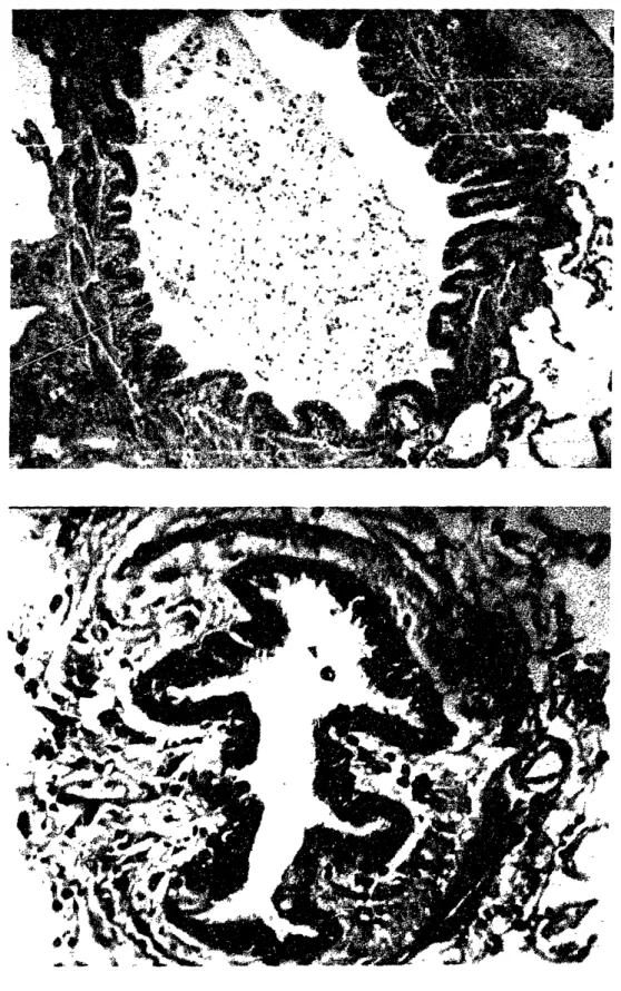
M
C
M
C
(top) Histology of a single fold of a constricted membranous bronchial airway. (from the University of British Columbia Pulmonary Research Laboratory) (bottom) Map indicating key components of the inner airway wall: lumen (L), epithelial cell layer (E), sub-epithelial collagen layer (C), internal submucosa (S), smooth muscle bundles (M). The thin "basement membrane" exists at the border between the sub-epithelial collagen layer and the epithelium.
Figure 1-4 shows a view of a single mucosal fold of a normal airway, from a slide
prepared with a Gomori trichrome stain. This stain makes the sub-epithelial collagen layer show up as a bright blue band, making it easier to distinguish it from the submucosal layer, where collagen is present but not as dense. Also, the epithelial layer, submucosal layer and intermittent smooth muscle layer are easily identified in Figure 1-4.
1.2.3 Airway Hyperresponsiveness
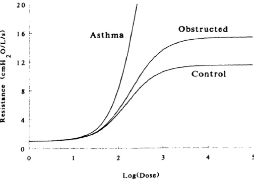
Asthrnatic bronchioles demonstrate "hyperresponsiveness." An airway is considered hyperresponsive when it constricts significantly more than a normal one would for a specified dose of smooth muscle agonist. Figure 1-5 is from paper by Wiggs et al.,50
showing the difference in response to smooth muscle agonist between airways under normal ("control"), non-asthmatic COPD ("obstructed"), and asthmatic conditions. The abscissa is the concentration of a dose of agonist (for example acetylcholine or
methacholine) which causes the airway smooth muscle to contract, constricting the airway. The ordinate is a measure of res;stance to airflow through the airway, normalized by the
1-.) U Ix u 16 12 Asthma Obstructed Control 4 V . 0 1 2 3 4 5 I og(Dose)
FIGURE 1-5: Theoretical dose response curves showing the marked increase in airflow resistance in asthma compared to normals and other chronic obstructive
pulmonary diseases. (from Wiggs et al.50) ,n
amount of resistance that would be exhibited by an airway that is completely open (and has not collapsed at all).
The plot shows the simulated response for typical normal, COPD and asthmatic airways. The plateau in the response of normal and obstructed airways indicates some type of mechanism for resisting high levels of constriction. During the plateau, the airway's stiffness to occlusion increases dramatically, so much so that the smooth muscle reaches its load-generating capacity and is unable to collapse the airway beyond a certain point.
Asthmatic airways do exhibit the same plateau in their response, but it is so markedly delayed to higher levels of constriction that it falls far off the axes of this plot. The smooth muscle is easily able to collapse such an asthmatic airway to very high airflow resistance levels under a comparable dose of agonist. Non-asthmatic COPD airways display a similar trend as asthmatic ones but to a far lesser degree.
1.2.4 Structural Differences Between Normal and Asthmatic Airways
The two cross-sections of human airways shown in Figure 1-6 are examples of typical normal (A) and asthmatic (B) bronchioles. Two more example airways are shown in Figure 1-7. They are not necessarily of exactly the same size, and are certainly not stained in the same way, but are presented here to show general trends in normal and asthmatic airway structure. In both cases the smooth muscle has contracted, and there is a resulting epithelial folding pattern. The change in airway structure from A to B in the course of chronic asthma is a type of "remodeling." Remodeling is the process of maintenance or turnover through which extracellular matrix is continually replaced.58 Certain mechanisms of remodeling exhibit enhanced synthesis and reduced degradation of matrix materials, resulting in larger and/or stiffer tissues. Others are oppositely more catabolic, resulting in smaller and/or more compliant tissues. Asthmatic airway wall remodeling is an example of the former.
Following are some of the notable structural-mechanical differences between normal and asthmatic airways, not all of which can be discerned from Figures 1-6 and 1-7:
* All layers of the asthmatic airway are thickened: the epithelial cell layer, the sub-epithelial collagen layer, the proteoglycan submucosal layer, the smooth muscle layer, etc.l 3,20,22,2 3,27,31 The sub-epithelial collagen layer is about twice its normal
Two cross-sections of airways from normal (A) and asthmatic (B) human subjects. (from the University of British Columbia Pulmonary Research Laboratory)
collagen is deposited by fibroblasts as opposed to epithelial cells,7although current studies from our research group (Ressler and Kamm) are investigating the biochemical messages that epithelial cells are sending out under the stress that results from excessive bronchoconstriction. Preliminary results suggest that indeed epithelial cells do send out biochemical messages that would enhance synthesis. While the growth of the
submucosal tissues requires ample time to reach its full extent, the growth of the sub-epithelial collagen has been known to occur within two weeks of asthmatic symptoms, and this thickening appears irreversible.2 6
@ One might suspect that in asthma the smooth muscle layer, because it has thickened due to cell hyperplasia, 8 can generate greater stress. Bramley et al. have measured
maximum stress capacities in asthmatic smooth muscle to be about 3 times as large as in normal smooth muscle.6 Other researchers, however, have indicated that there is no evidence to suggest greater smooth muscle stress capacity in asthma.
The lumen is far more occluded upon smooth muscle contraction in the asthmatic airway, not only from further constriction of the airway itself, but by interstitial fluid which had been squeezed out of the various airway layers, into the lumen.8,,19 5 6,57
* Though there has been no systematic study which has demonstrated it, there is some anecdotal evidence to suggest that the number of mucosal folds upon smooth muscle contraction is fewer in an asthmatic airway than in a normal one of comparable size (and location down the tracheobronchial tree). There has long existed evidence that the folds are deeper in asthmatic airways.22 It is in these large folds where fluid which has been squeezed out of the airway wall collects. Airways that collapse with fewer folds also tend to have an irregularly shaped lumen which appears to have collapsed greatly, while normal airways in general have more folds and a more circular, less obstructed lumen, as seen in Figures 1-6 and 1-7.
* Blood vessels which line the airway and run parallel to it through the submucosa tend to be fully open if they are located between two mucosal folds, but are shut closed if they are located at the tip of a fold.4 6
Asthmatic airways contain a much larger number of inflammatory cells, particularly throughout the submucosa, including TH2 lymphocytes, eosinophils and mast cells.12
FIGURE 1-7: Another two cross-sections of airways from relatively normal (top) and highly asthmatic (bottom) human subjects. (from the University of British Columbia Pulmonary Research Laboratory)
Epithelial desquamation is often observed in asthmatic airways. Those epithelial cells that have not been sloughed off are usually thicker and inflamed.3 3
All these changes occur in patches throughout the lungs in mild asthma, becoming more widespread in severe asthma.12
1.3
Previous Work
One feature that makes this work distinctive in the multitude of studies which attempt to elucidate airway wall mechanics is the special attention to the sub-epithelial collagen layer: how it folds and what possible ramifications its folding pattern has for an airway's load-resisting capacity. Most previous studies have considered only axisymmetric deformations
of airway components, thus neglecting mucosal folding.3,6,24,32,34,39, 5 0,51 Such models
would not consider buckling, and thus would grossly overestimate the stiffness of the airway to occlusion, perhaps dismissing the importance of the mode of collapse altogether. Some recent studies, mostly by Lambert et al. focus on the sub-epithelial collagen laer ind demonstrate its significance with regard to resistance to smooth muscle shortening anri to intraluminal liquid-filling of the mucosal folds.19,35,3 6 Our work is similar in that we also attempt to further understand mucosal folding, but our approach (which primarily uses finite element methods) is designed to better capture the interaction between the various layers internal to the constricting smooth muscle.
The simplest model which demonstrates this interaction is the planar sandwich panel, frequently used in structural design.' While incorporating circular geometries, earlier theoretical work by Lambert et al. obtains results based mostly on geometrical arguments, thus neglecting the full effect of material laws and force balance between the various airway layers.3 5,3 6 Lambert's models are indeed bi-layered, but the outer (submucosal) layer is assumed to be a fluid, and thus to have a negligible shear modulus. Our criticism of these models is that such a solid-on-fluid configuration should most likely only buckle in a peanut-shaped two-lobe collapse, as the solid is a single-layered ring responding to a uniform pressure with no shear. Higher frequency modes of collapse require significantly greater strain energy and thus are highly unlikely to be observed under simple pressure.
Our work is in effect somewhat of a hybrid between planar sandwich panels and Lambert's bi-layered circular models, obtaining results through numerical methods with some
analytical basis and experimental verification. The results that our models produce are significantly different from these previous modeling attempts.
1.4
Hypotheses
The implications of various hypothetical kinds of airway wall remodeling can be examined by varying the parameters of our two-layer model.
Firstly, one might hypothesize that growth of the proteoglycan-rich submucosal layer primarily stiffens the airway against luminal compromise. This thicker outer layer of loose connective tissue is likely to be the dominant source of resistance to smooth muscle
contraction (because it is so much thicker than the mucosal components). Without the presence of some type of confining stiffer inner layer, there would be only one possible mode of airway deformation upon smooth muscle constriction, a simple axisymmetrc contraction, which (if the assumptions of incompressibility and plane-strain are valid) would preserve the cross-sectional area of the airway wall tissue. At a given airway size (specified by its inner diameter when relaxed2 5), having more outer layer tissue implies that
more tissue has to be deformed to achieve the sane amount of luminal compromise, resulting in greater resistance to deformation, and consequently, a stiffer airway. If this hypothesis is true, submucosal thickening cannot be the only mechanism of importance in asthmatic airway wall remodeling.
Another major hypothesis that we wish to justify is the idea that any type of thickening or stiffening of the thin mucosal layer is detrimental to the stiffness of the airway at large deformations, thus predisposing the lumen to greater compromise upon maximal smooth muscle constriction. At first glance, this hypothesis may seem counter-intuitive and contrary to some previous reports. To quote Roberts, '"Thickening of the collagen-rich matrix beneath the basal lamina would be expected to increase both the tensile stiffness and resistance to deformation of the airway wall, thus tending to oppose the effect of smooth muscle shortening on airway narrowing."43
The argument is not really counter-intuitive after some examination of the mechanics governing the structure. It is inspired by thinking of the inner mucosal layer as a beam (on
an elastic foundation, the submucosa). Thickening or stiffening of the inner layer increases its intrinsic "beam stiffness" (EI, elastic modulus times moment of inertia). The inner layer's beam stiffness increases linearly with its elastic modulus, but with the cube of its thickness. Greater beam stiffness implies greater resistance to bending, but less curvature and larger wavelengths in the bending deformation. For a given airway size (same internal diameter & perimeter), this means fewer folds. We hypothesize that buckling in fewer folds requires a marginally larger buckling pressure, but once this pressure is met, collapse of the lumen can be more catastrophic than if the mucosa buckled into a greater number of smaller folds.
Furthermore, buckling in fewer folds implies that there is more constriction possible before the folds bend so much that they touch and eventually close up on themselves, providing more confinement of the surrounding submucosal tissue and markedly increasing the airway's stiffness. As a result, an airway with an increased mucosal layer thickness (or stiffness) under the same pressure generated by the surrounding smooth muscle would exhibit more iuminal compromise in its postbuckling response than one with a normal mucosal layer.
Figure 1-8 depicts the contraction of two hypothetical two-layer tubes (or airways), abbreviated to show only a mechanically stiffer mucosal layer and a thicker, more compliant proteoglycan layer (representing the internal submucosa). The smooth muscle and adventitia are not shown but are assumed to move in a simple axisymmetric, radially inward deformation as the smooth muscle contracts. Suppose that the intrinsic stiffness of the material that the mucosal layers are made of is the same. The only way in which these two hypothetical airways differ is that airway B has a thicker mucosal layer than airway A. Because the mucosal layer is thin but relatively stiff, at larger deformations it becomes energy-efficient to bend (or buckle) non-axisymmetrically instead of compressing circumferentially.
It has been suggested that the number of folds remains roughly constant throughout the constriction of in vivo airways. Therefore, assume for now that after buckling each of our
hypothetical airways continues to contract with the same number of folds until the sides of their folds are flat against one another. At this point, the mucosal layer cannot fold much more so the airway becomes stiffer to continued luminal occlusion. A different type of buckling mechanism is necessary to compress the airway further (perhaps buckling of the
outer proteoglycan layer), and that event will probably not occur because the load necessary would be greater than the smooth muscle's capacity.
The sketched plots to the right in Figure 1-8 give an idea of the load-displacement behavior of these hypothetical airways. The amount of lumen area available for airflow is plotted against the constricting smooth muscle pressure. Pmax is the maximum amount of pressure that the smooth muscle can provide, assumed for now to be the same for asthmatic as for normal airways. The prebuckling behavior of the two airways is such that the airway with the thickened mucosa is slightly stiffer to airway obstruction. However, when buckling
does occur, the consequence of buckling withfewerfolds is a more catastrophic collapse, resulting in less luminal area under maximal shortening of the smooth muscle, even if that
maximum were not to increase despite muscle hyperplasia.
A P Pmax A I I
P
PmaFIGURE 1-8: The major hypothesis of this work: having a relatively thin and/or compliant inner layer leads to buckling with relatively many folds (A), while having a thick and/or stiff inner layer causes buckling in relatively few folds (B). By the time the folds press up against one another, resisting further collapse, more cross-sectional lumen area is lost (in case B relative to A). The plots to the right show lumen area as a function of smooth muscle pressure applied throughout the contraction. P,,, indicates a hypothetical maximal smooth muscle pressure.
A
B
Further hypotheses can be cast addressing why the asthmatic airway would remodel the way it does using mechanical arguments. In case A of Figure 1-8, the mucosal layer undergoes much deformation while most of the submucosal layer undergoes a simple compression. Most of the strain energy of deformation is put into the mucosal layer and the nearby portion of the surrounding submucosal layer. This suggests that under repeated airway contraction (ue to an increasingly chronic asthmatic condition), cells in these regions (such as epithelial cells or fibroblasts in the nearby submucosa) might mediate the construction of more collagenous tissue to reduce stress levels locally. In case B, the increased thickness of the sub-epithelial collagen layer reduces stress on the epithelium and nearby cells by bending with less curvature, and more of the strain energy is devoted to larger deformations in the submucosa.
The purpose of the models to be presented in this work is not only to give credence to these hypotheses, but to attempt to quantify the effects of the possible structural changes
considered. Though the second hypothesis offers an explanation for how the increased structure of an asthmatic airway may result in greater luminal compromise upon maximal
smooth muscle activation, it is not necessarily the only mechanism which could cause such an effect.
1.5
Other Applications
There are several other biologic vessels of similar geometry and structure that also constrict, causing folds to appear at their luminal surfaces.
For instance, Lee and Chien have observed longitudinal ridges in the vascular endothelium and underlying internal elastic lamina of the canine carotid artery at reduced arterial
pressures.3 7 Razakamiadana et al. have studied the buckling instability of coronary microvessels, modeling them as thin extensible tubes embedded in a multi-phasic elastic medium. Their purpose was to study the reduction of coronary blood flow during cardiac contraction.4 2 It has also been suggested that fibrosis of the vascular basement membrane may be associated with diabetic ischemia.30 ,52, 53
Collagenous colitis, often characterized by inadequate absorption in the colon, involves thickening of the sub-epithelial collagen layer of the colon.1 5,16,2 8,41 Lewis et al. observed some shortening and broadening of the villi accompanying the enhanced collagen
deposition.3 8 Similar conditions of sub-epithelial fibrosis are known to exist in
neighboring digestive organs such as the small intestine (collagenous sprue)4 8and the stomach (collagenous gastritis).o The walls of the esophagus and urethra also contain
collagenous basement membranes which are known to fold and be prone to fibrosis.11,29
Because many biologic vessels share similar anatomical structure to airways, and because
they exhibit various medical conditions associated with thickening and stiffening of the
various vessel layers, the results of the simple models described here may have broader
implications for many kinds of vessels and their corresponding pathologies.
CHAPTER I REFERENCES
1. Allen HG. Analysis and Design of Structural Sandwich Panels. Pergamon Press, London, 1969. pp. 156-163.
. Bai A, Eidelman DH, Hogg JC, James AL, Lambert RK, Ludwig MS, Martin J, McDonald DM, Mitzner WA, Okazawa M, Pack RJ, Par6 PD, Schellenberg RR, Tiddens HAWM, Wagner EM, Yager D. Proposed nomenclature for quantifying subdivisions of the bronchial wall. J. Appl. Physiol. 77(2): 1011-1014, 1994.
3. Bates JHT, Martin JG. A theoretical study of the effect of airway smooth muscle orientation on bronchoconstriction. J. Appl. Physiol. 69(3): 995-1001, 1990.
4. Belilet L-P, Laviolette M, Turcotte H, Cartier A, Dugas M, Malo J-L, Boutet M. Bronchial subepithelial fibrosis correlates with airway responsiveness to methacholine. Chest. 112(1): 45-52, 1997.
5. Bousquet J, Chanez P, Lacoste JY, White R, Vic P, Godard P, Michel FB. Asthma: a disease remodeling the airways. Allergy. 47: 3-11, 1992.
6. Bramley AM, Thomson RJ, Roberts CR, Schellenberg RR. Hypothesis: excessive
bronchoconstriction in asthma is due to decreased airway elastance. Eur. Respir. J. 7: 337-341, 1994.
7. Brewster CEP, Howarth PH, Ratko D, Wilson J, Holgate ST, Roche WR. Myofibroblasts and subepithelial fibrosis in bronchial asthma. Am. J. Respir. Cell Mol. Biol. 3: 507-511, 1990.
8. Brown RH, Zerhouni EA, Mitzner W. Airway edema potentiates airway reactivity. J. Appl. Physiol. 79(4): 1242-1248, 1995.
9. Brown RE, Butler JP, Rogers RA, Leith DE. Mechanical connections between elastin and collagen.
Connective Tissue Research. 30: 295-308, 1994.
10. Colletti RB, Trainer TD. Collagenous gastritis. Gastroenterology. 97(6): 1552-1555, 1989.
11. De Carvalho HF, Line SRP. Basement membrane associated changes in the rat ventral prostate following castration. Biology International. 20(12): 809-819, 1996.
12. Drazen JM. Asthma, in Textbook of Medicine. ed. Cecil. ch. 51.
13. Dunnill MS. The pathology of asthma, with special reference to changes in the bronchial mucosa. J.
14. Ebina M, Yaegashi H, Takahashi T, Motomiya M, Tanemura M. Distribution of smooth muscles along the bronchial tree. Am. Rev. Resp. Dis. 141: 1322-1326, 1990.
15. Eckstein RP, Dowsett JF, Riley JW. Collagenous enterocolitis: a case of collagenous colitis with involvement of the small intestine. The American Journal of Gastroenterology. 83(7): 767-771, 1988.
16. Giardiello FM, Bayless TM, Jessurun J, Hamilton SR, Yardley JH. Collagenous colitis: physiologic and histopathologic studies in seven patients. Ann. Intern. Med. 106: 46-49, 1987.
17. Guyton AC. Textbook of Medical Physiology, 8th Ed. W.B. Saunders Co., Philadelphia, 1991. ch. 42.
18. Heard BE, Hossain S. Hyperplasia of bronchial muscle in asthma. J. Pathol. I 10: 319-33!, 1973. 19. Hill MJ, Wilson TA, Lambert RK. Effects of surface tension and intraluminal luid on mechanics ot
small airways. J. Appl. Physiol. 82(1): 233-239. '1997.
20. Hogg JC. Bronchiolitis in asthma and chronic obstructive pulmonary disease. Clinics in Chest
Medicine. 14(4): 733-740), 1993.
21. Hogg JC, Macklem P', Thurlbeck WM. Site and nature of airway obstruction in chronic obstructive disease. N. Engl. J. Med. 278: 1355-1360, 1968.
22. ttuber HL, Koessler KK. I'he pathology of bronchial asthma. Arch. nt. Med. 30(6): 689-760, 1922,
23. James AL. Relationship between airway wall thickness and airway hyperresponsiveness, in Airway
Wall Remodelling in Asthma. ed. Stewart AG. CRC Press, Boca Raton, 1997. ch. 1.
24. James AL, Pare PD, Hogg JC. The mechanics of airway narrowing in asthma. Am. Rev. Respir.
Dis. 139: 242-246, 1989.
25. James AL, Hogg JC, Dunn L, Pare PD. The use of internal perimeter to compare airway size and to calculate smooth muscle shortening. Amer. Rev. Resp. Dis. 138: 136-139, 1988.
26. Jeffrey PK, Godfrey RW, Adelroth E, Nelson F, Rogers A, Johanson S-A. Effects of treatment on airway inflammation and thickening of basement membrane reticular collagen in asthma. Am. Rev.
Respir. Dis. 145: 890-899, 1992.
27. Jeffery PK. Morphology of the airway wall n asthma and in chronic obstructive pulmonary disease.
Am. Rev. Respir. Dis. 143: 1152-1158, 1991.
28. Jessurun J, Yardley JH, Gardiello FM, Hamilton SR, Bayless TM. Chronic colitis with thickening of the subepithelial collagen layer (collagenous colitis): histopathologc findings in 15 patients. Human
Pathology. 18(8): 839-848, 1987.
29. Kalkay NM, Cordoba I, Plevy D. The nonreflux determinant of esophagitis. The American Journal of
Gastroenterology. 63(2): 135-146, 1975.
30. Katz MA, McCuskey P, Beggs JL, Johnson PC, Gaines JA. Relationships between microvascular function and capillary structure in diabetic and nondiabetic human skin. Diabetes. 38(10): 1245-1250,
1989.
31. Kuwano K, Bosken CH, Pare PD, Bai TR, Wiggs BR, Hogg JC. Small airways dimensions in asthma and in chronic obstructive pulmonary disease. Am. Rev. Respir. Dis. 148(5): 1220-1225,
32. Lai-Fook SJ, Hyatt RE, Rodarte JR, Wilson TA. Behavior of artificially produced holes in lung parenchyma. J. Appl. Physiol. 43(4): 648-655, 1977.
33. Laitinen LA, Laitinen A. Modulation of bronchial inflammation: corticosteroids and other theraputic agents. Am. Rev. Respir. Crit. Care. Med. 150: S87-S90, 1994.
34. Lambert RK, Wiggs BR, Kuwano K, Hogg JC, Pare PD. Functional significance of increased airway smooth muscle in asthma and COPD. J. Appl. Physiol. 74(6): 2771-2781, 1993.
35. Lambert RK, Codd SL, Alley MR, Pack RJ. Physical determinants of bronchial mucosal folding. J.
Appl. Physiol. 77(3): 1206-1216, 1994.
36. Lambert RK. Role of bronchial basement membrane in airway collapse. J. Appl. Physiol. 71(2): 666-673, 1991.
37. Lee MML, Chien S. Morphologic effects of pressure changes on canine carotid artery endothelium as observed by scanning electron microscopy. The Anatomical Record. 194(1): 1--14, 1979.
38. Lewis FW, Warren GH, Goff JS. Collagenous colitis with involvement of terminal ileum. Dig. Dis.
Sci. 36(8): 1161-1163, 1991.
39. Moreno RH, Hogg JC, Pard PD. Mechanics of airway narrowing. Am. Rev. Respir. Dis. 133: 1171-1180, 1986.
40. Okazawa M, Pare PD, Hogg JC, Lambert RK. Mechanical consequences of remodelling of the airway wall, in Airways and Vascular Remodelling. eds. Page C, Black J. Academic Press, Toronto, 1994. ch. 8.
41. Rams H, Rogers Al, Ghandur-Mnaymneh L. Collagenous colitis. Ann. Intern. Med. 106: 108-113, 1987.
42. Razakamiadana A, Oddou C, Zidi M, Geiger D. Buckling instability of thin tube stressed by
surrounding medium: application to coronary microvessels. Second World Congress of Biomechanics, Amsterdam, The Netherlands. July 10-15, 1994.
43. Roberts CR. Is asthma a fibrotic disease? Chest. 107(3): 11 1S-117S, 1995.
44. Roche WR, Beasley R, Williams JH, Holgate ST. Subepithelial fibrosis in the bronchi of asthmatics.
Lancet. 1: 520-524, 1989.
45. Vincent NJ, Knudsen R, Leith DE, Macklem PT, Mead J. Factors influencing pulmonary resistance.
J. Appl. Physiol. 29: 236-243, 1970.
46. Wagner EM, Mitzner W. Bronchial vascular engorgement and airway narrowing. Amer. Rev. Resp.
Dis. 149(4): A585, 1994.
47. Weibel E. Design of airways and blood vessels considered as branching trees, in The Lung: Scientific
Foundations. ed. Crystal R, et al. Vol. 1, pp. 711-720.
48. Weinstein WM, Saunders DR, Tytgat GN, Rubin CE. Collagenous sprue - an unrecognized type of malabsorption. N. Engl. J. Med. 283: 1297-1301, 1970.
49. Wiggs BR, Moreno R, James A, Hogg JC, Par PD. A model of the mechanics of airway narrowing in asthma, in Asthrma: Its Pathology and Treatment. eds. Kaliner MA, Barnes PJ, Persson CGA. Marcel Dekker, New York, 1991. ch. 3.
50. Wiggs BR, Bosken C, ParE PD, James A, Hogg JC. A model of airway narrowing in asthma and in chronic obstructive pulmonary disease. Am. Rev. Respir. Dis. 145: 1251-1258, 1992.
51. Wiggs BR, Moreno R, Hogg JC, Hilliam C, Pare PD. A model of the mechanics of airway narrowing. J. Appl. Physiol. 69(3): 849-860, 1990.
52. Williamson JR, Vogler NJ, Kilo C. Estimation of vascular basement membrane thickness: theoretical and practical considerations. Diabetes. 18: 567-578, 1969.
53. Williamson JR, Kilo C. Current studies of capillary basement-membrane disease in diabetes mellitus.
Diabetes. 26: 65-73, 1977.
54. Wilson JW, Li X, Pain MCF. The lack of distensibility of asthmatic airways. Am. Rev. Respir. Dis. 148: 806-809, 1993.
55. Yager D, Cloutier T, Feldman H, Bastacky J, Drazen JM, Kamm RD. Airway surface liquid thickness as a function of lung volume in small airways of the guinea pig. J. Appl. Physiol. 77(5): 2333-2340,
1994.
56. Yager D, Kamm RD, Drazen JM. Airway wail liquid. Chest. 107(3): 105S-I O(), I993.
57. Yager D, Butler JP, Bastacky J, Israel E, Smith G, Drazen JM. Amplification of airway constriction due to liquid filling of airway intersticies. J. Appl. Physiol. 66(6): 2873-2884, 1989.
58. Yannas IV, Spector M. Lecture notes for M.I.T. course 20785: Mechanical Forces in Organ Development and Remodeling. Spring 1993.
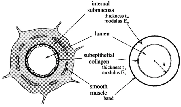
Development of the Two-Layer Model
2.1
Model Components
The simple two-layer structure of the models employed in these studies is based on the geometry of the passive vessel structure internal to the smooth muscle (Figure 2-1). The outer layer represents the loose connective tissue of the submucosa, mostly proteoglycans;
the inner layer represents the mucosa, whose primary structural component is the sub-epithelial layer of relatively well-ordered collagen. '8 Thus while the outer layer may be an order of magnitude thicker than the inner layer, the inner layer is assumed to have at least an order of magnitude more material stiffness than the outer layer.
internal
submucosa
cness to
ulus Eo
FIGURE 2-1: Sketch of a membranous bronchiole and the two-layer tube idealization,
showing correspondence between airway components and tube layers.
For simplicity, only these two layers are included, thus the epithelial cell layer is considered a part of the stiffer inner layer. The epithelium in particular, while comparable in thickness to the collagen layer beneath it, is perhaps an order of magnitude less stiff than the
submucosal layer, making it at least two orders of magnitude less stiff than the collagen
2.
I
layer.l 1 Therefore it is not included separately in the model because it cannot provide any significant resistance to collapse nor significantly affect the deformation behavior of the collapse. Similarly, airway fluids and gels which reside on and around the epithelium have a negligible elastic modulus and are therefore not included.
It is assumed that the smooth muscle layer and any other layers external to it move in a simple axisymmetric deformation, and therefore those layers do not need to be modeled in detail with continuum elements. The total effect of such external components is to impose a particular boundary condition on the internal submucosa (described in detail in the next section).
2.2
Mechanical Modeling Assumptions
2.2.1 Two-Dimensional Plane-Strain
All of the mechanical models presented here assume two-dimensional plane-strain
deformation. When excised, split axially and examined from the side, constricted airways exhibit long ridges that run axially down the wall (Figure 2-2). In cross-section, these ridges are the mucosal folds. The number of these folds does not seem to vary with axial distance along an airway, assuming that the length of airway observed is far enough away from bifurcations between airway generations where in general, the overall airway
dimensions change.
The smooth muscle in the airway wall is not a continuous band the way our simple model would suggest, but is intermittent around the cross-section of the airway. In fact, it is more like a randomly weaving mesh. It is assumed that the effect of the random wrapping is as if the muscle is continuous. Further examination of the airway cross-sections of Figures
1-6 and 1-7 reveals no appreciable difference in the deformation field in zones where the smooth muscle is present from where it is absent. This is because it is probable that a small distance axially up the airway there is muscle present where there wasn't previously, thus its effects tend to be averaged-out in the axial direction. Since there appears to be little variation with axial distance far enough away from bifurcations, our computations assume two-dimensional loading and deformation for simplicity and ease of computation.
FIGURE 2-2: Micrograph showing ridges (folds) running axially up and down the airways.
Given that there is two-dimensional deformation, an assumption must be made regarding the displacements in the third (axial) direction. The two extremes possible are so-called "plane-stress" and "plane-strain," implying that all non-zero stresses or strains lie in a plane (which is to say that in stress, there is no force in the axial direction and in plane-strain, there is no deformation in the axial direction). Of these two choices, plane-strain is more applicable to modeling airways because it is unlikely that they actually shorten or elongate axially during constriction. In such a case there would be motion of bifurcations toward and away from one another. The smooth muscle wraps around the airway at approximately a 13" angle to the plane of an airway cross-section (a relatively shallow angle), further supporting the idea that smooth muscle shortening should not produce any appreciable length change along the axis of the airway.18 Bates et al. have shown that
smooth muscle at angles of less than 30°has little effect on airway length during their constriction.4 Simplification to two-dimensional plane-strain, poses certain computational difficulties when incompressible (or nearly incompressible) material laws are used. How such difficulties are circumvented is described in the next section.
2.2.2 Homogeneous Isotropic Layers
It is assumed that each of the two layers is a homogeneous isotropic continuum. The only difference between the two layers is the assigned elastic modulus (or roughly speaking, the intrinsic material "stiffness").
At a relatively macroscopic level, homogeneity of material properties in each layer is a good assumption. However, at sufficiently small scales it becomes apparent that there are collagen and elastin bundles which would correspond to regions of slightly higher or lower E in the submucosa. The sizes of these bundles are very small compared to the gross thickness of the submucosa, and so it is assumed that their effects average out to produce a roughly constant value of E over the layer. The boundary between the two layers is not nearly as clear in an actual airway as it is in our two-layer model. However, in human airways there is ajump in collagen content within a small thickness under the epithelium, and the location of that jump is taken to be the interface between the two discrete layers, as reported by various morphometric studies.
Another simplification is the assumption of isotropy (or rotational invariance of material properties). The submucosa, being a random array of loose connected tissue, is probably
best modeled as isotropic. The sub-epithelial collagen layer, however, does have significantly more order to it. The collagen fibers appear to align themselves at
approximately 45° to the airway axis forming a loose helix.'8 The 45°angle orientation
suggests that there is an equal tendency to be stretched longitudinally along the airway axis as well as to be expanded or contracted circumferentially. Perhaps this is the normal state of loading associated with lung expansion and contraction due to breathing.
Though some attempts to model the inner layer as anisotropic were attempted, mixed success with them has prohibited further research in that direction at present. Any material with more than a 2-to- I parallel-to-perpendicular ratio of stiffnesses proved to be unstable
under compression. All models discussed henceforth have homogeneous isotropic material properties within their respective layers.
2.2.3 Incompressible Hookean and Neohookean Materials
In creating structural models of biological tissues, knowing the appropriate constitutive law is probably the most elusive issue. This is because the microstructure of tissues is not nearly so simple as that of more traditional engineering materials such as steel or even concrete. This is further complicated by the fact that tissues are generally hydrated and can have varying properties depending on the degree of hydration and the degree of
permeability, both of which may be affected by variations in the biochemical environment of the tissue. While it is more probable that the tissues modeled here demonstrate a nonlinear relationship between the strain they exhibit and the stress they develop, in the absence of proof to the contrary, they will be assumed linear. If not linear, are the materials strain-stiffening or strain-softening, and to what degree? Strain-stiffening and strain-softening material behaviors are contrasted pictorially with linear behavior in Figure 2-3. Under different conditions, tissues may exhibit either kind of nonlinear behavior. Some preliminary experiments on airway submucosa have yielded relatively linear results (see Section 6.2).
Even with the assumption of a linear material law, knowing the various moduli of these materials precisely is difficult. One could go to great lengths to develop sophisticated
/ / r behavior (E constant) i-stiffening (E increasing) -softening (E decreasing) strain (E)
FIGURE 2-3: Typical assumptions of stress versus strain behavior. The sketch
depicts linear, strain-stiffening and strain-softening material laws. E is
defined as the slope of this curve.
material models with many parameters, only to find that the space of possible material behaviors that is spanned by the best estimate of the parameters - plus or minus the large error in those parameters - could just as easily have been spanned by a simple model with
relatively few similarly uncertain parameters. That is why it is preferable in this case to model the materials involved with as few moduli as possible. Obtaining a reasonable guess at just two moduli has been difficult enough.
Our comnputational models assume the simplest material constitutive law possible, that of an incompressible linear-elastic (Hookean) material, characterized by a single parameter (E) for each layer. An isotropic, linear-elastic material is completely described by only two elastic moduli. For those two constants we will use Young's uniaxial elastic modulus (E) and the Poisson ratio (v). One might also have chosen some combination using the shear modulus (G), the bulk modulus (K) or the Lam6 constant (h), but our choice is a common one. Aside from some of the details in this section, the modeled materials will be described using E and v exclusively, though conversion to the other constants is entirely equivalent.
Young's uniaxial elastic modulus is so named because it is the ratio of stress to strain in a uniaxial unconfined tension or compression test. The Poisson ratio describes how the material will behave in the two unstressed directions during such a test. It ranges from 0 (when there is no displacement in the other two directions) to 1/2 (when there is half as much opposite displacement in each of the other two directions).
As v approaches /2 the material becomes more volume-preserving or "incompressible." This is because in a compression test where the material is completely confined in the untested directions, there will be no deformation - i.e. the material is "incompressible." Such a material has an infinite bulk compression modulus (K). The constituents of tissues (mostly water and proteins) do indeed appear to be incompressible when each is taken separately. When examining the combination of these constituents in a bi-phasic material, however, there is the possibility of a gross "compressible" behavior when the water is allowed to squeeze out of the solid matrix.
Most of the simulations presented here will make the leap of assuming that the gross materials involved are nearly incompressible. That is to assume that the interval of time over which the smooth muscle contracts is sufficiently short that the water which is inside the tissue does not have enough time to move any significant distance. It is as if the water is bound to the solid protein matrix and the material has a single phase. Some simulations have also been performed at different values of v, implying that water is squeezed out of the tissues, but this is a highly approximate way of incorporating water movement.
Chapter 8 investigates loosening the incompressibility assumption in a more rigorous way using a bi-phasic poroelastic material formulation. Until then, however, the material models used in the simulations presented will be the single-phase ones discussed in this section.
Stress and Strain Measures:
In describing the constitutive laws that have been used to develop the two-layer
computational models, there will be several references to stress and strain. The stress used here is the Cauchy or "true" stress, which is the force intensity per unit of current area in the deformed configuration. The stress is a full second-order tensor, which for two-dimensional computations is abbreviated as:
[;
:11 2 1 [T11 12T21 22 J T1T2 T22
Conservation of angular momentum dictates that the off-diagonal shear stresses (1 2 and ·2 1) be equal, making the stress tensor symmetric. If plane-strain is assumed, t3 3is not
necessarily zero, but it is typically not needed in computations and can be calculated afterward. For simplicity it is often preferable to express the three distinct stresses in a column matrix.
T22
x12
The strain can be defined in many ways. Presented below is a derivation of the nonlinear (B) and linear (E) strain measures which have been of use in this work. Note that in a plane-strain analysis, the deformation in the third direction is zero, and thus terms pertaining to that direction cancel out.
identitymatrix: 1
[
0[ 1current position as a function of reference position: y(x) displacement as a function of reference position: u(x) = y(x) - x
deformation gradient:
ayl
F Vy= = y_ Odx ax1 ax2 ax2 -displacement du gradient: I a aulaxl
au2 dxlau
1
aX2 au2 ax2left Cauchy-Green strain tensor: B F FT
dul axl aul ax2 aU2 aX1
]
au2 +dx2 = F-1aYI Y2 + OY ay2 ax1 axI ax2 ax2
/ 9y
2
(ay2\
2 kaxl) +aX 2) 1 + 22aul +ax +adul
taxl au2 aul aU2 + ax + ax1 axl /OUl\2+
(
a)2
+ul aU2 + aX2 aX2aul au2 aul au2 aul au2 ax2 ax1 axl axl ax 2 x2 1 + 22UaX2 2 /+ (u 2\2
+ ~,ax
2)
+ ( x
1)
I
B = FFT = (1 +H)(1 +H)T = 1 +H+HT+HHTBy design, this strain measure is symmetric (caused. oy multiplying a tensor by its transpose). While the left Cauchy-Green strain tensor is good for measuring nonlinear
strains, a simpler measure is desired for the small strains used in linear elasticity. The small strain tensor (£) is defined as follows:
=I2
(H +
HT) =[
aul ax1 1l au\22
ax
1l
+1 aU2\ 2ax
1) du2 ax21
]1
Note that B = 1 + 2 + quadratic strain terms.
As with the Cauchy stress, it is often convenient to write the three unique small strain terms in a column matrix:
IE22~
-£12JE~~~~~J adu axl au2 ax2(aul +
ax2 du2 IxiHookean (Linear-Elasticity) Constitutive Theory:
(
d
2
2 axi axl + (d 2 +ax
axYi Y2 2 aX2I
aul ax2Using the linear-elastic material description, the stresses at any particular point are a linear transformation of the strains at that point. For instance, the isotropic linear-elastic
constitutive behavior linking the stress components with the strain components in a two-dimensional plane-strain analysis can be expressed as follows:
E(1-v) (1+v)(1-2v) Ev (1+v)(1-2v) 0 Ev ( 1+v)(1-2v) E( -v) (l+v)(1-2v) 0
This is Hooke's law for two-dimensional plane-strain. Note that as v approaches 1/2, some of the constitutive terms give rise to singularities. This is because the average stress (or pressure) is becoming less dependent upon any of the strains taken independently. In finite element analyses, this problem manifests itself as non-physical mesh-dependent stress banding and element locking. To circumvent this problem, the so-called "u/p" or displacement-pressure formulation is introduced. Here is how the u/p formulation is derived for this situation:
For convenience, we can write Hooke's law for plane-strain in terms of the other elastic moduli, defined as follows:
shear modulus: E 2(1+v) bulk modulus: K E 3(1-2v) Ev Lam6 constant: - Ev (1+v)(1-2v)
Thus the above expression of Hooke's law for plane-strain is more simply written:
1 11] T22j T1 2J 0 E 1+v E22j