HAL Id: hal-01100508
https://hal.archives-ouvertes.fr/hal-01100508
Submitted on 8 Jan 2015HAL is a multi-disciplinary open access
archive for the deposit and dissemination of sci-entific research documents, whether they are pub-lished or not. The documents may come from teaching and research institutions in France or abroad, or from public or private research centers.
L’archive ouverte pluridisciplinaire HAL, est destinée au dépôt et à la diffusion de documents scientifiques de niveau recherche, publiés ou non, émanant des établissements d’enseignement et de recherche français ou étrangers, des laboratoires publics ou privés.
Proceedings of Surgetica’2014
Jocelyne Troccaz
To cite this version:
Jocelyne Troccaz. Proceedings of Surgetica’2014 : Computer-Assisted Medical Interventions: scientific problems, tools and clinical applications.. Jocelyne TROCCAZ. Surgetica’2014, Dec 2014, Chambéry, France. 2015. �hal-01100508�
Proceedings of
S
S
U
U
R
R
G
G
E
E
T
T
I
I
C
C
A
A
’
’
2
2
0
0
1
1
4
4
C
C
o
o
m
m
p
p
u
u
t
t
e
e
r
r
-
-
A
A
s
s
s
s
i
i
s
s
t
t
e
e
d
d
M
M
e
e
d
d
i
i
c
c
a
a
l
l
I
I
n
n
t
t
e
e
r
r
v
v
e
e
n
n
t
t
i
i
o
o
n
n
s
s
:
:
s
s
c
c
i
i
e
e
n
n
t
t
i
i
f
f
i
i
c
c
p
p
r
r
o
o
b
b
l
l
e
e
m
m
s
s
,
,
t
t
o
o
o
o
l
l
s
s
a
a
n
n
d
d
c
c
l
l
i
i
n
n
i
i
c
c
a
a
l
l
a
a
p
p
p
p
l
l
i
i
c
c
a
a
t
t
i
i
o
o
n
n
s
s
Editor : Jocelyne TROCCAZ
Chambéry, France, December, 2014
“Surgetica” conference series aims at gathering clinicians, scientists and industrial
actors in order to present and to discuss new computerized guidance tools and
their clinical applications. The 2014 conference has gathered French actors of
Computer Assisted Medical Interventions (CAMI) and three international invited
speakers Pr Alberto Arezzo (Department of Surgical Sciences, University of Torino,
Italy), Pr Nassir Navab (Technische Universität München, Germany) and Pr Russell
H. Taylor (Johns Hopkins University, Baltimore, USA) to discuss the recent progress
and the remaining clinical and scientific challenges of the field.
Surgetica is a single-track conference mixing scientific and clinical presentations
(oral and poster). The conference language was English. An industrial exhibit was
proposed for complementary exchange about existing tools and clinical practice.
Digital proceedings contain the accepted extended abstracts. The papers can be
separately uploaded from
http://surgetica2014-papers.imag.fr/
website.
The last morning of the conference was dedicated to the presentation of activities
developed in the CAMI excellence laboratory (cf.
http://cami-labex.fr
).
Scientific topics include among others: image processing and registration, modeling
and simulation, medical robotics and navigation systems, sensors and
instrumentation, specific man-machine interfaces and augmented reality, protocol
encoding and recognition, clinical evaluation of systems.
All clinical specialties are concerned among which: orthopedics and trauma,
urology, cardiac, vascular, digestive surgery, endoscopic surgery, interventional
imaging and radiology, neurosurgery, ENT, craniofacial surgery, radiation therapy.
Jocelyne TROCCAZ, Research Director CNRS
Organizing committee :
Chair : Jocelyne TROCCAZ, CNRS, TIMC-IMAG Laboratory
Pr Philippe MERLOZ, Grenoble University Hospital, TIMC-IMAG
Pr Eric STINDEL, Brest University Hospital, LATIM Laboratory
Jérôme SZEWCZYK, UPMC, ISIR Laboratory
Nabil ZEMITI, Montpellier II University, LIRMM Laboratory
Florent NAGEOTTE, Strasbourg University, ICUBE Laboratory
Antoine SIMON, Rennes I University, LTSI Laboratory
Program committee :
Chair : Jocelyne Troccaz, TIMC-IMAG, Grenoble
Bernard Bayle, ICube, Strasbourg
Georges Bettega, Cranio-facial surgery, Grenoble university hospital
Ivan Bricault, TIMC-IMAG, Radiology, Grenoble university hospital
Philippe Cinquin, TIMC-IMAG, CIC-IT Grenoble university hospital
Jean-Luc Descotes, TIMC-IMAG, Urologie, Grenoble university hospital
Jean-Yves Gauvrit, Radiology, Brest university hospital
Pascal Haigron, LTSI, Rennes
Julien Leboucher, LATIM, Brest
Antoine Lucas, vascular surgery, Rennes university hospital
Michel de Mathelin, ICube, Strasbourg
Philippe Merloz, TIMC-IMAG, Orthopedics-trauma, Grenoble university hospital
Guillaume Morel, ISIR, Paris
Pierre Mozer, ISIR, La Pitié Salpétrière Hospital, Paris
Florent Nageotte, ICube , Strasbourg
Yohan Payan, TIMC-IMAG, Grenoble
Philippe Poignet, LIRMM, Montpellier
Pierre Renaud, ICube, Strasbourg
Lotfi Senhadji, LTSI, Rennes
Antoine Simon, LTSI, Rennes
Eric Stindel, LATIM, Orthopedics-trauma, Brest university hospital
Jérôme Szewczyk, ISIR, Paris
Jean-Philippe Verhoye, Cardiac and vascular surgery, Rennes university hospital
Nabil Zemiti, LIRMM, Montpellier
Conference organization:
WEDNESDAY 3RD, DECEMBER 08:45 OPENING SESSION (J.Troccaz) 09:00-10:15 ORTHOPEDICS
Chairman: E.Stindel
09:00 Mid-term results of 29 computer-assisted osteotomies for genu valgum deformity.
D. Saragaglia, B. Chedal-Bornu Grenoble University Hospital
09:15 Functional hip center detection using only a tibial tracker for computer assisted tibial osteotomy:in vitro evaluation.
Z. Dib, G. Dardenne, N. Poirier, P-Y Huet, C.Lefevre, E. Stindel LATIM, B-com, OSTESYS SAS, Brest University Hospital
09:30 Three dimension fluoroscopy-based navigation for dorsal percutaneous instrumentation in traumatic vertebral fractures.
S. Ruatti G. Kerschbaumer, A. Moreau Gaudry, E. Chipon, C. Dubois, J. Tonetti, M. Milaire, P.Merloz
Grenoble University Hospital, CIC-IT Grenoble, TIMC-IMAG
09:45 Multi-modal Intra-articular Device for Virtual Biopsies of Cartilage.
A.Moreau-Gaudry, A. Meneses, F. Billet, D. Girardeau-Montaut, G. Custillon, B.Vettier, R. Adler, D. Saragaglia, P. Gaudin, P. Merloz, P. Cinquin
TIMC-IMAG, CIC-IT Grenoble, Grenoble University Hospital, NYU Langone Medical Center 10:00 Vector field interpolation for scapular position estimation from noisy acromial motion capture.
S. Bouvel, A. Roby-Brami, G. Morel, V. Pasqui ISIR
10:15-10:45 COFFEE BREAK
10:45-11H45 ORGAN DETECTION AND TRACKING Chairman: P.Haigron
10:45 Assistance to High Intensity Focused Ultrasound (HIFU) therapy: Real-time motion compensation using ultrafast ultrasound imaging.
L-A. Chanel, F. Nageotte, J. Vappou, J. Luo, L. Cuvillon, M. de Mathelin ICube, Department of Biomedical Engineering of Tsinghua University 11:00 Interactive Tracking of Soft Tissues in 2D Ultrasound Images.
L. Royer, M. Marchal, A. Le Bras, G. Dardenne, A. Krupa B-com, INRIA Rennes, Rennes University Hospital
11:15 Fully Automatic Organ Localization of Medical Images Using Improved Random Regression Forests.
P. Samarakoon, E. Promayon, C. Fouard TIMC-IMAG
11:30 Kidney 3D dynamic modeling and tumor tracking for therapeutic transcutaneous treatment.
V. Leonardi, M. Daniel, N. Olofsson, P. Souteyrand, V. Vidal, J-L. Mari LSIS, LIIE, Upssala University
11:45-12:30 POSTER TEASER (22x2mn)
14:00-15:00 INVITED SPEAKER 1 Chairman: Y.Payan
Robotic imaging for patient and process specific, multi-modal intra-operative, imaging and Visualization.
Pr Nassir NAVAB TUM University, Munich 15:00-16:30 REGISTRATION Chairwoman: C.Fouard
15:00 Fluorescence imaging of prostate cancer: when the surgeon looks beyond the visible.
R. Heus, M-P. Montmasson , A. Moreau-Gaudry, M. Hamel, F. Giroud, E. Chipon, O. Gaiffe , C.Pieralli ,M. Kassem, J-A. Long, J-L. Descotes, P. Cinquin, S. Voros
CIC-IT Grenoble, Grenoble University Hospital, TIMC-IMAG, FEMTO-ST
15:15 Registration of Preoperative Liver Model for Laparoscopic Surgery with Intraoperative 3D acquisition.
J. Bano , A. Hostettler, S. Nicolau, C. Doignon, L. Soler, J. Marescaux IRCAD, ICube, IHU Strasbourg
15:30 Video Synchronization: An Approach to Biopsy Site Re-localization.
A.S. Vemuri, A. Sportes, S. Nicolau, J. Marescaux, N. Ayache, L. Soler
IHU Strasbourg,IRCAD, INRIA Sophia-Antipolis, NHC, Strasbourg University Hospital 15:45 Phantom study of fiducial-free 3D-3D registration procedures for electromagnetic endovascular navigation.
LH.N'Guyen-Duc, A. de Lambert, S. Esneault, M. Castro, C. Göksu, J-L. Magne, P. Haigron LTSI, Therenva, Grenoble University Hospital, CIC-IT Grenoble
16:00 Percutaneous procedures with CT-Ultrasound fusion guiding imaging system: evaluation of the accuracy.
F. Bing, J. Garnon, I. Enescu, G. Tsoumakidou, M-A. Thenint, E. Breton, M. de Mathelin, M. Cardoso Saldanhas, M. Schaeffer, E. Sauleau, A. Gangi
Strasbourg University Hospital, ICube, Barretos Cancer Hospital
16:15 Evaluation of Human Factors in Neuronavigated Transcranial Magnetic Stimulation (TMS).
H. Chauvat, R. Ginhoux, B. Maurin ECE Paris, Axilum Robotics
16:30-17:00 COFFEE BREAK 17:00-17:45 POSTER SESSION
17:45-18:30 INSTRUMENT DETECTION AND TRACKING Chairman: J.Leboucher
17:45 Online registration applied to multi-modal tracking for real-time scan plane alignment in interventional MRI.
M. Neumann, L. Cuvillon, E. Breton, M. de Mathelin, L. Pan, A. Hengerer ICube, Siemens Research Corporate
18:00 Automatic detection of endoscope in intraoperative CT image : application to AR guidance in laparoscopic surgery .
S. Bernhardt , S. Nicolau, V. Agnus, L. Soler, C. Doignon, J. Marescaux IHU Strasbourg, ICube, IRCAD
P. Cabras, D. Goyard, F. Nageotte,P. Zanne, C. Doignon ICube
THURSDAY, 4TH, DECEMBER 09:00-10:00 BIOMECHANICS Chairman: M.Rochette
09:00 Biomechanical modeling to prevent soft tissues pressure ulcers.
V. Luboz, A. Perrier, M. Bucki, N. Vuillerme, F. Cannard, B. Diot, Y. Payan TIMC-IMAG, Texisense SAS, AGIM, IDS SAS
09:15 Predicting the consequences of tongue cancer surgery: design of a 3D patient-specific biomechanical model and evaluation.
P-Y. Rohan, A. Bijar, G. Bettega, P. Perrier, Y. Payan TIMC-IMAG, Gipsa-lab, Grenoble University Hospital
09:30 Biomechanical model of the fetal head for interactive childbirth simulation.
M. Bailet, F. Zara, E. Promayon LIRIS, TIMC-IMAG
09:45 An augmented reality approach integrating deformation simulation to assist EVAR procedures.
A.Duménil, A. Kaladji, J. Gindre, M. Rochette, C. Göksu, A. Lucas, P. Haigron LTSI, Therenva, CIC-IT Rennes, ANSYS France
10:00-10:45 CLINICAL DEVELOPMENTS Chairman: P.Poignet
10:00 Laparoscopic gastric bypass and gastric electrical stimulation with a robotically controlled needle holder (JaimyTM).
F. Reche, B. Trilling, T. Olshefski, P-A. Waroquet, C. Arvieux, J-L. Faucheron Grenoble University Hospital, Endocontrol Medical
10:15 Computer assisted transcatheter valve implantation for valve-in-valve procedures.
R. Belhaj, DLH. N’Guyen, M. Castro, S. Cadet, V. Auffret, VG. Ruggieri, J-P. Verhoye, P. Haigron LTSI, Rennes University Hospital, Therenva
10:30 Small pulmonary nodule localization with intraoperative CBCT during video-assisted thoracic surgery.
S. Rouzé, M. Castro, P. Haigron, J-P. Verhoye, B. de Latour LTSI, Rennes University Hospital
10:45-11:15 COFFEE BREAK 11:15-11:45 POSTER SESSION
11:45-12:45 COMPUTER-HUMAN INTERACTION Chairman: N.Zemiti
11:45 Haptic and visuo-haptic feedback for guiding laparoscopic surgery gestures.
T. Howard, J. Szewczyk ISIR
12:00 A novel contactless human-machine interface for laparoscopic telesurgery.
F. Despinoy, A. Sanchez, N. Zemiti, P. Jannin, P. Poignet LIRMM, LTSI
12:15 Evaluation of a new robotized needle holder on ergonomics and skills.
ISIR, Paris Montsouris Institute 12:45-14:00 LUNCH
14:00-15:00 INVITED SPEAKER 2 Chairman: P.Merloz
State of the art mini-robotic technology, clinician's perspectives and barriers.
Pr Alberto AREZZO, MD Torino University 15:00-16:00 ROBOTICS Chairman: P.Renaud
15:00 Endonasal Endoscopic Approach for Deep Brain Tumors Using Concentric Tube Robot.
M.N. Boushaki, C. Liu, V. Trevillot, P. Poignet LIRMM, Montpellier University Hospital
15:15 Towards clinical application of continuum active micro-endoscope robot based on EAP actuation.
M.T. Chikhaoui, K. Rabenorosoa, N. Andreff FEMTO-ST
15:30 Real-time FEM based control of soft surgical robots.
F. Largillière, E. Coevoet, L. Grisoni, C. Duriez LIFL, INRIA Lille
15:45 Achieving high precision in prostate biopsy thanks to robot closed loop control based on 3D ultrasound imaging.
C. Poquet, M-A. Vitrani, P. Mozer, G. Morel ISIR, La Pitié Salpétrière Hospital
16:00-16:30 TASK MODELLING OR MONITORING Chairwoman: S.Voros
16:00 OntoSPM: a core ontology of surgical procedure models.
B. Gibaud, C.Penet, P.Jannin LTSI
16:15 Analysis of dose monitoring uncertainties for prostate adaptive radiation therapy.
M. Nassef, A. Simon, G. Cazoulat, C. Lafond, O. Acosta, J. Balosso, R.d de Crevoisier, P. Haigron LTSI, Centre Eugène Marquis Rennes, Grenoble University Hospital
16:30-17:00 COFFEE BREAK 17:00-18:00 DEVICES Chairman: J.Szewczyk
17:00 Bidimensional Localization of Active Ultrasound Markers.
G. Custillon, S. Voros, P. Cinquin, A. Nguyen-Dinh, A. Moreau-Gaudry CIC-IT Grenoble, TIMC-IMAG, Grenoble University Hospital, Vermon 17:15 Dental implant stability assessment using quantitative ultrasound.
R. Vayron, G. Haiat MSME
17:30 An innovative NiTi based stent as an emergency treatment for acute urinary retention in case of benign prostatic hyperplasia.
G. Anthérieu, Y. Payan, D. Favier, N. Connesson, P. Mozer TIMC-IMAG, ISIR, La Pitié Salpétrière Hospital
17:45-18:30 VIDEO SESSION
FRIDAY, 5TH DECEMBER
09:00-10:00 INVITED SPEAKER 3: Chairman: G.Morel
Medical robotics and computer-integrated, interventional medicine.
Pr Russell H. TAYLOR
Johns Hopkins University, Baltimore 10:00-10:30 CAMI labex presentation
P. Cinquin (CAMI labex coordinator) 10:30-11:00 COFFEE BREAK
11:00-12:00 CAMI ACTIVITIES
11:00 CamiTK framework.
V. Leal, C. Fouard, E. Promayon (TIMC-IMAG) 11:15 Virtual Observatory.
A. Moreau-Gaudry (CIC-IT, Grenoble University Hospital)
11:30-12:00 Round table "CAMI interactions with other groups and initiatives"
Moderator: L. Senhadji (LTSI) 12:00 Closing session surgetica
12:15-13:00 Lunch
13:00-16:00 ANNUAL LABEX OFFICIAL MEETING
Labex members only
POSTERS
Patient specific guides for total knee arthroplasty. A cadaveric study. S. Dao-Lena, P.Merloz
Grenoble University Hospital, TIMC-IMAG, CIC-IT Grenoble
3D MRI/CT non-rigid registration for image-guided prostate brachytherapy. I.Hamdan, J. Bert, C. Hamitouche, G. Dardenne, D. Visvikis
B-com, LATIM
Robust point matching and outlier handling for minimally invasive Computer Aided Orthopedic Surgery (CAOS).
O. Haddad, J. Leboucher, J. Troccaz, E. Stindel LATIM, TIMC-IMAG, Brest University Hospital
Registration using wavelet coefficients in spectral domain. M. Ourak, B. Tamadazte, N. Andreff
FEMTO-ST
K. Wu, C. Garnier, H. Shu, J-L.Dillenseger LTSI, South University of Nanjing
Real-time simulation of soft tissue deformations for the childbirth related models using HEML. Z-W. Chen, F.Mourette
Mines Paris Tech
Biopsym : a virtual reality simulator integrating a learning environment for image-guided prostate biopsy.
S-Y. Selmi, G. Fiard, E. Promayon, L. Vadcard, J. Troccaz TIMC-IMAG, Grenoble University Hospital, LSE
Toward an MR-compatible needle holder with adaptive using an active tensegrity mechanism compliance.
Q. Boehler, M. Vedrines, S. Abdelaziz, P. Poignet,P. Renaud ICube, LIRMM
Robotized needle steering with 3D echographic feedback for prostate brachytherapy. P. Mignon, P. Poignet, J. Troccaz
TIMC-IMAG, LIRMM
3D Ultrasound Probe Calibration Using Robotic Arm and Image Registration. J. Sarrazin, E. Promayon, M. Baumann, J. Troccaz
TIMC-IMAG, Koelis SAS
Feature Comparison for Unsupervised Laparoscopic Video Retrieval. A.P. Twinanda, M. de Mathelin, N. Padoy
ICube, IHU Strasbourg
Surgical Process Model of laparoscopic rectopexy. (download paper
A.Huaulmé , S. Voros, F. Reche, J-L. Faucheron, P. Jannin, A. Moreau-Gaudry TIMC-IMAG, LTSI, Grenoble University Hospital, CIC-IT Grenoble
An Ontology-based Software Suite for the Analysis of Surgical Process Model. C. Garraud, B. Gibaud, C. Penet, G. Cazuguel, G. Dardenne, P. Jannin B-Com, LTSI,LATIM
Reconstruction of 3D dose distribution in external beam radiotherapy using portal imaging. L. Autret, J. Bert, S. Benhalouche, L. Desbat, D. Visvikis
LATIM, TIMC-IMAG
Observations of Semi-rigid Needle Deflection in 3D CT/MRI. E. Dorileo, N. Zemiti, P. Poignet, N. Hungr, I. Bricault, C. Fouard LIRMM, TIMC-IMAG, Grenoble University Hospital
Calibration with DCC in tomography. L. Desbat, B. Spencer
TIMC-IMAG
Direct model based needle trajectory generation for ultrasound-guided regional anaesthesia. N. Morette, C. Novales, A. Housni, P. Vieyres, O. Hadjerci, A. Hafiane
PRISME
Fusion on multi-modal data for Cardiac Resynchronization Therapy planification and guidance. S. Bruge, A. Simon, A. Hernandez, C. Leclercq, M. Garreau
LTSI, Rennes University Hospital
Virtual fracture reduction of the acetabulum using a rigid body biomechanical model. M. Boudissa, M. Chabanas, H. Oliveri, J. Tonetti
Towards a computer assisted system for dosimetry optimisation of intrapleural photodynamic therapy for malignant pleural mesothelomia.
C. Munck, S. Mordon, A, Scherpereel, H. Porte, X. Dhalluin, N. Betrouni INSERM U703, Calmette Hospital Lille
Evaluation of two Computed Assisted Medical Intervention (CAMI) systems in Forensic investigation. F. Grenier, S. Voros, J. Boutonnat, V. Scolan, F. Paysant, A. Moreau-Gaudry
TIMC-IMAG, Grenoble University Hospital
The CATANE project: a SMA-based approach of mini-invasive surgery. J-B. Cazeneuve, R. Blanc, J. Szewczyk
INVITED
SPEAKERS
Pr Nassir NAVAB – Surgetica’2014
invited talk
Robotic imaging for patient and process specific, multi-modal intra-operative, imaging
and Visualization.
Abstract:
In this talk, I will first discuss the needs for developing novel intra-operative personalized
imaging solutions. I will present my views on the future of intra-operative imaging and in
particular on the important role robotics and control need to play. I will then focus on some of
our latest results in patient and process specific multi-modal robotic imaging. I will introduce
the novel concept of “desired view” control for intra-operative X-ray, SPECT and Ultrasound
imaging. I will introduce: 1) the first intra-operative SPECT/CT imaging solution, 2) An
MR-based desired view control for Robotic Ultrasound imaging, and 3) the deployment of desired
view control concept for clinical applications in X-ray angiography. In terms of visualization
after presenting a quick overview of the development of Medical Augmented Reality since
early 90s, I will summarize our efforts in the last decade not only in improving depth
perception and user interaction within augmented environments, but also in introducing such
visualization techniques into real operating rooms. Finally, I will show some of our latest
results in relevance-based augmented reality visualization and advance UI for computer
assisted Interventions.
Short bio:
Nassir Navab is a Professor of Computer Science and founder and director of the Computer
Aided Medical Procedures (CAMP) Laboratories at TU Munich and Johns Hopkins
University. He is a fellow and member of board of directors of the MICCAI society. He is an
associated editor for IEEE transactions on Medical Imaging and member of the editorial board
of Medical Image Analysis and International Journal of Computer Vision. He received the
Siemens Inventor of the Year award in 2001 and the SMIT medical Innovation award in
2010. He is the inventor for 44 US and over 50 European patents. He has published hundreds
of papers and has co-authored over twenty papers awarded in most prestigious international
conferences. Nassir acts as Area Chair for ECCV 2014, Program Board for IPCAI 2014 and
General Chair for IEEE ISMAR 2014. He is the General Chair for MICCAI 2015, which will
be held in Munich, October 5-9. His current fields of interest include Patient and Process
Specific Robotic Imaging, Medical Augmented Reality and Computer Vision. (For more
details please visit: http://campar.in.tum.de and http://camp.lcsr.jhu.edu/ )
Pr Alberto AREZZO – Surgetica’2014
invited talk
State of the art mini-robotic technology, clinician's perspectives and barriers.
Abstract:
There are at least three good reasons to use robotics in surgery:
• to do better, things already done by standard laparoscopy
• to do more often procedures in a laparoscopic way, that require lots of skills under
standard condition, as they become easier.
• to enable procedures of evident benefit for the patient which are not possible today
with existing endoscopic technology
This last one is the case of mini-robotic technology.
Many prototypes have been proposed in the past decade.
We do have a novel idea, called RED - Robot for Endoscopic Dissection which should
overcome current limitations, and allow full compatibility with existing technologies currently
available world-wide.
We look forward to a robotic system which should be:
1. user friendly
2. force feed-back
3. miniaturization for NOTES applications
Short bio:
Professor Alberto Arezzo is Associate Professor of Surgery in the Department of Surgical
Sciences of the University of Torino.
General Surgeon and Digestive Endoscopists for operative procedures
Coordinator of several research projects sponsored by the European Commission and private
companies.
Former Scientist at the Section for Minimally Invasive Surgery at the Eberhart Karls
University of Tuebingen, Germany, Dir. Prof. Med. Gerhard Buess, pioneer of endoscopic
surgery.
Currently Associate Professor of Surgery at the Dept. of Surgical Sciences, Dir. Prof. Mario
Morino, pioneer of endoscopic surgery, he is covering a special role for Endoscopic Surgery.
He performed thousands of procedures, mainly colorectal surgery, open, laparoscopic and
transanal, including emergencies.
Expert in the field of proctology, particularly Transanal Endoscopic Microsurgery and
Operative procedures under Flexible Digestive Endoscopy, such as difficult polypectomies,
mucosectomies (EMR), endoscopic submucosal dissections (ESD).
Pr Russell H. TAYLOR – Surgetica’2014
invited talk
Medical robotics and computer-integrated, interventional medicine.
Abstract:
This talk will discuss ongoing research at the JHU Engineering Research Center for
Computer-Integrated Surgical Systems and Technology (CISST ERC) to develop CIIS
systems that combine innovative algorithms, robotic devices, imaging systems, sensors, and
human-machine interfaces to work cooperatively with surgeons in the planning and execution
of surgery and other interventional procedures. This talk will describe past and emerging
research themes and illustrate them with examples drawn from our current research activities
in medical robotics and computer-integrated interventional systems.
Short bio:
Russell H. Taylor received his Ph.D. in Computer Science from Stanford in 1976. He joined
IBM Research in 1976, where he developed the AML robot language and managed the
Automation Technology Department and (later) the Computer-Assisted Surgery Group before
moving in 1995 to Johns Hopkins, where he is the John C. Malone Professor of Computer
Science with joint appointments in Mechanical Engineering, Radiology, and Surgery and is
also Director of the Engineering Research Center for Computer-Integrated Surgical Systems
and Technology (CISST ERC) and of the Laboratory for Computational Sensing and Robotics
(LCSR). He is the author of over 350 peer-reviewed publications, a Fellow of the IEEE, of
the AIMBE, of the MICCAI Society, and of the Engineering School of the University of
Tokyo. He is also a recipient of numerous awards, including the IEEE Robotics Pioneer
Award, the MICCAI Society Enduring Impact Award, and the Maurice Müller Award for
Excellence in Computer-Assisted Orthopaedic Surgery.
Mid-term results of 29 computer-assisted osteotomies for genu valgum deformity.
D. Saragaglia*, B. Chedal-Bornu*
*Department of Orthopaedic Surgery and Sport Traumatology. Grenoble South Teaching
hospital. Avenue de Kimberley, 38130, Échirolles, France.
Introduction: Osteotomies for valgus deformity are much less frequent than those for
varus deformity as evidenced by published series which are, on one hand, less numerous
and on the other hand, based on far fewer cases. The principle of realignment osteotomy
of the lower limb is to discharge the injured compartment by transferring the load on
the opposite compartment, that must obviously be healthy. For genu varum deformity, it
has been proved that navigation allows to reach easier the preoperative correction goal
(1,2,3,4). Our hypothesis was that navigation for genu valgum could be as accurate as for
genu varum deformity. The aim of this paper was to present the mid-term results of 29
computer-assisted osteotomies for genu valgum deformity performed between
September 2001 and March 2013.
Material and methods: the series was composed of 27 patients (29 knees), 20 females
and 7 males, aged from 15 to 63 years (mean age: 42.4+/-14.3 years). The preoperative
functional status was evaluated according to the Lyshölm-Tegner score. The mean score
was of 64+/-20.5 points (18-100). The stages of osteoarthritis were evaluated according
to modified Ahlbäck’s criteria. We operated on 12 stage 1, 9 stage 2, 5 stage 3 and 1
stage 4. 2 female patients had no osteoarthritis but a particularly unesthetic deformity
(of which one was related to an overcorrected tibial osteotomy). The pre and
postoperative HKA angle was measured according to Ramadier’s protocol. We measured
also the medial tibial mechanical angle (MTMA) and the medial femoral mechanical
angle (MFMA). The mean preoperative HKA angle was 189.3°+/-3.9° (181° to 198°); the
mean MFMA was 97.2° +/- 2.6° (93° to 105°) and the mean MTMA was 90.1° +/- 2.8°
(86° to 95°). The goal of the osteotomies was to obtain an HKA angle of 179° +/- 2° and a
MTMA of 90°+/2° in order to avoid an oblique joint line. We performed 24 femoral
osteotomies (14 medial opening wedge and 10 lateral closing wedge) and 5 double
osteotomies (medial tibial closing wedge + lateral opening wedge osteotomy).
The functional results were evaluated according to Lyshölm-Tegner, IKS and KOO
Scores, which were obtained after revision or telephone call.
Results: We did not found any complication except a transient paralysis of the common
fibular nerve. 23 patients (4 lost to follow-up) were reviewed at a mean follow-up of
50.9+/-38.8 months (6-144). The mean Lyshölm-Tegner score was 92.9+/-4 points
(86-100), the mean KOO score 89.7+/-9.3 (68-(86-100), the mean IKS « knee» score 88.7 +/-11.4
points (60 à 100) and the mean « function » score 90.6 +/-13.3 points (55-100). 22 of
the 23 reviewed patients (25 knees) were very satisfied or satisfied of the result.
Regarding the radiological results, the mean HKA angle was of 180.1°+/-1.9° (176° to
185°), the mean MFMA of 90.7°+/-2.5° (86°-95°) and the mean MTMA of 89.1°+/-1.9°
(86°-92°). The preoperative goal was reached in 86.2% (25/29) of the cases for HKA
angle and in 100% of the cases for MTMA when performing double level osteotomy (5
cases). At this follow-up, no patient was revised to TKA.
Conclusion: computer-assisted osteotomies for genu valgum deformity lead to excellent
results a mid-term follow-up. Navigation is very useful to reach the preoperative goal.
References
1- Saragaglia D, Pradel P, Picard F. L’ostéotomie de valgisation assistée par
ordinateur dans le genu varum arthrosique : résultats radiologiques d’une étude
cas-témoin de 56 cas. E-mémoires de l’Académie Nationale de Chirurgie 2004 ; 3 :
21-25.
Disponible à :
http://www.bium.univ-paris5.fr/acad-chirurgie
2- Saragaglia D, Roberts J. Navigated osteotomies around the knee in 170 patients
with osteoarthritis secondary to genu varum. Orthopaedics 2005; 28, Suppl.
n°10: S1269-S1274.
3- Saragaglia D, Blaysat M, Mercier N, Grimaldi M. Results of forty two
computer-assisted double level osteotomies for severe genu varum deformity. Int Orthop
2012; 36:999-1003.
4- Saragaglia D, Mercier N, Colle PE. Computer-assisted osteotomies for genu varum
deformity: which osteotomy for which varus? Int Orthop 2010; 34:185-190.
5- Ramadier JO, Buard JE, Lortat-jacob A, Benoit J. Mesure radiologique des
déformations frontales du genou. Procédé du profil vrai radiologique. Rev Chir
Orthop 1982 ; 68 : 75-78.
Functional hip center detection using only a tibial tracker for computer
assisted tibial osteotomy: in vitro evaluation.
Zoheir DIB 1, 5, Guillaume DARDENNE 2, Nicolas POIRIER 4, 5, Pierre-Yves HUET 3, Christian LEFEVRE 1, 3, 4, 5, Eric STINDEL 1, 3, 4, 5
1
Laboratoire de Traitement de l’Information Médicale, (LaTIM - INSERM UMR 1101), Brest, France ;
2
B<>com, Rennes, France ;
3
OSTESYS SAS, Plouzané, France ;
4
Centre Hospitalier Régional et Universitaire de Brest, Service Orthopédie Traumatologie, Brest, France ;
5
Université de Bretagne Occidentale, Brest, France.
Introduction:
In orthopedic surgery, the lower limb alignment defined by the HKA parameter i.e. the angle between the hip, the knee and the ankle centers, is a crucial clinical criterion used for the achievement of several surgeries such as tibial osteotomy.
The hip center used for the HKA computation is defined by the experts as the anatomical center of the femoral head. The methods used in the CAOS systems allow the determination of the hip center without any direct access to the femur head anatomy by using functional methods [1, 2]. Therefore, the functional hip center can be computed with specific algorithms; the most common ones are the
Least Moving Point (LMP) [1] and The Pivoting (PIV) [3].
Usually, the acquisition of the functional hip center requires a femoral tracker [1, 2, 3]. This practice introduces additional invasivity to navigated tibial osteotomy comparing to the conventional surgery which is performed only on the proximal tibia [4]. The use of only tibial tracker may overcome this limit and reduce the invasivity during navigated tibial osteotomies; however, the accuracy of the functional hip center acquisition with only one tibial tracker has not been studied before.
This study shows in-vitro results concerning the difference between the HKA angle obtained with the anatomical hip center (HCANAT) and those obtained with the functional hip centers coming from the
LMP (HCLMP-T) and the PIV (HCPIV-T) algorithms and acquired using only a tibial tracker.
Materials and Methods:
Measurements have been performed by a surgeon at the anatomy lab of the University of Brest on six lower limbs. An ATRACSYS® camera (RMS precision = 0.3 mm) was used to acquire 3D positions of:
two trackers: one attached on the femur and the other on the tibia,
a digitizer allowing us to acquire 3D anatomical points. A software was implemented in C++, it allows the surgeon to acquire:
1. The medial and the lateral points of the femur condyles with the digitizer to compute the knee center.
2. The medial and the lateral points of the ankle with the digitizer to compute the ankle center. 3. The rotation motion of the lower limb around the pelvis with the tibial and the femoral
trackers, for the computation of the functional hip centers HCLMP and HCPIV using both
femoral and tibial trackers independently. 500 positions of the trackers have been recorded by acquisition.
4. 1000 points with a digitizer on the femoral head. A sphere is then fitted to these points to compute the sphere center corresponding to the anatomical hip center HCANAT.
All acquisitions have been repeated five times per lower limb. To give a better scan of the acquisitions, each acquisition is clipped into 10 frames with: 400 successive positions by frame and 10 positions between each frame.
The acquisition of the functional hip center has been performed in full extension of the lower limb and in different range of degrees of flexion between 0° and 45°, to assess the impact of flexion on the HKA. The differences DLMP-T and DPIV-T and the differences DLMP-F and DPIV-F have been computed for all
specimens i (0 < i < 6), all acquisitions j(0<j<5) and all frames f(0<f<10).
And Where:
is the HKA obtained with ,
is the average of all obtained for a given lower limb ;
is the HKA computed with HCLMP-T (i, j, f).
is the HKA computed with HCPIV-T (i, j, f).
is the HCLMP-T obtained with the fth frame of the jth acquisition of the ith lower
limb, from the tibial tracker;
is the HCPIV-T obtained with the fth frame of the jth acquisition of the ith lower
limb, from the tibial trakcer.
According to [1], the functional hip center acquisition had better results with a rotation motion, therefore, only this motion has been investigated in our paper to study the accuracy of the functional hip center acquisition.
Results:
Figure 1 shows the results concerning the HKA differences in degrees as defined by DLMP-T and DPIV-T
The average and standard deviation for the acquisitions with tibial flexion range between 35° and 45° are respectively 2.1° ± 1.4° and 2.3° ± 2.1° for LMP and PIV.
Figure 2 shows the results concerning the impact on the HKA in degrees for the hip center acquisition with tibial flexion <5° vs. the impact on the HKA for the hip center acquisition with the femoral tracker.
The average HKA differences and standard deviation are respectively for the LMP and the PIV 1.3° ± 1.0° and 1.0° ± 0.8° using the tibial tracker and there are 1.2± 0.9° and 1.1°± 0.9°using the femoral tracker.
Figure 1: impact of flexion on HKA for the functional hip center detection with only tibial tracker
Figure 2: impact on the HKA of the HJCacquisition with tibial tracker (T) and femoral tracker (F).
Discussion:
Several papers in the literature have studied the accuracy and the robustness of methods allowing CAOS systems to determine the functional hip center [1, 2, 3] with a femoral tracker. This study shows results coming from in-vitro data using only a tibial tracker for an application to a mini invasive navigated tibial osteotomy surgery.
We have therefore compared the HKA obtained with HCANAT with those obtained with HCLMP and HCPIV
in the presence of various degrees of tibial flexion around the femur, where the HCANAT average
(standard deviation) variations was 0.89(0.63) mm. We had found a direct relationship between the errors on HKA and the tibia flexion since the errors on the HKA computation increased by increasing the range of tibia flexion. Respectively for the LMP and the PIV methods, the errors increased from an average (SD) of 1.3(1.0)° and 1.0(0.8)° in flexions less than 5° to 3.1(1.8)° and 3.4(1.2)° in flexions between 25° and 35°, until 6.2(4.2)° and 7.5(5.3)° in flexions superior than 50°.
Given these results, the impact on the HKA of the hip center acquisition with the tibial tracker in flexion < 3° was compared to those with the femoral tracker acquisition. The functional hip center accuracy with the femoral tracker for the LMP method is better than those with the tibial trakcer, LMP and PIV errors were respectively 1.1(0.9)° and 1.2(0.9)° with femoral tracker and 1.3(1.0)° and 1.0(0.8)° with tibia tracker. However the differences in HKA are small for both the LMP and the PIV methods.
The results are extremely encouraging since, for all acquisition with flexion less than 5°, the impact on HKAs are less than 3° which is the gool of tibial osteotomy.
Acknowledgment: This work was partly supported by the French ANR within the Investissements
d'Avenir program (Labex CAMI) under reference ANR-11-LABX-0004.
References:
[1] Stindel E. et al., Detection of the center of hip joint in computer-assisted surgery: An evaluation study of the Surgetics algorithm. Computer Aided Surgery 10(3): 133-139, 2005.
[2] Zoheir D. et al., In Vitro comparison of two methods of detection of the functional hip center Vs. anatomical hip center in computer assisted. Bone & Joint Surgery Journal 95-B(SUPP 28): 19,2013. [3] Ehrig, R.M. et al., A survey of formal methods for determining the center of rotation of ball joints. Journal Of Biomechanics, 39(15):2798-2809, 2006.
[4] Natasha E. P. et al., Computer-Assisted Navigation in High Tibial Osteotomy: A Systematic Review of the Literature. The Open Orthopedics Journal 6(SUPP 2 M8): 305-312, 2012.
P a g e | 1
Three Dimension Fluoroscopy-Based Navigation For Dorsal
Percutaneous Instrumentation In Traumatic Vertebral Fractures.
Sebastien Ruatti, Gael Kerschbaumer, Alexandre Moreau Gaudry, Emilie Chipon, Caroline Dubois, Jerome Tonetti, Michel Milaire, Philippe Merloz.
Univ Dept Orthopaedic Surgery; CIC-IT, Grenoble; Lab TIMC – IMAG, (Univ. Joseph Fourier - CNRS UMR 5525), Pavillon Taillefer - Faculté de Médecine - 38706 La Tronche cedex - France
Introduction: In recent years internal fixation of the spine by using posterior approach with minimal-invasive and percutaneous technique were increasingly used in trauma. The percutaneous surgery lose information and navigation is supposed to provide better data because the lost information is found again. We hypothesize that a percutaneous minimal invasive dorsal procedure by using 3D intra-operative imaging for vertebral fractures allows short operating times with correct screw positioning and does not increase radiation exposure.
Objectives: The aim of this study was to perform a prospective, monocentric, randomized,
comparative and controlled trial study between three dimension (3D) fluoroscopy-based navigation (3D fluo) and conventional 2D fluoroscopy surgical procedure (CP) in order to check the effectiveness of 3D fluoroscopy-based navigation in terms of accuracy, radiation exposure and operative running time.
Methods: 59 patients were included in this study. 29 patients (108 implants) were operated on by using conventional surgical procedure (CP) and 30 patients (72 implants) were operated on by using a 3D fluoroscopy-based navigation system (3D fluo). In the two groups, a percutaneous approach was performed for transpedicular vertebroplasty or percutaneous pedicle screws insertion. In the two groups surgery was done from T4 level to L5 levels. Patients (54 years old on average) suffered trauma fractures, fragility fractures or degenerative instabilities. Evaluation of screw placement was done by using post-operative CT scan with two independent radiologists that used Youkilis criteria. Operative and radiation running time were also evaluated.
Results: With percutaneous surgery, the 3D fluo technique was less accurate with 13.88% of
misplaced pedicle screws (10/72) compared with 11.11% (12/108) observed with CP. The radiation running time for each vertebra level (two screws) reached on average 0,56 mSv with 3D fluo group compared to 1,57 mSv with the CP group. The time required for instrumentation (one vertebra, two screws) with 3D fluo was 19,75 minutes compared with CP group 9,19 minutes. The results were statistically significant in terms of radiation dose and operative running time (p < 0,05), but not in terms of accuracy (p=0,24).
Discussion and Conclusion: The operative running time with 3D fluo group is much longer than with CP procedure. This fact can be explained by navigation tools set up, calibration and image
acquisition. The low radiation dose observed with 3D fluo group (as compared with CP group) can be explained by the fact that image acquisition allows for implantation of three vertebral levels. The difference in terms of precision is not significant. With percutaneous procedures, 3D fluoroscopy-based navigation system has no superiority in terms of operative running time and to a lesser degree in terms of accuracy, as compared to 2D conventional procedure, but the benefit in terms of
radiation dose is important. Other advantages of the 3D fluo system are twofold: up-to-date image data of patient anatomy and immediate availability to assess the anatomical position of the
implanted screws. On a clinical point of view, the advantages of dorsal percutaneous pedicle screw insertion for the patient are the chances of early mobilization and reduction of postoperative pain. With the development of new percutaneous instrument systems short mono- segmental or bi-segmental instrumentations can be performed in addition to long percutaneous instrumentations.
Multi-modal Intra-articular Device for Virtual
Biopsies of Cartilage
A. Moreau-Gaudry1,2, A. Meneses3, F. Billet1, D. Girardeau-Montaut1, G. Custillon1, B.Vettier1, R. Adler4, D. Saragaglia5, P. Gaudin3, P. Merloz6,
and P. Cinquin1,2
1 TIMC-IMAG Laboratory, GMCAO team, Grenoble, France
2
CIC-IT, Centre d’Investigation Clinique - Innovation Technologique
3 Grenoble Hospital - Rheumatology
4
NYU Langone Medical Center, New York, USA
5 Grenoble Hospital - Orthopedy and Sports Traumatology Surgery - South
6
Grenoble Hospital - Orthopedy and Traumatology - North
Introduction
Different kinds of imaging techniques are currently used to evaluate osteoarthritis (OA): plain radiographs, MRI, arthroscopy, ultrasonography. Each of them shows its own advantages and drawbacks. OA is traditionally diagnosed on standard radiographs, as a joint space narrowing that testifies of the chondrolysis, with associated lesions like geodes and osteophytes. Nevertheless, all these radiological signs are only visible at advanced stages of the disease [1].
Qualitative MR scanning techniques (dGEMRIC, T2 mapping, T1rho map-ping, and sodium imaging) that evaluate deterioration of biochemical compo-nents of articular cartilage are being accepted into clinical routine. Less-validated methods for quantifying cartilage composition (Ultrashort Echo Time, diffusion-Weighted Imaging, and gagCEST ) are also emerging. Each technique corre-lates with biochemical cartilage components, with advantages and limitations for clinical and research use [2]. Articular cartilage can also be evaluated dur-ing arthroscopy. In arthroscopic classifications, parameters like lesion location, depth, and size can be visually and then subjectively estimated [3]. The classifi-cations of Collins, Outerbridge and the French Society of Arthroscopy all seem to suffer from a lack of inter and intra-observers reliability [4]. In the case of knee cartilage assessment, reliability has been improved by dividing the knee into sectors. This allows localization of the area of damage. The depth of the lesion can be evaluated with the International Cartilage Repair Society classifi-cation. However, arthroscopy limitations have also been reported. Among these, very early osteoarthritis cannot be detected and cartilage evaluation is limited to its surface only. The diagnosis of deep cartilage defects has a high validity but the diagnosis of low grade lesions is often inaccurate [5, 6]. Ultrasonogra-phy (US) has been increasingly used these past decades in routine, especially in case of musculoskeletal diseases. The international OMERACT group (Out-come Measures in Rheumatoid Arthritis Clinical Trials) is interested in out(Out-come measurement and technical standardization. Its Ultrasound group has developed
scoring systems to assess diagnosis and therapy responses in inflammatory arthri-tis, particularly rheumatoid arthritis and ankylosing spondylitis. Furthermore, ultrasound-detected cartilage pathology has recently been summarized in a sys-tematic review: cartilage thickness, sharpness and clarity are the items that are most frequently used to define cartilage pathology [7]. However, ultrasonography is infrequently used in everyday clinical practice for osteoarthritis because of the lack of well accepted ultrasound parameters in this pathology. Therefore, numer-ous challenges have to be addressed to improve not only the early diagnosis of cartilage alterations but also the monitoring of the cartilage disease evolution.
Focusing on the US research field, new leads are currently investigated. Quan-titative ultrasound parameters are currently described to qualify cartilage le-sions: thickness, speed of sound, ultrasound reflection coefficient, integrated re-flection coefficient, or ultrasound roughness index. These parameters seem sensi-tive enough to detect mechanical degradation, or fibrillation of cartilage surface. Intra-articular probes are now developed to perform such quantitative ultra-sound evaluation in arthroscopic conditions [8]. Neverteless, to the best of our knowledge, no 3D cartilage evaluation has ever been considered with such intra-articular approaches, although mature 3D navigation technologies are available and daily used in clinical practice.
A new medical device
We introduce a new, multimodal and navigated imaging device, usable during a standard arthroscopic procedure. This device enables the realization of on-the-fly virtual biopsies (non-destructive, without sample extraction), thanks to two innovations: the ability to register the instruments’ position with regards to pre-operative quantitative imaging, and the ability to enhance the evaluation of cartilage tissue by taking new imaging modes into account. Registration with pre-operative images (MRI with various sequences, arthroscanner) is performed through a two-step, non-rigid deformation process based on the relative position of some key anatomical landmarks, recorded thanks to a 3D-localizer pointer.
Three-dimensional localization (using a stereoscopic camera, with active LED markers on each tracked item, supplied by the French company BlueOrtho) al-lows to acquire a patient-specific bone volume model, which is key to integrating a user-friendly (automated parameter setup), intra-articular ultrasonic trans-ducer in this arthroscopic setup. It is then possible to precisely measure not only the shape of a defect (area, depth, ICRS grading) but also other features of the cartilage tissue (roughness, stiffness) using real-time data fusion. The ad-dition of other imaging modes (including, but not limited to, Optical Coherence Tomography) will further enhance this evaluation.
The imaging devices’ shape and size are similar to arthroscopes (diameter, length) and 3D-localized in the reference space of the patient’s anatomy, on a unique screen. This display consists in a 3D anatomic volume (see Figure 1), mapping multiple criteria, both pre-operative (GAG content for example) and per-operative (ICRS score, roughness index). Each per-operative criterion has
the advantage of being updated on-the-fly. Considering the example of defect debridement, it will allow the surgeon to visualize the amount to damaged tissue to remove, check on his progress, and precisely tailor a potential scaffold or implant.
First evaluation
First proofs of concept were obtained during pre-clinical trials on cadaver speci-mens, at the Grenoble Hospital’s anatomopathology laboratory and at the Lab-oratoire dAnatomie des Alpes Fran¸caises. These trials’ goal was to show on one hand that compared to histology (see Figure 1) and MRI (see Figure 2) the US data are correct, and on the other hand that such a new device can be used in an arthroscopic setup, its usage validated by surgeons.
Fig. 1. A cartilage thickness map was computed from the 2.5D US images (gathered under arthroscopic conditions), here on femoral condyles (left). A comparison between histology and ultrasound shows a statistically significant correlation (Kendall: p = 0.016) on tibial plateau samples. This comparison is made possible by the fact that the US images are spatially localized with submillimeter precision.
Conclusion
The feasibility of using a navigated, intra-articular ultrasonic transducer in an arthroscopic setup is demonstrated. A first in-vivo clinical trial is the next step. This work was supported by French state funds managed by the Agence Na-tionale de la Recherche within the Investissements d’Avenir programme (Labex CAMI), under reference ANR-11-LABX-0004.
Fig. 2. Nine tibial plateaus were scanned using both MRI and US. A low mean absolute error between imaging modes validates the use of US for the measurement of cartilage thickness, and the evenly distributed difference (here, the example of plateau n7 from the set) validates the hypothesis made regarding the ultrasound speed in cartilage.
References
1. Chan, W.P., P. Lang, M.P. Stevens, K. Sack, S. Majumdar, D.W. Stoller, et al., Osteoarthritis of the knee: comparison of radiography, CT, and MR imaging to assess extent and severity, in AJR Am J Roentgenol. 1991. p. 799-806.
2. Matzat SJ, van Tiel J, Gold GE, Oei EH.Quantitative MRI techniques of cartilage composition. Quant Imaging Med Surg. 2013 Jun;3(3):162-74. 3. Brittberg, M. and C.S. Winalski, Evaluation of cartilage injuries and repair.
J Bone Joint Surg Am, 2003. 85-A Suppl 2: p. 58-69.
4. Brismar, B.H., T. Wredmark, T. Movin, J. Leandersson and O. Svensson. Observer reliability in the arthroscopic classification of osteoarthritis of the knee. J Bone Joint Surg Br, 2002. 84(1): p. 42-7
5. Ayral, X., A. Gueguen, R.W. Ike, J.P. Bonvarlet, L. Frizziero, K. Kalunian, et al., Inter-observer reliability of the arthroscopic quantification of chondropa-thy of the knee. Osteoarthritis Cartilage, 1998. 6(3): p. 160-6.
6. Oakley, S.P., I. Portek, Z. Szomor, A. Turnbull, G.A. Murrell, B.W. Kirkham, et al., Poor accuracy and interobserver reliability of knee arthroscopy mea-surements are improved by the use of variable angle elongated probes. Ann Rheum Dis, 2002. 61(6): p. 540-3.
7. Keen, H.I., R.J. Wakefield and P.G. Conaghan. A systematic review of ultra-sonography in osteoarthritis. Ann Rheum Dis, 2009. 68(5): p. 611-9. 8. Podlipska J, Koski JM, Pulkkinen P, Saarakkala S. In vivo quantitative
ul-trasound image analysis of femoral subchondral bone in knee osteoarthritis. ScientificWorldJournal. 2013.
Vector field interpolation for scapular position and orientation
estimation from noisy acromial motion capture.
BOUVEL Simon (1,2,3), ROBY-BRAMI Agnès (1,2,3), MOREL Guillaume (1,2,3), PASQUI Viviane (1,2,3) 1- Sorbonne Universités, UPMC Univ Paris 06, UMR 7222, ISIR, F-75005, Paris, France
2- CNRS, UMR 7222, ISIR, F-75005, Paris, France 3- INSERM, U1150, Agathe-ISIR, F-75005, Paris France
1. Introduction
Several shoulder pathologies, such as impingement syndrome or rotator cuff tear, affect a large group of the population. Being able to prevent them or being able to design efficient shoulder prosthesis requires a good understanding of the shoulder mechanics, and therefore being able to measure the shoulder position and orientation accurately during motion. Measurement techniques all have a degree of invasivity and accuracy. An ideal measurement method would be accurate and non-invasive.
This study focuses on the estimation of the motion of the scapula using optoelectronic motion capture, therefore completely non-invasive. The main method for recording the scapular motion is to use a physical cluster of motion capture markers on the acromion (a flat part of the scapula). Yet, due to important skin tissue artifact, the measurement is not accurate after a certain degree of elevation of the arm [1,2]. However, another system usable with non-invasive motion capture is the scapula locator. It is more precise than the acromial cluster but can only be used as the subject remains motionless [1,2,3].
In this study, we separate the data acquisition in two parts: a first set of so-called prerecorded data is acquired, as the subject remains motionless, using both an acromial cluster (inaccurate) and a scapula locator (gold standard). This data is not sufficient for a motion analysis as there is no guarantee that the subject will go through the same trajectory during motion. A second set of data is acquired as the subject performs a motion, using only the acromial cluster.
From the prerecorded data, a set of transformations between the acromial cluster and the scapula locator can be computed. Our goal is to find an estimate of this transformation at every available configuration of the second set of data (during motion). Then, applying the estimated transformation to the measured acromial cluster during motion would give a better estimation of the scapula position and orientation during motion.
1. Methods
a. Problem formulation
This is a case of spatial interpolation of transformation problem. Knowing a set of transformations at certain points in space from the prerecorded data, the objective is to find an estimation of this transformation at other points. Due to complications induced by the orientations not being elements of a vector space, we focused on interpolating points from the acromion to points on the scapula, the resulting estimation of the scapular point being the mean of all estimations.
This is a problem of multivariate interpolation. After trying simple algorithms such as the inverse distance method or nearest neighbor method that turned out ineffective, the natural neighbors interpolation was used. This method is known for providing a smooth interpolation, so seems well suited for the problem.
b. Description of the algorithm
Consider , n elements of , on which are associated n functions(Fig. 1a). Our goal is to estimate this function at another point . The natural neighbors algorithm is a weighted averaging method. Computing the weights is performed in three steps :
The Voronoï tessellation of is computed (Fig. 1b). This provides for each an associated Voronoï cell
The Voronoï tessellation of is computed (Fig. 1c). Only the cell associated to will be used.
The weights can be computed by : (Fig. 1d) Then, the estimated function associated to is computed by :
.
Figure 1 : Description of natural neighbors interpolation.
Practically, the points are the positions of the points of the acromial cluster from the prerecorded data, the points are the positions of the points of the acromial cluster from data
during motion, and the functions are the vectors pointing from one point of the acromial cluster to another point of the scapula.
c. Experimental setup
For the validation of this method, we did not set up an experiment on human subjects. This is due to the absence of ground truth regarding the position of the scapula during motion. Instead, we tested the method on a 7 degrees on freedom WAM manipulator robot, on which we attached a piece of flexible material (Fig. 2). We used the optoelectronic CODAMOTION motion capture device to acquire data. We placed markers both on the acromial cluster placed on the soft material and on the arm of the robot (Fig. 2).
Figure 2 : experimental setup
A set of prerecorded data was setup, the markers on the soft tissue playing the role of the acromial cluster, and the ones on the arm of the robot play the role of the scapula locator. From there, the transformations between the markers on the soft tissue and the ones on the robot can be computed. Then, the robot is controlled to do a simple motion. From this second set of data, only the positions of the points on the soft tissue were considered. The algorithm was then applied on these points, with knowledge of the prerecorded data, to estimate the position of the points on the arm of the robot. An error can be computed between the estimated points positions and the measured ones.
There are large deformations of the soft tissue as the robot performs its movement. This ensures that the soft tissue displacement is greater than what it would be in the case of a scapular study. Note that the motion of the robot does not accurately match the motion of the shoulder, but the induced deformations with the robot are greater. So any error reduction with this experimental setup will be increased with a scapular study.
2. Experimental results
The mean position error between the estimated points and the measured points was 7.1mm (std 6.6mm), with a maximum value of 35mm and a minimum value of 0.1mm. Note that the deformation induced displacement was around 150mm, so the estimation seems efficient. Compared to what the error would be in case of rigid transformation between the markers on the soft tissue and the ones on the robot, there is up to 90% error reduction.
For an improvement on this method, we need not only to consider the interpolation of the positions, but also orientation, which brings its lot of problems. We could integrate in the
position/orientation of all the other joints available, such as the elbow, which probably gives valuable information. A priori knowledge of the joints kinematics could also be integrated in the algorithm in future works.
This work was supported by French state funds managed by the ANR within the Investissements d’Avenir programme (Labex CAMI) under reference ANR-11-LABX-0004
References
[1] S. Brochard, M. Lempereur, and O. Rémy-Néris, “Double calibration: an accurate, reliable and easy-to-use method for 3D scapular motion analysis.” Journal of biomechanics, vol. 44, no. 4, pp. 751–4, Feb. 2011.
[2] A. R. Karduna, P. W. McClure, L. a. Michener, and B. Sennett, “Dynamic Measurements of Three-Dimensional Scapular Kinematics: A Validation Study,” Journal of Biomechanical Engineering, vol. 123, no. 2, p. 184, 2001.
[3] J. a. I. Prinold, A. F. Shaheen, and A. M. J. Bull, “Skin-fixed scapula trackers: a comparison of two dynamic methods across a range of calibration positions.” Journal of biomechanics, vol. 44, no. 10, pp. 2004–7, July 2011
ORGAN DETECTION
AND TRACKING
Assistance to High Intensity Focused Ultrasound (HIFU) therapy:
Real-time motion compensation using ultrafast ultrasound imaging
Laure-Anaïs Chanel1, Florent Nageotte1, Jonathan Vappou1, Jianwen Luo2, Loïc Cuvillon1, Michel de Mathelin1
1: ICube, UMR 7357 Université de Strasbourg-CNRS
2: Department of Biomedical Engineering, School of Medicine, Tsinghua University, Beijing, China
Background:
HIFU therapy is a very promising non-invasive and non-ionizing method for ablation of solid tumors[1]. HIFU therapy relies on the absorption of the acoustic energy at the focal region, leading to localized tissue heating and necrosis subsequently. Some applications of this therapy such as ablation of prostate tumors and of uterine fibroids are already used in clinical routine. Other applications such as tumor ablation in intra-abdominal organs (liver, kidney, pancreas etc.) are still under research. One of the major challenges in intra-abdominal HIFU therapy is the need to find an adequate acoustic window, which is particularly complex due to the presence of bone (e.g. ribs) and air (e.g. intestine, lungs). Another major problem of HIFU therapy in intra-abdominal organs is their physiological motion, which is principally due to breathing[2], thus preventing the focused ultrasound beam from targeting correctly the tumor. The main solution to overcome this limitation is to perform HIFU sonication only during the patient apnea periods. However, this solution is associated with substantially longer treatment times and decreased efficiency. In this context, we are developing an all-in one robotized HIFU solution for active motion compensation in real-time using ultrafast ultrasound imaging. Motion compensation using standard US probes has already been proposed (see [3] for instance). In this study, we aim at (1) Improving the accuracy and spatial resolution of displacement estimates by working directly on raw radiofrequency (RF) ultrasonic signal, and (2) Coupling our ultrasound imaging system with a HIFU transducer for simultaneous displacement estimation during HIFU treatment.
Methods:
An ultrasound visual servoing system was developed in order to keep the distance between the HIFU transducer and the tumor constant during the whole treatment. An ultrasound probe is inserted into the spherical HIFU transducer through its central hole so that the position of the HIFU focal point is fixed in the conventional US image. The ultrasound probe is attached to the effector of a robot (Sinters) in an eye-in-hand configuration. A fast ultrasonic speckle tracking method was used to estimate motion in real time. This method consists in cross correlating ultrasound RF signal between the initial image and the current image. It does not require B-mode reconstruction and can be applied in any subregion of the image. An ultrafast displacement estimation method that relies on pre-calculated sum-tables, avoiding redundancy in cross-correlation calculations is used [4] . Ultrasound RF signals are acquired using a research ultrasound imaging system (Verasonics, WA, USA). A phased array ultrasound probe (f=2.5 MHz) was used in this study.
The central frequency of the HIFU transducer was 2 MHz and was driven at an electrical power equal to 40 W, which corresponds to an acoustic power of approximately 20 W. In order to avoid any interference between the HIFU and the imaging ultrasound signals, an interleaved sequence was used (8 ms imaging / 42 ms HIFU heating for a 50 ms cycle) The position of the HIFU transducer is controlled in real-time by the robotic arm. A Position-Based Visual Servoing loop working at 20 Hz is used, where the position error is given by the displacement corresponding to the “residual motion” measured by the speckle tracking algorithm. A proportional controller computes the velocity input sent to the robot, which has a 500 Hz internal control loop with a PID and a trajectory generator. The image acquisition and processing steps (reconstruction and motion estimation) introduce a delay estimated to 12 ms approximately.
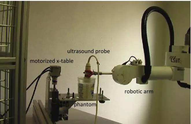
The visual servoing scheme was simulated on Simulink and then tested on a phantom made of silicon with cellulose particles for backscattering of the imaging ultrasound beams (see Figure 1). For a good transmission of ultrasound, this phantom was immersed in degased and demineralized water. The phantom was positioned on a motorized x-table to simulate the organ motion.
Figure 1: Experimental set up
motorized x-table
ultrasound probe
phantom
Results:
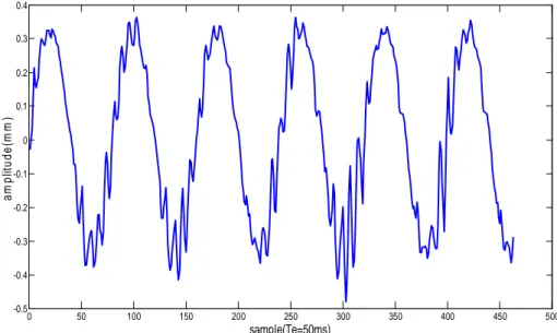
For a 1D motion of 2.3 mm of amplitude and 0.25 Hz of frequency and a controller gain of 12, simulation results on Simulink showed a residual motion of 0.3 mm in amplitude which corresponds to a reduction of 87 % of the object motion. As for the result of the experiment on the phantom, the mean residual motion was 0.33 mm in amplitude (maximum amplitude of 0.42 mm), which corresponds to a reduction of 86% of the phantom motion (see Figure 2).
0 50 100 150 200 250 300 350 400 450 500 -0.5 -0.4 -0.3 -0.2 -0.1 0 0.1 0.2 0.3 0.4 sample(Te=50ms) a m p lit u d e (m m )
Figure 2: residual motion estimated by ultrasound imaging for a 1D motion of 2.3 mm of amplitude
The actual amplitude of breathing motion is larger than the one used here (typically 1 to 2.5 cm for liver, 1 to 1.5 cm for kidney and less than 2 cm for pancreas peak to peak) [2]. We can however estimate that for a typical breathing motion of 2 cm of amplitude peak to peak, the residual motion would be 1.4 mm. This is deemed to be negligible in most of the clinical cases except for in the case of very small tumor (<1cm in diameter). Though breathing motion is predominant in the superior-inferior axis, motion in antero-posterior axis reaching a maximum of 10 mm and motion in lateral axis reaching a maximum of 5 mm peak to peak need to be taken into account.
Conclusion:
The experimental results show that the visual servoing allows for a significant reduction of the apparent motion. The robotized system is thus proved to be efficient for motion compensation. Optimizing this system is needed for a larger reduction of the motion, for instance by using a more advanced controller. For further studies, HIFU sonications will be performed in order to assess differences in terms of thermal lesion. First, sonication will be performed without any motion, then with motion but without motion compensation and finally with both motion and visual servoing.
Acknowledgment:
This work was partly supported by the French ANR within the Investissements d'Avenir program (Labex CAMI) under reference ANR-11-LABX-0004
References:
[1] J. W. Jenne, T. Preusser, and M. Günther, “High-intensity focused ultrasound: Principles, therapy guidance, simulations and applications”, Zeitschrift für Medizinische Physik, vol. 22, no. 4, pp. 311–322, Dec. 2012.
[2] A. Muller, L. Petrusca, V. Auboiroux, P. J. Valette, R. Salomir, and F. Cotton, “Management of Respiratory Motion in Extracorporeal High-Intensity Focused Ultrasound Treatment in Upper Abdominal Organs: Current Status and Perspectives”, Cardiovasc Intervent Radiol, vol. 36, no. 6, pp. 1464–1476, Dec. 2013.
[3] A. Krupa, G. Fichtinger, and G. D. Hager, “Real-time motion stabilization with B-mode ultrasound using image speckle information and visual servoing”, The International Journal of
Robotics Research, vol. 28, no. 10, pp. 1334–1354, 2009.
[4] J. Luo and E. E Konofagou, “A fast normalized cross-correlation calculation method for motion estimation”, Ultrasonics, Ferroelectrics and Frequency Control, IEEE Transactions on, vol. 57, no. 6, pp. 1347–1357, 2010.