HAL Id: inserm-01571688
https://www.hal.inserm.fr/inserm-01571688
Submitted on 3 Aug 2017
HAL is a multi-disciplinary open access archive for the deposit and dissemination of sci-entific research documents, whether they are pub-lished or not. The documents may come from teaching and research institutions in France or abroad, or from public or private research centers.
L’archive ouverte pluridisciplinaire HAL, est destinée au dépôt et à la diffusion de documents scientifiques de niveau recherche, publiés ou non, émanant des établissements d’enseignement et de recherche français ou étrangers, des laboratoires publics ou privés.
Distributed under a Creative Commons Attribution| 4.0 International License
Towards reproducible brain imaging research
Camille Maumet, Alexander Bowring, Thomas Nichols
To cite this version:
Camille Maumet, Alexander Bowring, Thomas Nichols. Towards reproducible brain imaging research. West Midlands Health Informatics Network annual conference, Jan 2017, Coventry, United Kingdom. �inserm-01571688�
Towards reproducible brain imaging research
Camille Maumet
1, Alexander Bowring
2, Thomas E. Nichols
1,2 1WMG, University of Warwick, Coventry, UK2Department of Statistics, University of Warwick, Coventry, UK
Abstract
Like many areas in scientific research, brain imaging is facing a “reproducibility crisis”. This has prompted a shift in focus within the neuroimaging community towards more transparent and open research. In this panel session, we will first introduce current practices in brain imaging research and their limitations. Then, we will discuss reproducibility across neuroimaging tools. Finally, we will review recent efforts by the Committee on Best Practice in Data Analysis and Sharing (COBIDAS) to improve reporting practices.
This panel session is composed of three talks discussing neuroimaging reproducibility:
1. Introduction to reproducible neuroimaging research: current practices and limitations (Camille Maumet)
2. Investigating reproducibility across neuroimaging software pipelines (Alexander Bowring)
3. COBIDAS: improving reporting guidelines (Thomas Nichols)
Content of the introductory talk is presented in the Introduction section below. The second and third talks are presented in more detail in the Methods and Results sections.
Introduction
Reproducibility in scientific research covers a broad spectrum ranging from the capacity to which researchers can recompute the results of an experiment using identical conditions, to investigating how general experimental results are under varying settings (e.g. different population, methodology or tool). New research builds on existing literature which makes the ability for a researcher to reproduce their peers’ results an essential aspect of their work. Scientific papers are still regarded as the main output of research experiments. But, with increasing complexity of research data and methods, the textual description included in publications to describe the methods has become insufficient to reproduce published results. Ideally, all the data, tools and scripts used to obtain the results should be published along with the papers to provide a complete description of the experiment1. Unfortunately, while there is a growing number of tools and platforms for data and code sharing (e.g. OpenfMRI2,3, NeuroVault4,5, GitHub6), they are still not commonly used in the neuroimaging community7. In this panel, we will first provide an overview of current practices in neuroimaging research and their limitations with respect to reproducibility. We will then present the results of an experiment investigating cross-software variability. Finally we will discuss how transparency can be improved through reporting guidelines as proposed by the COBIDAS8.
Methods
Investigating reproducibility across neuroimaging software pipelines
We selected three functional Magnetic Resonance Imaging (fMRI) studies from the publically accessible OpenfMRI data repository: ds0000019,10, ds00010911,12, ds00012013,14.
Each of these studies was based on a risk-taking task, social interaction task, and reward task respectively. The studies were chosen as they used relatively simple analysis pipelines, and each study had a supporting publication showing clear, well-defined regions of brain activation to which we could easily compare with our own results. Exclusion criteria included the use of custom software, activations defined using small volume correction, the absence of a related publication or only a data paper. We aimed to reproduce the main figure from each study, by separately reanalysing the data with the three main neuroimaging software packages SPM15,16, FSL17,18and AFNI19,20.
COBIDAS: improving reporting guidelines
The Committee on Best Practice in Data Analysis and Sharing (COBIDAS) was created by the Organisation of Human Brain Mapping 21 in 2014 and was composed of 15 experts in brain imaging research. The experts held a half-dozen teleconferences over 2 years to complete the report.
Results
Investigating reproducibility across neuroimaging software pipelines
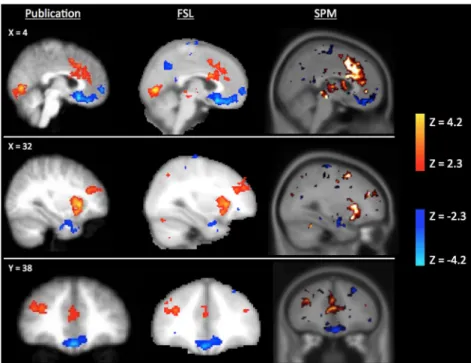
Scripts used to reanalyse the three experiments in each of the three software packages are publically available at: https://github.com/AlexBowring/Software_Comparison/. Fig. 1 presents the results obtained for SPM and FSL on the main contrast (“pump_demean>ctrl_demean”) for the ds000001 dataset. Strong similarities to the publication images can be seen in the images we obtained for the reproductions using both neuroimaging software packages.
Fig. 1. Significant activations (red) and deactivations (blue) for the contrast “pump_demean>ctrl_demean” of
COBIDAS: improving reporting guidelines
COBIDAS developed a set of best practices for reporting of a Magnetic Resonance Imaging (MRI) study, organised in seven sections: “Experimental design”, “Acquisition”, “Preprocessing”, “Statistical Modeling & Inference”, “Results”, “Data sharing” and “Reproducibility”. The document was discussed 22 and approved by the OHBM community and is now publically available online8.
Discussion & Conclusion
Reproducibility is gaining greater attention in the neuroimaging community and a number of tools, standards and platforms are now available to make data sharing as effortless as possible for neuroimaging researchers.
References
1. Gorgolewski KJ, Poldrack RA. A Practical Guide for Improving Transparency and Reproducibility in Neuroimaging Research. PLoS Biol 2016; 14: e1002506.
2. OpenfMRI https://openfmri.org (accessed 31 March 2016).
3. Poldrack RA, Barch DM, Mitchell JP, et al. Toward open sharing of task-based fMRI data: the OpenfMRI project. Front Neuroinform 2013; 7: 12–12.
4. NeuroVault: a new home for all brain statistical maps!http://neurovault.org (accessed 15 June 2016). 5. Gorgolewski KJ, Varoquaux G, Rivera G, et al. NeuroVault.org: a web-based repository for collecting and
sharing unthresholded statistical maps of the human brain. Front Neuroinform; 9. Epub ahead of print April 2015. DOI: 10.3389/fninf.2015.00008.
6. Build software better, together. GitHub http://github.com (accessed 7 November 2016).
7. Poline J-B, Breeze JL, Ghosh S, et al. Data sharing in neuroimaging research. Front Neuroinform 2012; 6: 9–9.
8. Nichols TE, Das S, Eickhoff SB, et al. Best Practices in Data Analysis and Sharing in Neuroimaging using
MRI. Epub ahead of print 20 May 2016. DOI: 10.1101/054262.
9. Balloon Analog Risk-taking Task https://openfmri.org/dataset/ds000001/ (accessed 11 November 2016). 10. Schonberg T, Fox CR, Mumford JA, et al. Decreasing ventromedial prefrontal cortex activity during
sequential risk-taking: an FMRI investigation of the balloon analog risk task. Front Neurosci 2012; 6: 80. 11. Symons DK. False Belief Task. In: Goldstein S, Naglieri JA (eds) Encyclopedia of Child Behavior and
Development. Springer US, 2011, pp. 637–639.
12. Moran JM, Jolly E, Mitchell JP. Social-cognitive deficits in normal aging. J Neurosci 2012; 32: 5553– 5561.
13. Developmental changes in brain function underlying the influence of reward processing on inhibitory control (Slot Reward)https://openfmri.org/dataset/ds000120 (accessed 11 November 2016).
14. Padmanabhan A, Geier CF, Ordaz SJ, et al. Developmental changes in brain function underlying the influence of reward processing on inhibitory control. Dev Cogn Neurosci 2011; 1: 517–529.
15. Penny WD, Friston KJ, Ashburner JT, et al. Statistical parametric mapping: the analysis of functional
brain images: the analysis of functional brain images. Academic press, 2011.
16. Wellcome Trust Centre. SPM - Statistical Parametric Mapping http://www.fil.ion.ucl.ac.uk/spm/ (accessed 15 June 2016).
17. Jenkinson M, Beckmann CF, Behrens TEJ, et al. FSL. Neuroimage 2012; 62: 782–790. 18. FSL - FslWiki http://fsl.fmrib.ox.ac.uk/fsl (accessed 15 June 2016).
19. Cox RW. AFNI: software for analysis and visualization of functional magnetic resonance neuroimages.
Comput Biomed Res 1996; 162–173.
20. AFNI. AFNI/NIfTI Server http://afni.nimh.nih.gov/ (2005, accessed 23 April 2015).
21. Home - Organization for Human Brain Mapping http://www.humanbrainmapping.org/ (accessed 7 November 2016).