‘This work was supported by Medical Research Council of Canada program grant PG-2 and National Institutes of Health grant NIH
15R0 l-NS 163 15-06.
1219
Original
Articles
Immunocytochemical
Localization
of
a Novel
Pituitary
Protein
(7B2)
within
the
Rat
Brain
and
Hypophysis’
MIECZYSLAW
MARCINKIEWICZ,
SUZANNE
BENJANNET,
NABIL
G.
SEIDAH,
MARC
CANTIN
and
MICHEL
CHRETIEN
Laboratories of Biochemical and Molecular Neuroendocrinology (MM. ;SB. ,‘N.G.S. ;M. Chr#{233}tieni, and Laboratory of Pathobtologj
(M. Cantin), Clinical Research Institute ofMontreal, I 10 Pine Azenue West, Montreal, Quebec, H2W 1R7, Canada
Received for publicationJanuary 28, 1985 and in revised form May 30, 1985; accepted June 1.1, 1985 (5A0333)
A novel pituitary protein called 7B2 was localized in rat
pituitary and brain by immunocytochemistry (unlabeled
antibody technique). Immunoreactive material was
pres-ent in the secretory cells of anterior and intermediate lobes and in neural structures of the posterior lobe of the
hy-pophysis. 7B2-immunoreactive neurons were evident within
the hypothalamus in the supraoptic nucleus, paraventric-ular nucleus (magnocellular and parvocellular parts), and
lateral hypothalamus. Immunoreactive nerve fibers were
seen within the internal and external zone of the median
Introduction
Systematic biochemical analysis of extracts from human and
porcine hypophysis has led to the isolation and characterization
of a novel pituitary protein (designated 7B2), belonging to a
new superfamily (5,16).
The 7B2 identified in extracts from human and porcine
pituitanies elutes at similar organic solvent compositions on
high-performance liquid chromatography (HPLC), and shows
similar isoelectric points (p1 4.9) and molecular weights (21,000)
as verified by sodium dodecyl sulfate-polyacrylamide
electro-phoresis (SDS-PAGE). Moreover, the amino acid
composi-tion and sequence of the N-terminal 77 and 81 amino acids
(human and porcine homologs respectively) revealed a high
degree of sequence conservation (16).
Immunocytochemical studies using antibodies generated
against a synthetic fragment of this protein show the presence of immunoreactive material within the anterior and posterior
lobes of the pituitary and within the neuronal soma of the
supraoptic nucleus of the hypothalamus (16).
eminence. Among extrahypothalamic regions, the
sub-stantia nigra, dorsal tegmental nucleus, cuneiform
nu-cleus, dorsal parabrachial nucleus, spinal tract trigeminal nerve, interior olive, solitary nucleus, and layers I and II ofthe spinal cord contained 7B2-immunoreactive material. This anatomical distribution suggests a role for 7B2 in endocrine and autonomic functions.
KEY WORDS: Immunocytochemistry ; Immunopreadsorption; Novel pituitary protein ;Anatomical organization ;Hypophysis; Brain.
Radioimmunoassay (RIA) studies confirmed the presence
of immunoreactive 7B2 (7B2-IR) in the hypothalamus and
pituitary, and also indicated its presence in other regions of
the brain as well as in the thyroid and adrenal glands (6,7).
We now report in detail the localization of 7B2-IR within
the brain and spinal cord.
Material
and
Methods
Animals. Male and female Sprague-Dawley rats 8 and 22 days
old and adult were used without any prior treatment or after colchicine administration ( 100 jagII() jal in distilled water into the lateral ventricle
48 hr before sacrifice). The coordinates were L-l.4 mm, H-7.0
mm, A-7. 3 mm, from the stereotaxic atlas of the rat brain ( I). The
animals were anesthetized with Somnotol prior to injection or at sacrifice.
Fixation and tissue processing. Cold Bouin’s solution was cho-sen from among the fixatives assessed, and administered by cardiac
perfusion after washing with ice-cold 0.9% NaCI. The whole brain
including the medulla oblongata and pituitary was removed, minced,
preserved in fixative for 12 hours at 4#{176}C,dehydrated via a series of alcohols followed by xylene, embedded in paraffin, cut in 5-jam see-tions, and mounted onto the microscopy slides. The sections from
different pituitaries were mounted together on the same slide.
Antiserum and controls. The antiserum against the synthetic
7B224 ,,, is as follows:
Glu-Gln-Leu-Gly-lle-Ala-Arg-Pro-Arg-Val-Glu-Tyr-Pro-Ala-His-GIn-Ala. The peptide was coupled to bovine
thy-roglobulin at a molar ratio of 10: 1 using a water-soluble carbodiimide method ( l6. Following three boosts, the titer in one of the animals
was found to be 1 : 10,000. This antibody was found to cross-react
(207 ) with the porcine and human 7B2, but did not recognize any
POMC segment including N-terminal, adrenocorticotropic hormone
(ACTH), n-melanocyte stimulating hormone (a-MSH), /3-LPH,
/3-endorphin (7,16), o-corticotropin releasing factor (oCRF), 8-arginine
vasopressin (AVP), or oxytocin (7).
For immunocytochemistry, immunopreadsorption was performed with synthetic 7B2 both in liquid and solid phase; 1 jag of synthetic
antigen in 100 pA of antiserum diluted I : 1000 resulted in total
in-hibition of immunostaining.
The specificity ofantibodies was verified by observation of possible cross-reactivity using solid-phase immunopreadsorption (22) using the following peptides bound to agarose beads: oxytocin, 8-arg-vasopres-sin, 8-lys-vasopres8-arg-vasopres-sin, neurophysin, substance P. met-enkephalin, and somatostatin 14; all peptides were used at a concentration of 2 jag/mI
in antiserum diluted I : 1000. The same test was performed using
thyroglobulin in the range of 0.0 1- 1 0 jag/mI, and the results were
compared with those obtained after immunopreadsorption using
syn-thetic 7B2. Ethanolamine was used to obtain a standard background. The beads were incubated with 4 ml of antiserum to 7B2 diluted
1 : 1000 for 1 6 hours at 4#{176}Cwith shaking. Antisera were then aspirated and used for incubation with tissue sections. The beads, washed three
times for 10 mm with phosphate-buffered saline (PBS), were exposed
to fluorescein isothiocyanate (FITC) in a dilution of 1 : 30 for 45 mm at 37#{176}C,washed again with PBS three times for 10 mm, resuspended
in I drop of 90% glycerol/PBS, and deposited on a glass slide, which
was examined by Leitz (UV equipped) microscopy. Photographs were
taken with the same exposure time for all samples (25 see).
Immunostaining. The unlabeled antibody technique of
Stern-berger (20) was used. The rehydrated sections were successively
cx-posed to normal goat serum (NGS) 3% in PBS for 15 mm and to
nonpreadsorbed and preadsorbed antisera diluted 1 : 1000 in PBS
con-taming 1% NGS for 24-48 hours at 4#{176}C.After three washes for 10
mm with PBS, the antirabbit IgG, diluted I :60 in 10% NGS/PBS,
was applied for 30 mm, followed by three washes for 10 mm with
PBS. The sections were exposed to the peroxidase-antiperoxidase
(PAP) complex diluted I :60 in 1% NGS for 30 mm, then washed
and allowed to react with diaminobenzidine (30 mg/lOO ml Tris-HCI
buffer + l00jalof30% H2O2)for 10mm. Asacontrol, theantiserum
was omitted and nonimmune rabbit serum was applied.
Figure 1. Solid-phase immunopread-sorption of7B2 antiserum diluted I : 1000
using ethanolamine as a standard (a) and the following peptides: oxytocin (b), arg-vasopressin (c), lys-vasopressin (d), neu-rophysin (e), substance P (1),
met-en-kephalin (g), and somatostatin (h). No
fluorescence was observed. Thyroglob-ulin bound to the agarose beads
gener-ated fluorescence, increasing over the
range 0.1-10 jag/I ml of diluted
anti-serum (i and j). Adsorption with
syn-thetic 7B2 produced a strong
fluores-cence (k) under the same conditions.
Results
Specificity of Antiserum
The antiserum incubated with different heterologous peptides
bound to agarose beads induced a faint fluorescence
compa-rable to that obtained with ethanolamine as a background
(Fig-ure la-h). Immunostaining was not affected by incubation of
sections with these preadsorbed antisera. This observation al-lowed us to conclude that there was no cross-reaction between
7B2 antiserum and oxytocin, lys-vasopressin, arg-vasopressin,
neurophysin, somatostatin, substance P, or met-enkephalin.
The thyroglobulin bound to agarose appeared to be more
potent in attracting some of the antibodies, since the intensity of generated fluorescence increased in a range of
concentra-tions running from 0. 1 to 10 mg/I ml (Figure Ii and
j),
butnever attained the intensity observed with 7B2 (Figure 1k).
The use of the thyroglobulin immunopreadsorbed antiserum
resulted in partial decrease of staining and background, but
did not affect the visualized structures. The synthetic 7B2
complexed with agarose induced a strong fluorescence (Figure
1k). The immunopreadsorbed antiserum did not show the
staining (Figure 2b). No reaction was found when the
anti-serum was omitted or when preimmune rabbit serum was used.
Distribution
of 7B2-IR
in
Pituitary
In all rat groups (8 and 22 days old and adult), an
immuno-reaction was observed in the anterior (AL), intermediate (IL),
and posterior (PL) lobes (Figure 2a,c,d). Immunoreactive cells
were sparsely distributed in the AL, attached to sinusoids and
within strongly stained cells lying on the border of the lobe
(Figure 2c).
All cells of the intermediate lobe were labeled. Sometimes the dorsal part ofthe lobe exhibited more intense PAP staining
(Figure 2d). The best staining was obtained after 48 hr of
incubation.
Immunostaining was localized in the posterior pituitary, associated with fibers surrounding the vessels and in adult rats
within the nerve swellings, which were densely packed with
At F =-. . . ,.t*...??’ ... . - . PL
L2:x;
C. IL 2bThe intensity of reaction varied with age. Among the
pi-tuitaries studied, those from animals of8 and 22 days exhibited
a more intense staining of the AL cells than in the adult. In contrast, the intensity of staining within the PL seems to
in-crease from moderate in neonatal and young rats to strong in
the adult rat (Table 1 ), whereas that in the intermediate lobe
does not seem to change with age.
Distribution
of
7B2-IR
in
Hypothalamus
In all groups of untreated animals, the immunoreactive
ma-terial was found in the neurons ofthe supraoptic nucleus (SON)
(Figure 3a and b), within the fibers of the median eminence
(ME) (Figure 4), and in the hypophyseal stalk. Excluding the
above regions, the PAP deposit was not observed in neonatal
and young rats, whereas in adults the immunoreaction was
found to be widely distributed. Granules of different forms
and sizes were seen in several areas of the hypothalamus: the
paraventriculan nucleus, both magnocellular (PVOm) and
par-vocellular (PVOp), the lateral hypothalamic area (LHy), and
the area penifonnicalis (AP) (Table 2).
Administration of colchicine resulted in the appearance of
strongly immunostained neuronal somas inside structures
ad-jacent to the ventricle. Colchicine treatment increased the
Figure 2. (a,b) Adult rat pituitary gland: paraffin-embedded specimen, 5 -jam successive sections. Low-power micrographs of immunostaining
obtained after .18 hr of incubation with 7B2 antiserum I : 1000. In a,
the sites ofreaction are largely distributed in the whole organ; b shows
the effect of immunopreadsorption of antiserum with synthetic 7B2
fragment. The reaction is completely abolished within the whole
or-gan, except for some nonspecific background in the anterior lobe.
AL = anterior lobe, IL intermediate lobe, PL posterior lobe.
Original magnification x 17. Bar = 1000 jam. (C) Adult rat: higher
magnification of adenohypophysis. The immunoreaction product is
found within the cells as dispersed or granular material surrounding
the nuclei. Note the apical accumulation of PAP deposit in cells
as-sociated with the sinusoid. Original magnification x 400. Bar = 25
jam. (d) Adult rat: higher magnification ofposterior lobe (right)
show-ing the sections of nerve fibers and terminal swellings labeled with
PAP reaction prc)duct. The intermediate lobe cells (left) exhibit a
rather moderate immunoreaction within the cytoplasm. Original
mag-nification x 400. Bar = 25 jam.
immunoreaction within the cells of the supraoptic nucleus
(Figure 3b). Sparsely distributed positive neurons were found
in the entire rostral hypothalamus. In particular, neurons
as-sociated with capillaries were frequently seen in the lateral
hypothalamus (Figure 3a), and supraoptic retrochiasmic area
7B2-L3a.’
3b
Table 1. Intensity of 7B2-IR labeling obserz’ed in pituitary ofneonatal. young, and adult rats”
Age of
rat
Pituitary lobe
Anterior Intermediate Posterior
8days +++++ ++ ++
22days +++++ ++ ++
Adult +++ ++ +++++
Inte’nsitvof1ahc’lingincrease-sfrom + to + + + + +. Resultswereobtained from four rats ,n each group. Sections representing all ages were mounted together 4fld scored comparatively.
IR within the magnocellular neurons, and parvocellular
neu-rons were also stained (Figure 3a and c).
Extrahypothalamic
Distribution
of
7B2-JR
The concentration of immunostained material was lower
out-side the hypothalamus. The regions that exhibited a positive
reaction in adult rats were the medial pars of the substantia
nigra, pars reticulata and compacta (SN) (Figure 5a). There
the reaction product formed irregular granules, often arranged
with faintly visible fibers in a reticular pattern (Figure Sb). In
the midbrain, the whole peniaqueductal gniseum contained a
weak, finely dispersed reaction product. The strongest
con-centration of immunoreaction was evident within the dorsal
tegmental nucleus (DTN), cuneiform nucleus (CN) and dorsal
parabrachialis nucleus (DPN) (Figure 6). The staining of all
these structures contrasted with that of the white matter, areas of the cortex, and inferior colliculus, which were unstained. At higher magnification, more on less dense irregular granules
were seen distributed through the parenchyma (not shown).
Among the pontine structures, the spinal tract tnigeminal
nerve (STI’N), inferior olive (10), and nucleus solitanius (NS) were strongly labeled (Figure 7a). Figure 7b shows an example of 10 staining to illustrate the typical aspect of the
varicosity-like and/or terminal-like structures crossing the parenchyma.
Such an aspect was similar to that of the cornu dorsale (CD)
of the spinal cord (Figure 8). The IR-7B2 was mainly
concen-trated in layers I and II, and little immunoreactivity was found
in deeper layers.
Discussion
This study shows the immunocytochemical localization of a
recently isolated and characterized pituitary protein, 7B2 ( 16),
in the pituitary gland and CNS. The results were obtained
using the antiserum previously shown by RIA not to
cross-react with adenohypophysis POMC peptides, oxytocin, or
va-sopressin (7,16). Although the larger 7B2 sequence shows
partial homology to proinsulin, Rous sarcoma virus
transform-ing protein, and pig secretin (5), the 23-39 7B2 fragment used
to raise the antiserum does not present homology to any of
these proteins, including secretin. Secretin immunoreactivity
is known to occur in brain and in pituitary gland; however, its
cerebral distribution (12) does not correlate with that of
7B2-.‘-‘.AP
F
PVOp
.. #{176} . ttfrChN
C
C.- #{149} ‘4j
, .)j
, :J”#{149}
. ,‘ , . #{149},I A ,iI’ 4 vc, ,.I ,1,
, ‘‘::
c\;:,
‘.;‘r’
Figure 3. (a) Adult rat treated with colchicine. Low-magnification
frontal section through rostral hypothalamus. The PAP deposit widely
distributed through the parenchyma contrasts with the negative
re-action ofwhite matter. Note the strong reaction within the supraoptic
nucleus (SON), the paraventricular nuclei both magnocellular (PVOm)
and parvocellular (PVOp), and the group of neurons associated with
capillaries in the lateral hypothalamus (LHy). AP = area perifornicalis,
ChO = optic chiasm, F = fornix, III V = third ventricle. Original
magnification x 20. Bar = 400 jam. (b) Higher magnification view of
SON magnocellular neurons. Original magnification x 187. Bar = 40
jam. (c) Detail showing the population of neurons within the PVNp.
Most of the neurons are parvocellular (arrowhead); only a few
mag-nocellular neurons are seen (arrow). Original magnification x 235.
.,
.. ‘ . .. . . .- .4#{149}
. . . . . t#{149} . F ‘ ....i...
.‘;,:.:
. . .. . ..:,!..i
p
M:I4
‘ 0 b.: ::“ : ‘. .‘:: “: : ‘ :‘t...4-;
:‘#{248}.:, ,. .-4
.:
‘ ‘Figure 4. B2-IR fibers within the median eminence. Both interna’
(MEi) and external (MEe) laminae exhibit immunoreaction. Note that
the strongest accumulation of B2-lR is around portal capillaries and
around the perivascular spaces (arrow). Original magnification x 392. Bar = 25 jam.
IR. Since the brain contains other peptides known to have in
some particular areas a similar localization to 7B2-IR (23),
immunopreadsorption tests were performed using
double-control observation on agarose beads treated for
immunoflu-orescence, and simultaneously on tissue sections. Under these
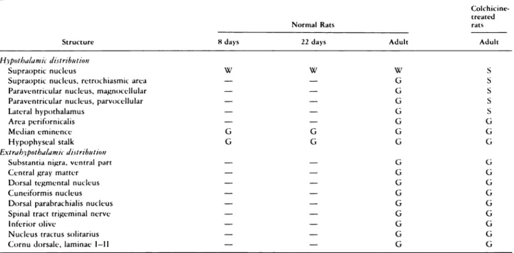
‘-No reaction; G = granular deposit of PAP product in cerebral parenchyma. PAP deposit within the neurons: W, weak afld S. strong intensity of re-ac turn.
conditions no cross reaction was detected with oxytocin,
lys-vasopressin, arg-vasopressin, neurophysin, substance P. r
met-enkephalin. The minor cross-reactivity with thyroglohulin had
. . -. no significant effect on specific immunostaining, since
tread-. . . sorption of antiserum resulted mainly in reducing the
hack-; A ‘ ground. Moreover, the liquid-phase and solid-phase
imniu-. . I nopreadsorption with synthetic B2 fragment totally blocked
the effect of immunocytostaining and induced strong
immu-nofluorescence of material hound to agarose heads. Therefre,
according to generally accepted criteria, the specificity of the
7B2 antiserum (2 1 ) was such that all antibodies recognized
the antigen and were able to detect antigenic sites in tissue.
The three lobes of the pituitary gland contained 7B2-IR
ma-terial. It was found in the anterior lobe in the cc’lls
character-ized as gonadotrophs (Capella, Buffa, and Magnoni, and Polak
and Bloom, personal communications), and in the posterior
lobe, where strongly immunoreactive sites correspond to nerve
fibers and endings. In contrast, uniform and moderate
ito-munocytostaining was seen within the intermediate lobe. Since
the reaction was displaceable by synthetic 7B2 (hut not by
thyroglobulin), this confirmed that the antigen is present in
all these areas, albeit in different local concentrations. Several
factors have led us to report now the immunostaining in the
intermediate lobe: 1 ) the antiserum actually used exhibits less
nondisplaceable background, 2) the maximum
immuno-staining intensity was obtained not earlier than after .18 hr of
incubation, 3) use ofSternherger’s technique has been shown
tO be more sensitive than the indirect immunoperoxidase
pro-cedure. These conditions most clearly permitted us to
appre-ciate the effect of staining and its total displacement. The
neurohypophysis B2-IR originates from hypothalamic nuclei.
Table 2. Distribution of 7B2-IR material in CNS in normal and co/chicine-treated rats”
Colchicine-treated
Structure
Normal Rats rats
Adult
8 days 22 days Adult
Hypothalamic distribution
Supraoptic nucleus W W W S
Supraoptic nucleus, retrochiasmic area - - G S
Paraventricular nucleus, magnocellular - - G S
Paraventricular nucleus, parvocellular - - G S
Lateral hypothalamus - - G S
Area perifornicalis - - G G
Median eminence G G G G
Hypophyseal stalk G G G G
Extrah3pothalamt distribution
Substantia nigra, ventral part - - G G
Central gray matter - - G G
Dorsal tegmental nucleus - - G U
Cuneiformis nucleus - - U G
Dorsal parabrachialis nucleus - - G U
Spinal tract trigeminal nerve - - U U
Inferior olive - - U U
Nucleus tractus solitarius - - U U
ic
..s. .4Wm
.6
:#{176}
‘:“
. .. 151
‘.ijx
.-,..,... . C. ‘C#{149}’ ‘4’ ,1#{149} . *,1
. .7a
c ,. S. - .-,Iyoung rats than in the adult, whereas the immunoreactivity of
Figure 5. (a) Low magnification frontal section through
mesenceph-alon. 7B2-IR labels the medial pars of the substantia nigra (SNc and
SNr). Original magnification x 24.5. Bar = 400 jam. (b) Higher
mag-nification view showing the PAP deposit in the SNc. The small and
irregular granules are arranged in the form of a varicosity-like
retic-ulum. Original magnification x 392. Bar = 25 jam.
It is not yet evident whether in the anterior and intermediate
lobes immunoreactive material originates from local synthesis
Or uptake. Although little is yet known about the origin,
syn-thesis, and function of 7B2 protein within the pituitary, the
protein seems to be linked to its secretory activities, since the
anatomic distribution selectively covers the sites of glandular
and neurosecretory structures. The ontogeny of 7B2 in the
AL and PL seems to be different, since the 7B2-1R staining
in the anterior hypophysis is more intense in neonatal and
r-
.#{149}:
. .#{149}-Figure 6. Macrophotograph through the midbrain frontal section. PAP
deposit is seen in the whole periaqueductal griseum (PU). The
strong-est labeling is observed within symmetrically located structures: the
dorsal tegmental nucleus (DTN), cuneiform nucleus (CN), and the
region of the dorsal parabrachialis nucleus (DPN ). Labeling was absent in the mass ofwhite matter (Wm) and inferior colliculus 1c. Original
magnification x 9. Bar - 1000 jam.
Figure 7. (a) Macrophotograph of pontine region. Spinal tract
tn-geminal nerve (SITN), infeniorolive (10) and nucleus solitarius (NS)
are labeled with 7B2 antiserum. Original magnification x 7.5. Bar = 1000 jam. (b). Higher magnification ofthe fragment ofthe 10 region illustrates the existence of vanicosity-like structures. Original
__4 .
Figure 8. Macrophotograph of section
from cervical part of spinal cord. Weak
7B2-IR labeling covers the whole area
of the gray matter. The strong reaction is localized within layers I and II of the dorsal horn containing the primary sen-sony neurons. In deeper layers only lam-mae
x
show moderate labelling. Onigi-nal magnification x 15. Bar = 1000 jam...
...,...i
H;, . - ., , -.5 1 \ . .. . . ...‘. ..‘... .. ‘N . .. .. . .. .,--‘-.‘---‘.- . . C ‘ - .,. .-‘ -.8
neural hypophysis increases with age. Moreover, there is a
good correlation between the increase of 7B2-lR in the whole
brain and in the neurohypophysis because the only sites clearly
visualized in newborn and young rats were the SON and ME.
Other cerebral regions containing 7B2-IR have been observed
only at maturity.
These regional and quantitative differences could indicate
the differential participation of this protein in glandular and neural activity. Several peptides are known to exert a different role in the hypophysis and in nervous tissue. Good examples
are oxytocin and vasopressin, hormones that can alter the
dcc-trical activity in the CNS (4,10,1 1).
Without any pretreatment, the brown granular PAP deposit
(concentrated or diffused) showed the presence of the 7B2
antigen in different cerebral regions. However, the cellular
localization made possible by colchicine administration
re-suited in the appearance of intense staining, mostly in
mag-nocellular neurons within the SON, SONr, PVNm, LHy, and
also in parvocellular neurons in the PVNp.
The interpretation ofPAP-positive results for regions distal
to the ventricle (i.e., the PA) and for the extrahypothalamic areas (which were not affected by colchicine) is still uncertain with regard to the 7B2-IR localization within the perikarya or within the terminals. Nevertheless, the effect of colchicine
indicates that 7B2-IR protein may be synthesized within the
neuronal somas of hypothalamic nuclei and transported via
axonal flow flux to their respective targets. The projections
coming from the SON are almost exclusively to the PL ( I 3),
but those of the PVO were clearly demonstrated to connect
in addition with the hindbrain and spinal cord areas (8,14).
Our observations, for which light microscopy does not provide
adequate resolution, will be corroborated by future study,
analogous to that of Conrath-Verrier et al. (3), with local col-chicine administration to the discrete CNS nuclei. Under these conditions and using electron microscopy it should be possible
to elucidate whether 7B2-IR is present within the perikarya
or within the terminals contacting the target neurons in the
regions described above (PA, SN, PG. DTN, CN, SITN, 10,
NS, CD).
The positively stained neurons are distributed throughout
the hypothalamic NSO and PVO nuclei. Both nuclei are known
to be related via an internal zone of the ME to the
neurohy-pophysis (15). Furthermore, the PVO projections contact the
portal capillaries ofthe ME external zone (2,9,28). This system
is involved in the regulation ofadenohypophysial activity.
Cer-tam
pathways of the PVO contact with autonomic centers ofthe brain system and spinal cord and, there, are involved in a range of aspects of central autonomic regulation, nociception, and behavior (18,19).
The correlation of this fact with the anatomic distribution
of 7B2-IR gives more support to the hypothesis that 7B2
protein could participate in endocrine and autonomic functions.
Immunoreactivity to several peptides has been
demon-strated in the magnocellular division of both the NSO and
the PVN, namely oxytocin, vasopressin, and their associated
neurophysins, met-enkephalins and leu-enkephalins, gastrin,
dynorphins, renin, and glucagon (see extensive review by
Swanson and Sawchenko, ref 23). Among these peptides, the
dynorphins, the differential proteolytic processing products of
prodynorphin B, have been reported within anterior and
neu-rointermediate pituitary lobes in various concentrations ( 17).
Moreover, the immunoreactivity of the
prodynorphin-B-derived peptides exhibited a common localization with
vaso-pressin in neurons and projections in the brain (24,25,26,27).
Furthermore, preliminary data in our laboratory show the
com-mon localization of7B2, arg-vasopressin, and dynorphin 1-13
immunoreactivities within the SON, PVO, and posterior
pi-tuitary. Interestingly, the whole area of 7B2-IR is larger and
extends to regions where arg-vasopressin is absent (the
28. Weigand SJ, Price JL: The cells of origin of the afferent fibers to the median eminence in the rat. J Comp Neurol 192:1, 1980
B2-lR may be in part associated with dynorphin and/or
va-sopressin systems; however, this association appears to be
ir-regular. We expect that further investigation will yield an
anal-ysis of the above question.
Acknowledgments
The author. thank Dr. Roy Sik.itrom for his reading and co,nnents. and
Al.i.
(Jarole ‘I’remhlay for typing the manu.cript.Literature
Cited
I. Albe-Fessand D, Stutinsky F, Libouban 5: Atlas St#{233}r#{233}otaxiquedu
Diencephale du Rat Blanc. 2nd Ed. CNRS, Paris, l9l
2. Antunes JL, Canmel PW, Zimmerman EA: Projections from the
panaventnicular nucleus to the zona externa of the median
cmi-ncncc of the rhesus monkey: An immunohistochemical study.
Brain Res l3:1, l9
3. Conrath-Vernier M, Dietl M, Arluison M, Cesselin F, Bourgoin
5, Hamon M: Localization of met-enkephalin-like
immunoreac-tivity within pain-related nuclei ofcervical spinal cord. Brain Res Bull 11:587, 1983
1. Uilhcy MP, Coote JH, Fleetwood-Walker 5, Peterson DF: The
influence of the paraventniculo-spinal pathway and oxytocin and
vasopressin on sympathetic preganglionic neurones. Brain Res
21 1 :43, 1982
5. Hsi KL, Seidah NG, Dc Serres G, Chr#{233}tien M: Isolation and
NH2-terminal sequence ofa novel porcine anterior pituitary poly-peptide. Homology to proinsulin, secretin and Rous sarcoma virus
transforming protein TVFV6O. FEBS Lett 147:261, 1982
6. Iguchi H, Chan JSD, Dennis M, Seidah NG, Chr#{233}tien M:
Re-gional distribution of a novel pituitary protein (7B2) in the rat brain. Brain Res, in press
7. Iguchi H, Chan,JSD, Seidah NG, Chr#{233}tien M: Tissue distribution and molecular forms of a novel pituitary protein in the rat. Neu-roendocninology 39:1 5 3, 1981
8. Kuypers HGJM, Maiskey VA: Retrograde axonal transpe)rt of
horseradish peroxidase from spinal cord to brain stem cell groups in the’ cat. Neurosci Lett 1:9, 1975
9. Lechan RM, Nestler JL, Jacobson 5, Reichling S: The
hypotha-lamic tuberoinfundibular system of the rat as demonstrated by
horseradish peroxidase (HRP) microiontophoresis. Brain Res
195:13, 198()
10. Morris R, Salt TE, Sofroniew MV, Hill RG: Actions of
microion-tophoretically applied oxytocin, and immunohistochemical
local-ization ofoxytocin, vasopressin and neurophysin in the rat caudal
medulla. Neurosci Lett 18: 163, 1980
I I. M#{252}hlethaler M, Dreifuss JJ, G#{228}hwilerBH: Vasopressin excites
hippocampal neurones. Nature (London) 296:7.19, 1982
12. O’Donohue ThL, Charlton CU, Miller RL, Boden G,Jacobowitz
DM: Identification, characterization, and distribution of secretin immunoreactivity in rat and pig brain. Proc NatI Acad USA 78:522 1,
1981
I 3. Rasmussen AT: Effect of hypophysectomy and hypophysial stalk
resection on the hypothalamic nuclei of animals and man. Assn
Res Nerv Ment Dis 20:215, 19i()
14. Saper CB, Loewy AD, Swanson LW, Cowan WM: Direct
hy-pothalamo-autonomic connections. Brain Res 1 l:305, 1976
15. Saper CB, Swanson LW, Cowan WM: The efferent connections
of the anterior hypothalamic area of the rat, cat and monkey. J
Comp Neurol l82:55, I98
16. Seidah NU, Hsi KL, Dc Serres G, Rochemont J, Hamelin J,
Antakly T, Cantin M, Chretien M: Isolation and NH-terminal
sequence of a highly conserved human and porcine pituitary
pro-tein belonging to a new superfamily. Immunocytochemical
lo-calization in pars distalis and pars nervosa of the pituitary and in
the supraoptic nucleus of the hypothalamus. Arch Biochem
Bio-phys 225:525, 1983
I7. Seizinger BR, Grimm C, H#{246}lltV. Hen A: Evidence for a selective processing of pro-enkephalin B into different opioid peptide forms in particular regions ofrat brain and pituitary.J Neurochem 42:147,
1984
18. Sofroniew MV: Morphology of vasopressin and oxytocin
rieu-nones and their central and vascular projections. In The
Neuro-hypophysis: Structure, Function and Control, Progress in Brain
Research. Edited by BA Cross, U Leng. Elsevier, New York,
1983, p 101-1 1.1
19. Sofroniew M: Vasopressin and oxytocin in the mammalian brain
and spinal cord. Trends Neurosci 6:467, 1983
20. Sternberger LA (ed): lmmunocytochemistry. 2nd Ed. Wiley, New
York, l99
21. Swaab DF: Comments on the validity of immunocytochemical
method. In Cytochemical Methods in Neuroanatomy, Neurology
and Neurobiology, Vol 1. Edited by V Chan-Palay and SL Palay.
Alan Liss, New York, 1982, p 423-440
22. Swaab DF, Pool CW: Specificity of oxytocin and vasopressin
im-munofluorescence. J Endocninol 66:263, 1975
23. Swanson LW, Sawchenko PE: Hypothalamic integration:
Orga-nization of the paraventnicular and supraoptic nuclei. Ann Rev
Neurosci 6:269, 1983
24. Watson SJ, Akil H, Fischli W, Goldstein A, Zimmerman E,
Ni-layer U, Van Wimersma Greidanus TB: Dynorphin and
vaso-pressin: Common localization in magnocellular neurons. Science
216:85, 1982
25. Watson SJ, Khachatunian H, Taylor L, Fischli W, Goldstein A,
Akil H: Pro-dynorphin peptides are found in the same neurons
throughout rat brain: Immunocytochemical study. Proc NatI Acad
Sci USA 80:891, 1983
26. Watson SJ, Seidah NU, Chr#{233}tien M: The carboxy terminus of
the precursor to vasopressin and neurophysin: Immunocytochem-istry in rat brain. Science 217:853, 1982
27. Weber E, Barchas J: Immunohistochemical distribution of
dy-norphin B in rat brain: Relation to dynorphin A and