Oxidation-Reduction Properties of Chloroplast Thioredoxins,
Ferredoxin:
Thioredoxin Reductase, and Thioredoxin f-Regulated Enzymes
†MasakazuHirasawa,‡PeterSchu¨rmann,§Jean-Pierre Jacquot,|
Wanda Manieri,§Pierre Jacquot,‡Eliane Keryer,⊥Fred C. Hartman,#and David B. Knaff*,‡
DepartmentofChemistryandBiochemistry,TexasTechUniVersity,Lubbock,Texas79409-1061 DepartmentofPlantBiochemistry,UniVersityofNeuchâtel,NeuchaˆtelCH-2007,Switzerland InstituteofBiotechnologyofPlants,UniVersityofParis-South,OrsayCedex91405,France
LaboratoryofForestBiology,AssocieINRA,UniVersityofNancy1, 54506VandoeuVreCedex,France
Life Sciences DiVision, Protein Engineering Program, Oak Ridge National Laboratory, Oak Ridge, Tennessee 37831
ABSTRACT
Oxidation-reduction midpoint potentials were determined, as a function of pH, for the disulfide/dithiol couples of spinach and pea thioredoxins f, for spinach and Chlamydomonas reinhardtii thioredoxins m, for spinach ferredoxin:thioredoxin reductase (FTR), and for two enzymes regulated by thioredoxin f, spinach phosphoribulokinase (PRK) and the fructose-1,6-bisphosphatases (FBPase) from pea and spinach. Midpoint oxidation-reduction potential (Em) values at pH 7.0 of -290 mV for both spinach and pea thioredoxin f, -300 mV for both C. reinhardtii and spinach thioredoxin m, -320 mV for spinach FTR, -290 mV for spinach PRK, -315 mV for pea FBPase, and -330 mV for spinach FBPase were obtained. With the exception of spinach FBPase, titrations showed a single two-electron component at all pH values tested. Spinach FBPase exhibited a more complicated behavior, with a single two-electron component being observed at pH values g 7.0, but with two components being present at pH values <7.0. The slopes of plots of Em versus pH were close to the -60 mV/pH unit value expected for a process that involves the uptake of two protons per two electrons (i.e., the reduction of a disulfide to two fully protonated thiols) for thioredoxins f and m, for FTR, and for pea FBPase. The slope of the Emversus pH profile for PRK shows three regions, consistent with the presence of pKa values for the two regulatory cysteines in the region between pH 7.5 and 9.0.
The ferredoxin/thioredoxin system of oxygenic photosyn-thetic organisms plays an important role in the regulation of the carbon metabolism of these organisms (1-3). The initial step in the thioredoxin regulatory pathway, which has been extensively characterized in spinach and pea chloroplasts, involves the reduction of ferredoxin:thioredoxin reductase (hereafter abbreviated FTR1) by the reduced ferredoxin generated during light-driven noncyclic electron flow (1-3). Spinach leaf FTR, the best characterized of these enzymes, is a 25.6 kDa heterodimeric protein located in the chloroplast stroma (1-3). FTR contains a unique [4Fe-4S] cluster that serves to stabilize the one-electron-reduced
intermediate formed after the first electron donation by ferredoxin, during the two-electron reduction of the active-site disulfide of the oxidized enzyme to the two cysteine thiols present in reduced FTR (4, 5). FTR reduces thioredoxin in a reaction in which the two cysteines at the active site of the reduced enzyme become oxidized to a cystine disulfide, while the active-site disulfide of the oxidized thioredoxin becomes reduced to two cysteine thiols (1-5). FTR reduces both of the thioredoxins found in chloroplasts, thioredoxin f and thioredoxin m (monomeric proteins with molecular masses of∼12 kDa that contain a conserved -WCGPC-active site), with equal efficiency. However, the two chlo-roplast thioredoxins display differential but overlapping reactivities among the array of identified target proteins (1-3). Although regulatory reduction by thioredoxin m appears to be restricted to glucose-6-phosphate dehydrogenase and the NADP+-linked malate dehydrogenase (hereafter abbrevi-ated MDH), MDH is even more efficiently activabbrevi-ated by thioredoxin f (6-8), which also regulates fructose-1,6-bisphosphatase (hereafter abbreviated FBPase),
phosphor-†Supported by a grant (DE-FG03-93ER20125 to D.B.K.) and a
contract (DE-AC05-96OR22464 with Lockheed Martin Energy Re-search Corp. for the Oak Ridge National Laboratory) from the U.S. Department of Energy, by grants from the Schweizerischer National-fonds (31.37725.93 and 31.47107.96 to P.S.), by a joint U.S. National Science Foundation/Centre National de la Recherche Scientific grant (to D.B.K., M.H., J.-P.J., and Myroslawa Miginiac-Maslow), and by the U.S. Department of Agriculture under Grant 96-35306-3412 (to F.C.H.) from the National Research Initiative Competitive Grants Program.
* Author to whom correspondence should be addressed [fax (806) 742-1289].
‡Texas Tech University. §University of Neuchaˆtel.
|
University of Nancy 1.
⊥University of ParissSouth.
#Oak Ridge National Laboratory.
1Abbreviations:
Bis-Tris,bis(2-hydroxyethyl)iminotris(hydroxymethyl)-methane; Bis-Tris propane, 1,3-bis[tris(hydroxymethy)methylamino]-propane; DTT, dithiothreitol; FBPase, fructose-1,6-bisphosphatase; FTR, ferredoxin:thioredoxin reductase; mBBr, monobromobimane; MES, 2-(N-morpholino)ethanesulfonic acid; MOPS, 3-(N-morpholino)pro-panesulfonic acid; PRK, phosphoribulokinase; Tricine, N-tris(hy-droxymethyl)methylglycine.
ibulokinase (hereafter abbreviated PRK), and sedoheptulose-1,7-bisphosphatase (1-3). A consideration of the chemistry involved in the net sulfhydryl-disulfide exchange reactions that occur between thioredoxins and target proteins suggested that these reactions necessarily entail the formation of intermolecular disulfides as transient intermediates, and such an intermediate has been directly demonstrated in the case of the reductive activation of spinach PRK by reduced thioredoxin f, with Cys46 of thioredoxin paired with Cys55 of PRK (9). Cys46 of thioredoxin f has also been identified as the primary nucleophile in the reduction of spinach FBPase (10).
Three-dimensional structures are available from both X-ray crystallography and NMR spectroscopy for chloroplast thioredoxins f and m (11-13), and a crystal structure is available for the spinach chloroplast FBPase (14). A recent crystal structure for PRK from the purple photosynthetic bacterium Rhodobacter sphaeroides, an enzyme that is not regulated by thioredoxin, has allowed modeling studies of the possible site of thioredoxin/PRK interactions in the plant system (15). Progress is being made toward solving the structures of a cyanobacterial FTR, an enzyme with proper-ties very similar to those of spinach FTR (16), and the thioredoxin m-regulated chloroplast MDH (17).
Attempts to model the kinetics of activation and deactiva-tion of various thioredoxin-regulated enzymes and to explain the differential effects of light intensity on these enzymes in situ (18) require an accurate knowledge of the oxidation-reduction midpoint potentials (Em) of the disulfide/dithiol couples involved in the regulatory pathway. Although a limited number of measurements of the midpoint potentials of some of these proteins had been reported in the years since the discovery of thioredoxin regulation of carbon metabolism (19-21), until recently there has been little systematic investigation of the oxidation-reduction properties of the proteins of the chloroplast thioredoxin regulation system. Work in our laboratories used cyclic voltammetry to provide the first published Emvalues for spinach thioredoxins f and m and for spinach FTR (22). Although cyclic voltammetry had proven to be useful for reliably measuring Em values for the disulfide/dithiol couples of glutathione, Escherichia coli thioredoxin, and the thioredoxin encoded by the genome of the bacteriophage T4T (23), the Emvalues obtained by cyclic voltammetry for spinach thioredoxins f and m were significantly more positive than those previously obtained for maize thioredoxin m (19) and spinach thioredoxin f (21) using chemical oxidation-reduction poising techniques. As it is possible that some or all of the spinach chloroplast proteins investigated could have undergone Em-altering conformational changes in the lipid bilayer at the surface of the electrode used for the cyclic voltammetry measurements (22), it appeared to be important to reinvestigate the oxidation-reduction properties of these proteins using an alternative technique. It also seemed to be important to measure the Emvalues for these proteins over as wide a range of pH values as possible [previous investigations, with the exception of our own study of spinach PRK (ref 24), involved measurements at a single pH] because of the changes that occur in the pH of the chloroplast stroma during illumination (25). Below we report Emvalues, measured over a range of pH values, for thioredoxins f from spinach and pea, for thioredoxins m from spinach and the green alga
Chlamy-domonas reinhardtii, for spinach FTR, for spinach PRK, and for spinach and pea FBPases.
EXPERIMENTAL PROCEDURES
All proteins used in this study, except for spinach FBPase, which was purified from spinach leaf, were recombinant proteins. Except for spinach PRK (see below), the recom-binant proteins were all expressed in E. coli. Spinach thioredoxins f and m and spinach FBPase (26) and spinach FTR (27) were purified as described previously, as were pea thioredoxin f (7), C. reinhardtii thioredoxin m (28), and pea FBPase (29, 30). Spinach PRK was isolated from a trans-formed strain of the yeast Pichia pastoris that carries the gene for the wild-type enzyme (31) and was purified as described previously (24). The reduced form of dithiothreitol (DTT) was obtained from Sigma Chemical Co.. Monobro-mobimane (mBBr) and the oxidized form of DTT were obtained from CalBiochem.
Oxidation-reduction titrations, using either enzymatic activity (in the case of the spinach and pea FBPases and spinach PRK) or mBBr fluorescence (for thioredoxins f and m and for FTR) to monitor the oxidation-reduction state of the disulfide/dithiol systems, were carried out at ambient temperature under aerobic conditions. Control experiments carried out under anaerobic conditions yielded results identi-cal to those reported below. Activities for FBPase (26, 28) and PRK (24) were assayed as described previously. Fluorescence was measured either using a Perkin-Elmer model MPF-3 spectrofluorometer with 380 nm excitation, 450 nm emission, and 5 nm spectral resolution (as described in ref 32) or using samples in microtiter plates (Costar Black Plate 3603) and a Bio-Tek model FL500 fluorescence plate reader with excitation at 360 nm and emission at 460 nm. Samples were allowed to equilibrate at ambient potentials (Eh) defined by different ratios of reduced:oxidized DTT, for times between 1.0 and 3.0 h (see figure legends for details), to maximize the probability for achieving oxidation-reduction equilibrium without compromising the stability of the proteins (33). Total DTT concentrations were 2 mM for thioredoxins f and m and FTR, 10 mM for FBPase, and 80 mM for PRK. Control experiments demonstrated that both mBBr fluorescence and activity were constant with time, in the time ranges centered around the times used to establish redox equilibration, and that the Em values obtained were independent of total DTT concentration. In the cases of the thioredoxins, PRK, and FTR, the proteins equilibrate directly with DTT, but in the cases of the spinach and pea FBPases, catalytic amounts of either spinach or pea thioredoxin f, respectively, were added to speed the approach to oxidation-reduction equilibrium (33). At the end of the equilibration period, aliquots were removed and either assayed for activity (in the case of spinach and pea FBPase and spinach PRK) or exposed to mBBr (in the case of thioredoxins f and m and FTR) under conditions causing this fluorescent probe to become covalently linked to thiol groups (32-34). For the mBBr titrations, a modification (32) of the method of Hutchinson and Ort (33) was used in which protein precipi-tation with trichloroacetic acid (32) replaced methylene chloride extraction (33) as the method for separating the protein from other reactants and products. Control experi-ments, in which mixtures containing different ratios of
reduced:oxidized DTT, but containing no protein, were treated with mBBr and then with trichloroacetic acid under conditions identical to those utilized in the protein titrations exhibited no detectable fluorescence, indicating that the separation was sufficiently effective so that no significant errors in assigning Emvalues resulted from mBBr that was not covalently linked to the protein being titrated.
Best-fit values for Emwere determined by fitting titration data to the Nernst equation using Kaleidagraph software (Synergy Software) and setting the value of n in the Nernst equation at 2, the value expected for a disulfide/dithiol two-electron-transfer process. Data fitting of single-component titrations in which the value of n was not fixed at 2 but in which the software selected the n value that gave the best fit to the Nernst equation, never gave n values that showed any significant deviations from 2. Titrations of spinach FBPase at pH values <7.0 gave poor fits to either single component n ) 1 or n ) 2 Nernst curves but gave excellent fits to plots containing two n ) 2 component, in which the two Em values and the contribution of the two n ) 2 components are selected by the software to give the best fit. All plots of extent of reduction versus Ehshown below set the value of either enzyme activity or fluorescence observed in the sample poised at the most positive Ehvalue used as corresponding to 100% oxidized and take the values mea-sured at the most negative Ehvalue used as 100% reduced. In the cases where multiple titrations were carried out for the same protein at a given pH, average values are given and the average deviation is reported to provide an estimate of the experimental uncertainties.
All Emvalue calculations were based on a value of -330 mV for the Emof DTT at pH 7.0. This value is an average of several closely agreeing values available in the literature (33). The value used for the pH dependence of the Emvalue of DTT was -60 mV/pH unit over the pH range from 5.5 to 8.2 (33, 35). A value of 9.2 was used for the pKaof the more acidic of the thiol groups of DTT (35) for calculating the Emvalue of DTT at pH values >8.2. Plots of Emversus pH were obtained using Kaleidagraph software, using slope values of either -60 mV/pH unit (corresponding to the uptake of two protons per disulfide reduced), -30 mV/pH unit (corresponding to the uptake of one proton per disulfide reduced), or 0 mV/pH unit (corresponding to the uptake of zero protons per disulfide reduced), as described by Chivers et al. (36).
RESULTS
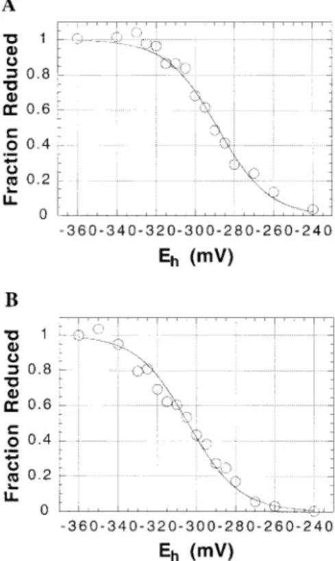
Figure 1 shows the results of titrations of spinach thio-redoxins f (Figure 1A) and m (Figure 1B) at pH 7.0. Both titrations give excellent fits to the Nernst equation for a single two-electron redox couple. Multiple titrations of these spinach proteins gave average values for Em, at pH 7.0, of -290 ( 10 mV (10 titrations) and -300 ( 10 mV (5 titrations) for thioredoxins f and m, respectively. Titrations of pea thioredoxin f and C. reinhardtii thioredoxin m at pH 7.0 gave Emvalues of -290 and -300 mV, respectively, values identical to those obtained for the corresponding spinach proteins. Figure 2 shows the results of a titration of the active-site disulfide of spinach FTR at pH 7.0. An average value of -320 ( 10 mV was obtained (nine titrations) for the Emvalue of the spinach enzyme at this pH. The Emvalues
obtained for the two spinach chloroplast thioredoxins at pH 7.0 in this study are 80-90 mV more negative than the values of -210 mV previously obtained by cyclic voltam-metry for both spinach thioredoxins f and m (22), a difference that is considerably greater than the sum of the (10 mV experimental uncertainties in either of the two types of measurements. The more reducing numbers obtained in the current study from mBBr titrations are, however, in good agreement with values reported for spinach thioredoxin f (21) and maize thioredoxin m (19) in titrations that used the ability of the reduced thioredoxins to activate thioredoxin-regulated enzymes to monitor the reduction state of the active-site disulfides. The Emvalue at pH 7.0 obtained for spinach FTR in this study is 90 mV more negative than that obtained
FIGURE1: Oxidation-reduction titrations of spinach thioredoxins f and m at pH 7.0. Thioredoxin f (A) or thioredoxin m (B), at a
final concentration of 270µg/mL, was incubated in 300 µL of 100
mM Bis-Tris-Cl buffer (pH 7.0) that contained DTT at a total concentration of 2 mM. After a 120 min incubation at ambient temperature, excess mBBr was added and the samples were prepared for fluorescence analysis as described under Experimental Procedures. Each point represents the average fluorescence from
two replicate 50µL samples, each of which was diluted with water
into microtiter plates to a total volume of 200µL.
FIGURE 2: Oxidation-reduction titration of spinach FTR at pH 7.0. Titrations were carried out as in Figure 1, using an FTR
previously by cyclic voltammetry. The fact that the apparent error in the cyclic voltammetry measurements is almost identical for all three of the proteins examined means that the thermodynamic driving force, at pH 7.0, of -30 mV estimated previously from cyclic voltammetry data (22) for the reduction of the active-site disulfides of thioredoxins f and m by the two cysteines at the active site of reduced FTR was apparently correct.
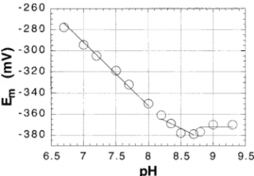
Given the rise in the stromal pH, from approximately pH 7.0 to pH 7.9, that occurs during illumination of chloroplasts (25), it was of interest to examine the pH dependence of the Emvalues of thioredoxins f and m, and of FTR, all of which are soluble proteins located in the stromal space of chloro-plasts (1-3). Figure 3 shows that plots of Emversus pH for both spinach thioredoxins f and m give good fits to straight lines with the -60 mV/pH unit dependence expected for a redox reaction that involves the uptake of two protons per two electrons (36) over the pH range from 6.0 to 8.5. Identical pH dependencies for Em were obtained for pea thioredoxin f over the pH range from 6.0 to 9.0 and for C. reinhardtii thioredoxin m over the pH range from 6.0 to 8.0 (not shown). Titrations of spinach FTR at pH 6.0 indicate that the enzyme also exhibits a -60 mV/pH unit dependence over the pH range between 6.0 and 7.0. Although titrations of FTR at pH 8.0 suggest that this Em versus pH unit dependence is also likely to extend to pH 8.0, the enzyme proved to be insufficiently stable at this pH to provide completely reliable Emvalues at alkaline pH values.
The simplest explanation for the observation that chloro-plast thioredoxins f and m both exhibit an Em versus pH dependence that does not deviate from the -60 mV/pH unit value expected for the uptake of two protons per reduction of the active-site disulfide up to pH values of 8.5 or 9.0 is that neither of the active-site cysteines of these proteins have pKavalues significantly below 8.0 (36). At pH values above the pKaof the more acidic cysteine, where only one proton would be taken up per disulfide reduced, an Emversus pH dependence of -30 mV/pH would be expected, and at pH values above the pKa values of both cysteines, where no protons are taken up on reduction of the disulfide, the Em would be expected to be independent of pH (36). It was also of interest to determine whether pKavalues for the regulatory cysteines of thioredoxin-regulated chloroplast enzymes could be detected from their Emversus pH profiles. Earlier studies of the pH dependence of the rate of alkylation of spinach PRK indicated that alkylation of Cys16, one of the two
regulatory cysteines in spinach PRK, reflects a process with a pKaof 7.9 ( 0.2 (37). One might thus expect a transition in the Emversus pH dependence for the reductive activation of PRK, centered at pH 7.9, from -60 to -30 mV/pH. A preliminary investigation of the Emversus pH dependence for spinach PRK in our laboratories failed to detect this transition, but it appeared to be likely that an insufficient number of pH values had been sampled to provide a rigorous test of whether the predicted change in Em versus pH dependence could be detected (24). Figure 4 shows an Em versus pH profile for the reductive activation of spinach PRK, combining data obtained in the earlier investigation (24) with new data obtained as part of this study, which is more extensive than was available earlier. The data could not be fitted to a single straight line with an Em versus pH dependence of -60 mV/pH. The Em versus pH data for spinach PRK were thus fitted instead using three regions (Figure 4), with slopes of -60, -30, and 0 mV/pH, respectively, as pH increases. Using the intersections of these three straight-line portions to estimate two pKavalues (36), this fitting procedure predicts values of 8.1 and 8.6 for pKa values of the more acidic (probably Cys16) and less acidic (probably Cys55) regulatory cysteines of PRK, respectively. The value of 8.1 for the pKaof the more acidic group agrees, within the experimental uncertainties of the measurements, with the value of 7.9 determined from the earlier alkylation studies (37). The data also give an equally good fit (not shown) using only two regions, with slopes of -60 and 0 mV/pH (i.e., the behavior that would occur if both regulatory cysteines had identical or very similar pKavalues). However, this fitting procedure yields a predicted value near pH 8.5 for the pKa of the two cysteines, in poorer agreement with the 7.9 value for the pKaof Cys16 obtained from the earlier alkylation studies (37) than the value of 8.1 obtained from the fit shown in Figure 4.
Figure 5 shows that Emvalues for the reductive activation of pea FBPase, over the range from pH 5.5 to 8.5, exhibit the -60 mV/pH unit dependence expected for a process in which two protons are taken up during a two-electron reduction. The Emvalue at pH 7.0, -315 (10 mV (three titrations), is somewhat less negative than the value of -330 mV obtained for spinach FBPase at this pH (see below), but this difference is within the (20 mV uncertainty for the
FIGURE3: Effect of pH on the Emvalues of spinach thioredoxins f and m. Titrations were carried out as in Figure 1. Buffers used were Bis-Tris-Cl (pH 6.0-7.0) and Tricine-NaOH (pH 7.5-8.5). The open circles represent the data for thioredoxin f, and the open squares represent the data for thioredoxin m.
FIGURE4: Effect of pH on the Emvalue of spinach PRK. PRK, at
a final protein concentration of 50µg/mL, was incubated in 1.0
mL of 100 mM buffer that contained DTT at a total concentration of 80 mM as described in ref 23. Buffers used were MOPS (pH 6.5-7.7), Tricine (pH 8.2-8.7), and Bis-Tris propane (pH 8.8-9.4). After a 150 min incubation at ambient temperature, aliquots were withdrawn and assayed for PRK activity as described under Experimental Procedures.
difference between two Em values, each of which has an experimental uncertainty of (10 mV. Titrations of pea FBPase at all pH values examined gave excellent fits to the Nernst equation for the reduction of a single two-electron component, despite the fact that the FBPase is known to undergo subunit dissociation in the alkaline pH range (38-42). Figure 6A shows a titration of spinach FBPase at pH 7.0. Titrations at this pH gave an Emvalue of -330 ( 10 mV (three titrations). All titrations of spinach FBPase carried out over the pH range from 7.0 to 7.8 gave excellent fits to the Nernst equation for a single two-electron component. As the spinach enzyme appears to be less stable at alkaline pH than the pea enzyme, it was not possible to obtain an unambiguous value for the slope of the Em versus pH dependence of the spinach enzyme at pH values >7.0. Figure
6B shows that at pH 6.0, titrations of the activity of spinach FBPase could not be fitted to the Nernst equation for a single two-electron process but instead required two components. An average of three titrations at pH 6.0 gave Emvalues of -230 ( 10 and -285 ( 10 mV, respectively, for the two components. A two-component fit was also necessary at pH 6.5 (average Em values of -270 and -325 mV were calculated for the two components at this pH). The behavior of the spinach enzyme is consistent with the involvement of two different disulfides, which exhibit different Emversus pH profiles, in the activation of the enzyme. The hypothesis that two different disulfides participate in the reductive activation of FBPase was previously suggested to explain the results of site-directed mutagenesis studies (29, 43).
DISCUSSION
As a result of this study, oxidation-reduction midpoint potentials are now available for all components of the spinach chloroplast ferredoxin/FTR/thioredoxin f,m system. It is also now possible to say, at least for the limited number of species examined, that there are few or no differences in Emvalues for chloroplast thioredoxins from different species and, at most, a small difference in the Emvalues for the regulatory disulfide of two higher plant FBPases. Furthermore, for the first time, the dependence of the Emvalues for thioredoxins and FTR on pH have been determined. Although the Em value for ferredoxin, the initial reductant in this regulatory pathway, is pH-independent (44), the Emvalues for the two chloroplast thioredoxins, and probably for FTR as well, exhibit a -60 mV/pH unit dependence for the pH range over which the pH of the chloroplast stromal space increases during illumination. Identical pH dependencies for FTR and the two thioredoxins reduced by the enzyme mean that the driving force for reduction of either thioredoxin f or m by FTR will not change during illumination. As pea FBPase and spinach PRK show little or no deviation from an Em versus pH dependence of -60 mV/pH unit over the range from pH 7.0 to 8.0, the driving force for reduction of these two thioredoxin f-regulated proteins will also be unaffected by illumination of the chloroplasts. However, the driving force for reduction of FTR by ferredoxin will decrease by ∼55 mV as a result of the light-induced increase in stromal pH by 0.9 pH unit (25), because the Emvalue of FTR will become more negative while that of ferredoxin will be unaffected.
Thioredoxins f and m, FTR, and PRK each contain only a single pair of cysteines involved in the thioredoxin regulatory pathway (1, 3), and thus the observation that all of the redox titrations of these proteins carried out as part of this study could be treated with a single-component fit to the two-electron Nernst equation was expected. In contrast to this simple situation, it appeared to be possible that the situation would be more complex for chloroplast FBPases. Recent site-directed mutagenesis studies on the pea (30) and rapeseed (43) FBPases have established the likelihood that three cysteine residues are involved in the thioredoxin f-regulated activation of this chloroplast enzyme. One of these regulatory cysteines (Cys153 in the pea enzyme, Cys157 in the rapeseed enzyme, and Cys155 in the spinach enzyme) appears to be able to form a disulfide bond with either of the other two regulatory cysteines (i.e., with either Cys173 or Cys178 in the pea enzyme and with either Cys174 or Cys179 in the
FIGURE5: Effect of pH on the Emvalue of pea FBPase.
Oxidation-reduction equilibration was carried out for 3 h at ambient temper-ature using 440 nM pea FBPase and a catalytic amount of pea
thioredoxin f (0.2µg/mL) in 100 mM buffer containing DTT at a
total concentration of 10 mM. Aliquots were withdrawn and assayed for FBPase activity as described under Experimental Procedures. Buffers used were MES (pH 6.0-7.0), MOPS (pH 7.5), Tricine (pH 7.8-8.2), and Bis-Tris propane (pH 8.5).
FIGURE6: Effect of pH on the oxidation-reduction properties of spinach FBPase. Reaction conditions were as in Figure 5 except that spinach FBPase and thioredoxin f replaced the pea proteins. Buffers used were (A) MOPS at pH 7.0 and (B) MES at pH 6.0.
rapeseed and spinach enzymes). The observation that two n ) 2 components are required to fit the activation redox titrations of spinach FBPase at pH values <7.0 (see Figure 6B) is consistent with this hypothesis and suggests that, at these pH values, samples of oxidized spinach FBPase are mixtures of enzyme with a Cys155-Cys174 disulfide and an enzyme with a Cys155-Cys179 disulfide. Although it is not yet possible to propose an unambiguous explanation for why only a single n ) 2 component is observed in redox titrations of spinach FBPase at pH values >7.0, this phenomenon could possibly result from the existence of a pH-dependent equilibrium between the two disulfides, from different Emversus pH dependencies for the two disulfides, or from a combination of these two effects. Our inability to observe two redox components during titrations of the highly homologous pea FBPase, regardless of pH, may be due to the fact that the Em values for the two disulfides in the spinach enzyme are identical to within the experimental uncertainties of our measurements. Redox titrations with site-directed Cys/Ser mutants of both pea and spinach FBPases are planned to further elucidate the redox properties of the enzymes.
ACKNOWLEDGMENT
We thank Prof. Donald Ort (University of Illinois at Urbana) for access to data prior to publication and for many helpful discussions, Prof. Myroslawa Miginiac-Maslow (Institute of Biotechnology of Plants, University of Paris-South) for her helpful suggestions on the manuscript, and Laurence Franchini and Soraya Balmelli (University of Neuchaˆtel) for their assistance in the preparation of the spinach proteins used in this study.
REFERENCES
1. Buchanan, B. B. (1991) Arch. Biochem. Biophys. 288, 1-9. 2. Knaff, D. B. (1996) in Oxygenic Photosynthesis: The Light
Reactions (Ort, D. R., and Yocum, C. F., Eds.) pp 333-361,
Kluwer Academic Publishers, Dordrecht, Holland.
3. Jacquot, J.-P., Lancelin, J.-M., and Meyer, Y. (1997) New
Phytol. 136, 543-570.
4. Staples, C. R., Ameyibor, E., Gardet-Salvi, L., Stritt-Etter, A.-L., Schu¨rmann, P., Knaff, D. B., and Johnson, M. K. (1996)
Biochemistry 35, 11425-11434.
5. Staples, C. R., Gaymard, E., Stritt-Etter, A.-L., Telser, J., Hoffman, B. M., Schu¨rmann, P., Knaff, D. B., and Johnson, M. K. (1998) Biochemistry 37, 4612-4620.
6. Geck, M. K., Larimer, F. W., and Hartman, F. C. (1996) J.
Biol. Chem. 271, 24736-24740.
7. Hodges, M., Miginiac-Maslow, M., Decottignies, P., Jacquot, J.-P., Stein, M., Lepiniec, L., Cre´tin, C., and Gadal, P. (1994)
Plant Mol. Biol. 26, 225-234.
8. Schwarz, O., Schu¨rmann, P., and Strotmann, H. (1997) J. Biol.
Chem. 272, 16924-16927.
9. Brandes, H. K., Larimer, F. W., and Hartman, F. C. (1996) J.
Biol. Chem. 271, 3333-3335.
10. Brandes, H. K, Larimer, F. W., Geck, M. K., Stringer, C. D., Schu¨rmann, P., and Hartman, F. C. (1993) J. Biol. Chem. 268, 18411-18414.
11. Ge´nove´sio-Taverne, J.-C., Jetzer, Y., Sauder, U., Hohenester, E., Hughet, C., Jansonius, J. N., Gardet-Salvi, L., and Schu¨rmann, P. (1991) J. Mol. Biol. 222, 459-461.
12. Lancelin, J,-M., Stein, M., and Jacquot, J.-P. (1993) J.
Biochem. 114, 421-431.
13. Capitani, G., Markovic-Housley, Z., Jansonius, J. N., Del Val, G., Morris, M., and Schu¨rmann, P. (1998) XI International
Photosynthesis Congress Abstracts, Poster SY9-P36, p 103
(in press).
14. Villeret, V., Huang, S., Zhang, Y., Xue, Y., and Lipscomb, W. N. (1995) Biochemistry 34, 4299-4306.
15. Harrison, D. H. T., Runquist, J. A., Holub, A., and Miziorko, H. M. (1998) Biochemistry 37, 5074-5085.
16. Dai, S., Schwendtmayer, C., Schu¨rmann, P., and Eklund, H. (1998) XI International Photosynthesis Congress Abstracts, Poster SY9-P10, p 99.
17. MacPherson, K. H. R., Ashton, A. R., Trevanion, S. J., Verger, D., and Ollis, D. L. (1998) Acta Crystallogr. D54, 654-656. 18. Kramer, D. M., Wise, R. R., Fredereick, J. R., Alm, D. M., Hesketh, D. R., Ort, D. R., and Crofts, A. R. (1990)
Photosynth. Res. 26, 213-222.
19. Rebeille, F., and Hatch, M. D. (1986) Arch. Biochem. Biophys.
249, 164-170.
20. Clancey, C. J., and Gilbert, H. F. (1987) J. Biol. Chem. 262, 13545-13549.
21. Hutchinson, R. S. (1994) Ph.D. Dissertation, University of Illinois.
22. Salamon, Z., Tollin, G., Hirasawa, M., Gardet-Salvi, L., Stritt-Etter, A.-L., Knaff, D. B., and Schu¨rmann, P. (1995) Biochim.
Biophys. Acta 1230, 114-118.
23. Salamon, Z., Gleason, F. K., and Tollin, G. (1992) Arch.
Biochem. Biophys. 299, 193-198.
24. Hirasawa, M., Brandes, H. K., Hartman, F. C., and Knaff, D. B. (1998) Arch. Biochem. Biophys. 350, 127-131.
25. Junge, W., and Jackson, J. B. (1982) in Photosynthesis: Energy
ConVersion by Plants and Bacteria (Govindjee, Ed.) Vol. 1,
pp 589-646, Academic Press, New York.
26. Schu¨rmann, P. (1995) Methods Enzymol. 252, 274-283. 27. Gaymard, E., and Schu¨rmann, P. (1995) in Photosynthesis:
From Light to Biosphere, Proceedings of the Xth International
Photosynthesis Congress, Montpellier, France (Mathis, P., Ed.) pp 761-764, Kluwer Academic Publishers, Dordrecht, The Netherlands.
28. Decottignies, P., Schmitter, J.-M., Jacquot, J.-P., Dutka, S., Picaud, A., and Gadal, P. (1990) Arch. Biochem. Biophys. 280, 112-121.
29. Jacquot, J.-P., Lopez-Jaramillo, I., Chueca, A., Cherfils, J., Lemaire, S., Chedozeau, B., Miginiac-Maslow, M., Decot-tignies, P., Wolosiuk, R. A., and Lopez-Gorge, J. (1995) Eur.
J. Biochem. 229, 675-681.
30. Jacquot, J.-P., Lopez-Jaramillo, J., Miginiac-Maslow, M., Lemaire, S., Cherfils, J., Chueca, A., and Lopez-Gorge, J. (1997) FEBS Lett. 401, 143-146.
31. Brandes, H. K., Hartman, F. C., Lu, T.-Y. S., and Larimer, F. W. (1996) J. Biol. Chem. 271, 6490-6496.
32. Krimm, I., Lemaire, S., Ruelland, E., Miginiac-Maslow, M., Jacquot, P., Hirasawa, M., Knaff, D. B., and Lancelin, J.-M. (1998) Eur. J. Biochem. 255, 185-195.
33. Hutchinson, R. S., and Ort, D. R. (1995) Methods Enzymol.
252, 220-228.
34. Kosower, N. S., and Kosower, E. M. (1987) Methods Enzymol.
143, 76-84.
35. Whitesides, G. M., Liburn, J. E., and Szajewski, R. D. (1977)
J. Org. Chem. 42, 332-338.
36. Chivers, P. T., Prehoda, K. E., and Raines, R. T. (1997)
Biochemistry 36, 4061-4065.
37. Porter, M. A., and Hartman, F. C. (1986) Biochemistry 25, 7314-7318.
38. Lazaro, J. J., Chueca, A., Lopez-Gorge, J., and Mayor, F. (1975) Plant Sci. Lett. 5, 49-55.
39. Buchanan, B. B., Schu¨rmann, P., and Wolosiuk, R. A. (1976)
Biochem. Biophys. Res. Commun. 69, 970-978.
40. Schu¨rmann, P., and Wolosiuk, R. A. (1978) Biochim. Biophys.
Acta 522, 130-138.
41. Zimmerman, G., Kelly, G. J., and Latzko, E. (1976) Eur. J.
Biochem. 70, 361-367.
42. Buc, J., Pradel, J., Meunier, J.-C., Soulie´, J.-M., and Ricard, J. (1980) FEBS Lett. 113, 285-288.
43. Rodriguez-Suarez, R. J., Mora-Ga´rcia, S., and Wolosiuk, R. A. (1997) Biochem. Biophys. Res. Commin. 232, 388-393. 44. Matsubara, H., and Sakai, K. (1992) AdV. Inorg. Chem. 38,