HAL Id: hal-01526980
https://hal-univ-rennes1.archives-ouvertes.fr/hal-01526980
Submitted on 23 May 2017
HAL is a multi-disciplinary open access
archive for the deposit and dissemination of
sci-entific research documents, whether they are
pub-lished or not. The documents may come from
teaching and research institutions in France or
abroad, or from public or private research centers.
L’archive ouverte pluridisciplinaire HAL, est
destinée au dépôt et à la diffusion de documents
scientifiques de niveau recherche, publiés ou non,
émanant des établissements d’enseignement et de
recherche français ou étrangers, des laboratoires
publics ou privés.
Distributed under a Creative Commons Attribution| 4.0 International License
Cinnamaldehyde loaded-microparticles obtained by
complex coacervation: Influence of the process
parameters on the morphology and the release of the
core material
Ilham Abdelmalek, Abderrezzak Mesli, Isabelle Svahn, Gerard Simonneaux
To cite this version:
Ilham Abdelmalek, Abderrezzak Mesli, Isabelle Svahn, Gerard Simonneaux. Cinnamaldehyde
loaded-microparticles obtained by complex coacervation: Influence of the process parameters on the
mor-phology and the release of the core material. Biointerface Research in Applied Chemistry, Comporter
SRL, 2017, 7, pp.1939-1944. �hal-01526980�
Ilham Abdelmalek, Abderrezzak Mesli, Isabelle Svahn, Gerard Simonneaux
Page | 1939
Cinnamaldehyde loaded-microparticles obtained by complex coacervation: Influence of the
process parameters on the morphology and the release of the core material
Ilham Abdelmalek
1, Abderrezzak Mesli
1, *, Isabelle Svahn
2, Gerard Simonneaux
31 Physical and Organic Macromolecular Chemistry Laboratory (LCOPM) Faculty of Exact Sciences, University "Djillali Liabès" of Sidi Bel-Abbes, LP 89, Algeria 2 Bordeaux Imaging Center –UMS 3420, Victor Segalen University of Bordeaux2, France
3 Chemical Sciences Laboratory of Rennes UMR CNRS 6226 University of Rennes-1, 25042 Rennes, France
*corresponding author e-mail address: abderrezzak-mesli@netcourrier.com
ABSTRACT
Encapsulation by complex coacervation includes several steps that have been developed in order to attain a better control over the whole process and to achieve an important delayed effect. This has been carried out for the encapsulation of cinnamaldehyde (CN) by the classical gelatin/acacia gum pair of coacervating polymers. This preparation was performed in different conditions (stirring speeds, cooling rate, emulsion and coacervate time, use of surfactant, and polymer concentration) in order to investigate their effect on the encapsulation efficiency and drug release kinetics. Optical microscopy studies showed spherical microcapsules. The yield of the encapsulation attains more than 88% of all prepared microcapsules. The mean Sauter diameter (d32) of obtained microparticles was in the
range from 124 to 200 µm. The microspheres were also characterized by the FTIR method; showing the presence of core and polymers in the microparticles. The release of cinnamaldehyde was performed in heterogeneous medium (Water / ethanol, v/v: 30/70) at 25±0.5°C using UV–Vis analysis. It was demonstrated that the drug release followed the Fickian diffusion mechanism. The data were best fitted to the Fick's law with high correlation coefficients (R²).
Keywords:Cinnamaldehyde; Microcapsules; Complex Coacervation; Fick's Diffusion.
1. INTRODUCTION
Several formulations of drug delivery are developed including spherical dosage forms [1], tablets [2], microparticles [3-5], nanoparticles [6] and liposomes [7]. These systems are formulated with different carrier polymers to allow modulation of the pharmacokinetics of the active agent and the achievement of a controlled release of the drug within a defined therapeutic period [8-10]. Microencapsulation is one of the most numerous techniques used to prepare drug delivery systems. It is used in order to achieve various goals: to protect sensitive ingredients from oxidation or hydrolysis, to mask unpleasant taste and to transform liquid droplets into solid particles [11, 12].
The process of coacervation is a physicochemical method of the microencapsulation; it may occur in polymer solutions containing a few tenths, or even a few hundredths, of a percent of the polymer, in which case the polymer concentration in the coacervate drops may be as high as several dozen percent. For this reason, coacervation is used as a means of concentrating and fractionating native and denatured biopolymers (in particular, water-soluble proteins) and synthetic polymers [13].
The coacervation method may be either simple or complex. Simple coacervation is the result of the interaction of a dissolved polymer with a low-molecular substance as gelatine with alcohol or sodium sulphate [14]. Complex coacervation consists in the precipitation of a hydrophobic substance, a coacervate, and its deposition at the surface of oil droplets so as to build a wall of polymer material [15-17].
Complex coacervation is a phase separation phenomenon induced by electrostatic interactions between polymers of opposite
electrical charge, most often hydrophilic biopolymers. Gelatin/acacia gum couple is the most widely used pair of biopolymers for this technique [18-22].
Encapsulation by complex coacervation has been used in different fields such as pharmaceutical, food, chemical, and cosmetics for the controlled release of several types of cores such as flavours, drugs, colorants and paints with a wide range of applications.
The aim of this paper is to encapsulate cinnamaldehyde using Gelatin/gum acacia system. This active agent is a natural compound isolated from the stem bark of Cinnamomum cassia [23, 24]. It has been shown various activities such as peripheral vasodilatory, antitumor, antifungal, cytotoxic and mutagenic. Cinnamadehyde is developed as food antimicrobial agent due to its demonstrated activity against both positive and gram-negative bacteria, including organisms that are of safety concern [23]. Cinnamaldehyde has been reported to inhibit the growth of Clostridium botulinum [25], Staphylococcus aureus [26], Escherichia coli [27] and Salmonella enterica serovar Typhimurium [28, 29]. Furthermore, cinnamaldehyde also found to show cyclin dependent kinases (CDKs) inhibition activity. It can inhibit the proliferation of several human cancer cell lines including those established from breast, ovarian lung and colon carcinomas and leukaemia's [30, 31].
We want to report herein, first the preparation and characterization of cinnamaldehyde-loaded microparticles with different processes parameters by complex coacervation and then, their release study in heterogeneous medium at 25°C.
Volume 7, Issue 1, 2017, 1939 - 1944
ISSN 2069-5837
Open Access Journal
Received: 25.12.2016 / Revised: 29.01.2017 / Accepted:12.02.2017 / Published on-line: 15.02.2017
Original Research Article
Biointerface Research in Applied Chemistry
Ilham Abdelmalek, Abderrezzak Mesli, Isabelle Svahn, Gerard Simonneaux
Page | 1940
2. EXPERIMENTAL SECTION
2.1. Materials. The following materials were used: bovine type B gelatin (GE) (Sigma-Aldrich, USA), gum acacia (GA) (Merck Chemicals, Germany), trans-cinnamaldehyde (Sigma-Aldrich, USA), glutaraldehyde (25% v/v, Sigma Aldrich, USA), tween20 (Sigma-Aldrich, USA), acetic acid (Wacker Chemie AG, Germany), NaOH (Wushi, China)
2.2. Experimental part. The microencapsulation method is based essentially on the procedure described in the literature [21, 32]. The simultaneous desolvation of two water-soluble polyelectrolytes with opposite charge (GE and GA) is used. A pH modification of the reaction medium induced an electrostatic attraction of these polymers. Once the coacervation is formed around the microdroplets of cinnamaldehyde, the temperature is decreased to 5 °C in order to obtain a gelling of the coating. The crosslinking of microcapsules obtained is carried out by adding glutaraldehyde in order to solidify the particles.
The process of microencapsulation by complex coacervation was realized in cover cylindrical glass reactor (volume of 1000 mL, external diameter = 80 mm) under mechanical stirring with four-bladed turbine impeller (blade length = 50 mm, blade width = 08 mm) by a homogenizer (IKA RW20 digital, UK) as described in the following steps:
Emulsion: the solution of gelatin (GE) (40g, 1%) is introduced into the reactor containing the solution of gum acacia (GA) (40g, 1%) at 40 °C, the mixture is allowed to stir at 500 rpm. After 15 min, cinnamaldehyde (CN) is gradually added to the reactor. The pH of the mixture is 6.2.
Coacervation: After 30 min, the pH is decreased to 3.9 by adding 2.4 mL of acetic acid solution CH3COOH (1M). The stirring speed is reduced to 250 rpm.
Consolidation: After 45 min, 0.08 mL of 25% glutaraldehyde was introduced to consolidate the membranes by crosslinking. The temperature of mixture was reduced to 5°C by cooling water for 45min (∆T / ∆t =1.2 °C/min). The mixture is left stirring at 5 ° C. for 1 hour.
Alkalinization: The crosslinking is improved by alkalinization of the reaction medium at pH = 8.9 by adding 2.6 mL of sodium hydroxide solution (1M), the temperature is then raised to 40 ° C, under stirring.
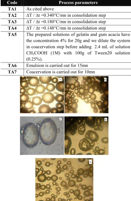
After 15 hours, the pH was lowered from 8.9 to 4.5 to form the microcapsules, by adding 4.7 mL of acetic acid solution (1M). The observations of each step were released by optical microscope (OPTIKA 4083. B1). They are presented in figure1.
The microcapsules prepared were named TA1; the other formulations were carried out by following the same protocol of TA1 with modifying process parameters to see their effect on the morphology of the microcapsules as well as the release of the active agent from these microcapsules. The process parameters are listed in Table 1.
Table 1. The microencapsulation process parameters CN by complex
coacervation.
Code Process parameters
TA1 As cited above
TA2 ∆T / ∆t =0.340°C/mn in consolidation step TA3 ∆T / ∆t =0.180°C/mn in consolidation step TA4 ∆T / ∆t =0.148°C/mn in consolidation step
TA5 The prepared solutions of gelatin and gum acacia have the concentration 4% for 20g and we dilute the system in coacervation step before adding 2.4 mL of solution CH3COOH (1M) with 100g of Tween20 solution (0.25%).
TA6 Emulsion is carried out for 15mn TA7 Coacervation is carried out for 10mn
Figure 1. Stages of microencapsulation by complex coacervation (a:
Emulsion, b: Coacervation, c: Consolidation, d: Alkalinization, e: The obtained microcapsules).
3. RESULTS SECTION
3.1. Characterization of the coacervated microcapsules. 3.1.1.Encapsulation efficiency: The drug amount in microcapsules was determined by Soxhlet extraction using isopropanol as solvent: 1g of drained and dried microcapsules was introduced into a Soxhlet cartridge. Extraction was carried out with 200 mL of isopropanol under reflux for 6 hours (the heating is fixed to obtain 7 cycles/hour). The resulting solution was analyzed by UV-VIS spectroscopy (Shimadzu UV-2401 PC,
Shimadzu, Japan) at λmax=305 nm. The drug concentration was determined from the standard curve. The encapsulation efficiency (Yield) of the microcapsules was calculated using the equation (1).
Yield% = ) x 100 (1)
3.1.2. Mean diameter and size distribution of the particles. The mean particle size of the active agent microcapsules was determined by optical microscopy (OPTIKA 4083. B1). At least
morphology and the release of the core material
Page | 1941 500 microspheres were analyzed for each preparation and the
mean diameter was calculated. Each sample was measured in triplicate. The particle-size distribution was calculated from several equations [33] (See Figure 2).
3.1.3. FTIR Spectroscopy. The microcapsules were characterised by infrared spectroscopy. The infrared spectra of cinnamaldehyde, gelatin, gum acacia, and the corresponding microparticles were compared. The FTIR spectra were recorded using an FTIR-8300 Shimadzu spectrophotometer (Shimadzu, Japan).
3.1.4. Morphology. An optical microscope (OPTIKA 4083. B1) was used to obtain images from moist microparticles. A sample (TA2) was dried by lyophilisation and then analyzed using Scanning Electron Microscopy (Quanta 200 FEI, France) at Bordeaux Center Imaging (University Bordeaux-1). The sample was mounted on a double-scotched carbon film fixed on a metal support.
3.2. In vitro drug release studies. The in-vitro release study from the microcapsules was carried out using a cylindrical double-wall glass reactor equipped with a fritted glass extremity immersed in the solution. This allows the ascent of the solution without passage of microparticles. The release kinetics of the active agent from microcapsules were followed by using an UV-Vis spectrometer 2401PC SCHIMADZU (The apparatus was calibrated at the maximum wavelength of cinnamaldehyde λmax= 305nm). 100mg of drained and dried microparticles was soaked in 100 mL of heterogeneous solution (Water / ethanol, v/v: 30/70). The dispersion medium was stirred magnetically at a rotation speed of 500 rpm at T=25°C; the dosage of released active agent is obtained on taking out 1mL of solution containing the support (reading of the optical density).
3.3. Results and discussion. The microcapsules containing cinnamaldehyde were prepared by complex coacervation in order to study the effect of parameters on the formulation of different samples, and to control the concentration of drugs in living microparticles. The obtained microcapsules have a different morphology as indicated in Table 2.
Table 2. The yield and the morphology of obtained microcapsules.
Code Yield % Morphology of microcapsules TA1 92 ± 0.85 Individual spherical microcapsules,
individual with a fine-looking membrane (aggregates: 1%)
TA2 93 ± 1.25 Individual spherical microcapsules with a clearly visible membrane (aggregates: 0%) TA3 92 ± 1.12 Aggregated microcapsules (aggregates:
18%)
TA4 92 ± 0.97 Aggregated microcapsules (aggregates: 26%)
TA5 96 ± 1.03 The microcapsules are very small and individual with a thin membrane (aggregates: 1%)
TA6 90 ± 1.45 Spherical microcapsules with a thick membrane.
(aggregates: 0%)
TA7 89 ± 0.43 Individual spherical microcapsules with a visible membrane
(Aggregates: 0%)
The optical microscopy analysis of various samples was carried out. The study indicated that the surface of the microcapsules were spherical with different sizes. The mean
diameter of the microcapsules was kept in average 90 to 200 µm (Figure2). It can be noted that smaller microspheres particle sizes were obtained with addition of emulsifier (tween20). It is well known that the surfactant reduces the surface tension of continuous phase, and prevents the coalescence and agglomeration of drops by stabilizing the emulsion [34]. The presence of Tween20 favors the formation of uniform small spherical microcapsules. The value of the dispersiona is less than 1.6, indicating adjacent sizes of obtained microcapsules (Figure3 & Figure4). The observation by scanning electron microscopy of the freeze-dried microcapsules (TA2) shows that they have an ovoid shape, and the "gum acacia-gelatin" matrix well surrounds the encapsulated "cinnamaldehyde" agent. The membrane appears wrinkled (Figure5).
The percentage of polymer was chosen according to the work of Xiao J-X et al [35]. The authors showed that this percentage allows the total encapsulation of the active agent. The agitation rate was kept constant in the first step (emulsion) for all samples because it affects the size of the microdroplets and the size of the microcapsules [36]. Thus, the decrease the rotation speed was chosen in the second step. Accordingly, the size of the microdroplets in the emulsion step and the size of microcapsules were similar. The same result is obtained if the speed is kept constant in both steps. The size is maintained when the surface of the cinnamaldehyde microdroplets was completely covered by matrices.
Figure 2. Size distribution of different microcapsules (the arithmetic
mean: d10=∑ ni di / ∑ ni, the volume-surface mean, d32= ∑ ni di3/ ∑ ni di2, the volume-moment mean: d43= ∑ ni di4/ ∑ ni di3. Where, ∑ ni, i is an index of the population, and di is the particle diameter of population i).
Ilham Abdelmalek, Abderrezzak Mesli, Isabelle Svahn, Gerard Simonneaux
Page | 1942
Figure 4. Size distribution curves of microcapsules TA1.
The emulsion time is an important parameter for the formulation of microparticles. Agnihotri N et al proved that the emulsification time must be more than 5 minutes [37]. The diameter of the microcapsules decreases when the emulsion time increases from 15 to 30 minutes. However, the decreasing time of coacervation does not lead to any variation in microparticle morphology. With decreasing cooling rate, the microcapsules were aggregated without any significant influence on the size of microparticles or the cinnamaldehyde content. This situation may suggest that deposition of the matrices on the cinnamaldehyde microdroplets is limited by the complete formation of the envelope.
The crosslinking agent used (glutaraldehyde) is a dialdehyde giving possible formation of "imine" bridges between the gelatin macromolecules. This reaction is more favorable in low acid medium because the amine functions are free. By carrying out the crosslinking in the basic medium, the microcapsules lost the previous spherical shape obtained in acid medium; and become ovoid with a wrinkled membrane. By varying the concentration of the polymer, the size of microparticles remains unchanged between 1% and 4%.
It should be noted that changing the cooling rate has no influence on the yield in TA1-TA4. However, when both the emulsion and coacervation times are reduced a low yield is obtained. This effect may be due to the incomplete process of emulsion or to a coacervate polymer deposit onto the microdroplets. A maximum yield is obtained after addition of the surfactant due to a more stable emulsion step (Table2).
Figure 5. SEM of microcapsules (TA2).
The infrared spectrum of the microcapsules is compared with spectrum of free CN, polymer matrix GE and GA in Figure 6. The analysis showed the presence of similar characteristic bands of the polymers and CN in the CN-loaded microcapsules TA1 spectrum: the OH functions of GE and GA appeared at 3400 cm–1, the C-N and N-H gelatin functions appeared at 1308cm–1 and 1577 cm–1, the C-OH and C-O-C acacia gum functions appeared respectively at 1200cm–1 and 1125cm–1. We find also the characteristic bands of cinnamaldehyde: C=O, C-H and the aromatic vibration at 1671 cm-1, 2748 cm-1 and 1626 cm-1 respectively.
The spectrum of the CN-loaded microspheres appeared as the sum of the spectra of pure CN and pair of polymers GE/GA.
Figure 6. Infrared Spectra (a: gelatin, b: acacia gum, c: cinnamaldehyde,
d: TA1).
The release of CN from the reservoir systems (microcapsules) was carried out in a water / alcohol mixture at T=25 °C. The determination of the release concentrations were carried out by UV-VIS at λmax of CN. Figure 7 shows CN release profiles from the prepared formulations (TA1-TA7).
In drug release curves of CN, two stages are noted: first, the percentage released of CN increases quickly and then, becomes constant. Different Drug delivery systems cannot be described by a classical kinetic equation. This process is related to a phenomenon of mass transfer controlled by diffusion according to the curves shown in Figure 8. Thus, a vertical tangent is observed at the beginning of the process. A linear effect is observed (at short time). Transfer of the CN from microcapsules to the medium represents the slowest step because in the reservoir systems the release of the active agent is linked to the width of membrane. The release is therefore governed by the diffusion of encapsulated material in the microcapsules.
morphology and the release of the core material
Page | 1943 The influence of the parameters (cooling rate, coacervation
time, addition of a crosslinker and emulsion time) is weak because the release of CN remains rapid in most cases.
The comparison of the release of CN from patches (TA1-TA4) shows that the release of CN decreases with the cooling rate of the process. This observation could be explained by a decrease in porosity caused by the lowering of the cooling rate and the diffusion difficulties of CN from coacervated microcapsules.
In the presence of the surfactant, we obtained an important released of CN, this is may be explained by the small size of corresponding microcapsules which leads to a large surface contact with the medium.
Decreasing emulsion time to 15 min allowed to obtain great microcapsules. This is due to the shearing time of the microdroplets formed during the emulsion. As a result, the contact surface with the study medium is low in TA6 giving a low release percentage of CN. However, drug release profiles are not modified by changing the coacervation time.
From the different curves, it can be observed that all the releases have an important delayed effect due to their small diameters and the thinness of their membranes, thus facilitating the passage (by diffusion) of the active agent. The linearity of the percentage with the square root of the time confirms that the release of CN was controlled by diffusion inside the solid polymer particles dispersed in the solution according to the simplified Fick’s equation for the short times (Table3)[38, 39]. However, it was not possible to get the diffusion coefficients due to unknown the membrane thickness, in the case of microcapsules.
Figure 7. Cumulated percentage of CN released from TA1-TA7.
0 2 4 6 8 10 12 0 5 10 15 20 25 30 35 40 45 CN % t1/2 (mn1/2 ) TA1 TA2 TA3 TA4 TA5 TA6 TA7
Figure 8. Cumulated percentage of CN released from TA1-TA7 as a
function of square root of time.
Table 3. Release kinetic results of microcapsules.
Code CN%= f (t1/2) R² TA1 y 4.24 x - 3.01 0.97 TA2 y 3.17 x + 1.81 0.97 TA3 y 2.62 x - 2.20 0.98 TA4 y 2.62 x - 4.13 0.93 TA5 y 3.10 x - 0.65 0.98 TA6 y 3.00 x - 2.78 0.95 TA7 y 3.21 x - 3.18 0.95
4. CONCLUSIONS
The present study emphasized the effects of formulation process parameters on the characteristics and in vitro release behavior of CN-loaded microcapsules prepared by microencapsulation using the complex coacervation method. This process presented high encapsulation efficiency, with spherical microparticles.
The concentration ratio of polymers matrix 1/1 with the presence of 0.25% tween20 as surfactant ensured the stability of the emulsion, and we obtained spherical microcapsules without aggregation with accelerating cool. The rotation speed of 250 rpm applied for 30 min makes it possible to obtain microcapsules with
average diameters of 90 μm. Releases of the hydrophobic core for moist samples were varied due to different formulation conditions. The release profiles showed a Fickian diffusion mechanism and the release rate could be controlled by adjusting the microencapsulation processing parameters that have significant effects on the particle size.
In the present study, we demonstrated that the microencapsulation provided a sustained release of cinnamaldehyde-induced apoptosis of human cancer cells, and we want to test those CN-loaded microparticles in living systems whether can be further developed into a chemotherapeutic agent.
5. REFERENCES
[1]. Pahwa R., Chhabra L., Kaur Lamba A., Jindal S., Rathour A., Formulation and in-vitro evaluation of effervescent floating tablets of an antiulcer agent, Journal of Chemical and Pharmaceutical Research, 4(2), 1066-1073, 2012.
[2]. Preeti K., Kasture P.V., Formulation and in vitro evaluation of Bilayer Tablets of Zolpidem Tartrate for Biphasic Drug Release, International
Journal of Pharm Tech Research,4(3), 1919-1929, 2011.
0 100 200 30 0 40 0 500 600 0 10 20 30 40 50 t (mn) TA1 TA2 TA3 TA4 TA5 TA6 TA7 CN%
Ilham Abdelmalek, Abderrezzak Mesli, Isabelle Svahn, Gerard Simonneaux
Page | 1944 [3]. Grama S., Zdenek P., Trchova M., Kovarova J., Benes J., Horak D.,
Monodisperse macroporous poly(glycidyl methacrylate) microspheres coated with silica: Design, preparation and characterization, Reactive &
functional polymers, 77, 11-17, 2014.
[4]. Mouffok M., Mesli A., Abdelmalek I, Gontier E., Effect of the formulation parameters on the encapsulation efficiency and release behavior of p-aminobenzoic acid-loaded ethylcellulose microspheres, J.
Serb. Chem. Soc., 81 (10), 1183–1198, 2016.
[5]. Abdelmalek I., Svahn I., Mesli S., Simonneaux S., Mesli A., Formulation, evaluation and microbiological activity of ampicillin and amoxicillin microspheres, J. Mater. Environ. Sci., 5 (6), 1799-1807, 2014. [6]. Gomez-Gaete C., Fattal E., Silvia L., Besnard M., Tsapis N., Dexamethasone acetate encapsulation into Trojan particles, Journal of
Controlled Release, 128, 41-49, 2008.
[7]. Bekkara-Aounallah F., Gref R., Othman M., Reddy H., Pili B., Allain V., Bourgaux C., Hillaireau H., Lepetre-Mouelhi S., Desmaele D., Nicolas J., Chafi N., Couvreur P., Novel PEGylated nanoassemblies made of self-assembled squalenoyl nucleoside analogues, Advanced Functional
Materials,18, 1-11, 2008.
[8]. Xu T., He B., Mechanism of sustained drug release in diffusion-controlled polymer matrix-application of percolation theory, International
Journal of Pharmaceutics, 170, 139–149, 1998.
[9]. Queiroz A.C., Santos J.D., Monteiro F.J., Gibson I.R., Knowles J.C., Adsorption and release studies of sodium ampicillin from hydroxyapatite and glass-reinforced hydroxyapatite composites, Biomaterials, 22, 1393-1400, 2001.
[10]. Alcorn J., McNamara P.J., Pharmacokinetic sin the newborn,
Advanced Drug Delivery Reviews, 55, 667–686, 2003.
[11]. Wilson N., Shah N.P., Microencapsulation of Vitamins, ASEAN
Food Journal, 14 (1), 1-14, 2007.
[12]. Vidhyalakshmi R., Bhakyaraj R., Subhasrre R.S., Encapsulation "The future of probiotics", Advances in Biological Reseach, 3(3-4), 96-103, 2009.
[13]. Dong S.H., Herbert J.W., Matthew T., Promotion of osteoblast proliferation on complex coacervation-based hyaluronic acid-recombinant mussel adhesive protein coatings on titanium, Biomaterials, 31, 1080-1084, 2010.
[14]. Biswaranjan M., Aswal V.K., Kohlbrecher J., Bohidar H.B., Synthesis of gelatin nanoparticles via simple coacervation, J. Surface Sci.
Technol., 21(3-4), 149-160, 2005.
[15]. Kizilay E., Kayitmazer A.B., Dubin P.L., Complexation and coacervation of polyelectrolytes with oppositely charged colloids,
Advances in Colloid and Surface Science, 167, 24-37, 2011.
[16]. Bhattacharyya A., Argillier J-F, Microencapsulation by complex coacervation: effect of cationic surfactants, J. Surface Sci. Technol., 21(3-4), 161-168, 2005.
[17]. Dutra Alvim I., Ferreira Grosso C.R., Microparticles obtained by complex coacervation: influence of the type of reticulation and the drying process on the release of the core material, Ciência. Tecnologia de
Alimentos Campina, 30(4), 1069-1076, 2010.
[18]. Schmitt C., Turgeon S.I., Protein/polysaccharide complexes and coacervates in food systems, Advances in Colloid and Surface Science, 167, 73-70, 2011.
[19]. Lee-Fong S., Chee-Sian O., Effect of pH on Garlic Oil Encapsulation by Complex Coacervation, J Food Process Technol, 4(1), 1-5, 2013.
[20]. Bo W., Benu A., Colin J.B., Optimisation of the microencapsulation of tuna oil in gelatin-sodium hexametaphosphate using complex coacervation, Food Chemistry,158, 358-365, 2014.
[21] Nori M. P., Favaro-Trindale C. S., Matias de Alencar S., Thomazini M., Camargo Balieiro J. C., Contreras Castillo J.C., Microencapsulation of propolis extract by complex coacervation, LWT-Food Science and
Technology, 44, 429-435, 2011.
[22]. Cousin F., Gummel, S., Combet F.B., The model lysozyme-PSSNa system for electrostatic complexation: Similarities and differences with complex coacervation, Advances in Colloid and Surface Science, 167, 71-84, 2011.
[23]. Varukattu N.B., Soundarapandian K., Enhanced delivery of baicalein using cinnamaldehyde cross-linked chitosan nanoparticle including apoptosis, International Journal of Biological Macromolecules,51, 1103-1108, 2012.
[24]. Yan S., Liu-Nan J., Natsumi H., Takashi H., Toyohiko A., Taiichiro S., Beneficial Effects of Cinnamon on the Metabolic Syndrome, Inflammation, and Pain, and Mechanisms Underlying These Effects,
Journal of Traditional and Complementary Medicine, 2, 27-32, 2012.
[25]. Dima C., Cotarlet M., Tiberius B., Bahrim G, Alexe P, Dima S., Encapsulation of Coriander Essential Oil in Beta-Cyclodextrin: Antioxidant and Antimicrobial Properties Evaluation, Romanian
Biotechnological Letters,19(2), 9128-9140, 2014.
[26].Hongyan C., Hongbing J., Xiantai Z., Lefu W., Green Synthesis of natural benzaldehyde from cinnamon oil catalyzed by hydroxypropyl-β-Cyclodextrin, Tetrahedron, 66, 9888-9893, 2010.
[27]. Suxia S., Tiehua Z., Yuan Y., Songyi L., Jingyue X., Haiqing Y, Effects of cinnamaldehyde on Escherichia coli and Staphylococcus aureus membrane, Food Control, 47, 196-202, 2015.
[28]. Han J., Castell-Perez M.E., Moreira R.G., The Influence of Electron Beam Irradiation on the Effectiveness of Trans-cinnamaldehyde-coated LDPE/Polyamide Film, Food Engineering and Physical Properties, 71(5), 245-251, 2006.
[29]. Sivakumar J.T.G., Devaraj H., Cinnamaldehyde Induces Behavioral and Biochemical Changes in the Male Albino Wistar Rat, Journal of
Medical Sciences, 3(2), 101-109, 2010.
[30]. Hyeon K., Hee-Juhn P., Hyun-Ju J., Jong-Won C., Kyu-Seok C., Joohun H., Kyung-Tae L., Cinnamaldehyde induces apoptosis by ROS-mediated mitochondrial permeability transition in human promyelocytic leukemia HL-60 cells, Cancer Letters, 196, 143–152, 2003.
[31]. Shih-Hua F., Yerra K.R., Yew-Min T., Cytotoxic Effect of trans-Cinnamaldehyde from Cinnamomum osmophloeum Leaves on Human Cancer Cell Lines, International Journal of Applied Science and
Engineering, 2 (2), 136-147, 2004.
[32]. Lamoudi L., Chaumeil J.C., Daoud K., Effect of the microencapsulation process parameters piroxicam by complex coacervation, Ann Pharm Fr, 73(1), 37-42, 2015.
[33]. Kaczmarski K., Bellot J. Ch., Effect of particle-size distribution and particle porosity changes on mass-transfer kinetics, Acta Chromatographica, 13, 22-37, 2003.
[34]. Li M., Rouaud O., Poncelet, D., Microencapsulation by solvent evaporation: state of the art for process engineering approaches.
International Journal of Pharmaceutics, 363, 26–39, 2008.
[35]. Xiao J-X., Yu H-Y., Yang J., Microencapsulation of sweet orange oil by complex coacervation with soybean protein isolate/gum arabic,
Food Chemistry, 125, 1267-1272, 2011.
[36]. Jegat C., Taverdet J-L., Stirring speed influence study on microencapsulation process and the drug release from microcapsules,
Polymer Bulletin, 44, 345–351, 2000.
[37]. Agnihotri N., Mishra R., Goda C., Arora M., Microencapsulation – A Novel Approach in Drug Delivery, Indo Global Journal of
Pharmaceutical Sciences, 2(1), 1-20, 2012.
[38]. Muhammad Sajid H.A., Furqan I., Musa R., Kanwal R., Shabbir A., Yasser S., Syed Nisar H.S., Characterization of Ethylcellulose and Hydroxypropyl Methylcellulose Microspheres for Controlled Release of Flurbiprofen, J Pharm Drug Deliv Res, 2(1), 1-10, 2013.
[39] Abdelmalek I., Mesli A., Bendahmane M., Chafi N., Simonneaux G., Evaluation of anilines models release kinetics from dosage forms using Eudragit-RL as matrix, J. Mater. Environ. Sci., 5 (1), 49-56, 2014.