Feasibility of the Engager
™ aortic transcatheter valve system using a
flexible over-the-wire design
Simon H. Sündermann
a,*, Jürg Grünenfelder
a, Roberto Corti
b, Ardawan J. Rastan
c, Axel Linke
d,
Rüdiger Lange
e, Volkmar Falk
aand Sabine Bleiziffer
ea
Division of Cardiac and Vascular Surgery, University Hospital Zurich, Zurich, Switzerland
b Division of Cardiology, University Hospital Zurich, Zurich, Switzerland
c
Department of Cardiac Surgery, Heart Center Leipzig, Leipzig, Germany
d Department of Cardiology, Heart Center Leipzig, Leipzig, Germany
e
Department of Cardiovascular Surgery, German Heart Centre Munich, Munich, Germany
* Corresponding author. Division of Cardiac and Vascular Surgery, University Hospital Zürich, Rämistrasse 100, 8091 Zürich, Switzerland. Tel: +41-442554775; fax: +41-442554446; e-mail: [email protected] (S.H. Sündermann).
Received 26 March 2012; received in revised form 16 May 2012; accepted 24 May 2012
Abstract
OBJECTIVES: The aim was to investigate the safety and feasibility of the redesigned Engager™ transcatheter aortic valve implantation (TAVI) system.
METHODS: Transapical aortic valve implantation with the Engager™ valve prosthesis was intended in 11 patients, and performed in 10. Endpoints were defined according to the valve academic research consortium recommendations for reporting outcomes of TAVI in clinical trials. RESULTS: All 10 patients were implanted successfully. No devicerelated or delivery system complications like coronary obstruction or aortic dis-section emerged. One patient (10%) died from non-device-related reasons at post-operative day 23 of multi-organ failure. The invasively mea-sured peak-to-peak gradient after valve implantation was 7.1 ± 3.5 mmHg. In 90%, there was no or only trivial (≤grad I) aortic regurgitation due to paravalvular leakage. In 10% of the patients, aortic regurgitation grade I–II was observed. At 30-day follow up, the mean gradient was 15.6 ± 4.9 mmHg, and no more than a mild transvalvular and paravalvular aortic regurgitation was seen as assessed by transthoracic echocardiography. CONCLUSIONS: Application of the Engager™ TAVI system is safe and feasible. Prosthesis deployment in an anatomically correct position was facilitated by the design of the valve prosthesis and was successful in all patients. No device or delivery-system-related complications emerged. Safety and feasibility endpoints were met. Good results concerning the aortic valve performance after implantation and at 30-day follow up were ascertained. These results encouraged the start of a European Pivotal trial including patients to date.
Keywords:Transapical aortic valve replacement• Minimally invasive surgery • Aortic valve disease
INTRODUCTION
Several prostheses for transcatheter aortic valve implantation (TAVI) have been developed [1–5]. A multicentre study with thefirst gen-eration of the Medtronic Engager™ Aortic Valve (formerly called Ventor Embracer) prosthesis established the feasibility of implant-ation into the correct anatomical position of this self-expandable valve. However, following aortic dissections in 4 out of 30 patients caused by non-covered commissural posts and a rigid straight de-livery catheter, a redesign of the dede-livery system was completed [6]. A key feature of the redesigned system is a flexible soft-tip over-the-wire delivery-catheter that covers the entire prosthesis up to the stage of deployment. Here, we describe the 30-day results of the feasibility study with the new Engager™ TAVI system.
METHODS
Approval by competent authorities as well as the local ethical com-mittees in three European centres was obtained for a multicentre
feasibility study in elderly patients (≥75 years of age) with severe (mean gradient >40 mmHg, jet velocity >4.0 m/s, valve area≤0.8 cm2or indexed valve area≤0.5 cm2/m2) symptomatic aortic sten-osis who were considered to be at high risk (logistic EuroSCORE ≥15%) for surgical aortic valve replacement. All patients gave written informed consent. The endpoints were defined according to the valve academic research consortium recommendations for reporting outcomes of TAVI in clinical trials [7].
The valve
The Engager™ Aortic Valve bioprosthesis (Medtronic, Inc., Minneapolis, MN, USA), shown in Fig. 1, is a biological heart valve prosthesis composed of three leaflets, cut from tissue-fixated bovine pericardium, sewn to a polyester sleeve and mounted on a compressible and self-expanding Nitinol frame. The stent assembly consists of a shaped main frame and a support frame, which are coupled together so as to form the commissural posts of the valve. Two types of sewing materials are
used: polyester and expanded polytetrafluoroethylene. For this study, the bioprosthesis was available in two sizes, labelled as 23 and 26 according to the diameter at the commissural outlet (in mm); the Engager 23 and 26 have a total deployed frame length of 25.5, and 27.5 mm, respectively. These two sizes are designed tofit an effective aortic annulus diameter by computed tomog-raphy (CT) between 21 and 26.5 mm, typically corresponding to an echocardiographic annulus diameter between 19 and 26. The valve is sterilized and stored in a glutaraldehyde solution. To achieve an anatomically correct position, a defined height of im-plantation and to minimize the risk of coronary obstruction, the side armsfixed at the main frame of the prosthesis are designed to be placed into the sinuses of the aortic root.
Implant procedure
The procedures were performed from September 2010 to July 2011. All procedures were performed in a surgical hybrid suite and with cardiopulmonary bypass on stand-by. The delivery system is composed of a 29Fr (inner diameter) introducer and aflexible de-livery catheter with a 13Fr shaft, which form one integral unit (Fig. 2). Prior to the procedure, the valve was crimped and mounted onto the delivery system and handed pre-flashed to the operator. A temporary trans-venous pacemaker wire was inserted for rapid pacing. A 6Fr femoral arterial sheath was inserted into one femoral artery and a pigtail catheter was placed into the aortic root for contrast aortography. A 6F introducer sheath was inserted into one femoral vein and a guidewire placed in the right atrium to establish access for fast cannulation in case conversion to extracor-poreal circulation became necessary (safety precaution). Low-dose heparin was given with a target activated clotting time of 250 s.
A standard transapical approach was applied. After exposure of the apex, pledgeted purse string sutures were placed. The left ventricu-lar apex was punctured with an 18G Seldinger-type needle and a 6Fr soft sheath was inserted. A standard soft guidewire was advanced across the aortic arch into the descending aorta with the help of a right coronary Judkins or Amplatz catheter and exchanged for a 0.0035 super-stiff guidewire. The 6Fr sheath was exchanged for a 14Fr sheath and a balloon valvuloplasty catheter, selected according to the annulus diameter, was positioned and balloon valvuloplasty was performed during a brief period of rapid ventricular pacing. The delivery system with the mounted Engager valve was then inserted over the guidewire and with the introducer heldfixed at the desired depth, the delivery catheter was advanced across the aortic valve underfluoroscopic guidance. Commissural alignment was performed using the technique of rotational posi-tioning underfluoroscopical control (Fig.3). The Engager support arms were then gradually exposed by controlling a blue rotating knob at the handle. The support arms were then positioned in the aortic sinuses by withdrawing the delivery catheter under fluoroscopical and tactile control. Before deployment, correct sub-coronary positioning was verified by aortic root angiography. Repositioning (if necessary with recapture of the support arms) could be performed at this stage. After verifying that the desired deployment position was achieved, a safety-button was unlocked, allowing the uncovering of the commissural posts by further rotat-ing the blue knob. Self-expandable deployment was controlled by rotating a second (black) knob at the handle while holding the support arms engaged against the valve. This was performed under rapid ventricular pacing in seven patients and without pacing in three. With the last turn of the knob, the device was released. The delivery system was reconnected with the introducer tube and the whole system including the guide wire, was removed and the apex was closed with the purse string sutures. Valve pos-ition and function were immediately assessed using angiographical and echocardiographical imaging as well as by simultaneous recording of the left ventricular and aortic pressure curves. The pericardium was partially closed over the apex and a left lateral chest tube inserted. Intercostal blockade was performed using a local anaesthetic. The intercostal incision was closed in a standard fashion. Femoral sheaths were removed. Post-operative device-specific medical therapy consisted of aspirin 100 mg daily indefin-itely and clopidogrel 75 mg daily for at least 3 months.
Figure 1:Engager™ valve prosthesis.
Figure 2:Redesignedflexible prosthesis delivery system with soft tip: loaded
device before implantation.
AD UL T C ARDIA C
RESULTS
Patient baseline characteristics
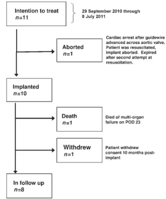
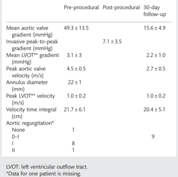
Eleven patients were brought to the operating room (OR) with the intention to treat, and 10 patients underwent the implant-ation procedure, all of them successfully. The patientflowchart is summarized in Fig. 4: the baseline characteristics of the 11 patients brought to the OR are summarized in Table1. Mean age was 82.5 ± 3.6 years (range from 76 to 88 years). Nine were female. The majority of patients were in New York Heart Classification class III (eight patients) or IV (two patients) and one patient had undergone previous cardiac surgery. Mean logistic EuroSCORE was 24.6 ± 13.6% (range from 14.9 to 52.5%). Mean aortic annulus diameter as assessed by transoesophageal echo-cardiography was 21.0 ± 0.8 mm. Aortic annulus perimeter and derived effective mean diameter by CT were 74 ± 2.3, and 23.6 ± 0.7 mm, respectively. Mean aortic valve pressure gradient was 49.3 ± 13.5 mmHg assessed by continuous-wave doppler.
Procedural details
Procedural details and outcomes are summarized in Table 2. Accurate valve placement was achieved in all 10 implanted patients. Mean fluoroscopy time was 8.6 ± 2.5 min; 103 ± 32 ml of contrast medium was used per procedure. Skin-to-skin time was 94.5 ± 16.7 min. No device related or delivery-system-related complications occurred in any of the 10 implanted patients.
Safety and ef
ficacy
Death within 30 days post-implant occurred in 2 of 11 patients (18%) brought to the OR and 1 of the 10 patients (10%) in whom the valve implantation was attempted.
One patient (logistic EuroSCORE 16.6%) experienced hypoten-sion and cardiac arrest during insertion of the guidewire through the apex to the aortic arch. The patient was stabilized through the use of extracorporeal membrane oxygenation and balloon valvuloplasty was performed. The procedure was aborted without implantation of any prosthetic valve because the patient deteriorated again and expired after a second attempt at resuscitation.
Figure 3:Rotational positioning of the device. Commissural post with window is centred and its posterior position is verified by observing its movement to the
left of the image upon mild clockwise rotation.
The second patient (logistic EuroSCORE 48.9%) became haemodynamically unstable upon introduction of the balloon valvuloplasty sheath, and experienced cardiac arrest. The patient was resuscitated and the Engager™ valve bioprosthesis was suc-cessfully deployed. The patient was transferred to the ICU in a stable condition, but had a complicated post-operative course (including respiratory insufficiency requiring reintubation, tra-cheal perforation, third degree atrioventricular (AV) block requir-ing pacemaker implantation, renal insufficiency requiring dialysis and bowel ischaemia requiring resection), and ultimately died on post-implant day 23 of multi-organ failure. There were no strokes or myocardial infarctions during the first 30 days after implantation. One patient (10%) with chronic renal insufficiency required dialysis in the early post-operative period.
Prosthetic valve performance
The invasively measured peak-to-peak gradient after valve im-plantation was 7.1 ± 3.5 mmHg. In nine of the implanted
patients, there was no or only trivial (grad I or less) aortic regur-gitation due to paravalvular leak. In one patient, aortic regurgita-tion grade I–II was observed. There were no cases of aortic insufficiency greater than grade II. At 30-day follow up, average mean aortic gradient was 15.6 ± 4.9 mmHg (one patient who may have benefited from a post-dilatation had a mean gradient of 24 mmHg; all other values ranged between range 8.0 and 17 mmHg). No more than mild transvalvular and paravalvular aortic regurgitation was assessed by transthoracic echocardiography (Table3).
Prosthetic valve-associated complications
Two of 10 implanted patients (20%) required permanent pace-maker implantation for complete atrioventricular block. One patient developed complete atrioventricular block for a short period of time followed by resuscitation, and during the follow-ing day appeared to have stable sinus rhythm with AV-block grade I. One patient with known episodes of atrial fibrillation pre-implantation developed atrial fibrillation requiring electro-cardioversion at Day 5. No coronary obstructions or aortic dis-sections occurred, no undesired mitral interference, or any other prosthesis-related adverse events were observed and none of the implanted prostheses was explanted.
DISCUSSION
The purpose of this feasibility study was to assess the safety and clinical performance of the re-designed Engager™ TAVI system prior to commencing a multicentre pivotal study. Ten of 11 patients who were considered high risk for conventional aortic valve replacement have been successfully implanted through a Table 2: Procedural details
Characteristic FIM* [6] (n = 30) Feasibility
(n = 10)
Aortic annulus diameter (mm)
21.8 ± 1.4 21.0 ± 0.8
Accurate device placement (patients)
29 (97%) 10 (100%)
Used prosthesis size (patients)
23 mm 30 (100%, only
available size)
4 (40%)
26 mm 6 (60%)
Skin-to-skin time (min) 74 ± 16 94.5 ± 16.7
Contrast medium volume (ml)
130 ± 58 103 ± 32
Fluoroscopy time (min) 7.5 ± 2.6 8.6 ± 2.5
*FIM: first-in-man.
Table 3: Haemodynamic parameters pre- and post-implantation
Pre-procedural Post-procedural 30-day follow-up Mean aortic valve
gradient (mmHg)
49.3 ± 13.5 15.6 ± 4.9
Invasive peak-to-peak gradient (mmHg)
7.1 ± 3.5 Mean LVOT** gradient
(mmHg)
3.1 ± 3 2.2 ± 1.0
Peak aortic valve velocity (m/s)
4.5 ± 0.5 2.7 ± 0.5
Annulus diameter (mm)
22 ± 1 Peak LVOT** velocity
(m/s)
1.0 ± 0.2 1.0 ± 0.2
Velocity time integral (cm) 21.7 ± 6.1 20.4 ± 5.1 Aortic regurgitation* None 1 0–I 9 I 8 II 1
LVOT: left ventricular outflow tract. *Data for one patient is missing. Table 1: Baseline characteristics of patients brought
to the OR
Characteristic No. of patients Percentage
n 11
Age (years) 82.5 ± 3.6
Female 9 80
Logistic EuroSCORE (%) 24.6 ± 13.6
New York Heart Classification
II 1 9
III 8 73
IV 2 18
Chronic pulmonary disease 5 45
Impaired renal function 0 0
Neurological dysfunction 1 9
Previous cardiac surgery 1 9
AD UL T C ARDIA C
using the new delivery system. One patient died in the OR before valve implantation was attempted and 1 of the 10 implanted patients died on post-procedure day 23 after experi-encing multiple serious adverse events resulting in an as-treated mortality rate of 10%. Theflexible over-the-wire delivery system adapted well to the aortic anatomy, and allowed for safe and quick implantation without evidence of aortic injury or any other delivery-system-related complications. No patient experi-enced a stroke after implantation and until 30 days of follow up. No vascular complication occurred in our patient population. These results confirm the findings of other trials reporting a low incidence of major vascular complications [8,9] for the transapi-cal access. Although the number of patients in this feasibility study is small, the current results suggest that the redesign of the delivery system, as aflexible, fully covered over-the-wire system, has effectively corrected the root cause of the aortic dissections observed in thefirst-in-man study.
At the end of the procedure, 9 of 10 patients had no more than grade I perivalvular regurgitation, and none had grade II or more. No patient was documented with more than mild trans-valvular or paratrans-valvular regurgitation by colour flow Doppler echocardiography at 30-day follow-up. Different values for the mean transprosthetic gradients were obtained with two different methods and under different haemodynamic conditions: the peak-to-peak pressure gradient during general anaesthesia and the transthoracic echocardiographically measured gradient based on flow velocity and calculated with the modified Bernoulli equation. The geometry of the support arms facilitates predictable deployment of the prosthesis into an anatomical correct positioning with a defined height of implantation, making the procedure both intuitive and reliable. Adverse events like valve dislocation and coronary obstruction as described for other prostheses [8] can potentially be avoided by the design, and were indeed not observed. The incidence and type of reported serious adverse events could be primarily attributed to the study’s patient population with a mean logistic EuroSCORE of 24.5%.
In conclusion, the current results of the Engager™ feasibility study demonstrated the successful deployment of the Engager™ bioprosthesis into an anatomically correct position without peri-operative delivery system-related complications using the rede-signed Engager™ transapical catheter delivery system. As a result, patients are currently being enrolled into the multicentre,
safety and clinical performance of the Engager™ bioprosthesis in a larger patient population.
ACKNOWLEDGEMENTS
We thank Ehud Schwammenthal for his invaluable work as proctor of the Engager™ TAVI system. As a co-founder of Ventor technologies Ltd that primarily introduced the valve, he partici-pated in all procedures and gave indispensable input during the implantations and significantly participated in completing this manuscript.
Conflict of interest: none declared.
REFERENCES
[1] Rodés-Cabau J, Webb JG, Cheung A, Ye J, Dumont E, Feindel CMet al.
Transcatheter aortic valve implantation for the treatment of severe symp-tomatic aortic stenosis in patients at very high or prohibitive surgical risk.
J Am Coll Cardiol 2010;55:1080–90.
[2] Thomas M, Schymik G, Walther T, Himbert D, Lefèvre T, Treede Het al.
Thirty-day results of the SAPIEN aortic Bioprosthesis European Outcome (SOURCE) Registry: a European registry of transcatheter aortic valve
im-plantation using the Edwards SAPIEN valve. Circulation 2010;122:62–9.
[3] Kempfert J, Rastan AJ, Mohr FW, Walther T. A new self-expanding
trans-catheter aortic valve for transapical implantation—first in man
implant-ation of the JenaValve™. Eur J Cardiothorac Surg 2011;40:761–3.
[4] Kempfert J, Rastan AJ, Beyersdorf F, Schönburg M, Schuler G, Sorg Set al.
Trans-apical aortic valve implantation using a new self-expandable
bio-prosthesis: initial outcomes. Eur J Cardiothorac Surg 2011;40:1114–9.
[5] Lange R, Schreiber C, Götz W, Hettich I, Will A, Libera Pet al. First
suc-cessful transapical aortic valve implantation with the Corevalve Revalving system: a case report. Heart Surg Forum 2007;10:E478–9.
[6] Falk V, Walther T, Schwammenthal E, Strauch J, Aicher D, Wahlers Tet al.
Transapical aortic valve implantation with a self-expanding anatomically
oriented valve. Eur Heart J 2011;32:878–87.
[7] Leon MB, Piazza N, Nikolsky E, Blackstone EH, Cutlip DE, Kappetein P et al. Standardized endpoint definitions for transcatheter aortic valve im-plantation clinical trials. JACC 2011;57:253–69.
[8] Walther T, Falk V, Kempfert J, Borger MA, Fassl J, Chu MW et al.
Transapical minimally invasive aortic valve implantation; the initial 50
patients. Eur J Cardiothorac Surg 2008;33:983–8.
[9] Ewe SH, Delgado V, Ng AC, Antoni ML, van der Kley F, Marsan NAet al.
Outcomes after transcatheter aortic valve implantation: transfemoral versus transapical approach. Ann Thorac Surg 2011;92:1244–51.