Abstract. The fatality of cancer predominantly results
from the dissemination of primary tumor cells to distant sites and the subsequent formation of metastases. During tumor progression, some of the primary tumor cells as well as the tumor microenvironment undergo charac-teristic molecular changes, which are essential for the metastatic dissemination of tumor cells. In this review, we will discuss recent insights into pro-metastatic events oc-curring in tumor cells themselves and in the tumor stroma. Tumor cell-intrinsic alterations include the loss of cell polarity and alterations in cell-cell and cell-matrix
DOI 10.1007/s00018-005-5296-8 © Birkhäuser Verlag, Basel, 2006
adhesion as well as deregulated receptor kinase signaling, which together support detachment, migration and inva-sion of tumor cells. On the other hand, the tumor stroma, including endothelial cells, fibroblasts and cells of the immune system, is engaged in an active molecular crosstalk within the tumor microenvironment. Subse-quent activation of blood vessel and lymph vessel angio-genesis together with inflammatory and immune-sup-pressive responses further promotes cancer cell migration and invasion, as well as initiation of the metastatic pro-cess.
Key words: Angiogenesis; lymphangiogenesis; metastasis; tumorigenesis; tumor stroma.
Introduction
Carcinomas are the most frequent type of human malig-nancies, and the vast majority of cancer deaths are caused by the formation of metastases rather than by the primary tumor itself. Since existing metastases are difficult to tar-get by conventional cancer therapies, a curative regimen requires the detection of the disease before the primary tumor has spread. Yet, despite evolving clinical diagnos-tic tools, a large proportion of tumors have already metas-tasized by the time of diagnosis.
The selective process of metastasis requires that cancer cells successfully complete several sequential, rate-limit-ing steps. They must detach from the primary tumor, in-vade the host stroma, intravasate into lymphatic or blood vessels, spread to the capillary bed of distant organs,
ex-travasate and proliferate in the receptive organ parench-yma [1]. The succeeding metastatic cells have undergone changes in their proliferative, survival, migratory and in-vasive abilities and can be seen as winners of a ‘metasta-sis decathlon’ [2]. It is now well established that tumor cell-autonomous changes are not sufficient to allow tu-mor progression and metastasis to occur. By analogy with the architecture of organs, tumors are not only composed of a ‘parenchyma’ formed by the neoplastic cells, but also of a supportive ‘stroma’, consisting of specific extra-cel-lular matrix (ECM) components, fibroblasts, adipocytes, vascular cells, smooth muscle cells and cells of the haematopoietic system [3]. During tumor progression and metastasis, an active crosstalk occurs between tumor cells and their stroma, mainly mediated by direct cell-cell contact or paracrine cytokine and growth factor signal-ing, reminiscent of the communication between epithelial and mesenchymal cells during embryonic development.
Review
Metastasis: cell-autonomous mechanisms versus
contributions by the tumor microenvironment
L. Kopfstein and G. Christofori*Institute of Biochemistry and Genetics, Department of Clinical-Biological Sciences, University of Basel, Mattenstrasse 28, 4058 Basel (Switzerland), Fax: +41 61 267 3566, e-mail: [email protected] Received 4 July 2005; received after revision 3 November 2005; accepted 14 November 2005
Online First 16 January 2006
Such signaling may activate the tumor microenvironment at the primary and secondary tumor sites, thereby under-going morphological changes (desmoplasia’) and allow-ing or even supportallow-ing tumor outgrowth, invasion and metastasis [4]. Hence, tumor cell-stroma interaction is an important new focus of research in the treatment of me-tastatic disease.
Many excellent reviews cover particular aspects of mole-cular pathways known to be involved in the metastatic process. This review more broadly summarizes recent progress in the elucidation of various pro-metastatic mol-ecular events and pathways occurring during tumor pro-gression. In particular, we emphasize the distinction be-tween tumor cell and tumor stroma contribution, and we discuss the role of tumor cell polarity, adhesion and deregulated receptor kinase signaling, as well as endothe-lial cells, fibroblasts and immune cells in invasion and metastatic dissemination of tumor cells.
Tumor cell-intrinsic changes
Changes in cell-cell and cell-matrix adhesion Loss of E-cadherin function and epithelial-mesenchymal transition
E-cadherin is a central player in the makeup of cell polar-ity and epithelial organization. With its extracellular do-mains, this cadherin mediates calcium-dependent ho-mophilic cell-cell contact in adhesion junctions, while linking adhesion junctions to the actin cytoskeleton via a cytoplasmic cell adhesion complex consisting of a-, b-, g- and p120 catenins [5]. Thereby cell-cell adhesion can affect localization and function of cytoskeletal regulators and influence actin-guided cell motility [6]. In most can-cers of epithelial origin, E-cadherin-mediated cell-cell adhesion is lost concomitantly with tumor progression and is correlated with advanced tumor grades and poor patient survival [7]. Consistent with this observation, forced downregulation of E-cadherin function in a mouse model of carcinogenesis promotes tumor progression, in-vasion and metastasis [8]. These and many more results from a variety of in vivo and in vitro experimental sys-tems demonstrate that E-cadherin is an important mole-cule in tumor progression [5].
How does the loss of E-cadherin promote metastasis? In order to leave the coherent epithelial cell assembly of the primary tumor, malignant tumor cells have to acquire a fibroblastoid, migratory and invasive phenotype. A simi-lar process physiologically occurs during embryonic de-velopment, tissue remodeling, wound healing and in-flammation and is termed epithelial-to-mesenchymal transition (EMT) [9]. A number of in vitro studies (see below) collectively support the idea that morphologic and molecular changes of tumor cells required for the first
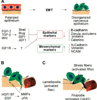
steps of metastasis mimic physiological EMT. In fact, full EMT of in vitro cultured tumor cells is thought to resem-ble, at least in part, the metastatic process in cancer pa-tients. Nevertheless, this hypothesis is still under debate and evidence for carcinoma EMT in vivo has yet to be found [10]. We support the view that EMT-like events oc-cur in tumors and that studying these events may con-tribute to our understanding of metastasis formation. Oncogenic events in tumor cells as well as growth factors secreted by tumor and stromal cells, including transform-ing growth factor-b (TGF-b) and fibroblast growth factor 2 (FGF-2), induce EMT (fig. 1). During EMT, cells pro-gressively redistribute or downregulate their apical and basolateral epithelial-specific proteins, such as tight and
Figure 1. Mechanisms of tumor invasion and metastatic dissemina-tion. During scattering and epithelial-to-mesenchymal transition (EMT), cells undergo major changes in morphology and lose cell-cell contacts. (A) EMT of tumor cell-cells. Loss of E-cadherin leads to disruption of adhesion junctions and is essential for EMT. TGF-b, FGF-2 and Wnt induce or stabilize Snail1, Snail2, dEF1 and SIP1, all direct or indirect transcriptional repressors of E-cadherin. Con-comitant with the loss of E-cadherin function, the expression of mesenchymal marker proteins is induced. (B) Degradation of base-ment membrane (BM) and extra-cellular matrix (ECM). HGF/SF and EGF activate fibroblasts and endothelial cells to secrete matrix metalloproteases and urokinase-type plasminogen activator. The BM and ECM are thereby degraded, allowing tumor cells to trans-migrate into the subepithelial ECM and to finally intravasate into blood or lymphatic vessels. (C) Activation of the tumor cell actin cytoskeleton, migration and invasion of metastatic cells into blood vessels. Upon loss of cell polarity, activated Rac, Cdc42 and RhoA are no longer sequestered at adhesion junctions and exert pro-mi-gratory activities: Rac induces lamellipodia and membrane ruffles at the leading edge of the cell, Cdc42 bundles actin into filopodia, Rho assembles actin into stress fibers. These cytoskeletal changes result in increased cell motility.
adherens junction proteins (including E-cadherin), and re-express mesenchymal molecules, such as vimentin and N-cadherin. These changes lead to the abrogation of cell-cell contacts and the gain of cell-cell motility necessary for in-vasion (reviewed in [11]). EMT is in contrast to the process of cell scattering, where different epithelial mark-ers are revmark-ersibly downregulated and expression of mes-enchymal proteins is not induced. Scattering can be me-diated by a number of growth factors, including hepato-cyte growth factor/scatter factor (HGF/SF), TGF-b, FGF-2, TGF-a and epidermal growth factor (EGF) via the PI3K or the Raf/MAPK signaling pathways [9, 12]. Loss of E-cadherin promotes, whereas maintenance of its expression inhibits, EMT and metastasis [5, 8]. E-cad-herin is therefore considered a metastasis suppressor gene. Downregulation of E-cadherin mostly occurs at the transcriptional level. The E-cadherin promoter is fre-quently repressed by specific transcriptional repressors, including Snail1 (previously Snail), Snail2 (previously Slug), SIP1, dEF1, Twist and E12/E47, and by subse-quent promoter hypermethylation [13, 14]. Consistent with this observation, some of these repressors have been found expressed specifically at the invasive front of human invasive hepatocellular and breast carcinoma [15, 16]. The expression of these repressors seems to be highly regulated by pathways, including canonical Wnt signaling, TGF-b (see below), FGF, EGF, Stat3 and nu-clear factor k-B (NFk-B) signaling [17–19]). Notably, Snail1 is a highly unstable protein. It is rapidly phospho-rylated by glycogen synthase kinase 3b (GSK3b) and subsequently ubiquitylated and degraded by the protea-some pathway [20]. As a result of transcriptional repres-sion, the E-cadherin promoter is frequently found hyper-methylated in a large subset of cancer cases [21]. E-cad-herin can also be downregulated at the protein level. Receptor tyrosine kinases (RTKs), such as EGFR, c-Met, IGF1R, FGFR and the non-receptor tyrosine kinase c-Src can induce phosphorylation of E-cadherin and catenins, resulting in their ubiquitylation by the E3 lig-ase Hakai and subsequent endocytosis and degradation [22–25]. Finally, secreted proteases such as matrix met-alloprotease (MMP)-9, for example induced by TGF-b and HGF/SF, can cleave E-cadherin and disrupt cad-herin-mediated cell-cell contacts [26].
Loss of E-cadherin during EMT disrupts adhesion junc-tions between neighboring cells and thereby supports de-tachment of malignant cells from the epithelial cell layer. Yet, migration and invasion of tumor cells is also pro-moted by the loss of interaction of E-cadherin with the cytoskeleton, subsequent changes in the activities of Rho family GTPases, most prominently Rac1, Cdc42 and RhoA, and the concomitant reorganization of the actin cytoskeleton (fig. 1). In epithelial cells, E-cadherin-me-diated assembly of adherens junctions recruits and ac-tivates Rac1 and Cdc42. Thereby, E-cadherin-bound
a-and p120-catenin directly interact with Rho family-spe-cific guanine nucleotide exchange factors (GEFs) and with Rho family members [27, 28]. Moreover, activated Rac1 and Cdc42 promote and consolidate E-cadherin-mediated adhesion by sequestering their downstream ef-fector IQGAP1, which in its free form inhibits the inter-action of b-catenin with a-catenin/E-cadherin [29]. After an early activation phase, where RhoA is required for E-cadherin clustering, RhoA gets gradually downregulated with a concomitant repression of cell migration [30]. In contrast, forced expression of RhoA (and also RhoC) in tumor cells induces focal adhesions and stress fibers, pro-moting invasion and metastasis [31, 32]. Upon loss of E-cadherin function, RhoA, Rac1 and Cdc42 are released from adhesion junctions and promote cell migration and invasion. Consistent with the dual role of these mole-cules, the Rac1-specific GEF Tiam1 localizes to adhesion junctions and promotes cell adhesion on substrates im-peding cell migration (fibronectin, laminin), whereas it is found in lamellipodia when cells are cultured on cell motility-supporting collagen type I [33]. However, the mechanisms, by which ECM and thus integrin-mediated signaling affect the activity of RhoA, Rac1 and Cdc42, and with it E-cadherin function, remain to be elucidated. Recent studies have shown that mesenchymal cadherins, in particular N-cadherin, enhance tumor cell motility and migration (reviewed in [5]), thus exhibiting an opposite effect compared with E-cadherin. N-cadherin-induced tu-mor cell invasion can even overcome E-cadherin-medi-ated cell-cell adhesion. A novel concept based on the above observations is that a ‘cadherin switch’ from ep-ithelial to mesenchymal cadherins exists which supports the transition from a benign to an invasive, malignant tu-mor phenotype. Hence, in addition to the loss of E-cad-herin, the gain of N-cadherin may critically contribute to tumor invasion and metastatic dissemination. In fact, N-cadherin has been shown to induce tumor cell migration, most likely by stimulating FGF receptor-mediated signal transduction [34, 35]. Hence, changes in the expression of cadherins not only modulate cell-cell adhesion but also induce pro-metastatic signaling pathways.
Integrins and tumor progression
In order to detach and migrate, tumor cells depend on changes in both cell-cell and cell-matrix interactions. It is tacitly assumed that strong cell-matrix adhesions need to be dissolved, whereas transient and weak adhesions are a prerequisite for migration. This complex situation has made it difficult to experimentally determine the func-tional contribution of cell-matrix adhesion to tumor pro-gression. With integrins being the prototype mediators of cell-matrix adhesion, they have been studied in great de-tail, also in tumor progression [36–38]. It is beyond the scope of this review to discuss the detailed mechanisms by which integrins affect tumor cell adhesion and
migra-tion. Instead, we focus on the functional contribution of integrins to tumor metastasis. Moreover, the following sections present several mechanistic links of integrins to signaling pathways.
Integrins are heterodimeric cell surface receptors consist-ing of two type-I transmembrane subunits, a and b. They provide the essential link between the actin cytoskeleton and the ECM during cell migration. Different a and b subunits form distinct integrin subtypes, which link ECM ligands, such as fibronectin, vitronectin, laminin and col-lagen, to the intracellular actin cytoskeleton [38]. Impor-tantly, binding to these ECM components activates inte-grins, which in turn induce intracellular signaling cas-cades modulating cell proliferation, survival, polarity, motility and differentiation [36, 38, 39]. Thereby inte-grins mediate anchorage dependence, since they allow these processes only in appropriately adhering cells. However, during tumor progression, mutations in onco-genes and tumor-suppressor onco-genes can enable non-adher-ent neoplastic cells to survive and proliferate in the ab-sence of proper cell-matrix adhesion [40]. Cancer cells that profit from integrin signals are being selected due to their survival and proliferation advantage. Many cancer types, including melanoma, glioblastoma, prostate, breast and ovarian cancer, exhibit increased expression of avb3, a2b1, a4b1and a6b1integrins during tumor
progres-sion [41–43]. Tumor cell migration and metastasis is supported by integrin-mediated focal adhesion and actomyosdependent contractility, as demonstrated for in-stance for integrins a6b4, a2b1and aVb3[44–46].
Further-more, activated integrins can recruit proteases, such as MMP-9, towards the site of attachment, and the subse-quent ECM degradation supports migration and invasion of cells into the surrounding tissue [38, 43]. Unfortu-nately, various subsets of integrins seem to mediate dif-ferent functions during progression of difdif-ferent cancer types, and more experimental work will be required to unravel their actual functional contribution.
Endothelial cell integrins have been studied in great de-tail during physiological and pathological angiogenesis and have been shown to play an important role in tumor angiogenesis. Distinct subtypes, including a1b1and a2b1
integrins, support vascular endothelial growth factor (VEGF)-mediated angiogenesis in tumor xenotransplan-tion experiments [47]. Endothelial a5b1, aVb3and aVb5
integrins are critically involved in both VEGF- and FGF-mediated angiogenesis. Peptide antagonists against these integrins are able to interfere with tumor angiogenesis in a number of experimental model systems, and initial clin-ical trials with some of these compounds are under way [37]. Interestingly, experiments with mice lacking aVb3
and aVb5 integrins have demonstrated increased rather
than reduced tumor growth, raising the possibility of a dual function of integrins at least in the angiogenic process [48]. Thus, changes in integrin-mediated matrix
adhesion on tumor cells, endothelial cells and certainly other cells of the tumor stroma can influence tumor cell migration, invasion and angiogenesis, all rate-limiting processes in tumor metastasis.
Immunoglobulin family adhesion molecules: NCAM and L1
Among many other cell adhesion molecules (CAMs), two have been specifically implicated in promoting tumor cell invasion and metastatic spread, NCAM (CD56) and L1 (CD171, L1CAM). Both structurally belong to the im-munoglobulin (Ig) superfamily and are critical for central nervous system (CNS) development. They not only me-diate static neuron-neuron adhesion but also are particu-larly involved in neurite outgrowth, axon guidance and neural cell migration. In addition to providing mechani-cal cell-cell and cell-ECM adhesion, they also activate signaling receptors and induce intracellular signaling cas-cades [5, 49].
NCAM has been shown to play an important role in the progression to tumor malignancy. Both the shift of the adult NCAM 120-kDa isoform to the embryonic 140-and 180-kDa isoforms 140-and a general downregulation of NCAM expression are associated with poor prognosis in a few cancer types [50, 51]. In a transgenic mouse tumor model of pancreatic b cell carcinogenesis (Rip1Tag2), loss of NCAM leads to the formation of lymph node metastasis mediated by the increased expression of the lymphangiogenic factors VEGF-C and D and with it up-regulated peri-tumoral lymphangiogenesis [52, 53]. Sim-ilar to its function in neurons, NCAM also modulates cell-matrix adhesion of tumor cells by binding to FGFR and inducing a variety of signaling pathways that lead to b1 integrin-mediated cell-matrix adhesion and neurite
outgrowth [34].
While L1 exerts functions similar to NCAM in neurons, L1 is specifically expressed at the invasive front of glio-mas, melanoglio-mas, lung, prostate, renal, ovarian and en-dometrial carcinoma, and its expression appears to corre-late with metastasis [54–59]. Similar to NCAM, L1 has been shown to enhance migration and invasion of tumor cells in vitro. L1 promotes cell-matrix adhesion and mi-gration of tumor cells on ECM proteins, such as fibro-nectin and laminin, by associating with integrins through an integrin recognition motif. Namely, integrins avb5, avb3
and a5b1have been found to associate with L1 [49, 60–
62]. In these studies, integrin-mediated signaling events induced by associated L1 seem to be responsible for pro-moted cell migration. Interestingly, L1-integrin interac-tion may not only occur in cis at the cell surface but also by ectodomain shedding of L1 and subsequent autocrine/ paracrine binding of soluble L1 to avb5 integrin. Inde-pendent of integrin binding and activation, L1 has also been shown to promote growth factor-induced mitogen-activated protein kinase (MAPK) signaling and to
subse-quently stimulate extracellular regulated kinase (Erk)1/2-mediated transcriptional changes in genes associated with cell motility and invasion and matrix remodeling [49]. Such targets include the genes for cathepsin-L and -B, os-teopontin (OPN), RhoC and CD44, all proteins implicated in tumor invasion and metastasis (CD44 and RhoC are dis-cussed below) [49, 63]. Finally, L1 was recently identified as a direct target gene of activated Wnt signaling, together with the metalloprotease ADAM10. Co-induction of ADAM10 and L1 leads to proteolytic shedding of the L1 extracellular domain, which in turn induces tumor cell motility and enhanced tumorigenesis [64].
Together, these selected examples indicate that detach-ment and migration of tumor cells critically depends on altered cell-cell and cell-matrix adhesion. Yet, such alter-ations appear not to only affect cell adhesion but also a variety of signal transduction pathways, mediated by ty-rosine kinase receptors or by integrins, which all seem to lead to increased tumor cell invasiveness and metastatic dissemination.
Changes in signaling pathways during EMT and scattering
Malignant transformation with deregulation of cell growth is frequently induced by somatic mutations or overex-pression of receptor tyrosine kinases [65]. Importantly, deregulated signaling of some receptor tyrosine kinases not only exerts transformation of tumor cells but also pro-motes their invasion and metastatic dissemination. The following sections address some of the newly emerging aspects of how receptor tyrosine kinases contribute to tu-mor metastasis (fig. 1).
c-Met and hepatocyte growth factor/scatter factor (HGF/SF)
Overexpression of and activating mutations in c-Met have been reported in many human tumors, and xenotransplant and transgenic mouse models demonstrate a profound pro-metastatic activity of HGF/SF and Met [66–68]. c-Met, encoded by the MET proto-oncogene, is a dimeric transmembrane tyrosine kinase receptor for HGF/SF. In-tracellular transducers of c-Met activity include Ras/ MAPK, PI3K, phospholipase g (PLC-g), Src-related ty-rosine kinases and growth-factor-receptor-bound protein 2 (Grb2)-associated binder 1 (Gab-1). Internalization is a typical mechanism by which signaling of receptors such as c-Met is terminated, and poly-ubiquitylation promoted by c-Cbl is critical for c-Met downregulation by targeting it to the proteasome [69, 70].
Although the precise mechanisms underlying c-Met-me-diated tumor cell migration and invasion are not com-pletely understood, several critical events have been recently elucidated. Upon ligand binding, c-Met induces invasive growth, both during physiological tissue
morph-ogenesis and tumor progression. HGF/SF-mediated ef-fects appear to depend on the differentiation state of the c-Met-expressing cells. Whereas HGF/SF induces mor-phogenesis of E-cadherin-positive polarized epithelial cells, it promotes scattering and metastasis of cells that have lost their epithelial characteristics [69]. The metasta-tic potential of c-Met is mediated via a signal transducer docking site, which can bind and activate multiple Src homology region 2 (SH2)-containing intracellular effec-tors. Interestingly, a distinct mutation of this domain (H1351N) increases the transforming but in parallel abol-ishes the metastatic properties of c-Met [71]. c-Met-in-duced dissociation of adherens junctions, scattering and metastasis appear to be primarily mediated via the PI3K pathway in a MAPK-dependent, yet protein kinase B (PKB/Akt)- or Rac-independent manner [72, 73]. Through activation of the MAPK pathway, c-Met signaling in-duces Ets1 transcription factors, which control the ex-pression of genes involved in ECM remodeling and cell scattering [74]. In fact, Ets1 expression is known to cor-relate with metastasis incidence in a number of cancer types [75, 76].
Recent results indicate that the pro-metastatic activity of c-Met also critically depends on its ability to associate with other cell surface proteins, notably a6b4integrin,
CD44 and Plexin B1. c-Met is constitutively associated with integrin a6b4, which in turn is phosphorylated by
activated c-Met. Subsequently, phosphorylated integrin a6b4 recruits the signaling adaptor Shc and PI3K and
po-tentiates HGF/SF-induced activation of the Ras and PI3K pathways [77]. c-Met also associates with and is functionally modulated by CD44, a hyaluronan receptor involved in cell-cell/cell-matrix interactions, cell migra-tion and metastasis [78]. Upon HGF/SF stimulamigra-tion, c-Met associates with a variant CD44 isoform (CD44v6) and recruits ezrin, radixin and moesin proteins (ERMs) to the complex [79]. ERMs link adhesion molecules to filamentous F-actin and participate in membrane and cy-toskeletal remodeling during cell migration. Both CD44 and the ERM proteins are known to promote metastasis by various mechanisms. For instance, ezrin can stabilize mammalian target of rapamycin (mTOR) downstream signaling molecules, enhancing metastasis [80]. c-Met-mediated tumor cell invasion is CD44-dependent and may also require ERMs, since signal transduction by ac-tivated c-Met via the MAPK pathways depends on the ERM binding domain of CD44 [79, 81, 82]. Further-more, HGF/SF specifically induces the expression of OPN and its association with the hyaluronan-CD44-ERM complex at the leading edge of metastatic cells [83, 84]. OPN is a protein of the ECM with a wide variety of cell surface binding proteins and has been shown to pro-mote tumor cell invasiveness [85]. The functional coop-eration of c-Met, CD44 and OPN thus may be critical for metastasis [83].
Plexins, which share structural homology with c-Met, are widely expressed receptors for Semaphorins (Sema). The Sema family consists of secreted and membrane-bound members that act as guidance signals for neurons [86, 87]. Recently, Plexin B1 has been shown to be overex-pressed in several tumor cell lines and to be physically as-sociated with c-Met [87]. Notably, binding of Sema 4D to Plexin B1 transactivates the tyrosine kinase activity of c-Met independent of the presence of HGF/SF [88]. Inter-estingly, the identical signaling pathway in endothelial cells leads to angiogenesis [89].
In addition to inducing a motile tumor cell phenotype, c-Met promotes invasion of neoplastic cells by upregulat-ing proteases such as plasminogen activator (uPA) and MMPs via MAPK and PKC, respectively. These pro-teases are critical in degrading the ECM and allowing mi-gration of tumor cells through the basement membrane and the surrounding stroma [90].
Based on the multitude of activities and interactions dur-ing tumor progression, c-Met represents an important mediator of cell migration and metastasis and, hence, c-Met-mediated signaling pathways are in the spotlight as potential targets for the development of anti-metastatic therapy [91].
Wnt signaling and tumor progression
In many cancer types, Wnt signaling is activated by mu-tations in a number of effector genes, including the genes encoding for adenomatous polyposis coli (APC), axin-1 and 2, and b-catenin, which predispose to cancer [92–94]. Besides their critical role in assembling the E-cadherin-mediated cell adhesion complex, b-catenin and g-catenin also have important functions in the canonical Wnt-sig-naling pathway. Non-sequestered, free b- and g-catenin are rapidly phosphorylated by GSK-3b in the APC/axin/ GSK-3b complex and subsequently degraded by the ubiquitin-proteasome pathway. If the tumor suppressor APC is non-functional, as in many colon-cancer cells, or if GSK-3b activity is blocked by the activated Wnt-sig-naling pathway, b-catenin accumulates at high levels in the cytoplasm. Subsequently, it translocates to the nu-cleus, where it binds to members of the Tcf/Lef-1 family of transcription factors and modulates the expression of Tcf/Lef-1-target genes, including c-Myc, cyclin D1, fi-bronectin, MMP-7, Id2, CD44, axin-2, Tcf-1 and others, all genes implicated in cell proliferation, transformation and tumor progression.
The dual function of b-catenin has motivated a multitude of experiments to assess whether the loss of E-cadherin function would subsequently lead to the activation of the Wnt-signaling pathway. In a number of cellular systems, it has been demonstrated that sequestration of b-catenin by E-cadherin can compete with the b-catenin/TCF-me-diated transcriptional activity of the canonical Wnt-sig-naling pathway. The fact that E-cadherin does not
com-pletely deplete the cytoplasmic catenin suggests that b-catenin exists in different functional pools [95, 96]. Inter-estingly, in breast and prostate carcinoma cell lines, E-cadherin suppresses tumor cell invasion by binding b-catenin without repressing b-b-catenin/TCF transcriptional activity, indicating that a novel, as yet unknown, addi-tional function of b-catenin may be required for cellular invasiveness [97, 98].
Furthermore, activated Wnt signaling inhibits E-cad-herin-mediated cell adhesion by inducing expression of Snail1, a transcriptional E-cadherin repressor. Snail1 in turn synergizes with the Wnt/b-catenin pathway by in-ducing Tcf expression, and b-catenin/Tcf can in turn repress E-cadherin transcription in co-operation with Snail1 [99, 100].
Signals elicited by ErbB receptors
Many metastatic human carcinomas are characterized by the overexpression or constitutive activation of ErbB ty-rosine kinase receptor (EGFR) family members involving activating mutations of the receptor kinases or an au-tocrine loop with EGF family ligands. In addition to stim-ulating cell differentiation and proliferation, EGF pro-motes tumor cell motility, invasion and metastasis by af-fecting a large number of protein functions and signaling pathways, including Wnt signaling, Snail1 expression, focal adhesion kinase (FAK) activity and expression of MMPs [101].
Interestingly, ErbB receptors and Wnt signaling cooper-ate during tumorigenesis, which may be critical for me-tastasis formation [102]. In tumors of mouse mammary tumor virus (MMTV)-Wnt-1 transgenic mice, two mem-bers of the ErbB family of transmembrane receptor tyro-sine kinases, ErbB1 (EGF receptor) and ErbB2 (Her-2/c-Neu), interact with and phosphorylate b-catenin. Interest-ingly, the b-catenin/ErbB interaction correlates with the incidence of pulmonary metastases, indicating a func-tional role of these interactions in metastasis formation [102]. Tyrosine phosphorylation of b-catenin by ErbB1 and by other receptor tyrosine kinases is known to desta-bilize its binding to E-cadherin. As a result, adhesion junctions disassemble, the liberated b-catenin accumu-lates in the cytoplasm, transfers to the nucleus and, in combination with Tcf/Lef1 transcription factors, modu-lates gene expression. Such activation of the Wnt path-way may result in increased tumor cell migration, inva-sion and metastasis (reviewed in [103]).
Notably, Snail1 and caveolin-1 seem to play antagonistic roles during EMT induced by EGF. Caveolins are struc-tural proteins of caveolae, the invaginations of the plasma membrane that function as regulators of signal transduction. Transient EGF signaling induces caveolin-1-dependent endocytosis of E-cadherin. Long-term EGF treatment downregulates caveolin-1 and induces Snail1. Concomitantly, b-catenin/Tcf/Lef-1 transcriptional
ac-tivity is enhanced [19]. Interestingly, both EGF-medi-ated upregulation of Snail1 transcription as well as in-creased transactivation by b-catenin are dependent on caveolin-1 downregulation. EGF-induced downregula-tion of caveol1 may therefore play a central role in in-vasion and metastasis.
Insulin-like growth factor 1 receptor (IGF1R)
Insulin-like growth factors (IGFs) and their cognate sig-naling receptor IGF1R are involved in cellular and organ-ismal growth, embryonic development and numerous pathological states, including cancer and metastasis [104]. IGF1R overexpression in the primary tumor correlates with the increased incidence of metastases in patients with gastric cancer, gastrinoma, gastrointestinal stromal tu-mors, thyroid cancer and other malignancies [105–108]. Forced expression of IGF1R in a transgenic mouse model of pancreatic b cell carcinogenesis (Rip1Tag2) results in accelerated tumor progression, with increased tumor ma-lignancy and distant tumor metastasis to various organs [109]. Conversely, a small chemical inhibitor of IGF1R ty-rosine kinase activity (NVP-AEW541) is able to repress tumor cell survival and tumor growth of a number of dif-ferent tumor types in culture or in xenograft transplanta-tion experiments, yet its effects on tumor progression and metastasis have not been explicitly assessed [110, 111]. The mechanisms of IGF1R-mediated invasive and metastatic abilities of tumor cells have been recently studied. IGF1R signaling has been famous for mediating anti-apoptotic and mitogenic effects via PI3K/PKB and Ras/MAPK pathways. Yet, IGF1R also modulates cell-substrate adhesion, migration, invasion and cell-cell in-teraction by signaling via FAK [112, 113]. IGF1R has also been shown to modulate the expression and function of different junctional proteins, including cadherins, catenins, ZO-1 and the small GTPases RhoA, Rac1 and Cdc42 [114]. Notably, upon IGF-II binding, activated IGF1R associates with E-cadherin and b-catenin, thereby inducing reversible scattering [115]. Through internaliza-tion of the entire complex, E-cadherin is sequestered from the plasma membrane and targeted to late endo-somes and lysoendo-somes. As a consequence, the released b-catenin translocates to the nucleus and induces Tcf/Lef1-mediated transcription of target genes (see also the previ-ous sections). Conversely, overexpression of E-cadherin antagonizes the effects of IGF-II. Such mechanisms are currently being investigated to resolve the actual contri-bution of IGF1R to tumor metastasis.
TGF-b signaling
TGF-b is a member of the TGF-b superfamily of ligands and binds to a heterodimeric receptor consisting of type I and II transmembrane receptor serine-threonine kinases. TGF-b-mediated activation of the receptors induces a ca-nonical signaling pathway via Smad proteins, which
re-sults in nuclear translocation of receptor-Smads (Smad2/ 3) together with common-Smad (Smad4) and in the mod-ulation of expression of various target genes [116, 117]. Depending on the cell type, alternative signaling path-ways, including MAPK, PI3K and PKC, can also be acti-vated [118–120].
Depending on the differentiation and transformation sta-tus of a cell, TGF-b exerts two directly opposing func-tions; it acts as an anti-proliferative and pro-apoptotic growth factor on differentiated cells, but induces prolif-eration and EMT of undifferentiated or transformed cells. Hence, at early stages of tumor development, TGF-b’s cy-tostatic action helps to suppress tumor growth. At later tu-mor stages, however, transformed cells develop resis-tance to the growth-inhibitory effect of TGF-b and re-spond to TGF-b by undergoing EMT (reviewed in [121]). Moreover, TGF-b exerts immunosuppressive functions that further promote tumor progression.
Resistance of tumor cells to TGF-b-mediated growth re-straint and induction of EMT is mostly due to functional changes downstream of the TGF-b signaling pathway [9]. At the transcriptional level, TGF-b cooperates with vari-ous signaling pathways stimulated by EGF, interferon g (IFNg) and tumor necrosis factor (TNF)-a. Activated MAPK signaling, for instance, can inactivate Smads, in-duce inhibitory Smads or inin-duce an autocrine TGF-b pro-duction (for detailed reviews see [117, 122]). In immor-talized (non-transformed) mammary epithelial EpH4 cells, oncogenic Ras and TGF-bR signaling are required for the induction and maintenance of TGF-b-induced EMT [123]. In Ha-Ras-transformed EpH4 cells (EpRas), upregulation of the PI3K pathway prevents TGF-b-in-duced apoptosis and promotes scattering. Full EMT, however, depends on the activation of the MAPK path-way. Similarly, an activated PI3K pathway correlates with tumor formation of these cells, whereas a hyperac-tive MAPK pathway is required for both tumorigenesis and metastasis [9, 12]). Moreover, TGF-b-induced EMT seems to depend on b1 integrin-mediated signals and
RhoA-induced actin cytoskeleton rearrangements [124]. Such cooperative activities of TGF-b together with its dual role as tumor promoter and tumor suppressor ob-scure a direct therapeutic exploitation of this pathway and stimulate further studies into the mechanistic details of these activities.
Genes mediating metastasis
An ongoing interesting debate in the field of cancer metastasis concerns the question whether the metastatic potential of a tumor cell is primed with the first initial ge-netic events of transformation or whether tumor cells by clonal selection progress to tumor malignancy. It seems that genetic alterations acquired early during tumorigen-esis (including gain of oncogenes and loss of
tumor-sup-pressor genes) are sufficient for certain tumor cells to spread [125, 126]. For instance, gene microarray analysis has identified specific expression profiles in metastatic versus non-metastatic primary human carcinomas [127– 130]. Yet, the pro-metastatic efficacy of those genes re-mains to be shown. On the other hand, circulating tumor cells are found to carry various genetic alterations, sug-gesting that additional genetic events are required for tu-mor cell dissemination [131]. Moreover, many genes are expressed and seem to be functional only during the metastatic process. Even more impressive, transgenic ex-pression of MMP3 is sufficient to induce full-blown carcinogenesis, indicating that ‘invasive or metastatic’ processes can even affect the transformation status of a cell [132].
More refined approaches of gene expression analysis combined with functional experiments have now pro-vided novel insights into such events. For example, work published this year has not only identified genes that may specifically mediate lung metastasis of breast cancer cells but also confirmed their pro-metastatic functionality in vitro and in vivo [133]. The genes were tested by overex-pression in poorly metastatic cells or by RNA interfer-ence (RNAi) knock-down in highly metastatic cells. These were observed to specifically metastasize to the lung and not the bone when orthotopically injected into the mammary fat pad of immunodeficient mice. Nine genes have been found to be overexpressed in cells medi-ating lung metastagenicity in in vivo experiments, encod-ing the pan-ErbB receptor ligand epiregulin, the chemo-kine CXCL1, the interleukin decoy receptor IL13Ra2, the proteases MMP1 and 2, cyclooxygenase-2 (COX2), the transcriptional inhibitor of cell differentiation-1 (ID1) and the cell adhesion molecules VCAM1 and SPARC [133]. Recently, the chemokine receptor CXCR4 was shown to be highly expressed in breast cancer cells, pri-mary breast tumors and metastases. Its ligand CXCL12/ stromal cell-derived factor (SDF)1a, on the other hand, is predominantly found in organs known to be the main tar-gets of breast cancer metastasis, namely lymph nodes, lung, bone marrow and liver. Notably, treatment with neu-tralizing antibodies against CXCR4 reduced the capabil-ity of a metastatic breast cancer cell line to metastasize to lymph nodes and the lung [134].
Several other genes with various functions have been iden-tified in different gene expression profiling experiments on non-metastatic versus metastatic tumor cell lines. Some of these are already confirmed as being required for metasta-sis in vivo. For instance, the transcriptional repressor Twist is overexpressed in human invasive lobular breast cancer and has been shown to abrogate E-cadherin-mediated cell-cell adhesion as well as to induce EMT in vitro. RNAi knock-down of Twist in a highly metastatic breast cancer cell line reduces the number of circulating tumor cells in the blood vasculature and dramatically decreases the
inci-dence of lung metastases when tumor cells are injected or-thotopically in the mammary fat pad [135].
The neurotrophic tyrosine kinase receptor TrkB has been identified as a potent metastasis-inducing gene in an elegant expression cloning experiment [136]. TrkB spe-cifically suppresses caspase-induced anoikis of non-ma-lignant epithelial cells. Anoikis is a physiological mecha-nism eliminating cells that have lost an adequate in-teraction with the ECM, a process that is apparently repressed in metastasizing cells. Forced expression of TrkB in normal epithelial cells confers resistance to anoikis and allows their metastatic growth in the lung and heart upon tail vein injection into nude mice. Cells expressing both TrkB and its ligand BDNF even cause metastasis throughout the body. In contrast, control-transfected epithelial cells colonize these organs but un-dergo apoptosis after a short period of time [136]. Rho-family GTPases function in cytoskeletal reorganiza-tion, cell migrareorganiza-tion, stress fiber formation and focal ad-hesion [137]. It is not surprising that enhanced expression of one of the Rho GTPase family members, RhoC, corre-lates with the progression of various tumor types to a metastatic phenotype [138, 139]. In gene expression pro-filing experiments with melanoma variants of low or high metastatic potential, RhoC has been identified as a candidate involved in the spread of malignant cells. In fact, melanoma cells expressing high levels of RhoC show a markedly increased metastatic potential compared with control cells [32]. Cells lacking RhoC display re-duced motility and invasiveness in vitro. Moreover, when crossed to RhoC-deficient mice, transgenic mice devel-oping metastatic breast cancer (MMTV-PymT) exhibit a significant decrease in metastasis together with increased apoptosis of disseminating cells [140].
Protease-activated receptors (PARs) constitute a family of G-protein-coupled receptors and are involved in various physiological processes. They are frequently expressed in the context of metastasis and are currently subject to in-tense research. PARs seem to be causally involved in the metastatic process of many human cancers, and a clear pro-migratory and pro-metastatic effect has been demon-strated for PAR1 in vitro and in vivo [141]. Activating cleavage of PAR1 by various proteases induces increased migration and invasion of cells, notably by the engage-ment of aVb5integrins and changes in cell adhesion [142].
In a xenograft mouse model, expression of PAR1 is both required and sufficient to promote growth and invasion of breast carcinoma cells. In this model, fibroblast-derived MMP1 cleaves and activates PAR1, thereby inducing Ca+
-dependent signaling and promoting tumor cell migration and invasion [141]. Proteases known to activate PAR1 and 2 include thrombin and plasmin, which may account for one mechanism by which the coagulation system, fre-quently activated during tumorigenesis, promotes cancer cell motility and metastasis [143, 144].
Some of the above-mentioned genes may not only be use-ful predictive screening markers for cancer patients in or-der to adapt a more or less aggressive therapy, but may also serve as potential targets for anti-metastatic mea-sures. Fortunately, the number of genes and factors in-volved in cancer metastasis is growing daily, and, after adequate functional characterization, a number of useful novel therapeutic targets can be expected.
Metastasis-promoting activities of the tumor microenvironment
Mounting evidence from various experimental systems has demonstrated in the past years that the tumor mi-croenvironment contributes in a pivotal way not only to tumor initiation but also to tumor progression. Normal stroma modulates cell homeostasis in order to exert tis-sue-specific physiological functions and to prevent inap-propriate growth, apoptosis and differentiation. In con-trast, aberrant stroma supports de-differentiation and transformation of cells. Tumor formation thus depends on both tumor cell-intrinsic genetic events and changes in the tumor microenvironment [40, 145].
In the following chapters, we call attention to cells of the tumor stroma and discuss some selected examples of the molecular mechanisms by which the tumor microenvi-ronment promotes tumor cell invasiveness and spread to distant organs (fig. 2).
Tumor angiogenesis
The entry of tumor cells into blood vessels (intravasa-tion) initiates dissemination of cancer cells from the pri-mary tumor to distant organs via the blood vasculature (hematogenous metastasis). This first step not only de-pends on tumor-cell-intrinsic invasive properties but ob-viously also on the presence of a tumoral vascular net-work, which itself constitutes part of the tumor stroma. Stimulated sprouting of new tumor blood vessels from a pre-existing vascular network (angiogenesis) represents a risk factor for the development of distant metastases in at least two ways. First, an increasing number of tumor blood vessels augment the contact area between tumor cells and their potential escape routes, thereby increas-ing the probability of intravasation. Second, tumor blood vessels display a distinct morphology in compar-ison to the physiological vascular bed, facilitating entry of cells: they are larger, tortuous and leaky due to a frag-mented basement membrane and an incomplete pericyte lining [146]. Furthermore, hematogenous spread of can-cer cells can also occur as a result of lymphogenous metastasis. Since the lymph is drained into the venous system and lymph nodes display afferent and efferent blood vessels, tumor cells circulating in the lymphatic vasculature can successively enter the systemic circula-tion.
Angiogenesis is orchestrated by a fine-tuned balance be-tween secreted pro- and anti-angiogenic factors [147]. The contribution of the tumor stroma to the regulation of angiogenesis has also complicated the simplistic view that angiogenesis is mainly induced by transforming sig-naling pathways and by tumor hypoxia, resulting in the tumor cells’ expression of angiogenic factors, such as VEGF-A and PlGF. Rather, several cell types of the tu-mor stroma appear to contribute in a significant way: (i) endothelial cells by secreting angiogenic factors, such as angiopoietin-2, which affect the activation status of the endothelium and its differentiation into mature vessels, (ii) infiltrating macrophages and mast cells by secreting additional angiogenic factors, including VEGF-A, FGF-2, TGF-b and IL-8, and MMPs that activate latent forms of these growth factors, (iii) cancer-associated fibroblasts (CAFs) by secreting additional growth factors, cytokines and chemokines and by modulating the ECM and (iv) ad-ditional cells of the innate and adaptive immune system or of tissue homeostasis (see below) [148, 149].
During tumor outgrowth, tumor cells and cells of the tu-mor stroma are stimulated for example by tutu-mor hypoxia or lack of nutrition to produce angiogenic factors, such as VEGF-A and PlGF. These factors, together with vari-ous inflammatory chemokines and cytokines and other stimuli secreted by both neoplastic and tumor stroma
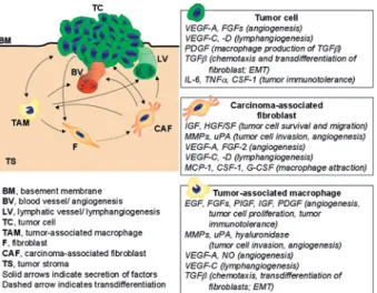
Figure 2. Contribution by the tumor microenvironment. Stromal and tumor cells cross-talk via soluble growth factors, cytokines and chemokines as schematically exemplified in this figure for tumor-associated macrophages (TAMs) and carcinoma-tumor-associated fibrob-lasts (CAFs). Stromal cells and tumor cells thus mutually influence their behavior in a way that promotes metastasis. For instance, tu-mor cell-secreted PDGF stimulates macrophages to produce
TGF-b, which induces the transdifferentiation of fibroblasts into CAFs. CAFs in turn secrete macrophage chemoattractants, which promote the infiltration of macrophages into the tumor. Both TAMs and CAFs secrete growth factors, cytokines and proteases, which pro-mote tumor cell survival, migration and invasion into the surround-ing tissue. Moreover, a variety of tumor and stromal cell-secreted factors induce the formation of blood and lymphatic vessels and also modulate the immune system to tolerate the transformed cells.
cells, tilt the balance between angiogenesis inhibitors and inducers towards the stimulation of angiogenesis (angiogenic switch). Thereby, cell and tumor-stroma-induced angiogenesis not only promotes primary (and secondary) tumor growth, but also the hematoge-nous dissemination of tumor cells and the outgrowth of metastasis.
Most, though not all, clinical studies demonstrate a di-rect correlation of high primary tumor blood micro-vessel densities (MVDs) with increased incidence of metastases [150, 151]. Tumor MVD is a significant and independent prognostic indicator for relapse-free and overall survival of cancer patients [152]. Similarly, ele-vated tumor or serum levels of the pro-angiogenic fac-tors VEGF-A, interleukin (IL) and FGF-2 as well as a low ratio between the angiogenesis inhibitor throm-bospond2 and VEGF-A are associated with an in-creased incidence of metastasis in cancer patients [153– 155]. Xenograft animal models confirm the correlation of tumoral overexpression of angiogenic growth factors, increased MVD of primary and secondary tumors and metastasis formation [156–158]. Consistent with these results, transgenic mice developing prostate adenocarci-noma (TRAMP) exhibit impaired metastasis when crossed into a FGF-2 knockout background [159]. Sim-ilarly, in xenotransplant and transgenic mouse models, increased tumor and/or serum levels of angiogenesis inhibitors, such as IL-10, tissue inhibitor-1 of metallo-proteinases (TIMP-1), thrombospondin-1 or endostatin, reduce the incidence of metastasis [158, 160–162]. Yet, upregulated tumor angiogenesis may not necessarily lead to metastasis formation. For example, forced ex-pression of VEGF-A in a transgenic mouse model (Rip1Tag2) accelerates tumor angiogenesis and growth of primary tumors but does not result in increased meta-stasis formation [163]. Although tumorigenic transfor-mation, mediated for example by Ras or ErbB2, can up-regulate the expression of angiogenic growth factors and repress the production of anti-angiogenic mole-cules, the direct contribution of the angiogenic process to tumor metastasis is difficult to demonstrate [164]. In-hibition of angiogenesis during tumor progression by specific angiogenesis inhibitors also affects primary tu-mor growth and thus indirectly the metastatic dissemi-nation of tumor cells. One should also be aware of the observation that intra-tumoral hypoxia and concomitant upregulation of the expression of angiogenic factors may occur only intermittently, leading to variations in the expression in tumors and in serum levels at different time points of measurement. Moreover, methods and markers for the determination of MVD have not been standardized, hindering a direct comparison of indepen-dent studies [165]. In any case, interference with angio-genesis will not only lead to a repression of tumor growth but may also reduce tumor metastasis.
Tumor-induced lymphangiogenesis
Some tumor types, including melanoma and cancers of the breast, lung and gastrointestinal tract, preferably spread via lymphatics [166]. Sentinel lymph node biopsy of patients with these tumors is routinely used for re-finement of prognostic and therapeutic measures. Since the lymphatic vasculature is specialized for entry and transport of immune cells, lymphogenous spread is more efficient than that via blood vessels. Lymphatic capillar-ies are larger, lack a basal lamina and display an en-dothelial cell arrangement facilitating intravasation of cells. Moreover, the composition of lymph is similar to interstitial fluid and flow velocities are low, thus allow-ing better cell viability as compared with the serum tox-icity and shear stress encountered in the bloodstream [167]. Histological analyses of human primary tumor samples demonstrate a consistent correlation between the presence of enlarged peri-tumoral lymphatics and the incidence of regional lymph node metastases [168, 169]. Moreover, animal models clearly demonstrate a signifi-cant promotion of lymphogenic metastasis by tumor-in-duced lymphangiogenesis (reviewed in [170]). VEGF-C and D are the two most-investigated secreted lymphan-giogenic factors. During secretion from the producing cell, these glycoproteins are proteolytically processed by plasmin and other (unknown) proteases. Unprocessed forms can only bind to VEGF receptor-3 (VEGFR-3), which is usually found on both normal and tumor lym-phatic endothelial cells. Processing of VEGF-C and D increases the binding affinity for VEGFR-3 and allows binding to VEGFR-2, which is specifically expressed on blood vessel endothelial cells. Therefore, expression and the processing status of VEGF-C and D in a given tissue appear to determine the lymphangiogenic and angio-genic activities of these growth factors [171]. Expression of VEGF-C in tumor tissue serves as a reliable marker for ongoing tumor lymphangiogenesis and increased risk of regional lymph node metastasis in many cancer types [172, 173]. Data for VEGF-D are less consistent sug-gesting that its ability to promote metastatic spread via lymphatics depends on the investigated cancer type and/ or grade [174–176].
The question remains whether VEGF-C and D may alter adhesive properties of both tumor and endothelial cells or modify functional features of lymphatic vessels, thus en-hancing tumor cell adhesion to and intravasation into lymphatics. In addition, it is conceivable that VEGF-C and D activate lymphatic endothelial cells to secrete chemotactic factors for tumor cells. Although it is known that both tumor and stromal cells can express VEGF-C and D [53, 177], it is not clear to what extent and upon which signals cells of the tumor stroma contribute to tu-mor-associated lymphangiogenesis [178]. Moreover, the genetic background of a given patient may modulate ex-pression levels and processing of VEGF family members
and thereby determine the biological behavior of a tumor [179, 180].
Carcinoma-associated fibroblasts
Besides endothelial cells of nearby vessels, neoplastic cells can also activate resident fibroblasts, which then cooperate in tumor progression (fig. 2). Activated fi-broblasts or myofifi-broblasts (also called carcinoma-asso-ciated fibroblasts, CAFs) are often encountered at the in-vasive front of many different cancer types. Myofibrob-lasts share features of both smooth muscle cells and fibroblasts and are important promoters of growth and differentiation during embryogenesis, wound healing and other tissue-remodeling processes [181]. It is pre-dominantly fibroblasts that transdifferentiate into myofi-broblasts, but also vascular smooth muscle cells, peri-cytes, hematopoietic bone marrow-derived precursor cells and even cancer cells themselves are discussed as putative progenitors. Tumor cell-secreted platelet-de-rived growth factor (PDGF) stimulates fibroblast prolif-eration and the release of TGF-b from macrophages, which itself is chemotactic for fibroblasts at lower con-centrations and induces their transdifferentiation into myofibroblasts at high concentrations [181]. Further-more, tumor cells often express TGF-b themselves [121]. Myofibroblasts appear shortly before the invasive stage of tumors and promote degradation of basement membranes and ECM by secreting serine proteases, MMPs and urokinase plasminogen activator. Moreover, they express IGFs and HGF/SF, thus promoting cell-sur-vival and migration, as well as the pro-angiogenic factors FGF-2 and VEGF and the pro-inflammatory cytokines IL-1, 6, 8 and TNF-a. By doing so, myofibroblasts not only stimulate their own migration into the tumor, but also promote survival, proliferation and invasion of adja-cent cancer cells as well as angiogenesis, collectively en-hancing metastasis [182–184].
Fibroblasts of the tumor stroma participate in the deci-sion whether neighboring tumor cells proliferate and in-vade into surrounding tissue and metastasize to distant sites [185]. For example, epithelial cells and fibroblasts in tissues that line portals of entry to the body secrete high levels of IFNb, whereas fibroblasts of internal or-gans do not. It has been demonstrated in xenotransplant and in vitro experiments that IFNb secreted by skin fi-broblasts downregulates expression of FGF-2 and se-creted type IV collagenase in transplanted colon and re-nal carcinoma cells, thereby inhibiting their angiogenic and invasive capabilities in the skin. In contrast, when transplanted orthotopically, tumor cells express high lev-els of FGF-2 and secreted type IV collagenase, resulting in the development of highly vascularized and metastatic tumors [186, 187]. Moreover, transgenic mice in which TGF-b receptor type II is selectively inactivated in
fi-broblasts develop prostate and gastric cancer even in the absence of mutations in the respective epithelial cells [188]. Notably, the mutated fibroblasts secrete high lev-els of HGF/SF, thereby conferring a paracrine tumori-genic signal on the epithelial cells.
Contribution of the immune system to tumor metastasis
The increased incidence of cancer in immunosuppressed patients and intense research in tumor immunology over the past years provide evidence that the immune system can to some degree recognize and eliminate tumor cells [190, 191]. Nevertheless, an increasing body of evidence indicates that immune cells represent a double-edged sword during tumorigenesis. Although many tumors are potentially immunogenic, they evidently develop nisms to escape immunosurveillance by various mecha-nisms and can even ‘educate’ immune cells to support tu-mor cell survival and proliferation. Many recent reports have addressed these functions of the immune system and suggest new strategies how to therapeutically boost a can-cer patient’s immune response against the cancan-cer. How-ever, only limited insights have been gained into the role of immune cells during late-stage tumor progression and metastasis. Apparently, tumor-induced immunotolerance not only allows primary tumor outgrowth but also metas-tasis. Such ‘immunological’ involvement of the immune system in preventing carcinogenesis has been recently summarized [192]. Here, we restrict ourselves to the question of whether ‘tumor-educated’ immune cells can actively contribute to tumor cell migration, invasion and metastatic dissemination.
The innate immune system
The innate immune system recognizes and eliminates many exogenous infectious agents as well as altered host cells independent of preexisting specific (adaptive) im-munity. Macrophages, dendritic cells (DCs) and natural killer cells (NKs) are cellular components of the innate immune system. Whereas macrophages have been re-peatedly shown to promote tumor growth and metastasis by various mechanisms, NKs are usually associated with suppression of tumor metastasis, and DCs exert both anti-tumor and anti-tumor immunosuppressive activities.
Tumor-associated macrophages
Macrophages are critical players in inflammation, wound healing, tissue repair and remodeling. Due to their anti-gen-presenting activity, they play a central role in cell-based immunity and can execute cells via cytotoxic mechanisms [193, 194]. Tumor cell damage and hypoxia are general attractants for circulating monocytes, and the presence of leukocytes in hypoxic areas of tumors is a well-known phenomenon [195–197]. In addition,
neo-plastic cells, tumoral fibroblasts and smooth muscle cells produce distinct monocyte-attracting factors, including monocyte chemoattractant protein-1 (MCP-1), colony-stimulating factor-1 (CSF-1), granulocyte-colony stimu-lating factor (G-CSF), VEGF-A, PlGF and VEGF-C [198–200]. Upon invasion into the tumor tissue, mono-cytes differentiate into macrophages and are specifically activated or de-activated, depending on the cytokine mi-lieu encountered.
Consistent with their antigen-presenting and cytotoxic role in inflammation, high numbers of macrophages within or in the periphery of tumors (tumor-associated macrophages, TAMs) can repress tumor growth and pro-gression [201, 202]. Surprisingly however, most clinical and experimental data correlate high TAM counts with reduced disease-free and overall survival. This discrep-ancy has been explained by a tumor type-specific para-crine cytokine crosstalk between tumor cells and TAMs: tumors secreting granulocyte-monocyte colony stimulat-ing factor (GM-CSF), IFNg and IL-12 promote the anti-gen-presenting and cytotoxic activities of macrophages (reviewed in [198, 203, 204]). In contrast, most tumor cells secrete VEGF-A, TGF-b, IL-6, CSF-1 and low-dose TNFa, all factors that inhibit the TAMs’ antigen-present-ing activities and rather support immunosuppression, an-giogenesis, tumor cell proliferation and invasion [205– 208]. In turn, these TAMs promote tumor cell prolifera-tion by secreting a variety of tumor promoting factors, in-cluding nitric oxide (NO), EGF, FGF-8b, HGF/SF, IGF-I, TGF-b, VEGF-C and PDGF. Alternatively, activated ma-crophages can stimulate tumor cells to secrete these fac-tors in an autocrine fashion [209–214]. Furthermore, TAMs themselves potently stimulate tumor angiogenesis and lymphangiogenesis by secreting growth factors, cyto-kines and chemocyto-kines [215–217]. Interestingly, in a cor-nea model of inflammation-induced lymphangiogenesis, macrophages transdifferentiate and directly incorporate into the endothelial layer of nascent lymphatic vessels [217]. By releasing MMPs, plasminogen activators, hyal-uronidase and other enzymes involved in the breakdown of the ECM, TAMs liberate matrix-bound angiogenic molecules, including VEGF-A and IL-1b, thus further enhancing angiogenesis and tumor cell migration and in-vasion (fig. 2) [203, 218, 219]. Notably, TAMs have also been found to actively associate with tumor cells via ad-hesion molecules, such as ICAM-1 and sialoadhesin, and to convey them into efferent blood vessels [220]. Since macrophage activity is associated with the release of re-active oxygen and other mutagenic compounds, they might also indirectly induce additional genetic pro-tu-morigenic and pro-metastatic alterations [3, 4, 221]. NKs and DCs
NKs are a derivative of the CD8+ T-lymphocyte lineage, but lack the rearrangement of T-cell receptor genes. NKT
cells represent a subtype sharing characteristics with NK and T cells. It is well-established that NKs and NKTs dis-play important anti-tumor activity by inducing death of tumor cells through direct contact or by the secretion of pro-apoptotic cytokines [222, 223]. In parallel, NKs stim-ulate the maturation of DCs, which are first-line antigen-presenting cells, and thereby promote specific anti-tumor immunity mediated by cytotoxic CD8+ T lymphocytes (CTLs) and B lymphocytes [224]. In cancer patients, high peripheral blood NK activity significantly correlates with longer metastasis-free survival [225]. Experiments using perforin-deficient mice clearly demonstrate that NK-me-diated cytotoxicity is one of the essential anti-metastatic mechanisms performed by the innate immune system [226–228]. Further details on NK-mediated tumor sur-veillance and anti-metastatic activity have been recently summarized [229]. To date, here is no evidence indicating a potential pro-metastatic role of NKs.
DCs are classical antigen-presenting cells, which can initiate strong CTL-mediated anti-tumor immune re-sponses. They are generally divided into myeloid and plasmacytoid DCs with differences in cell-surface recep-tors, anatomic localization and function [192]. Recently, a third type of DCs, vascular DCs (VDCs), was identi-fied, which simultaneously expresses endothelial and DC markers. Strikingly, VDCs can assemble into func-tional blood vessels [230]. Depending on the cytokine milieu encountered in tumors, DCs can either mediate anti-tumor activity or support tumor growth and metas-tasis. In fact, increasing evidence from analyses of hu-man ovarian, breast, prostate and renal cell carcinoma re-veals the presence of tumor-promoting rather than tu-mor-suppressing dendritic cells, and altered dendritic cell function and differentiation is likely to be one of the most fundamental mechanisms by which tumors escape immune responses [231–235]. Pro-tumorigenic den-dritic cells can favor metastasis by inducing tumor im-munotolerance and by promoting tumor angiogenesis. Whereas mature myeloid DCs act as classical antigen-presenting cells, immature or partially differentiated myeloid DCs can tolerize T cells to self-antigens by in-ducing either suppressive T cells or T-cell unresponsive-ness. This mechanism of immunomodulation is particu-larly important to counteract autoimmune reactions, but can be abused by tumors as well [236, 237]. Tumor cells, TAMs and tumoral immunosuppressive T cells can se-crete high amounts of VEGF-A, IL-6, macrophage colony-stimulating factor (M-CSF), COX2, IL-10, TGF-b and gangliosides, which suppress maturation of DCs [192]. Most myeloid DCs found in human ovarian, breast, prostate and renal cell carcinoma are therefore immature and may induce tumor immunotolerance [231–234]. Tumor-secreted IL-10 and VEGF also induce expression of B7-H1 on myeloid dendritic cells, a ligand for PD-1 receptor expressed on suppressor T cells [238]. A study
published this year demonstrates that the metastatic abil-ity of melanoma and colon cancer cells expressing B7-H1 is abolished in PD-1-deficient mice and that tumors also grow more slowly [239]. These and other experi-ments indirectly show that tumor cell escape from im-munosurveillance – in this case mediated by modulation of myeloid DC and suppressive T cell function – is es-sential for tumor growth and metastasis.
The involvement of dendritic cells in tumor angiogenesis has recently drawn major attention. It is thought that ma-ture myeloid DCs suppress tumor angiogenesis and thereby impede both tumor growth and metastasis by se-creting or stimulating the production of anti-angiogenic factors, such as IL-12, IFNg and IL-10 [240]. Conversely, the tumor-induced suppression of myeloid DC matura-tion described above therefore enhances tumor angiogen-esis. Moreover, tumors contain significant numbers of plasmacytoid and vascular DCs secreting pro-angiogenic molecules, such as TNFa and IL-8 [230, 240, 241]. Based on this rather complex situation, the involvement of DCs in tumor angiogenesis and metastasis and their therapeutic exploitation certainly deserves future investi-gation.
The adaptive immune system and metastasis
After a primary contact with an antigen, the adaptive im-mune system confers longterm antigen-specific immuno-logical memory. Its basic mediators are T and B lympho-cytes. CD8+ cytotoxic T cells (CTLs) together with NKs are the fundamental effectors of immune response-medi-ated tumor cell elimination via the release of cytokines that activate death receptors on the tumor cell surface. CD4+ T helper cells are central to the development of im-mune responses by activating antigen-specific effector cells and recruiting cells of the innate immune system [242, 243]. As mentioned earlier, suppressor T cells play an important role in inducing tumor immunotolerance, which might not only allow tumor growth but also metas-tasis [237, 244]. Nevertheless, stimulation of anti-tumor T cell response has been shown to be a promising strategy to counteract tumor growth and metastasis in mouse tu-mor models and in human cancer [245–247].
B lymphocytes are antigen-presenting cells which can differentiate into antibody-producing plasma cells. There-by they can stimulate both cytotoxic and humoral anti-tumor immune responses. On the other hand, there is mounting evidence that B cells can also promote tumor cell invasiveness and metastasis. In one study, tumor cells producing antigenic Secreted/shed Tumor Glyco-Proteins (STGPs) were transplanted into immune-com-petent mice or mice deficient in T cells [248]. In both mouse lines, increased tumor cell invasion correlated with an increase in serum anti-STPG IgG levels and tu-moral infiltration of cells of the innate and adaptive im-mune system. Experimental infiltration of specific
anti-STGP-IgG-secreting plasmocytoma cells further pro-moted tumor invasion, angiogenesis and metastasis. Ap-parently, the humoral anti-STGP response stimulated the tumor infiltrating stromal cells to release proinflamma-tory cytokines and VEGF, thereby promoting invasion and metastasis.
Recent work with a transgenic mouse model of multi-stage skin carcinogenesis (K14-HPV16) demonstrated a similar surprising pro-tumorigenic role of B but not T lymphocytes in the development of inflammation-asso-ciated de novo epithelial cancer. Upon crossing K14-HPV16 mice with T and B lymphocyte-deficient RAG–/–
mice resulted in reduced tumor progression [249]. Con-versely, adoptive transfer of B lymphocytes or serum from K14-HPV16 mice restored the infiltration of innate immune cells into premalignant tissue and the progres-sion to malignancy. These studies indicate that B cells and even antibodies produced by B cells contribute to tu-mor progression and metastasis. Of course, these sur-prising results raise an important caveat for the applica-tion of vaccinaapplica-tion-based cancer therapies that aim to stimulate B cell responses in patients with pre-malignant disease.
Conclusions
Apparently, cancer needs to be treated at an early tumor stage or grade in order to prevent the formation of metas-tases. Nevertheless, even when cancer may have already spread throughout the body, targeting the growth of pri-mary and secondary tumors as well as further tumor cell spread may significantly improve quality of life and over-all survival of patients. Although ‘curing cancer’ may be an unrealistic notion in advanced-stage tumors, a signifi-cant fraction of patients may nevertheless profit from a cocktail of drugs inhibiting tumor progression at different levels. Most of the experimental results presented here highlight many pathways and mechanisms that may be appropriate targets for the development of such therapeu-tic interventions. Prevention of further metastatherapeu-tic dissem-ination, in addition to the reduction of tumor load, is cer-tainly a prerequisite to convert a rapidly fatal disease into a chronic illness.
Acknowledgements. The authors are grateful to Dr. Miguel Cabrita and Dr. Tibor Schomber for critically reading the manuscript. We apologize to all colleagues whose important work we could not cite due to space limitations. Research in the laboratory of the authors is supported by the Swiss National Science Foundation, Swiss Bridge Award, Krebsliga Beider Basel, EU-FP6 framework programmes LYMPHANGIOGENOMICS LSHG-CT-2004-503573 and BRE-COSM LSHC-CT-2004-503224, and Novartis Pharma Inc.
1 Chambers A. F., Groom A. C. and MacDonald I. C. (2002) Dissemination and growth of cancer cells in metastatic sites. Nat. Rev. Cancer 2: 563–572