HAL Id: hal-01780642
https://hal.archives-ouvertes.fr/hal-01780642
Submitted on 22 Aug 2018
HAL is a multi-disciplinary open access
archive for the deposit and dissemination of
sci-entific research documents, whether they are
pub-lished or not. The documents may come from
teaching and research institutions in France or
abroad, or from public or private research centers.
L’archive ouverte pluridisciplinaire HAL, est
destinée au dépôt et à la diffusion de documents
scientifiques de niveau recherche, publiés ou non,
émanant des établissements d’enseignement et de
recherche français ou étrangers, des laboratoires
publics ou privés.
translational apparatus of the known virosphere
Jonatas Abrahao, Lorena Silva, Ludmila Santos Silva, Jacques Yaacoub Bou
Khalil, Rodrigo Rodrigues, Thalita Arantes, Felipe Assis, Paulo Boratto,
Miguel Andrade, Erna Geessien Kroon, et al.
To cite this version:
Jonatas Abrahao, Lorena Silva, Ludmila Santos Silva, Jacques Yaacoub Bou Khalil, Rodrigo
Ro-drigues, et al..
Tailed giant Tupanvirus possesses the most complete translational apparatus
of the known virosphere.
Nature Communications, Nature Publishing Group, 2018, 9, pp.749.
�10.1038/s41467-018-03168-1�. �hal-01780642�
Tailed giant Tupanvirus possesses the most
complete translational apparatus of the known
virosphere
Jônatas Abrahão
1,2
, Lorena Silva
1,2
, Ludmila Santos Silva
1,2
, Jacques Yaacoub Bou Khalil
3
, Rodrigo Rodrigues
2
,
Thalita Arantes
2
, Felipe Assis
2
, Paulo Boratto
2
, Miguel Andrade
4
, Erna Geessien Kroon
2
, Bergmann Ribeiro
4
,
Ivan Bergier
5
, Herve Seligmann
1
, Eric Ghigo
1
, Philippe Colson
1
, Anthony Levasseur
1
,
Guido Kroemer
6,7,8,9,10,11,12
, Didier Raoult
1
& Bernard La Scola
1
Here we report the discovery of two Tupanvirus strains, the longest tailed Mimiviridae
members isolated in amoebae. Their genomes are 1.44
–1.51 Mb linear double-strand DNA
coding for 1276
–1425 predicted proteins. Tupanviruses share the same ancestors with
mimivirus lineages and these giant viruses present the largest translational apparatus within
the known virosphere, with up to 70 tRNA, 20 aaRS, 11 factors for all translation steps, and
factors related to tRNA/mRNA maturation and ribosome protein modi
fication. Moreover,
two sequences with signi
ficant similarity to intronic regions of 18 S rRNA genes are encoded
by the tupanviruses and highly expressed. In this translation-associated gene set, only the
ribosome is lacking. At high multiplicity of infections, tupanvirus is also cytotoxic and causes
a severe shutdown of ribosomal RNA and a progressive degradation of the nucleus in host
and non-host cells. The analysis of tupanviruses constitutes a new step toward understanding
the evolution of giant viruses.
DOI: 10.1038/s41467-018-03168-1
OPEN
1MEPHI, APHM, IRD 198, Aix Marseille Univ, IHU-Méditerranee Infection, 19-21 Bd Jean Moulin, 13005 Marseille, France.2Laboratório de Vírus, Instituto de
Ciências Biológicas, Departamento de Microbiologia, Universidade Federal de Minas Gerais, Belo Horizonte 31270-901, Brazil.3CNRS, 13005 Marseille,
France.4Laboratório de Microscopia Eletrônica e Virologia, Departamento de Biologia Celular, Instituto de Ciências Biológicas, Universidade de Brasília, Asa
Norte, Brasília 70910-900, Brazil.5Lab. Biomass Conversion, Embrapa Pantanal, R. 21 de Setembro 1880, 79320-900 Corumbá/MS, Brazil.6Cell Biology
and Metabolomics Platforms, Gustave Roussy Cancer Campus, Villejuif 94805, France.7Equipe 11 labellisée Ligue Nationale contre le Cancer, Centre de
Recherche des Cordeliers, Paris 75006, France.8Institut National de la Santé et de la Recherche Médicale (INSERM), Paris 75654, France.9Université Paris
Descartes, Sorbonne Paris Cité, Paris 75015, France.10Université Pierre et Marie Curie, Paris 75005, France.11Pôle de Biologie, Hôpital Européen Georges
Pompidou, AP-HP, Paris 75015, France.12Department of Women’s and Children’s Health, Karolinska University Hospital, Stockholm SE-171 76, Sweden.
Jônatas Abrahão, Lorena Silva and Ludmila Santos Silva contributed equally to this work. Correspondence and requests for materials should be addressed to
D.R. (email:didier.raoult@gmail.com) or to B.S. (email:bernard.la-scola@univ-amu.fr)
123456789
T
ranslation is one of the canonical frontiers between the cell
world and the virosphere. Even the simplest non-viral
obligatory intracellular parasites present a wealthy set of
translation apparatus, including aminoacyl tRNA synthetases,
tRNAs, peptide synthesis factors and ribosomal proteins
1–3. The
nature of the parasitism of the most of non-viral obligatory
intracellular parasites relies on partial or severe deficiency of
genes related to energy production. Although sharing a similar
lifestyle with those organisms, the most of the virus lack not only
energy production genes, but also translation-related genes
1–3.
In this context, the discovery of mimiviruses and other
amoeba-infecting giant viruses surprised the scientific community
due their unusual sizes and long genomes, able to encode from
hundreds to thousands of genes, including tRNAs, peptide
synthesis factors and, for the
first time seen in the virosphere,
aminoacyl tRNA synthetases (aaRS)
4. Although the very
first
discovered mimivirus (Acanthamoeba polyphaga mimivirus)
encodes four different types of aaRS (Arg, Cys, Met, TyrRS), other
mimivirus isolates genomes were described, containing up to
seven types of aaRS, as Megavirus chilensis and LBA111 (Arg,
Asn, Cys, Ile, Met, Trp, TyrRS)
5. While amoeba-infecting
mimiviruses present up to six copies of tRNA, Cafeteria
roen-bergensis virus (CroV), a group II algae-infecting of Mimiviridae,
encodes 22 sequences for
five different tRNAs
5. Even more
sur-prisingly, metagenomics data reveal that klosneuvirus genome
may encodes aaRS with specificities for 19 different amino acids,
over 10 translation factors and several tRNA-modifying
enzymes
6. However, klosneuviruses were not isolated and
there-fore there is no data regarding important biological features as
virus particles, morphogenesis and host-range.
Here we report the discovery of two Tupanvirus strains, the
longest tailed Mimiviridae members isolated in amoebae. Their
genomes are 1.44–1.51 Mb linear double-strand DNA coding for
1276–1425 predicted proteins. Tupanviruses share the same
ancestors with mimivirus lineages and these giant viruses present
the largest translational apparatus within the known virosphere,
with up to 70 tRNA, 20 aaRS, 11 factors for all translation steps,
and factors related to tRNA/mRNA maturation and ribosome
protein modification. Moreover, two sequences with significant
similarity to intronic regions of 18 S rRNA genes are encoded by
the tupanviruses and highly expressed. In this
translation-associated gene set, only the ribosome is lacking. Tupanvirus is
also cytotoxic and causes a severe shutdown of ribosomal RNA
and a progressive degradation of the nucleus in host and
non-host cells. The analysis of tupanviruses constitutes a new step
towards understanding the evolution of giant viruses.
Results
Tupanviruses description and cycle. While attempting to
find
new and distant relatives of currently known giant viruses, we
performed prospecting studies in special environments. Soda
lakes (Nhecolândia, Pantanal biome, Brazil) are known as
environments that conserve and/or mimic ancient life conditions
(extremely high salinity and pH) and are considered some of the
most extreme aquatic environments on Earth
7. We also
pros-pected giant viruses in ocean sediments collected at a depth of
3000 m (Campos dos Goytacazes, Brazilian Atlantic Ocean).
Both in soda lake and deep ocean samples, we found optically
visible Mimiviridae members that surprisingly harbored a long,
thick tail (Fig.
1
a, b) as they grew on Acanthamoeba castellanii
and Vermamoeba vermiformis. We named these strains
Tupan-virus soda lake and TupanTupan-virus deep ocean as a tribute to the
South American Guarani Indigenous tribes, for whom Tupan—or
Tupã—(the God of Thunder) is one of the main mythological
figures. Electron microscopy analyses revealed a remarkable
virion structure. Tupanviruses present a capsid similar to that of
amoebal mimiviruses in size (~450 nm) and structure, including a
stargate vertex and
fibrils
8(Figs.
1
a–d,
2
a–q). However, the
Tupanvirus virion presents a large cylindrical tail (~550 nm
extension; ~450 nm diameter, including
fibrils) attached to the
base of the capsid (Figs.
1
b–d,
2
a,i–k). This tail is the longest
described in the virosphere
9. Microscopic analysis suggests that
the capsid and tail are not tightly attached (Figs.
1
e, f,
2
a, i;
Supplementary Movies
1
and
2
), although sonication and
enzymatic treatment of purified particles were not able to
separate the two parts (Fig.
2
f–h). The average length of a
complete virion is ~1.2 µm, although some particles can reach
lengths of up to 2.3 µm because of the variation in the tail’s size;
this makes them the one of the longest viral particles described to
date (Figs.
1
,
2
i–k). Furthermore, there is a lipid membrane inside
the capsid, which is associated with the fusion with the
cellular membrane and the release of capsid content (Fig.
1
f).
Tail content appears to be released after an invagination of
the phagosome membrane inside the tail (Fig.
1
e). In contrast
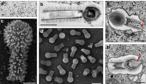
a
b
c
d
e
f
Fig. 1 Tupanvirus soda lake particles and cycle. a Optical microscopy of Tupanvirus particles after haemacolour staining (1000 × ). Scale bar, 2µm. b Super
particle (>1000 nm) observed by transmission electron microscopy (TEM). Scale bar, 500 nm.c, d Scanning electron microscopy (SEM) of Tupanvirus
particles. Scale bars 250 nm and 1µm, respectively. e, f The initial steps of infection in A. castellanii involve the release of both capsid (e) and tail (f) content
a
i
l
o
p
q
m
n
j
k
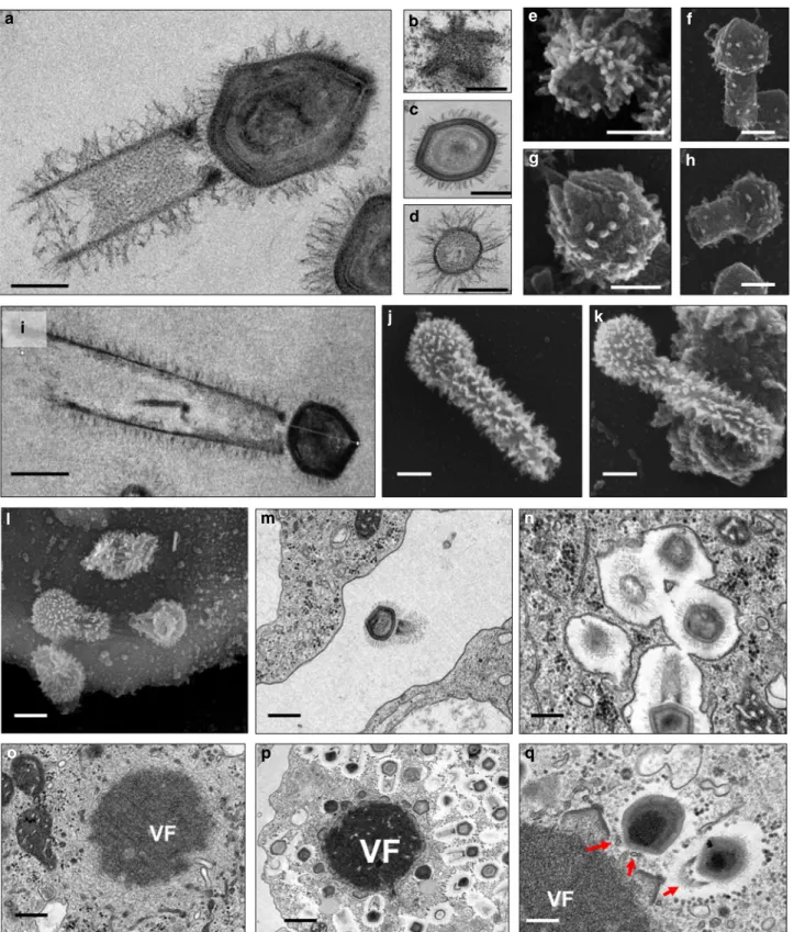
b
e
g
h
f
c
d
Fig. 2 Tupanvirus soda lake particles and cycle features. a Transmission electron microscopy (TEM) highlights the inner elements of the whole particle.
Scale bar, 200 nm.b Star-gate vertex transversally cut. Scale bar, 100 nm. c Capsid transversally cut. Scale bar, 100 nm. d Tail transversally cut. Scale bar,
200 nm.e–h Scanning electron microscopy (SEM) of purified particles. Scale bars, 250 nm. The treatment of particles with lysozyme, bromelain and
proteinase-K removed most of thefibers, revealing head and tail junction details. Super particles (>2000 nm) could be observed by TEM (i) and SEM (j, k).
Scale bars, 400 nm. Cycle steps are shown froml–r. l Viral particle attachment to Acanthamoeba castellanii surface; scale bar, 500 nm; m phagocytosis;
scale bar, 500 nm;n particles in a phagosome; scale bar, 500 nm; o early viral factory; scale bar, 500 nm; and p, q mature viral factories. Scale bars 1µm
to other giant viruses
10–14, Tupanviruses similarly replicate both
in A. castellanii and V. vermiformis. Viral particles attach to the
host-cell surface and enter through phagocytosis (1 h.p.i.) (Fig.
2
l,m,n). The inner membrane of the capsid merges with
the amoebal host phagosome membrane, releasing the genome
(2–6 h.p.i.) (Fig.
1
f). A viral factory (VF) is then formed (7–12 h.
p.i.)
15where
particle
morphogenesis
occurs
(Fig.
2
o–q;
Supplementary Movies
1
and
2
). During this step, the virion
tail is attached to the capsid after its formation and closure. Later
in the process (16–24 h.p.i.), the amoebal cytoplasm is filled with
viral particles, followed by cell lysis and the release of viruses
(Supplementary Fig.
1
). This nucleus-like viral factory has also
recently been reported in bacteria and fuels the concept of a
virocell
16,17. In that perspective, viral factories actively producing
the progeny could be considered as the nuclei of virocells
16,17.
The genomes of tupanviruses. The tupanvirus soda lake and
tupanvirus deep ocean genomes (GenBank accession number
KY523104 and MF405918) are linear dsDNA molecules
measur-ing 1,439,508 bp and 1,516,267 bp (GC% ~28%), respectively—the
fourth largest viral genomes described to date
10,18—containing a
total of 1276 and 1425 predicted ORFs, 375 and 378 of which are
ORFans (ORFs with no matches in current databases),
respec-tively. To date, the largest genomes belong to pandoravirus
iso-lates, and the largest one, P. salinus, has 2,473,870 bp and encodes
2,556 putative proteins
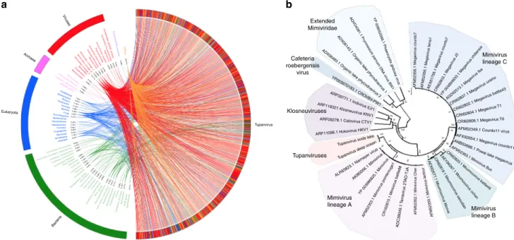
10. The rhizome
19of tupanvirus (graphical
representation of gene-by-gene best hits) revealed sequences from
mimiviruses of amoebae (~42%) and klosneuviruses (~8%) as
their main best hits. Other best hits were mostly sequences from
eukaryotes (~11%) and bacteria (~8%) (Fig.
3
a; Supplementary
Fig.
2
A). Tupanviruses exhibited relatively close numbers of best
matches to amoebal mimiviruses from lineages A (~10%), B
(~18%) and C (~14%). These data and phylogenetic analyses
demonstrate that they cluster with amoebal mimiviruses,
sug-gesting that tupanviruses are distant relatives of, and comprise a
sister group to, mimiviruses of amoeba (Figs.
3
b,
4
a). The
‘AAAATTGA’ promoter motif was found ~410 times in
Tupan-virus deep ocean intergenic regions, a frequency similar to that of
other Mimiviridae members, and ~600% more frequent than those
coding regions (p < 10
−95, Fisher exact test)
20–22(Fig.
4
b). The
pangenome of the family Mimiviridae, when taking into account
the gene contents of tupanviruses, klosneuviruses and distant
relatives to amoebal mimiviruses, was found by Proteinortho to
comprise 8,753 groups of orthologues (n
= 3588) or virus unique
genes (n
= 5165). A total of 189 groups were shared by at least one
tupanvirus; one mimivirus of Acanthamoeba of each lineage A, B
and C; and one klosneuvirus, and 100 of them were also shared
with Cafeteria roenbergensis virus. In addition, a total of 757
groups were shared by a tupanvirus and at least another
mimi-virus: 477 were shared with Megavirus chiliensis, 434 with
Mou-mouvirus, 431 with Mimivirus, 287 with Klosneuvirus, 126 with
Cafeteria roenbergensis virus, and 59 with Phaeocystis globosa
virus 12 T. Among these 757 groups, 583 corresponded to clusters
of orthologous groups previously delineated for mimiviruses
19.
Finally, a total of 775 tupanvirus genes were absent from all other
mimivirus genomes (Supplementary Data
1
).
Proteomic analysis. Proteomic analysis of Tupanvirus soda lake
particles revealed 127 proteins, nearly half (67/127
= 52.8%) of
which are unknown and eight of which are encoded by ORFans
(11/127
= 8.6%) (Supplementary Data
2
; Supplementary Note
1
).
Although 62 Tupanvirus virion proteins homologous were not
found by proteomics, either in Mimivirus or in Cafeteria
roenber-gensis virus particles, there are no distinct clues about which protein
(s) could be associated with the tupanvirus
fibrils and tail structure.
The most complete translational apparatus of the virosphere.
Analyses of the tupanvirus gene sets related to energy production
revealed a clear dependence of these viruses on host energy
production mechanisms, similarly to other mimiviruses, because
genes involved in glycolysis, the Krebs cycle and the respiratory
chain are mostly lacking
22–24. Astonishingly, Tupanvirus soda
lake and Tupanvirus deep ocean exhibit the largest viral sets of
genes involved in translation, with 20 ORFs related to
aminoa-cylation (aaRS) and transport, and 67 and 70 tRNA associated
with 46 and 47 codons, respectively (Fig.
5
a; Supplementary
Vir uses oRF ans 29.31% 0.08% 0.24% 0.24% 0.08% 0.08% 0.39% 0.08% 7.45% 14.58% 17.55% 10.11% 0.16% 0.08% 0.08% 0.08% 0.08% 1.02% 0.24% 0.16% 0.16% 0.08% 2.98% 3.45% 0.31% 0.24% 0.16% 0.08% 0.08% 0.24% 1.18% 0.94% 0.08% 0.16% 0.08% 0.08% 0.08% 0.08% 1.49% 0.63% 0.08% 0.39% 0.24% 0.08% 0.16% 0.08% 0.24% 0.08% 1.02% 0.78%0.31% 0.71% 0.08% 0.16% 0.71% 0.31% En vironmental_samples Unclassified_dsDNA_vir uses Unclassified_Ph ycodna vir idae Unclassified_Mimivir idae Extended_Mimivir idae Klosneuvir inae Raphido vir us En vironmental_samples Mimivir idae_Lineage_C Mimivir idae_Lineage_B Mimivir idae_Lineage_A Mimivir idae Caudo vir ales Baculo viridae Eur yarchaeota Unclassified_Archaea Candidatus_Micr archaeota DPANN_g roup Viridiplantae Stramenopiles Rhizar ia Parabasalia Fonticula Metazoa Fungi Choanoflagellida Opisthokonta Heterolobosea Haptophyceae Fornicata Cryptophyta Apusozoa Amoebozoa Alveolata Unclassified_P arcubacter ia Unclassified_Candidatus_DependentiaeCandidatus_Kaiserbacter ia Candidatus_Eisenbacter ia Unclassified_Bacter ia Ther motogae Tenericutes Firmicutes Cyanobacter ia Chlorofle xi Actinobacter ia Spirochaetes Ve rrucomicrobia Planctom ycetes Lentisphaer ae Chlam ydiae Candidatus_OmnitrophicaGammaproteobacter ia Delta_epsilon_subdivisions Betaproteobacter ia Alphaproteobacter ia Proteobacter ia Chlorobi Candidatus_Kr yotonia BacteroidetesFCB_g roup Acidobacter ia PVC_g roup Terrabacter ia_g roup Ph ycodna viridae Archaea Eukaryota Bacter ia Extended Mimiviridae
YP 008052566.1 Phaeocystis globosa vir
us
AQ VO
4381.1 Pr ymnesium par
vum DNA vir us BW1 ADX06143.1 Organic lak
e ph ycodna
vir us 1
ADX06483.1 Organic lak
e ph ycodna vir us 2 YP003970183.1 CR OVBV -PW1 ARF09771.1 Indivir us lL V1 ARF118321 Klosneuvir us KNV1 ARF09278.1 Cato virus CTV1
ARF11096.1 Hokovirus HKV1
Tupanvirus soda lak
e
Tupan virus deep ocean
ALR83823.1 Nieme yer vir us AKl80056.1 Mimivir us YP 003986825.1 Mimivir us AFM52353.1 Mimivir us pointerouge1 CR162815.1 Mimivir us battle88 ADC39049.1 T err av irus 2T AO -TJ A AFM52352.1 Mimivir u s Cher AFM52359.1 Mimivir u s lactour AEX62677.1 Moumouvir us Mon ve AEY99267.1 Moumouvir us ochan AFM52363.1 Mimivir us Bus ANB50688.1 P owai lak e mega virus AFX92654.1 Mega virus courdo11 AFM52349.1 Courdo11 vir
us
CRI62806.1 Megavirus T6
CRI62804.1 Mega virus T1 CRI62802.1 Mega virus battle43 CRI62807.1 Mega virus ursino AGD92513.1 Mega virus Iba YP 004894633.1 Mega virus chillensis CRI62803.1 Mega virus J3 AEX61758.1 Mega viru s courdo7 AFM52356.1 Mega vir us terr a1 AFM52358.1 Mega vir u s courdo7 CRI62819.1 Moumouvir us saoudian CRI62820.1 Moumouvir us battle49 Cafeteria roebergensis virus Klosneuviruses Tupanvirus Tupanviruses Mimivirus lineage A Mimivirus lineage B Mimivirus lineage C 10 92 99 83 78 89 100 100 61 67 71 100 100 88 99 99 66 64 100 51 40 52 63
a
b
Fig. 3 Tupanvirus soda lake rhizome and Mimiviridae family B DNA polymerase tree. a The rhizome shows that most Tupanvirus soda lake genes have mimiviruses as best hits. However, correspondence among Tupanvirus and Archaea, Eukaryota, Bacteria and other viruses was also observed. b Family B DNA polymerase maximum likelihood phylogenetic tree demonstrating the position of Tupanvirus among Mimiviridae members, likely forming a new genus
Fig.
2
B, C,
3
and
7
; Supplementary Datas
3
and
4
). Tupanvirus
deep ocean encodes an ambiguous tRNA related to the rare
amino-acid pyrrolysine. Only selenocysteine-related genes are
lacking, as previously observed for many cellular organisms
25.
Several translation factors were identified, including eight
trans-lation initiation factors (IF2 alpha, IF2 beta, IF2 gamma, IF4e (2
copies in Tupanvirus soda lake), IF5a (2 copies in Tupanvirus
deep ocean), SUI1, IF4a), one elongation/initiation factor
(GTP-binding elongation/initiation), one elongation factor (Ef-aef-2),
and one release factor (ERF1) (Fig.
5
a; Supplementary Fig
2
B, C
and
3
; Supplementary Datas
3
and
4
). Furthermore, we detected
additional translation-related genes: factors related to tRNA
maturation and stabilization (tRNA nucleotidyltransferase, tRNA
guanylyltransferase, cytidine deaminase, RNA methyl
transfer-ase); mRNA maturation (poly(A) polymerase, mRNA capping
enzyme) and splicing (RNA 2—phosphotransferase Tpt1 family
protein); and ribosomal protein modification
(ribosomal-protein-alanine N-acetyltransferase, FtsJ-like methyl transferase) (Fig.
5
a;
Supplementary Fig
2
B; Supplementary Datas
3
,
4
). Phylogenies
based on aaRS are acknowledged to be complex as they depend
on sequence sampling, and natural and canonical taxonomical
groups can be separated into different clades, as previously
observed
6. To reduce such disturbances in the trees during their
construction, we selected the 100 best hits related to Tupanvirus
soda lake, plus the sequences of 5–10 amoebozoa and those of
klosneuviruses. In most of the trees, sequences from natural
clades were clustered together, although some amoebozoa were
separated into different clusters. Based on the 20 aaRS trees, it is
not possible to state that the origin of most of these tupanvirus
aaRS genes is cellular (Supplementary Fig.
4
). Furthermore, the
mosaic structure of aaRS gene reinforced the difficult to state on
the origin of these genes, as illustrated in Supplementary Fig.
5
.
This contrasting result to that reported by Schultz et al.
6, 2017 for
klosneuviruses may also be explained by the different sampling
used for alignments and trees construction. In addition, the
codon and amino-acid usage of tupanviruses is substantially
different from that of Acanthamoeba spp. In tupanviruses, we
observed a high correlation between tRNA isoacceptors and the
most used codons, as these viruses present more tRNA
iso-acceptors for highly abundant codons (Fig.
4
c; Supplementary
Figs.
2
C and
3
). Surprisingly, we found two different copies of an
18 S rRNA intronic region in tupanviruses (Supplementary Fig.
6
;
Fig.
6
a–e). In fact, such 18 S rRNA intronic regions are
wide-spread in all mimiviruses (lineages A and B present only one copy
in an intronic region and next to a self-splicing group I intron
endonuclease) but are not found in klosneuviruses. Phylogenetic
analyses revealed that the two copies found in tupanviruses had
separate and different origins (Fig.
6
f). Although Tupanvirus 18 S
rRNA intronic sequences are located in intergenic regions, qPCR
and FISH demonstrated that Tupanvirus soda lake 18 S rRNA
intronic sequences are highly expressed during the entire
replicative cycle but particularly during intermediate and late
phases (6 and 12 h post-infection) (Fig.
7
a; Supplementary Fig.
8
).
Furthermore, Tupanvirus soda lake is more tolerant to the
translation-inhibiting drugs geneticin and cycloheximide than
Mimivirus, an impressive characteristic considering the natural
ribosomal RNA shutdown it performs in the permissive host
(Fig.
8
h). The functions of these 18 S rRNA intronic region
sequences require further clarification. No exonic region of 18 S
rRNA was found in the genomes of tupanviruses or any
previously described mimivirus. The comparison between
contents in translation-related categories of genes present in
tupanviruses and cellular organisms reveals that tupanviruses
present a richer gene set than Candidatus Carsonella ruddii
(Bacteria)
and
Nanoarchaeum
equitans
(Archaea)
(not
considering ribosomal proteins). Tupanvirus deep ocean has even
Yellowstone lake mimivirus
a
b
c
Organic lake phycodnavirus 1
Distantly related mimiviruses Klosneuviruses Cafeteria roenbergensisvirus Tupanviruses Mimiviruses of Acanthamoeba 1000 100 10 10 9 8 7 6 % codon/aa usage % codon/aa usage 4 3 2 1 0 F Y H Q N K D E C W R G Stop codon L l M V Motif S P T A TTT TAT TA C CA T CA C CAA CA G AA T AA C AA G GA T GA C GAA GA G TGT TGC TGG CGT CGC CGA CGG AG G GGT GGC GGA GGG TA A TA G TGA AG A AAA TTC TT A CTT CTC CT A CTG AT T AT C ATA AT G GTT GTC GT A GTG TCT AG T AG C TCC TCA TCG CCT CCC CCA CCG AC T AC C AC A AC G GCC GCA GCG GCT TTG 5 10 9 8 7 6 4 3 2 1 0 5 1 AAAA TTGA AAAA TTAA TAAA TTGA AAA TTTGA AAAAA TGA AATA TTGA AAAA TTGT AAAA TAGA AAAA TTGG AAAA TTGC AAA CTTGA GAAA TTGA CAAA TTGA AAA GTTGA AAAA TGGA AACA TTGA AAAA GTGA AGAA TTGA ACAA TTGA AAAA TCGA AAGA TTGA AAAA CTGA AAAA TTCA ATAA TTGA AAAA TTT A
Number of copies of the motif
(intergenic regions)
Log10
Organic lake phycodnavirus 2 Phaeocystis globosa virus14T Phaeocystis globosa virus12T Phaeocystis globosa virus16T Chrysochromulina ericina virus Kloseneuvirus Indivirus Catovirus Hokovirus Cafeteria roenbergensisvirus
Tupanvirus soda lake Tupan deep ocean
Sambavirus Niemeyer virus Lentille virus Terra2 virus Kroon virus Mamavirus Hirudovirus Mimivirus Mimivirus shirakomae Tupan APMV APMouV MCHV A.castellanii Mimivirus kasaii Mimivirus Bombay Moumouvirus goulette Moumouvirus Saudi moumouvirus Courdo7 virus Terra1 virus Powai lake megavirus Courdo11 virus
Megavirus chiliensis LBA111 virus 0.5
Monve virus
Fig. 4 Tupanvirus soda lake hierarchical clustering, promoter’s motifs and amino-acid usage analysis. a Hierarchical clustering tree based on
presence-absence matrix of cluster of orthologous genes shared by Mimiviridae members. b Frequency of mimivirus AAAATTGA canonical promoter motifs in
Tupanvirus intergenic regions. We also analyzed the presence of the AAAATTGA motif with SNPs, considering each motif position.c Comparative
amino-acid usage analysis of Tupan, A. castellanii and mimivirus lineages a, b and c. The amino-amino-acid usage for protein sequences was calculated using the CGUA (General Codon Usage Analysis) tool
more such genes than Encephalitozoon cuniculi, a eukaryotic
organism (Fig.
5
b). Even if the impressive translation gene set
recently described for klosneuviruses is taken into account,
tupanviruses are the
first viruses reported to harbor the complete
set of the 20 aaRS (Fig.
5
; Supplementary Figs.
3
,
6
and
7
).
Host range and host-ribosomal shutdown characterization. In
contrast to other mimiviruses, Tupanvirus soda lake was able to
infect a broad range of protist organisms in vitro. Surprisingly, we
observed four distinct profiles of infectiveness: (i) productive
cycle in permissive cells; (ii) abortive cycle; (iii) refractory cells;
and (iv) most surprisingly, non-host cells exhibiting a cytotoxic
phenotype in the presence of Tupanvirus without multiplication,
a circumstance never previously reported—to the best of our
knowledge (Supplementary table
1
; Supplementary Notes 1). This
latter profile was intriguing because toxicity was observed in
Tetrahymena sp., a ravenous free-living protist
26. This unusual
phenotype was also observed in A. castellanii but only at higher
multiplicities of infection (50 and 100; Fig.
8
a). No such
cyto-toxicity was observed for Mimivirus (Fig.
8
a). This toxic profile
associated specifically with Tupanvirus appears to be related to
shutting down host ribosomal RNA abundance, insofar that
Tupanvirus leads to a reduction of host rRNA amounts by a
mechanism not related to the canonical ribophagy/autophagy
process (Figs.
7
b–d,
8
b–d; Supplementary Fig.
9
). A remarkable
acidification of amoebal cytoplasm is induced by Tupanvirus
infection (but not mimivirus) (Fig.
7
b; Supplementary Fig.
9
A, B).
The treatment of acanthamoeba with chloroquine, a lysosomal
toxin, or bafilomycin did not prevent the Tupanvirus-induced
rRNA shutdown (Supplementary Fig.
9
A, C–H). Transfection
with an siRNA targeting Atg8-2, a protein required for
ribo-phagy/autophagy,
failed
to
prevent
Tupanvirus-induced
ribosomal shutdown in A. castellanii (Fig.
7
c, d). Transmission
electron microscopy (TEM) of vesicles containing ribosomes after
Tupanvirus infection (2 h.p.i.) revealed that these structures were
formed close to the nuclear membrane after invagination and
engulfment of ribosomes, mostly by single-membrane vesicles,
rarely by double-membrane vesicles (Fig.
7
e). These small vesicles
then aggregated, accumulating more ribosomes and forming large
structures containing ribosomes (Fig.
8
d), which were fully
depleted from the amoebal cytoplasm 6–9 h.p.i, when strong
cellular rRNA shutdown could be detected (Fig.
8
c). In addition,
Tupanvirus infection induces nuclear/nucleolar progressive
degradation, which is temporally associated with cellular rRNA
shutdown (Fig.
7
f). Mimivirus infection also caused changes in
nucleolus architecture, but such alterations were not comparable
to those observed upon Tupanvirus infection (Fig.
7
e, f).
Although the formation of vesicles containing ribosomes and
nuclear degradation can be related to cellular rRNA shutdown
during Tupanvirus infection, the mechanisms involved in cellular
rRNA degradation after the formation of large vesicles containing
ribosomes remain to be investigated. One possibility that should
be explored is that Tupanvirus might preferentially target some
ribosomes to favor the translation of its own (as opposed to
cellular) proteins, as previously described for poxviruses
27.
The toxicity effect and rRNA shutdown are independent of
Tupanvirus replication; instead, they are caused by the viral
particle (Fig.
8
e–g). UV-light-inactivated particles continued to
induce the depletion of Acanthamoeba rRNA (Fig.
8
g). Although
Tupanvirus is not able to replicate within Tetrahymena sp., it is
phagocytosed in a voracious manner (as are other available
macromolecular structures) and forms large intracellular vesicles,
where the capsid and tail release their content inside the protist
cytoplasm (Fig.
8
i–k). The virus induces gradual vacuolization
(Fig.
8
i), loss of motility, a decrease in the phagocytosis rate
tRNA-Val(gac) tRNA-Ala(agc) tRNA-Gly(tcc) tRNA-Ala(agc) tRNA-Ala(tgc) tRNA-Pro(tgg) tRNA-Pro(agg) tRNA-His(gtg) tRNA-Met(cat) tRNA-Trp(cca) tRNA-Met(cat) tRNA–Met(cat) tRNA-Thr(agt) tRNA-Asn(gtt) tRNA-Asn(gtt) tRNA-Tyr(gta) tRNA-Leu(tag) tRNA-Val(tac) tRNA-Pro(cgg) tRNA-Ala(cgc) tRNA-Val(gac) tRNA-Leu(taa) tRNA-Thr(tgt) tRNA-Gln(ctg) tRNA-Gln(ttg) tRNA-Gln(ttg) tRNA-Ser(gct) tRNA-Tyr(gta) tRNA-Tyr(gta) tRNA-Phe(gaa) tRNA-Phe(gaa) tRNA-Arg(acg) tRNA-Thr(cgt) tRNA-Cys(gca) tRNA-Arg(tct) tRNA-lle(gat) tRNA-Arg(tct) tRNA-Asn(gtt) tRNA-Gln(ttg) tRNA-Gln(ttg) tRNA-Ser(gct) tRNA-Ser(tga) tRNA-Ser(gga) tRNA-Ser(aga) tRNA-Ser(cga) tRNA-Pro(tgg) tRNA-Arg(gcg) tRNA-Gly(tcc) tRNA-Asp(gtc) tRNA-Leu(taa) tRNA-Leu(caa) tRNA-Leu(cag) tRNA-Leu(aag) tRNA-Val(cac) tRNA-Arg(cct) tRNA-Arg(tcg) tRNA-lle(tat) tRNA-lle(gat) tRNA-Lys(ctt) tRNA-Lys(ttt) tRNA-Lys(ttt) tRNA-Glu(ttc) tRNA-Glu(ttc) tRNA-Glu(gcc) tRNA-Glu(ttc) PSF PSF pro-RS Ser-RS asp-RS PSF PSF PSF PSF PSF PSF CDS tRNA aaRS PSF
Regions with BLAST match
1 2 3 4 5 aaRS
aaRS Other translational-related genes
Protein synthesis factor (PSF) Transfer RNAs 20 aaRS (targeting 20 amino acids) 5 genes related to tRNA maturation 8 initiation factors 1 initiation/ elongation factor 1 elongation factor 1 release factor 3 genes related to mRNA maturation and splicing 2 genes related to ribosomal proteins maturation 67–70 tRNAs (targeting 21 amino acids) tRNA Initiation factors Elongation factors Release factors 1) Tupanvirus deep ocean
3) Tupanvirus soda lake
5) Klosneuvirus
7) Catovirus 8) Hokovirus
10) Indivirus 4) Nanoarchaeum equitans
6) Candidatus carsonella ruddii
9) Acanthamoeba polyphaga mimivirus 2) Encephalitozoon cuniculi 6 7 9 8 10
Tupanvirus
soda lake (1,439,508 bp) GC content GC skew+ GC skew– PSF 1200 kbpa
b
1300 kbp 1400 kbp PSF PSF PSF 100 kbp 200 kbp 300 kbp 400 kbp 500 kbp 600 kbp 700 kbp 800 kbp 900 kbp 1000 kbp PSF iso–RS Thr-RS cys-RS arg-RSleu-RS tyr-RSmet-RS asp-RS Tryp-RS lys-RS glu/pro-RS his-RS gly-RS 1100 kbp PSF PSF PSF PSF PSF phen-RS ala-RS Val-RS glut-RS tRNA-Asp(gtc) tRNA-Asp(gtc)
Fig. 5 Tupanvirus genome-translation-related factors. a Circular representation of Tupanvirus soda lake genome highlighting its translation-related factors
(aaRS, tRNAs and PSF). The box (upright) summarizes this information and considers the Tupanvirus deep ocean data set.b Network of shared categories
of translation-related genes (not considering ribosomal proteins) present in tupanviruses, Mimivirus (APMV), Klosneuvirus, Catovirus, Hokovirus, Indivirus
and cellular world organism—Encephalitozoon cuniculi (Eukaryota), Nanoarchaeum equitans (Archaea) and Candidatus Carsonella ruddii (Bacteria). The diameter
of the organism’s circles (numbers) is proportional to the number of translation-related genes present in those genomes. CDS coding sequences, tRNA
(Fig.
8
m), a decrease in rRNA abundance (Fig.
8
l) and triggers
nuclear degradation (Fig.
7
g), similar to the effects observed in A.
castellanii cells with high multiplicities of infection (Fig.
8
a–d).
We performed in vitro simulations to determine whether this
toxicity could affect the maintenance of Tupanvirus populations
in a solution containing both Tetrahymena and Acanthamoeba.
Our data suggest that at M.O.I. of 10 the reduction of the
physiological activity of Tetrahymena sp., a (non-host) predator,
decreases the ingestion of Tupanvirus particles (Fig.
8
n),
improving their chances to
find its host, Acanthamoeba, and
keeping viral titers constant in the system. In contrast, mimivirus
particles, which do not present any toxicity to Tetrahymena sp.,
are quickly predated, and the virus is eliminated from the system
after some days (Fig.
8
n). Although we have no clues about the
hosts of tupanviruses at their original habitats nor if such high M.
O.I. would be expected in nature, to our knowledge, a viral
particle responsible for the modulation of host and non-host
organisms independent of viral replication has not been
previously described.
Discussion
Considering that tupanviruses comprise a sister group to amoebal
mimiviruses, we can hypothesize that the ancestors of these clades
of Mimiviridae could had a more generalist lifestyle and were able
to infect a wide variety of hosts. In this view, the ancestors of
tupanviruses (and maybe of amoebal mimiviruses) might have
already been giant viruses that underwent reductive evolution,
JX997160.1 Paramecium bursaria Chlorella virus CVB-1 partial genome
DNA-directed RNA polymerase subunit 1 / R501 (667832 – 673837)
a
f
b
c
d
e
DNA-directed RNA polymerase subunit 1 / 301 (316761 – 322915) Intron (669336 – 670730) Intron (319743 – 321382) Intron (360064 – 361827) 18s like region (669497 – 669598)
Group / intron endonuclease
/ R502 (669706 – 670476) 18s like region (321183 – 321284) 18s like region (361598 – 361698) 18s like region (798441 – 798541) 18s like region (692605 – 692692) 18s like region (810511 – 810611) 18s like region (697086 – 697173) Copy 2 Copy 1 Copy 2 Copy 1 Copy 2 Copy 1 Hypothetical protein / R678 (796804 – 798129) Hypothetical protein / 719 (808875 – 810200) Capsid protein 1 / L679 (798560 – 800524) Capsid protein 1 / 720 (810630 – 812594) 18s like region (476938 – 477028) DNA-directed RNA polymerase subnit 1 / 361
(357100 – 363339)
Group / intron endonuclease
/302
(320330 – 321100)
Group / intron endonuclease / 302
(360722 – 361498)
Putative intron encoded nuclease / 482
(476250 – 476534)
Putative ORFan / R580
(692327 – 692488)
Putative ORFan / 611
(696808 – 696969)
Putative heat shock 70 kDa protein
/ L581 (692723 – 693340)
Putative heat shock 70 kDa protein / 612
(697344 – 697961)
Capsid protein 1 483
(477240 – 478937)
JX997154.1 Paramecium bursaria Chlorella virus AP110A partial genome JX997160.1 Paramecium bursaria Chlorella virus CVB-1 partial genome(2) JX997176.1 Paramecium bursaria Chlorella virus NE-JV-1 partial genome(3) JX997164.1 Paramecium bursaria Chlorella virus CVR-1 partial genome
JX997176.1 Paramecium bursaria Chlorella virus NE-JV-1 partial genome(2) JX997159.1 Paramecium bursaria Chlorella virus CVA-1 partial genome JX997163.1 Paramecium bursaria Chlorella virus CVM-1 partial genome KX857749.1 Only Syngen Nebraska virus 5 complete genome
JX997158.1 Acanthocystis turfacea Chlorella virus Canal-1 partial genome JX997176.1 Paramecium bursaria Chlorella virus NE-JV-1 partial genome(6) JX997176.1 Paramecium bursaria Chlorella virus NE-JV-1 partial genome(5)
JX997176.1 Paramecium bursaria Chlorella virus NE-JV-1 partial genome(4)
Mimivirus, lineage C, copy 2 Phycodnaviridae Phycodnaviridae Eukarya, fungi Tupanviruses, copy 1 Mimivirus, lineages A, B and C, copy 1 Fungal mitochondrion Tupanviruses, copy 2
AB006978.1 Chlorella virus DNA for major capsid protein Vp54 complete cds JX997166.1 Paramecium bursaria Chlorella virus CZ-2 partial genome JX997161.1 Paramecium bursaria Chlorella virus CVG-1 partial genome JX997160.1 Paramecium bursaria Chlorella virus CVB-1 partial genome(3) JX997154.1 Paramecium bursaria Chlorella virus AP110A partial genome(2) JX997164.1 Paramecium bursaria Chlorella virus CVR-1 partial genome(2) JX997159.1 Paramecium bursaria Chlorella virus CVA-1 partial genome(2) JX997181.1 Paramecium bursaria Chlorella virus NW665.2 partial genome
JX997186.1 Acanthocystis turfacea Chlorella virus TN603.4.2 partial genome JX997180.1 Acanthocystis turfacea Chlorella virus NTS-1 partial genome JX997177.1 Acanthocystis turfacea Chlorella virus NE-JV-2 partial genome JX997155.1 Acanthocystis turfacea Chlorella virus Br0604L partial genome JX997168.1 Acanthocystis turfacea Chlorella virus GM0701.1 partial genome AB006979.1 Chlorella virus DNA for URF14.2 complete cds strain:CVB11 D29631.1 Chlorella virus gene for URF14.2 partial cds AB006980.1 Chlorella virus DNA for URF14.2 complete cds strain:CVN2-2 AB006981.1 Chlorella virus DNA for URF14.2 complete cds strain:CVH1
DQ491002.1 Paramecium bursaria Chlorella virus NY2A Chlorella virus NY2A ctg 13 genomic sequence DQ491003.2 Paramecium bursaria Chlorella virus AR158 genomic sequence
AB006982.1 Chlorella virus DNA for URF14.2 complete cds strain:CVEN5 JN258408.1 Megavirus chiliensis complete genome(2) JX975216.1 Megavirus courdo11 complete genome(2) JN885991.1 Megavirus courdo7 isolate Mv13-c7 partial genome(2) JX885207.1 Megavirus Iba isolate LBA111 complete genome(2) KF527229.1 Megavirus terra1 genome(2) JX997153.1 Paramecium bursaria Chlorella virus AN69C partial genome
DQ491002.1 Paramecium bursaria Chlorella virus NY2A Chiarella virus NY2A ctg 13 genomic sequence(2) JX997169.1 Paramecium bursaria Chlorella virus IL-3A partial genome
JX997179.1 Paramecium bursaria Chlorella virus NE-JV-4 partial genome JX997172.1 Paramecium bursaria Chlorella virus MA-1D partial genome JX997170.1 Paramecium bursaria Chlorella virus IL-5-2s1 partial genome
JF411744.1 Paramecium bursaria Chlorella virus 1 (PBCV-1) complete genome D17367.1 Chiarella virus TFIIS gene for transcription elongation factor SII complete cds
HE860250.1 Chlorochytrium lemnae partial 18S rRNA gene culture collection CAUP:H6903 HE860249.1 Chlorochytrium lemnae partial 18S rRNA gene culture coliection CAUP:H6902
JN941 731.1 Elaphocordyceps paradoxa strain NBRC 100945 18S ribosomal RNA (SSU) gene partial se AB027323.1 Elaphocordyceps paradoxa gene for 18S ribosomal RNA
GU479960.1 Uncultured Nebela clone PR4 4E 85 18S ribosomal RNA gene partial sequence EU392144.1 Nebela carinata from Switzerland small subunit ribosomal RNA gene partial sequence HE565386.1 Cylindrocystis brebissonii 18S rRNA gene group I intron strain ACOI 55 X75763.1 M.caldariorum nuclear encoded 16S-like small subunit rRNA gene
KM658065.1 Phialophora verrucosa strain BMU 00117 18S ribosomal RNA gene partial sequence KM658062.1 Phialophora verrucosa strain BMU 06000 18S ribosomal RNA gene partial sequence
H0336222.2 Acanthamoeba polyphaga mimivirus complete genome AY653733.1 Acanthamoeba polyphaga mimivirus complete genome JN036606.1 Acanthamoeba polyphaga mimivirus isolate M4 complete genome KF493731.1 Hirudovirusstrain Sangsue complete genome KF527228.1 Mimivirus terra2 genome KT599914.1 Niemeyer virus partial genome KF959826.2 Samba virus complete genome KU761889.1 Mimivirus Bombay isolate 1 genomic sequence
AP017644.1 Acanthamoeba castellanii mimivirus DNA nearly complete genome strain: Mimivirus kasai 99 99 99 45 85 99 99 99 52 49 98 76 40 20 99 97 99 99 99 13 21 27 14 14 7 27 60 84 61 75 86 25 99 99 90 44 65 92 87 44 46 99 72 79 63 67
AP017645.1 Acanthamoeba castellanii mimivirus DNA nearly complete genome strain: Mimivirus shirai JX962719.1 Acanthamoeba polyphaga moumouvirus complete genome
JN885998.1 Moumouvirus Monve isolate Mv13-mv partial genome KU877344.1 Powai lake megavirus isolate 1 complete genome
KF527229.1 Megavirus terra1 genome JX975216.1 Megavirus courdo11 complete genome JN885991.1 Megavirus courdo7 isolate Mv13-c7 partial genome JX885207.1 Megavirus Iba isolate LBA111 complete genome JN258408.1 Megavirus chiliensis complete genome Tupanvirus soda lake copy 2
Tupanvirus deep ocean copy 2
KX066185.1 Epichloe typhina strain E8 mitochondrion complete genome(3) KU666552.1 Hypomyces aurantius mitochondrion complete genome(2)
KU666552.1 Hypomyces aurantius mitochondrion complete genome(3) KX066185.1 Epichloe typhina strain E8 mitochondrion complete genome KX455872.1 Tolypocladium ophioglossoides isolate L2 mitochondrion complete genome
KU666552.1 Hypomyces aurantius mitochondrion complete genome EU100743.1 Cordyceps brongniartii isolate IMBST95031 mitochondrion complete genome KF432176.1 Cordyceps militaris strain EFCC-C2 mitochondrion complete genome KP722513.2 Cordyceps militaris isolate CM09-31-28 mitochondrion complete genome KP722512.2 Cordyceps militaris isolate V40-4 mitochondrion complete genome KP722507.2 Cordyceps militaris isolate CMB mitochondrion complete genome KP722505.2 Cordyceps militaris isolate F02 mitochondrion complete genome KP722502.2 Cordyceps militaris isolate CM552 mitochondrion complete genome 54 58 89 53 99 65 97 59 43 82 89
KP722500.2 Cordyceps militaris isolate CM06 mitochondrion complete genome KP722496.2 Cordyceps militaris isolate CM09-9-24 mitochondrion complete genome KP722501.2 Cordyceps militaris isolate V40-5 mitochondrion complete genome KP722511 .1 Cordyceps militaris isolate V26-16 mitochondrion partial genome KP722510.1 Cordyceps militaris isolate CM08 mitochondrion partial genome 92
KP722509.1 Cordyceps militaris isolate CM05 mitochondrion partial genome KP722508.1 Cordyceps militaris isolate CM02 mitochondrion partial genome KP722506.1 Cordyceps militaris isolate F03 mitochondrion partial genome KP722504.1 Cordyceps militaris isolate CM494 mitochondrion partial genome KP722503.1 Cordyceps militaris isolate CM556 mitochondrion partial genome KP722499.1 Cordyceps militaris isolate F01 mitochondrion partial genome Tupanvlrus soda lake copy 1
Tupanvirus deep ocean copy 1
Fig. 6 Adjacent regions of 18S rRNA intronic sequences in the genus Mimivirus and Tupanvirus and maximum likelihood phylogenetic tree of 18S rRNA intronic region. Core sequences are represented for lineages A (a), B (b), C (c), Tupanvirus soda lake (d) and Tupanvirus deep ocean (e). Phylogenetic tree
although some genes could have been acquired over time, as
previously hypothesized for other mimiviruses
5,23,28,29. A
reductive evolution pattern is typical among obligatory
intracel-lular parasites
30–32. In these cases, the organisms lose genes
related to energy production, which is one of the main reasons for
their obligatory parasitic lifestyle. In an alternative scenario, a
simpler ancestor could had substantially acquired genes over time
and became more resourceful, being able to infect a broader host
range
5,33. Nevertheless, tupanvirus presents the most complete
translational apparatus among viruses, and its discovery takes us
one step forward in understanding the evolutionary history of
giant viruses.
Methods
Virus isolation and host-range determination. In April 2014, a total of 12 sedi-ment samples were collected from soda lakes in Southern Nhecolândia, Pantanal, Brazil. In 2012, 8 ocean sediments samples were collected from 3000 meters below water line surface at Bacia de Campos, in Campos dos Goytacazes, Rio de Janeiro, Brazil. The collection was performed by a submarine robot, during petroleum prospection studies performed by the Petrobras Company, and kindly provided to our group. The samples were stored at 4 °C until the inoculation process. The
samples were transferred to 15 mLflasks, and 5 mL of Page’s Amoebae Saline
(PAS) was added. The solution was stored for 24 h to decant the sediment. The
liquid was then subjected to a series offiltrations: first through paper filter and then
through a 5μm filter to remove large particles of sediment and to concentrate any
giant viruses present. For co-culture, the cells used were A. castellanii (strain NEFF) and V. vermiformis (strain CDC 19), purchased from ATCC. These cell strains
Acanthamoeba 18S Tupanvirus 18S-like
Tupanvirus T upan vir us Tupanvirus Tupanvirus T upan vir us T upan vir us APMV APMV APMV APMV Uninfected
a
c
d
f
g
b
e
Uninfected 28S 18S Tupanvirus APMVUninfected Uninfected APMV Tupanvirus
Uninf ected Uninfected Uninf ected Uninf ected Uninf ected APMV 0 20 40 60 80 100 120 F
old of induction (arbitr
a ry units) 140160 180 200 220 240 Untreated Untreated + Lipofectamine + Lipofectamine + Lipofectamine +siRNA + Lipofectamine +siAtg8-2 TPV Uninf ected APMV TPV Uninf ected APMV TPV APMV Untreated Bafiom ycin Merge
Fig. 7 Tupanvirus soda lake biological features in a host (A. castellanii) and non-host (Tetrahymena sp.). a Expression of Tupanvirus 18S intronic
sequence-copy 1 transcript, 12 h post-infection observed byfluorescence in situ hybridization (FISH) (red). Tupanvirus-induced shutdown of A. castellanii ribosomal
RNA18S transcripts (green). Scale bars, 10µm. b Tupanvirus, but not mimivirus, induces strong acidification of A. castellanii cytoplasm (9 h.p.i.), even in the
presence of bafilomycin A1. Scale bars, 10 µm. c, d The silencing of the canonical autophagy protein Atg8-2 does not prevent rRNA shutdown induced by
Tupanvirus infection. Error bars, standard deviation.e Electron microscopy of A. castellanii infected by Tupanvirus (2 h.p.i.), highlighting the degradation of
the nucleolus, nuclear disorganization and the formation of ribosome-containing vesicles near nuclear membranes. Scale bar, 500 nm. Red arrow: single-membrane vesicles containing ribosomes; orange arrow: double-single-membraned vesicles containing ribosomes; asterisk: ribosomes wrapping by the external nuclear membrane. Right: electron microscopy of uninfected amoebae and APMV infected cell (8 h.p.i.) showing a mild nucleolar disorganization in the
presence of the virus. Scale bars: 1µm. f Haemacolour staining showing the nuclear degradation of A. castellanii induced by Tupanvirus infection (9 h.p.i.)
compared with infection by mimivirus (APMV) and uninfected cells. Tupanvirus-infected cells are purplish because of cytoplasm acidification. Scale bars, 5
µm. Arrows: nucleus, when present; VF: viral factory. g Haemacolour staining showing the nuclear degradation in Tetrahymena sp. induced by Tupanvirus infection (4 days post-inoculation) compared with mimivirus (APMV)-inoculated cells and uninfected cells. Tupanvirus-infected cells present an atypical
shape and intense vacuolization, and some cells lack a nucleus (asterisk). Arrows: nucleus under degradation. Scale bars, 5µm. The experiments were
were stored in 75 cm2cell cultureflasks containing 30 mL of peptone yeast extract
glucose medium (PYG) at 28 °C. After 24 h of growth, cells were harvested and pelleted by centrifugation. The supernatant was removed, and the amoebae were resuspended three times in sterile PAS. After the third washing, 500,000 A. castellanii or V. vermiformis were resuspended in PAS or TS solutions and seeded in 24-well plates. The amoebae suspensions were added to an antibiotic mix
containing ciprofloxacin (20 μg/mL; Panpharma, Z.I., Clairay, France), vancomycin
(10μg/mL; Mylan, Saint-Priest, France), imipenem (10 μg/mL; Mylan, Saint-Priest,
France), doxycycline (20μg/mL; Mylan, Saint-Priest, France), and voriconazole (20
μg/mL; Mylan, Saint-Priest, France). Each 100 μL of sample was mixed and inoculated in the numbered (1–12) wells and incubated at 30 °C in a humid chamber. A negative control was used in each plate. The wells were observed daily under an optical microscope. After 3 days, new passages of the inoculated wells were performed in the same manner until the third passage. In this passage, the content of the wells presenting lysis and cytopathic effects were collected and stored for production and analysis of the possible isolates by haemacolour staining and electron microscopy using the negative stain technique. Of the twelve tested samples from Pantanal, we found Tupanvirus (soda lake) in three. In only one
4
a
e
f
g
m
i
h
j
k
l
n
b
120c
d
100 80 60 40 20 0 120 100 80 60 40 20 0 3 2 28S MOI: 103 hpi 9 hpi 3 hpi 9 hpi
3 hpi 9 hpi 3 hpi 9 hpi MOI: 100 MOI: 10 MOI: 100 Control Control 18S 28S 18S 28S 18S 110 100 90 80 70 60 50 40 30 20 10 50 40 30 20 10 0 1 2 Days APMV APMV Tupan Tupan a a b b Relativ e titer Relativ e titer 40000 35000 30000 25000 20000 15000 10000 5000 250000 225000 200000 175000 150000 125000 100000 75000 50000 25000 0 0 1 2 3 4 5 6 7 Days Days 8 9 10 11 12 1 2 3 4 5 6 7 8 9 10 11 12 3 4 1 2 Days 3 4 50
Control Day 1 Day 2 Day 3 Day 4 40 30 20 10 0 0
APMV Tupan Tupan UV inact.
Tupan heat inact.
Control 3 h.p.i.9 h.p.i.3 h.p.i.9 h.p.i.3 h.p.i.9 h.p.i.3 h.p.i.9 h.p.i.
Increase of vir al titer (TCID50/ml) Increase of vir al titer (TCID50/ml) 1 0
1 10 50 100 Control 3 hpi 9 hpi 3 hpi 9 hpi
Control 3 hpi 9 hpi 3 hpi
110 100 90 80 70 60 50 40 30 20 10 0 Da y 1 Da y 2 Da y 3 Da y 4 120 110 100 90 80 70 60 50 40 30 20 10 0 120 110 100 90 80 70 60 50 40 30 20 10 0 0 0.0 0.5 1.0 5 10 15 10 20 Geneticin (µg/ml) Cycloheximide (µg/ml) Relativ e titer (%) Relativ e titer (%) 30 ** ** ** * * * * * 40 50 0.1 R R R R R 1 Control Tupan APMV Tupan APMV Inactivated (UV) Inactivated (heat) 5 10 50 m.o.i. 100 9 hpi MOI: 10 MOI: 100 Tupan Tupan T u pan APMV * * ** ** F
old of induction (arbitr
ar
y units)
F
old of induction (arbitr
ar
y units)
F
old of induction (arbitr
ar y units) Incor por ation of vir al par ticles/cell Incor por ation of vir al par ticles/cell APMV APMV T upan APMV m.o.i. 1 Control UV inactivated Heat inactivated
Cells presenting CPE
after 8 hours (%) 10 50 100 m.o.i. 8 6 4 2 0
Fig. 8 rRNA shutdown induced by Tupanvirus and toxicity assays. a Tupanvirus and mimivirus titers (log10values) 24 h.p.i. in Acanthamoeba castellanii at
distinct MOIs.b Ribosomal 18S RNA relative measure by qPCR from A. castellanii infected by tupanvirus or mimivirus at an MOI of 10 or 100, 3 and 9 h
post-infection.c Electrophoresis gel showing ribosomal 18S and 28S RNA from A. castellanii under the same conditions described in b. d Vesicle containing
a large amount of A. castellanii ribosomes (R) 6 h after Tupanvirus infection. Scale bar, 1 µm. (e) Cytopathic effect of A. castellanii inoculated with
Tupanvirus or mimivirus after UV or heat inactivation, MOI of 100, 8 h post-inoculation. Scale bar, 20µm. f Counting of A. castellanii presenting cytopathic
effect 8 h post-inoculation with tupanvirus inactivated by UV or heating under different MOIs.g Ribosomal 18S RNA relative measure by qPCR from A.
castellanii infected by UV-inativated or heat-inactivated Tupanvirus, or APMV, at an MOI of 100, 3 and 9 h post-infection. h Dose–response of Tupanvirus and APMV in A. castellanii pre-treated with distinct doses of geneticin or cycloheximide. (i) Progressive vacuolization of tetrahymena after infection with
Tupanvirus (days 1–4). Scale bars, 10 µm. j Tupanvirus tail content releasing in tetrahymena 1 h post-inoculation (TEM). Scale bar, 200 nm. k Vesicles
containing large amounts of Tetrahymena ribosomes (R) after Tupanvirus infection. Scale bar, 3.5 µm. l Electrophoresis gel showing rRNA shutdown in
tetrahymena inoculated with Tupanvirus at an MOI of 10 (days 1–4). m Rate of particles incorporation per tetrahymena cell (days 1–4). n Simulations
showing the decrease in APMV and maintenance of Tupanvirus populations over the analyzed days after infection of a mix of Acanthamoeba castellanii and
tetrahymena at an MOI of 10 (lines indicated by‘b’ in both graphs of n). At days 4 and 8, fresh PYG medium and 105A. castellanii were added to the
systems (arrows). For the negative control of this experiment we pre-treated tetrahymena with 20µg/ml of geneticin (lines indicated by ‘a’ in both graphs
ofn). In this case, both APMV and Tupanvirus were able to grow in the system. Statistical analyses in b and h: t-test based on control groups (b) or
corresponding virus/drug concentrations (h). *:p < 0.05; **:p < 0.01. The experiments were performed 3 times independently, with two replicates each.
ocean sample we did isolate Tupanvirus (deep ocean). To evaluate the Tupanvirus soda lake host range, a panel of cell lines was subjected to virus infection at an multiplicity of infection (MOI) of 5: Acanthamoeba castellanii (ATCC30010), Acanthamoeba royreba (ATCC30884), Acanthamoeba griffin (ATCC50702), Acanthamoeba sp. E4 (IHU isolate), Acanthamoeba sp. Micheline (IHU isolate), Vermamoeba vermiformis (ATCC50237), Dictyostelium discoideum (ATCC44841), Willartia magna (ATCC50035), Tetrahymena hyperangularis (ATCC 50254), Trichomonas tenax (ATCC 30207), RAW264.7 (Mouse leukemic monocyte-mac-rophage) (ATCCTIB71) and THP-1 (human monocytic cell line) (ATCCTIB202). Cell lines were tested for mycoplasma. The assays were carried out in 24-well plates, and cells were incubated for 24 or 48 h. The tupanvirus titer was measured
in A. castellanii by end-point and calculated by the Reed-Muench method34. In
parallel, the samples were subjected to qPCR, targeting the tupanvirus tyrosyl RNA synthetase (5ʹ-CGCAATGTGTGGAGCCTTTC-3ʹ and
5ʹ-CCAAGA-GATCCGGCGTAGTC-3ʹ) and aiming to verify viral genome replication (Biorad,
CA, USA). Tupanvirus was propagated in 20 A. castellanii 175 cm2cell culture
flasks in 50 mL PYG medium. The particles were purified by centrifugation
through a sucrose cushion (50%), suspended in PAS and stored at−80 °C. Purified
particles were used for genome sequencing, proteomic analysis10, and microscopic
and biological assays.
Cycle and virion characterization. All biological tests were performed with Tupanvirus soda lake only. To investigate the viral replication cycle by TEM, 25
cm2cell cultureflasks were filled with 10 × 106A. castellanii perflask, infected by
Tupanvirus at a multiplicity of infection of 10 and incubated at 30 °C for 0, 2, 4, 6, 8, 12, 15, 18, and 24 h. One hour after virus-cell incubation, the amoeba monolayer was washed three times with PAS buffer to eliminate non-internalized viruses. A
total of 10 mL of the infected cultures was distributed into new cultureflasks. A
cultureflask containing only amoeba was used as the negative control. The infected
cells and control sample werefixed and prepared for electron microscopy14. For
immunofluorescence, A. castellanii cells were grown, infected by Tupanvirus at a
multiplicity of infection of 1 as described and added to coverslips for 0, 2, 4, 6, 8, 12, 15, 18, and 24 h. After infection, the cells were rinsed in cold
phosphate-buffered saline (PBS) andfixed with 4% paraformaldehyde (PFA) in PAS for 10
mins. Afterfixation, cells were permeabilized with 0.2% Triton X-100 in 3% bovine
serum albumin (BSA)–PAS for 5 min, followed by rinsing with 3% BSA–PBS three times. Cells were then stained for 1 h at room temperature with an anti-tupanvirus antibody produced in mice (According Aix Marseille University ethics committee
rules). After incubation with an anti-mouse secondary antibody,fluorescently
labeled cells were viewed using a Leica DMI600b microscope. For Tupanvirus
virion characterization, we also used scanning electron microscopy (SEM)35.
Chemical treatment with proteases and sonication was performed as described
elsewhere35to investigatefiber composition and the attachment between capsid
and tail. For tomography videos, tilt series were acquired on a Tecnai G2 trans-mission electron microscope (FEI) operated at 200 keV and equipped with a 4096 × 4096 pixel resolution Eagle camera (FEI) and Explore 3D (FEI) software.
The tilt range was 100°, scanned in 1° increments. The magnification ranged
between 6,500 and 25,000, corresponding to pixel sizes between 1.64 and 0.45 nm, respectively. The image size was 4,096 × 4,096 pixels. The average thickness of the
obtained tomograms was 298 ± 131 nm (n= 11). The tilt series were aligned using
ETomo from the IMOD software package (University of Colorado, USA) by
cross-correlation (http://bio3d.colorado.edu/imod/). The tomograms were reconstructed
using the weighted-back projection algorithm in ETomo from IMOD. ImageJ software was used for image processing.
Genomes sequencing and analyses. The tupanviruses genomes were sequenced using an Illumina MiSeq instrument (Illumina Inc., San Diego, CA, USA) with the paired end application. The sequence reads were assembled de novo using ABYSS software and SPADES, and the resulting contigs were ordered by the Python-based CONTIGuator.py software. The obtained draft genomes were mapped back to verify the read assembly and close gaps. The best assembled genome was retained, and the few remaining gaps (three) were closed by Sanger sequencing. The gene predictions were performed using the RAST (Rapid Annotation using Subsystem Technology) and GeneMarkS tools. Transfer RNA (tRNA) sequences were
iden-tified using the ARAGORN tool. The functional annotations were inferred by
BLAST searches against the GenBank NCBI non-redundant protein sequence
database (nr) (e-value < 1 × 10⁻³) and by searching specialized databases through
the Blast2GO platform. Finally, the genome annotation was manually revised and curated. The predicted ORFs that were smaller than 50 amino acids and had no hits in any database were ruled out. Tupanvirus codon and aa usages were compared with those of A. castellanii and other lineages of mimiviruses. Sequences were obtained from NCBI GenBank and subjected to CGUA (General Codon Usage Analysis). The global distribution of Tupanvirus tRNAs was analyzed and com-pared manually with viral aa usage considering the corresponding canonical codons related to each aa. Phylogenetic analyses were carried out based on the separate alignments of several genes, including family B DNA polymerase, 18 S rRNA intronic regions (copies 1/2) and 20 aminoacyl-tRNA synthetases (aaRS). The predicted aa sequences were obtained from NCBI GenBank and aligned using Clustal W in the Mega 7.0 software program. Trees were constructed using the maximum likelihood evolution method and 1000 replicates. The analysis of aaRS
domains was carried out using NCBI Conserved Domain Search (https://www.ncbi.
nlm.nih.gov/Structure/cdd/wrpsb.cgi). A search for promoter sequences was per-formed in intergenic regions based on a search for the mimivirus canonical
AAAATTGA promoter sequence, as previously described20. Single-nucleotide
polymorphisms (SNP) in the AAAATTGA promoter sequence were also considered for each base, considering all possibilities. Gene sets available for members of the family Mimiviridae and those of Tupanvirus soda lake and Tupanvirus deep ocean were used for analyses of the mimivirus pangenome. Groups of orthologues were determined using the Proteinortho tool V51 with 1e−3 and 50% as the e-value and coverage thresholds, respectively. Concurrently, BLAST searches were performed using ORFs of all mimivirus genomes available in the NCBI GenBank nucleotide sequence database against the set of clusters of orthologous groups previously
delineated for mimiviruses (mimiCOGs) (n= 898), with 1e−3 and 50% as the
e-value and coverage thresholds, respectively. For rhizome preparations, all coding
sequences were blasted against the NR database, and results werefiltered to retain
the best hits. Taxonomic affiliation was retrieved from NCBI. For the construction
of a translation-associated elements network, the different classes of translation elements of each organism included in the analysis were obtained by searching for each component within their genome, according to protein function annotation using Blastp searches against the GenBank NCBI non-redundant protein sequence database. The tRNA components were obtained using the ARAGORN tool. Dif-ferent tRNA molecules were included in the analysis, considering the anti-codon sequence. Repeated elements were eliminated to avoid analysis of duplicate events. The layout of the network was generated by a force-directed algorithm—followed by local rearrangement for visual clarity, leaving the network’s overall layout
unper-turbed—using the program Gephi (https://gephi.org).
Ribosomal RNA shutdown and toxicity assays. To investigate the toxicity of Tupanvirus particles, 1 million A. castellanii cells were infected with Tupanvirus or mimivirus at a multiplicity of infection of 1, 10, 50, or 100 and incubated at 32 °C. At 0 and 24 h post-infection, the cell suspensions were collected and titered as previously described. A fraction of this suspension (200 µL) was subjected to RNA extraction (Qiagen RNA extraction Kit, Hilden, Germany). The RNA was subjected to reverse transcription by using Vilo enzymes (Invitrogen, CA, USA) and then
used as a template in qPCR targeting A. castellanii 18 S rRNA (5
ʹ-TCCAATTTTCTGCCACCGAA-3ʹ and 5ʹ-ATCATTACCCTAGTCCTCGCG C-3ʹ). The values were expressed as arbitrary units (delta-Ct). Normalized amounts of the original RNA extracted from each sample were electrophoresed in 1% agarose gel with TBE buffer and run at 150 V. TEM over the entire testing period was performed to evaluate the presence of ribosome-containing vesicles and other
cytological alterations. To investigate the nature of virion toxicity, purified
Tupanvirus was inactivated by UV light (1 h of exposure, 60 W/m2) or heating
(80 °C, 1 h)—inactivation was confirmed by inoculation on Acanthamoeba castellanii, CPE was observed for 5 days and lack of replication was confirmed by
qPCR—and inoculated onto A. castellanii containing 500,000 cells at multiplicities
of infection of 0.1, 1, 5, 10, 50, and 100. The assays were performed in PAS solution. The cytopathic effect was documented and quantified in a counting-cells chamber. Inactivated mimivirus was used for comparison. To determine whether Tupanvirus-induced shutdown of amoebal 18 S rRNA even after inactivation, 500,000 cells were infected (at a multiplicity of infection of 100) and collected at 3 and 9 h post-infection, and amoebal 18 S rRNA levels were measured by qPCR. APMV was used as the control. The sensitivity of Tupanvirus and mimivirus to the translation-inhibiting drugs geneticin and cycloheximide was tested. A total of 500,000 A. castellanii cells were pre-treated with different concentrations of the
drugs (0–50 and 0–15 µg/ml, respectively) for 8 h and then infected at a multiplicity
of infection of 10. Twenty-four hour post-infection, cells were collected, and the viral titers were measured. To investigate the toxicity effect of Tupanvirus particles in the non-host Tetrahymena sp., 1 million fresh cells were infected at a multi-plicity of infection of 10 in a medium composed of 50% PYG and 50% PAS. The cytopathic effect was monitored for 4 days post-infection, given the reduction of cell movement and vacuolization (lysis or viral replication was not observed). Each day post-infection, 100 µL of infected cell suspension was collected and subjected to cytospin and haemacolour staining to observe vacuolization and other cytological alterations induced by the virus. Other 100 µL aliquots were used to investigate the occurrence of rRNA shutdown induced by Tupanvirus. To this end, the samples were subjected to RNA extraction and electrophoresis. Viral infection in Tetrahymena sp. was also observed by TEM at a multiplicity of infection of 10. To determine whether Tupanvirus particles affect the rate of Tetrahymena sp. phagocytosis because of toxicity, the rate of viral particle incorporation per cell was calculated during the period of infection. The ratio of TCID50 (infectious entities)
and total particles wasfirst calculated by counting the number of viral particles in a
counting chamber (approximately 1 TCID50to 63 total particles). One million
Tetrahymena cells were infected by Tupanvirus or mimivirus at an MOI of 10
TCID50. Twelve hour post-infection, the number of viral particles in the medium
was estimated by counting the remaining (non-phagocytized) particles. An input of
10 TCID50per cell was added each day post-infection (in separateflasks, one for
each day), and the rate of particles phagocytosis was calculated 12 h post-input. For the calculation, the remaining particles from the day before were considered (counted immediately before the input). Considering the toxicity caused by Tupanvirus, but not APMV, in tetrahymena, we conducted an in vitro experiment