HAL Id: inserm-00093974
https://www.hal.inserm.fr/inserm-00093974
Submitted on 14 Sep 2006
HAL is a multi-disciplinary open access
archive for the deposit and dissemination of
sci-entific research documents, whether they are
pub-lished or not. The documents may come from
teaching and research institutions in France or
abroad, or from public or private research centers.
L’archive ouverte pluridisciplinaire HAL, est
destinée au dépôt et à la diffusion de documents
scientifiques de niveau recherche, publiés ou non,
émanant des établissements d’enseignement et de
recherche français ou étrangers, des laboratoires
publics ou privés.
protects mice against bleomycin-induced pulmonary
fibrosis.
Boris Manoury, Soazig Nenan, Olivier Leclerc, Isabelle Guenon, Elisabeth
Boichot, Jean-Michel Planquois, Claude Bertrand, Vincent Lagente
To cite this version:
Boris Manoury, Soazig Nenan, Olivier Leclerc, Isabelle Guenon, Elisabeth Boichot, et al.. The absence
of reactive oxygen species production protects mice against bleomycin-induced pulmonary fibrosis..
Respiratory Research, BioMed Central, 2005, 6, pp.11. �10.1186/1465-9921-6-11�. �inserm-00093974�
Open Access
Research
The absence of reactive oxygen species production protects mice
against bleomycin-induced pulmonary fibrosis
Boris Manoury
1, Soazig Nenan
1,2, Olivier Leclerc
2, Isabelle Guenon
1,
Elisabeth Boichot
1, Jean-Michel Planquois
2, Claude P Bertrand
2and
Vincent Lagente*
1Address: 1Laboratoire de Pharmacodynamie et de Pharmacologie Moléculaire, INSERM U620, Université de Rennes 1, Rennes, France and 2Pfizer Global R&D, Fresnes Laboratories, Fresnes, France
Email: Boris Manoury - vincent.lagente@univ-rennes1.fr; Soazig Nenan - vincent.lagente@univ-rennes1.fr;
Olivier Leclerc - rennes1.fr; Isabelle Guenon - rennes1.fr; Elisabeth Boichot - vincent.lagente@univ-rennes1.fr; Jean-Michel Planquois - vincent.lagente@univ-vincent.lagente@univ-rennes1.fr; Claude P Bertrand - vincent.lagente@univ-vincent.lagente@univ-rennes1.fr;
Vincent Lagente* - vincent.lagente@univ-rennes1.fr * Corresponding author
Abstract
Background: Reactive oxygen species and tissue remodeling regulators, such as
metalloproteinases (MMPs) and their inhibitors (TIMPs), are thought to be involved in the development of pulmonary fibrosis. We investigated these factors in the fibrotic response to bleomycin of p47phox -/- (KO) mice, deficient for ROS production through the NADPH-oxidase
pathway.
Methods: Mice are administered by intranasal instillation of 0.1 mg bleomycin. Either 24 h or 14
days after, mice were anesthetized and underwent either bronchoalveolar lavage (BAL) or lung removal.
Results: BAL cells from bleomycin treated WT mice showed enhanced ROS production after PMA
stimulation, whereas no change was observed with BAL cells from p47phox -/- mice. At day 1, the
bleomycin-induced acute inflammatory response (increased neutrophil count and MMP-9 activity in the BAL fluid) was strikingly greater in KO than wild-type (WT) mice, while IL-6 levels increased significantly more in the latter. Hydroxyproline assays in the lung tissue 14 days after bleomycin administration revealed the absence of collagen deposition in the lungs of the KO mice, which had significantly lower hydroxyproline levels than the WT mice. The MMP-9/TIMP-1 ratio did not change at day 1 after bleomycin administration in WT mice, but increased significantly in the KO mice. By day 14, the ratio fell significantly from baseline in both strains, but more in the WT than KO strains.
Conclusions: These results suggest that NADPH-oxidase-derived ROS are essential to the
development of pulmonary fibrosis. The absence of collagen deposition in KO mice seems to be associated with an elevated MMP-9/TIMP-1 ratio in the lungs. This finding highlights the importance of metalloproteinases and protease/anti-protease imbalances in pulmonary fibrosis.
Published: 21 January 2005
Respiratory Research 2005, 6:11 doi:10.1186/1465-9921-6-11
Received: 01 December 2004 Accepted: 21 January 2005 This article is available from: http://respiratory-research.com/content/6/1/11
© 2005 Manoury et al; licensee BioMed Central Ltd.
This is an Open Access article distributed under the terms of the Creative Commons Attribution License (http://creativecommons.org/licenses/by/2.0), which permits unrestricted use, distribution, and reproduction in any medium, provided the original work is properly cited.
Background
Pulmonary fibrosis is a severe chronic disease with various causes and poor prognosis. Its main histological features include lesions of the alveolar septa, fibroblast and myofi-broblast proliferation in lung parenchyma, abnormal reepithelialization, and excessive extracellular matrix macromolecule deposition [1-3]. Lung fibrosis is associ-ated with chronic inflammation and is characterized by the recruitment of macrophages, neutrophils, and lym-phocytes in the airways [4]. During lung inflammation, activated phagocytes release large amounts of reactive oxygen species (ROS), which may be involved in tissue injury and in impeding tissue repair, both of which lead to pulmonary fibrosis [4-6]. Recent studies show that anti-oxidant compounds such as N-acetylcysteine and bilirubin protect rats against the tissue damage and pul-monary fibrosis induced by bleomycin, an antineoplastic antibiotic commonly used in such experimental models [7,8]. Because these compounds can attenuate the oxidant burden in tissue, they may prevent the lung damage caused by ROS and subsequent fibrosis.
Metalloproteinases (MMPs) and their specific inhibitors, the tissue inhibitors of MMPs (TIMPs), are the hallmark of this fibrogenic microenvironment. MMPs are key enzymes that regulate tissue remodeling through turnover of the extracellular matrix in both normal and pathologi-cal conditions (for review see [9]). They play a crucial role in the fibrogenic process, as demonstrated recently through the marked reduction of bleomycin-induced pul-monary fibrosis in mice by batimastat, a selective MMP inhibitor [10]. Gelatinase A (MMP-2) and gelatinase B (MMP-9) are two MMPs that appear to be involved in pul-monary fibrosis, but their specific roles in the process remain unclear [9]. While MMP-9 is released primarily by inflammatory cells, MMP-2 is synthesized by structural cells including fibroblasts and endothelial and epithelial cells. Both may be associated with chronic impairment of tissue remodeling and abnormal collagen deposition [9]. Strong evidence indicates that various MMP/TIMP imbal-ances are crucial elements in the fibrogenic process. Sev-eral authors suggest that a "nondegrading microenvironment" induces fibrogenicity, that is, more specifically, that various events cause TIMP-1 levels to rise in lung tissue, which in turn lowers MMP/TIMP ratios [2,11,12]. Bleomycin-induced pulmonary fibrosis, for example, causes the expression of significant levels of TIMP-1 [13,14].
Further study is needed to illuminate the pathway that leads from lung injury, associated with ROS and acute inflammation, to initiation of the fibrogenic process, which involves remodeling mediators such as MMPs and TIMPs. The aim of the present study was to investigate the
involvement of the ROS released by inflammatory cells during the development of pulmonary fibrosis and to consider the consequence on MMP/TIMP balances. We therefore examined the fibrogenic response to bleomycin administration in mice deficient for the p47phox subunit of
NADPH-oxidase [15] and analyzed the variations in the MMP/TIMP balance during this process.
Materials and methods
MaterialsThis study used the following materials, from the manu-facturers mentioned: bleomycin sulfate from Bellon Lab-oratories (Montrouge, France); gelatin, Triton X-100, Coomassie Brilliant Blue, EDTA, Tween 20 solution, hydroxyproline, and trypan blue from Sigma (St Louis, MO, USA); May-Grünwald and Giemsa stains from RAL (Paris, France); sodium pentobarbital from Sanofi Santé Animale (Libourne, France); etomidate (Hypnomidate®, 2
mg/mL) from Janssen-Cilag (Issy-les-Moulineaux, France); acrylamide, sodium dodecyl sulfate (SDS), Tris, and BSA from Eurobio (Les Ulis, France); ELISA kits for IL-6, TIMP-1, and pro-MMP-9 detection from R&D Systems (Minneapolis, MN, USA); formaldehyde from Merck (Darmstadt, Germany); isopentane from Prolabo (Fonte-nay-sous-Bois, France); a low-range weight marker for SDS-PAGE from Biorad (Munich, Germany); and an ABEL® chemiluminescence kit for measurement of in vitro
ROS release from Knight Scientific Limited (Plymouth, UK).
Bleomycin administration
Ten week-old p47phox +/+ "wild-type" (WT) and p47phox
-/- "knockout" (KO) mice (origin: LHD/NIAID/NIH, Bethesda, MD, USA) with C57BL/6J backgrounds [15] were housed under controlled and ethical conditions that complied with the Interdisciplinary Principles and Guide-lines for the Use of Animals in Research, Marketing and Education, New York Academy of Sciences' Ad Hoc Com-mittee on Animal Research.
Pulmonary fibrosis was induced by intranasal (i.n.) instil-lation (under etomidate anesthesia, 15 mg i.p.) of 0.1 mg bleomycin sulfate in saline solution (50 µL/mouse). Con-trol mice received saline vehicle only. Either 24 h or 14 days after i.n. administration, mice were quickly anesthe-tized by an i.p. injection of sodium pentobarbital (60 mg/ kg) and underwent either bronchoalveolar lavage (BAL) or lung removal. These samples were stored at -80°C until either hydroxyproline measurement or homogenization for zymography.
Bronchoalveolar lavage and preparation of tissue homogenates
Mice were anesthetized with an i.p. administration (20 mL/kg) of sodium pentobarbital 0.6%. The BAL protocol
called for washing the airways 10 times with 0.5 mL of 0.9% NaCl solution at 37°C with a 1 mL syringe. The BAL fluid was centrifuged (600 g for 10 min, 4°C), and the supernatant of the first two fractions (1 mL) divided into aliquots and frozen at -80°C until analysis. The cell pellets were then pooled with the last fractions. Total cells were counted with a Coulter Z2® (particle counter and size
dis-tribution analyzer, Beckman Coulter). Red blood cells were eliminated by adding 3 mL of distilled water for 30 seconds and then 1 mL of KCl 0.6 M onto the pellets. After centrifugation (600 g for 10 min, 4°C), supernatant was eliminated and the cells were suspended in 1 mL of PBS. They were then cytospun at 700 rpm for 10 minutes (Cyt-ospin 3 ®, Thermo Shandon, Ltd, Astmoor, United
King-dom) and stained with the May-Grünwald Giemsa method. Differential cell counts of 200 cells used standard morphological criteria.
After in vitro stimulation with phorbol 12-myristate 13-acetate (PMA), 0.8 µM, ROS production was assayed with a chemiluminescence technique that used the ABEL®
detection kit.
At day 14, following BAL processing, lungs were removed and homogenized with an adapted grinder (Fast-Prep FP 120 cell disrupter, QBiogene Inc., Illkirch, France). Lung tissue homogenates were then stored at -80°C until analysis.
Zymographic analysis of MMPs
Since MMPs can degrade gelatin, zymographic techniques were used to detect MMPs in both BAL (day 1 and day 14) and lung homogenates (day 14). In nonreducing condi-tions and in the presence of SDS, as previously described [10], aliquots of BAL fluid or lung homogenate under-went electrophoresis onto a 6% acrylamide stacking gel/ 10% acrylamide separating gel containing 1 mg/mL gela-tin. After electrophoresis, gels were washed twice with 2.5% Triton X-100, rinsed with water, and incubated over-night at 37°C in 50 mM Tris, 5 mM CaCl2, 2 µM ZnCl2, pH = 8. The gels were stained with Coomassie Brilliant Blue in a solution of 25% ethanol-10% acetic acid in water and rinsed in an identical solution. Gelatinase activ-ity appeared as clear bands against blue background. We used recombinant protein molecular weight markers (20 kDa-112 kDa) to estimate the molecular weights of the gelatinolytic bands. Relative enzyme amounts were quan-tified by measuring the intensity of the bands with a den-sitometric analyzer (Bio-Profile, Vilber-Lourmat, Marne la Vallée, France). Results were expressed as a percentage of the band of migration of one control BAL sample loaded onto each gel. This sample was used as an internal stand-ard to allow further comparisons between gels.
Determination of IL-6, TIMP-1 and pro-MMP9 levels in BAL
The amounts of total IL-6, TIMP-1, and pro-MMP-9 were determined with ELISA methods, performed according to the manufacturer's recommendations. Assay sensitivity was 31 pg/mL for TIMP-1, 15 pg/mL for IL-6, and 8 pg/mL for pro-MMP-9.
Hydroxyproline measurement
Lung tissue was lyophilized, weighed, ground to a fine powder with a mortar and pestle, homogenized in PBS pH = 7.4, and divided into aliquots. After hydrolysis for 45 min in NaOH 2 N, the hydroxyproline content was assayed in duplicate aliquots, as previously described [16].
Expression of the results and statistical analysis
Results were expressed as means ± SEM. The differences between the groups for treatment and strain effects were analyzed with a nonparametric Mann-Whitney U test. Correlations between the BAL analysis data and the MMP levels and activity were assessed with the nonparametric Spearman correlation test. For each analysis, P values less than 0.05 were considered to be statistically significant.
Results
Reactive oxygen species production and cell recruitment in BAL fluids
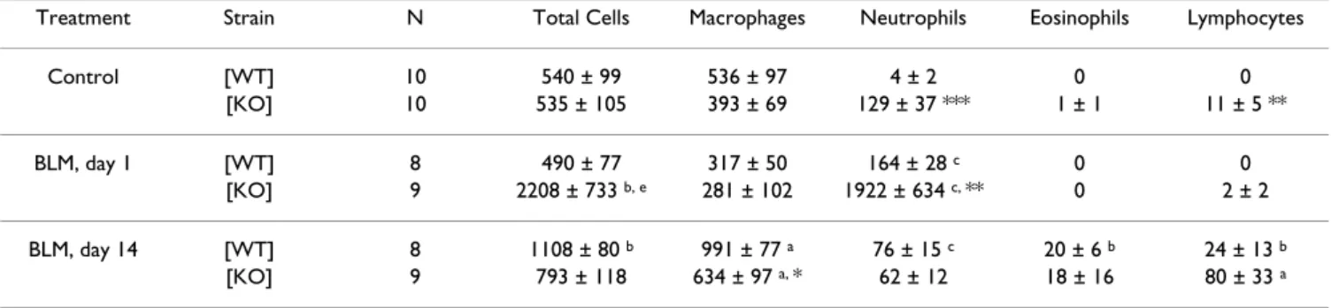
At both day 1 and day 14, bleomycin treatment elicited enhanced ROS production by BAL cells from WT mice stimulated in vitro with PMA (Figure 1). In contrast, nei-ther the cells removed from control mice (WT, not treated with bleomycin) nor those from KO mice (regardless of bleomycin treatment) could produce ROS in vitro after PMA stimulation. No chemiluminescence was detected when BAL cells were not stimulated by PMA in vitro. At day 1, ROS release by BAL cells from bleomycin-treated WT mice was accompanied by a significant neutrophil influx, but the total BAL cell count did not rise (Table 1). This bleomycin-induced neutrophil influx was quite noticeable in BAL from KO mice, significantly greater than in WT mice.
Fourteen days after bleomycin administration, the total cell count in the WT mouse BAL increased significantly. Specifically, the alveolar macrophage count rose mark-edly, as did the neutrophil, eosinophil, and lymphocyte counts, although to a lesser extent. The total cell count in the BAL fluid of the KO mice did not increase signifi-cantly, but the number of macrophages and lymphocytes did. The WT mice had significantly more alveolar macro-phages than the KO mice (Table 1).
Production of reactive oxygen species (ROS) by bronchoalveolar lavage (BAL) cells, 1 day (A) and 14 days (B) after intranasal
administration of bleomycin (0.1 mg, BLM) or saline (Control) to mice with the p47phox subunit of NADPH-oxidase deleted
(p47phox -/- knockout mice (KO): solid bars), compared with "Wild Type" p47phox +/+ mice (WT: blank bars)
Figure 1
Production of reactive oxygen species (ROS) by bronchoalveolar lavage (BAL) cells, 1 day (A) and 14 days (B) after intranasal administration of bleomycin (0.1 mg, BLM) or saline (Control) to mice with the p47phox subunit of NADPH-oxidase deleted
(p47phox -/- knockout mice (KO): solid bars), compared with "Wild Type" p47phox +/+ mice (WT: blank bars). Cells were
stim-ulated in vitro with PMA (0.8 µM) and ROS production was evaluated by chemiluminescence. Data were collected at the time of maximum light emission. Results are expressed as relative light units (mean ± SEM). ***: p < 0.001 comparison with control mice exposed to saline solution alone. ###: p < 0.001 for KO mice compared with WT mice. n = 5–9.
1 0 20000 40000 60000 80000 100000 120000 Control BLM R ela tiv e Li g h t U n it s *** *** ### ### 0 5000 10000 15000 20000 25000 30000 35000 Control BLM R ela tiv e Li g h t U n its ### ### *** *** A : Day 1 B : Day 14
Lung hydroxyproline measurement
Lung hydroxyproline concentration, which reflects colla-gen deposition in lungs, was measured 14 days after bleo-mycin administration to quantify pulmonary fibrosis (Table 2). Hydroxyproline levels did not increase in the KO mice. Moreover, although the hydroxyproline level was similar in both strains of mice at baseline, it was sig-nificantly higher in WT than in KO mice at day 14.
IL-6 levels in BAL fluids
IL-6 levels in the BAL fluids of WT and KO mice rose one day after bleomycin administration (figure 2) and were significantly higher in WT than KO mice. Fourteen days after bleomycin administration, IL-6 levels were not sig-nificantly different from baseline.
MMP activity in BAL fluids and lung homogenates
Zymography identified the following gelatinolytic bands as MMP activity: pro-MMP-9 (105 kDa), MMP-9 (86 kDa), pro-MMP-2 (70 kDa), and MMP-2 (64 kDa). At day one, pro-MMP-9 activity was significantly higher in the BAL of bleomycin-treated KO mice than in that of their bleomycin-treated WT counterparts, which in turn was significantly higher than in the control mouse BAL (figure 3 and figure 4A). At day 14, no pro-MMP-9 activity was observed in any of the mice. The active form of MMP-9 (86 kDa) was detected only in KO mouse BAL fluid at day
1 (figure 3). Pro-MMP-9 was significantly correlated with the neutrophil count in the BAL fluids of both WT (P = 0.001) and KO (P = 7 × 10⁻⁶) mice.
At one day and 14 days after bleomycin administration, MMP-2 activity was observed in both its latent (70 kDa) and active (64 kDa) forms (figure 3). Although densitom-etry analysis could detect only the 64-kD form (figure 4B) at day one, bleomycin elicited a significant increase in the 64-kDa MMP-2 activity in both strains, compared with the control; and this activity was substantially stronger in the BAL of KO than WT mice. At day 14, MMP-2 activity remained higher in both strains of bleomycin-treated mice (compared with controls) and did not differ signifi-cantly between them (figure 4B).
MMP activity was also evaluated at day 14 in lung homogenates (figure 5). Homogenate from both WT and KO mice showed similar levels of pro-MMP-9 activity lev-els, unexpected lower than in the homogenate from the control mice. In contrast, MMP-2 activity increased in the lungs of WT mice only (figure 5).
Pro-MMP-9/TIMP-1 ratio in BAL fluids
We also evaluated pro-MMP-9 as well as TIMP-1 with ELISA (table 3). The results for pro-MMP-9 confirm those obtained with zymography. In WT mice, TIMP-1 in BAL
Table 1: Total and differential cell counts of BAL fluid from p47phox +/+ WT and p47phox -/- KO mice, at day 1 and day 14 after intranasal
administration of bleomycin (BLM, 0.1 mg/mouse) or saline vehicle (Control, NaCl 0.9%). Results are presented as the mean (.103cells)
± SEM. n: number of mice. WT: wild-type; KO: knockout; a: P < 0.05, b: P < 0.01, c: P < 0.001 compared with control mice exposed to saline solution only. *: P < 0.05, **: P < 0.01, ***: P < 0.001 for KO mice compared with WT mice.
Treatment Strain N Total Cells Macrophages Neutrophils Eosinophils Lymphocytes Control [WT] 10 540 ± 99 536 ± 97 4 ± 2 0 0 [KO] 10 535 ± 105 393 ± 69 129 ± 37 *** 1 ± 1 11 ± 5 ** BLM, day 1 [WT] 8 490 ± 77 317 ± 50 164 ± 28 c 0 0 [KO] 9 2208 ± 733 b, e 281 ± 102 1922 ± 634 c, ** 0 2 ± 2 BLM, day 14 [WT] 8 1108 ± 80 b 991 ± 77 a 76 ± 15 c 20 ± 6 b 24 ± 13 b [KO] 9 793 ± 118 634 ± 97 a, * 62 ± 12 18 ± 16 80 ± 33 a
Table 2: Hydroxyproline content (mg/g of dry tissue) in lung homogenate from p47phox +/+ WT and p47phox -/- KO mice, at day 14 after
intranasal administration of bleomycin (BLM, 0.1 mg/mouse) or saline vehicle (Control, NaCl 0.9%). Results are presented as mean ± SEM. WT: wild-type; KO: knockout; ** : P < 0.01 for WT mice compared with KO mice. n = 3–6.
Strain Control BLM p47phox +/+ [WT] 1.20 ± 0.26 1.74 ± 0.10**
fluid was markedly higher at day one and day 14 after ble-omycin administration than at baseline. In contrast, the TIMP-1 level in the BAL fluid of KO mice was not signifi-cantly modified by bleomycin administration at either day 1 or day 14 (table 3).
Table 3 presents the calculation of the pro-MMP-9/TIMP-1 ratio. At day one after bleomycin administration, this ratio remained stable in the BAL of WT mice, both treated and untreated, but rose significantly in that of the KO mice (table 3). At day 14, the ratio was significantly lower in both strains of treated mice than in the control mice, and levels in the WT mice were significantly lower than those in the KO mice.
Discussion
This study shows that mice deficient in the p47phox
subu-nit of the NADPH oxidase complex do not develop pul-monary fibrosis after intranasal administration of bleomycin. It also suggests that an imbalance of the molar MMP-9/TIMP-1 ratio may influence the fibrogenic proc-ess in this model.
Several studies previously reported that antioxidant treat-ment attenuates the bleomycin-induced oxidative burden and subsequent pulmonary fibrosis [7,8,17]. Moreover, the absence of extracellular superoxide dismutase exacer-bates conditions that lead to inflammation and pulmonary fibrosis [18]. Although these studies suggest that ROS contribute to lung damage and fibrosis, they do not clearly indicate the mechanisms of the antioxidant effect. That is, antioxidant compounds may attenuate oxi-dative damage caused directly by bleomycin [19], or they may limit the impact of ROS produced by phagocytes such as macrophages and neutrophils [5] and thus inter-fere with the inflammatory process. To clarify the role of ROS produced by phagocytes in the development of pul-monary fibrosis, we induced pulpul-monary fibrosis by i.n. bleomycin administration to p47phox-/- KO mice. Unlike
antioxidant compounds, which nonspecifically target all ROS sources in the tissue, knocking out the p47phox
subu-nit of the NADPH oxidase complex shuts down only the main pathway of phagocytic ROS production. In this study, in vitro PMA stimulation of BAL cells from KO mice produced no detectable ROS, while BAL cells from WT
Level of IL-6 (pg/mL) in BAL fluids, 1 day after intranasal administration of bleomycin (BLM, 0.1 mg/mouse) or saline vehicle
(Control), to p47phox +/+ WT mice (blank bars) and p47phox -/- KO mice (solid bars)
Figure 2
Level of IL-6 (pg/mL) in BAL fluids, 1 day after intranasal administration of bleomycin (BLM, 0.1 mg/mouse) or saline vehicle (Control), to p47phox +/+ WT mice (blank bars) and p47phox -/- KO mice (solid bars). Results are presented as the mean ± SEM.
** : p < 0.01 compared with control mice exposed to saline solution alone. #: p < 0.05 for p47phox -/- KO mice compared with
p47phox +/+ WT mice. n = 4–9.
0
50
100
150
200
250
300
350
Control
BLM
IL
-6
(pg/
m
L
)
**
**
#
mice did produce ROS, as previous studies of circulating neutrophils from these mice have shown [15].
Hydroxyproline content did not increase in the lungs of these KO mice. This finding reveals the absence of fibrosis and thus provides strong evidence that phagocytic ROS production is an important component of the fibrogenic environment.
Although no ROS production was detected in KO mice, their BAL gave evidence of an acute inflammatory
response, as did that from WT mice: bleomycin adminis-tration elicited acute inflammation, characterized by an influx of neutrophils and associated with increased pro-MMP-9 activity. Moreover, the response of the KO mice to the bleomycin resembled the abnormal "exuberant" inflammation in vivo previously described in such KO mice [15,20,21]. It is, however, possible that this neu-trophil influx and the large amounts of MMP-9 it releases have a protective effect against the development of pulmonary fibrosis. This exaggerated inflammatory response may be caused by defective down-regulation of
Representative gelatin zymogram of BAL supernatant fluids, 1 and 14 days after intranasal administration of bleomycin (BLM,
0.1 mg/mouse) or saline vehicle (Control) to p47phox +/+ WT mice (A) and p47phox -/- KO mice (B)
Figure 3
Representative gelatin zymogram of BAL supernatant fluids, 1 and 14 days after intranasal administration of bleomycin (BLM, 0.1 mg/mouse) or saline vehicle (Control) to p47phox +/+ WT mice (A) and p47phox -/- KO mice (B). The following gelatinolytic
bands were identified as MMP activity: pro-MMP-9 (105 kDa), MMP-9 (86 kDa), pro-MMP-2 (70 kDa), and MMP-2 (64 kDa). M: molecular weight marker.
A :
p47
phox+/+ [WT]
M
Control
BLM, Day 1
BLM, Day 14
M
Control
BLM, Day 1
BLM, Day 14
pro-MMP2
MMP-2
112 kDa
pro-MMP9
81 kDa
B :
p47
phox-/- [KO]
112 kDa
pro-MMP9
MMP9
81 kDa
pro-MMP2
MMP2
Quantification by densitometry of 105-kDa pro-MMP-9 (A), and 64-kDa MMP-2 (B) gelatinase activity on zymograms of BAL fluid, performed 1 day or 14 days after intranasal administration of bleomycin (BLM, 0.1 mg/mouse) or saline vehicle (Control),
to p47phox +/+ WT mice (blank bars) and p47phox -/- KO mice (solid bars)
Figure 4
Quantification by densitometry of 105-kDa pro-MMP-9 (A), and 64-kDa MMP-2 (B) gelatinase activity on zymograms of BAL fluid, performed 1 day or 14 days after intranasal administration of bleomycin (BLM, 0.1 mg/mouse) or saline vehicle (Control), to p47phox +/+ WT mice (blank bars) and p47phox -/- KO mice (solid bars). Results are represented as the mean of relative
intensity ± SEM. * : p < 0.05, ** : p < 0.01, *** : p < 0.001 compared with control mice exposed to saline solution alone. ###: p < 0.001 for p47phox -/- KO mice compared with p47phox +/+ WT mice. n = 8–10.
1
0
50
100
150
200
250
300
350
Control
BLM, Day 1
P
ro-M
M
P
-9
(105 kD
a,
r
el
at
iv
e i
nt
ens
it
y)
**
***
###
0 20 40 60 80 100 120 140 160 180 200Control BLM, Day 1 BLM, Day 14
MMP-2 (64 kDa, relative intensity) *
*** *** *** ### A : 105 kDa pro-MMP-9 B : 64 kDa MMP-2
Quantification by densitometry of Pro-MMP-9 (105 kDa, A) and MMP-2 (64 kDa, B) gelatinase activity on zymograms of lung homogenates, performed 14 days after intranasal administration of bleomycin (BLM, 0.1 mg/mouse) or saline vehicle (Control)
to p47phox -/- KO mice (solid bars) and p47phox +/+ WT mice (blank bars)
Figure 5
Quantification by densitometry of Pro-MMP-9 (105 kDa, A) and MMP-2 (64 kDa, B) gelatinase activity on zymograms of lung homogenates, performed 14 days after intranasal administration of bleomycin (BLM, 0.1 mg/mouse) or saline vehicle (Control) to p47phox -/- KO mice (solid bars) and p47phox +/+ WT mice (blank bars). Results are represented as the mean of relative
intensity ± SEM. * : p < 0.05, ** : p < 0.01, compared with control mice exposed to saline solution alone. ##: p < 0.01 for p47phox -/- [KO] mice compared with p47phox +/+ WT mice. n = 3–5.
1
0
200
400
600
800
1000
1200
Control
BLM
pro-MMP9 (105 k
D
, relative intensity)
**
*
0
100
200
300
400
500
600
Control
BLM
M
M
P
2
(
64
kD
a,
r
el
at
ive
i
nt
ens
it
y)
**
##
A : 105 kDa pro-MMP-9 B : 64 kDa MMP-2the inflammatory process in these KO mice, perhaps due to the absence of ROS and the failure to degrade chemo-tactic signals [22].
This acute inflammation was accompanied by signifi-cantly elevated IL-6 levels in the BAL fluids of both strains of mice on the day after bleomycin administration, levels significantly higher in WT than KO mice. Given that IL-6 is secreted primarily by mononuclear cells, specifically macrophages, the difference between strains suggests that these cells are activated more weakly in the KO than in the WT mice. Moreover, in SP-D -/- alveolar macrophages, the NADPH oxidase inhibitor apocynin inactivates NF-kappa B, the transcription factor that regulates numerous proin-flammatory responses, including IL-6 release [23]. Simi-larly, lipopolysaccharide-induced NF-kappa B activation is impaired in nuclear protein extracts of lung tissue from p47phox-/- KO mice [24]. This would explain why IL-6
response seems to be redox-sensitive in our experiment. MMP induction, assessed by gelatinase release, has been reported in various cases of pulmonary fibrosis in human and experimental models [13,25,26]. We therefore ana-lyzed the gelatinolytic activities of MMPs, in both BAL fluid and lung homogenate. MMP-9 and MMP-2 activities in the BAL fluid of the KO mice reveal an intense response to bleomycin. MMP-9 levels were highly correlated with neutrophil infiltration, while MMP-2 is known to be pro-duced mainly by epithelial [27] and mesenchymal cells, such as fibroblasts, which are involved in collagen pro-duction and deposition [9,25]. This suggests that the exag-gerated response observed in the BAL fluid of KO mice involves a wide spectrum of cell types. Surprisingly, at day 14, gelatinase profiles were different in the lung homoge-nates than in BAL fluids. Although MMP-2 activity was equivalent in BAL of both strains, MMP-2 activity
increased substantially in the lung homogenate of the WT mice. One possible explanation is that the increased release of MMP-2 in the inner lung parenchyma may result from downstream events caused by phagocyte acti-vation and ROS production during inflammation. This is consistent with another study, mentioned above, in which apocynin, an inhibitor of NADPH oxidase, also inhibited the release of MMP-9 and MMP-2 in SP-D -/-alveolar macrophages [23]. Moreover, Pardo et al. [28] report increased levels of gelatinases (2 and MMP-9) in isolated type II alveolar cells from hyperoxic rats; these increases are associated with alterations in the bal-ance between MMPs and TIMPs and finally lead to diffuse alveolitis and its progression to pulmonary fibrosis. It is thus difficult to reach a definitive conclusion at this time about the exact function of MMPs. They play a role in promoting tissue remodeling and counterbalancing excessive matrix deposition, but may also facilitate tissue damage and disruption. Their involvement in bleomycin-induced pulmonary fibrosis has been demonstrated with the inhibition of collagen deposition by bastimastat, a nonselective MMP-inhibitor [10]. Complete understand-ing of the dynamic process of remodelunderstand-ing nonetheless requires consideration of TIMPs, which are natural MMP inhibitors.
Increased TIMP-1 expression has been observed in lung extracts and in BAL fluids after bleomycin administration and after the transfer of the active TGF-beta gene to "fibro-sis-prone" C57BL/6 mice [11,14]. In humans, increased levels of TIMP protein and RNA are observed in lungs of patients with idiopathic pulmonary fibrosis, and TIMP expression there exceeds that of MMP [12]. A reduced molar MMP/TIMP ratio seems to be a hallmark of pulmo-nary fibrosis, distinguishing it from other reversible
inter-Table 3: Levels of pro-MMP-9 and TIMP-1, and pro-MMP-9 / TIMP-1 ratio in BAL supernatant fluids, recovered from p47phox +/+ [WT]
and p47phox -/- [KO] mice, 1 day or 14 days after intranasal administration of bleomycin (BLM, 0.1 mg/mouse) or saline vehicle
(Control, NaCl 0.9%). For Pro-MMP-9 and TIMP-1, results are represented as the mean (pg/ml) ± SEM. N : number of mice. a : P < 0.05, b : P < 0.01, in comparison to control mice exposed to saline solution only. * : P < 0.05, ** : P < 0.01, for [KO] mice in comparison to [WT] mice.
Treatment Strain N Pro-MMP-9 TIMP-1 Ratio Pro-MMP-9 / TIMP-1 Control [WT] 5 46.3 ± 14.9 38.7 ± 13.5 2.7 ± 1.3 [KO] 5 1314.7 ± 798.2 ** 325.2 ± 173.6 10.4 ± 4.7 BLM, day 1 [WT] 7 2645.9 ± 1 011.6 b 781.8 ± 158.9 b 3.8 ± 1.3 [KO] 8 23646.8 ± 2 359.7 b, ** 1 213.8 ± 396.4 60.8 ± 23.7 a, ** BLM, day 14 [WT] 7 37.7 ± 9.1 2693.3 ± 378.3 b 0.02 ± 0.00 b [KO] 6 137.9 ± 15.5 b 567.9 ± 163.5** 0.2 ± 0.1 b, **
stitial lung diseases [26] and from chronic obstructive pulmonary disease (COPD) [29]. This ratio might be con-sidered to be a "snapshot" of the dynamic matrix remod-eling in lung tissue.
Interestingly, in our study, the pro-MMP-9/TIMP-1 ratio was significantly higher for KO than WT mice, at both day one and day 14. At day 1 this was due to the lower MMP-9 level and higher TIMP-1 level in the BAL from WT mice, and at day 14, only to the latter. The correlation of these levels with differences in hydroxyproline levels in the lungs of bleomycin-treated mice strongly suggests that a reduced molar pro-MMP-9/TIMP-1 ratio in BAL fluid is associated with collagen deposition, beginning as early as the inflammatory events at day 1 after bleomycin administration.
The usefulness of the pro-MMP-9/TIMP-1 ratio as a marker of fibrosis nonetheless requires discussion. Although a molar ratio appears to play a protective role against fibrotic changes, MMP-9 is considered primarily to be an inflammatory mediator released by leukocytes dur-ing acute inflammatory events to facilitate their progres-sion across the basement membrane [30]. Moreover, MMP-9 depletion in KO mice does not substantially alter the extent of either pulmonary fibrosis or lung inflamma-tion after bleomycin administrainflamma-tion [31]. TIMPs may also counterbalance the activity of MMP-2 or other protein-ases, such as collagenases. Ruiz et al. [32] recently observed that MMP-8 and MMP-13 RNA levels decreased and TIMP-1 RNA increased in the paraquat- and hyper-oxia-induced pulmonary fibrosis rat model. Matrilysin (MMP-7), which can degrade various substrates, seems to have a crucial role in pulmonary fibrosis [33]. Finally, the exuberant neutrophil influx observed at day one in p47phox-/- KO mice could provide great amount of other
kind of protease, such as serine proteases. Indeed, neu-trophil elastase was shown to have an impact on the sever-ity of bleomycin-induced pulmonary fibrosis [34,35]
Conclusion
In summary, this study demonstrates that the inability of phagocytes from p47phox-/- KO mice to produce large
quantities of ROS via the NADPH oxidase pathway inhib-its the development of bleomycin-induced pulmonary fibrosis. This inhibition is associated with changes in IL-6 production and in the molar MMP-9/TIMP-1 ratio, both probably key factors in airway remodeling and fibrosis. These rapidity of these differences after bleomycin admin-istration suggests that early inflammatory events and remodeling events may establish a favorable environment for further chronic fibrogenic processes.
Abbreviations
BAL: Bronchoalveolar lavage
IL: interleukin KO: knock out
MMP: matrix metalloproteinase
TIMP: tissue inhibitor of metalloproteinase
ROS: reactive oxygen species
WT: wild type
Authors' contributions
BM, SN, OL, IG:and EB have made substantial contribu-tions to acquisition and analysis of data
BM, SN, EB and VL have made substantial contributions to conception and design
BM, EB and VL have been involved in drafting the article JMP and CPB have been involved in revising it critically for important intellectual content
Acknowledgements
The authors thank C. Chesné and O. Minella (Biopredic Int., Rennes, France) for supplying the ABEL® chemiluminescence kit. This work is sup-ported by a research grant (ref #2004797) from Conseil Régional de Bre-tagne and by a collaborative project INSERM/FIOCRUZ.
References
1. Crouch E: Pathobiology of pulmonary fibrosis. Am J Physiol 1990,
259:L159-184.
2. Selman M, King TE, Pardo A: Idiopathic pulmonary fibrosis:
pre-vailing and evolving hypotheses about its pathogenesis and implications for therapy. Ann Intern Med 2001, 134:136-151.
3. Katzenstein AL, Myers JL: Idiopathic pulmonary fibrosis: clinical
relevance of pathologic classification. Am J Respir Crit Care Med
1998, 157:1301-1315.
4. Ward PA, Hunninghake GW: Lung inflammation and fibrosis. Am
J Respir Crit Care Med 1998, 157:S123-129.
5. Strausz J, Muller-Quernheim J, Steppling H, Nagel M, Ferlinz R:
Oxy-gen radical production by alveolar macrophages in sarcoido-sis in relation to activity status of bronchoalveolar lavage lymphocytes. Pneumologie 1990, 44(Suppl 1):222-223.
6. Rahman I, Skwarska E, Henry M, Davis M, O'Connor CM, FitzGerald MX, Greening A, MacNee W: Systemic and pulmonary
oxida-tive stress in idiopathic pulmonary fibrosis. Free Radic Biol Med
1999, 27:60-68.
7. Serrano-Mollar A, Closa D, Prats N, Blesa S, Martinez-Losa M, Cortijo J, Estrela JM, Morcillo EJ, Bulbena O: In vivo antioxidant
treat-ment protects against bleomycin-induced lung damage in rats. Br J Pharmacol 2003, 138:1037-1048.
8. Wang HD, Yamaya M, Okinaga S, Jia YX, Kamanaka M, Takahashi H, Guo LY, Ohrui T, Sasaki H: Bilirubin ameliorates
bleomycin-induced pulmonary fibrosis in rats. Am J Respir Crit Care Med
2002, 165:406-411.
9. Corbel M, Belleguic C, Boichot E, Lagente V: Involvement of
gela-tinases (MMP-2 and MMP-9) in the development of airway inflammation and pulmonary fibrosis. Cell Biol Toxicol 2002, 18:51-61.
10. Corbel M, Caulet-Maugendre S, Germain N, Molet S, Lagente V, Boi-chot E: Inhibition of bleomycin-induced pulmonary fibrosis in
mice by the matrix metalloproteinase inhibitor batimastat. J Pathol 2001, 193:538-545.
Publish with BioMed Central and every scientist can read your work free of charge "BioMed Central will be the most significant development for disseminating the results of biomedical researc h in our lifetime."
Sir Paul Nurse, Cancer Research UK
Your research papers will be:
available free of charge to the entire biomedical community peer reviewed and published immediately upon acceptance cited in PubMed and archived on PubMed Central yours — you keep the copyright
Submit your manuscript here:
http://www.biomedcentral.com/info/publishing_adv.asp
BioMedcentral
11. Kolb M, Bonniaud P, Galt T, Sime PJ, Kelly MM, Margetts PJ, Gauldie J: Differences in the fibrogenic response after transfer of
active transforming growth factor-beta1 gene to lungs of "fibrosis-prone" and "fibrosis-resistant" mouse strains. Am J Respir Cell Mol Biol 2002, 27:141-150.
12. Selman M, Ruiz V, Cabrera S, Segura L, Ramirez R, Barrios R, Pardo A: TIMP-1, -2, -3, and -4 in idiopathic pulmonary fibrosis. A
prevailing nondegradative lung microenvironment? Am J Phys-iol Lung Cell Mol PhysPhys-iol 2002, 279:L562-574.
13. Swiderski RE, Dencoff JE, Floerchinger CS, Shapiro SD, Hunninghake GW: Differential expression of extracellular matrix
remode-ling genes in a murine model of bleomycin-induced pulmo-nary fibrosis. Am J Pathol 1998, 152:821-828.
14. Madtes DK, Elston AL, Kaback LA, Clark JG: Selective induction of
tissue inhibitor of metalloproteinase-1 in bleomycin-induced pulmonary fibrosis. Am J Respir Cell Mol Biol 2001, 24:599-607.
15. Jackson SH, Gallin JI, Holland SM: The p47phox mouse knock-out
model of chronic granulomatous disease. J Exp Med 1995, 182:751-758.
16. Reddy GK, Enwemeka CS: A simplified method for the analysis
of hydroxyproline in biological tissues. Clin Biochem 1996, 29:225-229.
17. Punithavathi D, Venkatesan N, Babu M: Curcumin inhibition of
bleomycin-induced pulmonary fibrosis in rats. Br J Pharmacol
2000, 131:169-172.
18. Fattman CL, Chang LY, Termin TA, Petersen L, Enghild JJ, Oury TD:
Enhanced bleomycin-induced pulmonary damage in mice lacking extracellular superoxide dismutase. Free Rad Biol Med
2003, 35:763-771.
19. Hay J, Shahzeidi S, Laurent G: Mechanisms of bleomycin-induced
lung damage. Arch Toxicol 1991, 65:81-94.
20. Planquois JM, Leclerc O, Bertrand C: Enhancement of the airway
inflammatory response induced by LPS challenge in p47phox knock out (KO) mice [Abstract]. Am J Respir Crit Care Med 2001, 163:A347.
21. Gao XP, Standiford TJ, Rahman A, Newstead M, Holland SM, Dinauer MC, Liu QH, Malik AB: Role of NADPH oxidase in the
mecha-nism of lung neutrophil sequestration and microvessel injury induced by Gram-negative sepsis: studies in p47phox-/- and gp91phox-/- mice. J Immunol 2002, 168:3974-3982.
22. Gallin JI, Buescher ES: Abnormal regulation of inflammatory
skin responses in male patients with chronic granulomatous disease. Inflammation 1983, 7:227-232.
23. Yoshida M, Korfhagen TR, Whitsett JA: Surfactant protein D
reg-ulates NF-kappa B and matrix metalloproteinase production in alveolar macrophages via oxidant-sensitive pathways. J Immunol 2001, 166:7514-7519.
24. Koay MA, Christman JW, Segal BH, Venkatakrishnan A, Blackwell TR, Holland SM, Blackwell TS: Impaired pulmonary NF-kappaB
acti-vation in response to lipopolysaccharide in NADPH oxidase-deficient mice. Infect Immun 2001, 69:5991-5996.
25. Yaguchi T, Fukuda Y, Ishizaki M, Yamanaka N:
Immunohistochem-ical and gelatin zymography studies for matrix metallopro-teinases in bleomycin-induced pulmonary fibrosis. Pathol Int
1998, 48:954-963.
26. Suga M, Iyonaga K, Okamoto T, Gushima Y, Miyakawa H, Akaike T, Ando M: Characteristic elevation of matrix
metalloprotein-ase activity in idiopathic interstitial pneumonias. Am J Respir Crit Care Med 2000, 162:1949-1956.
27. Kunugi S, Fukuda Y, Ishizaki M, Yamanaka N: Role of MMP-2 in
alveolar epithelial cell repair after bleomycin administration in rabbits. Lab Invest 2001, 81:1309-1318.
28. Pardo A, Barrios R, Maldonado V, Melendez J, Pérez J, Ruiz V, Segura-Valdez L, Sznajder JI, Selman M: Gelatinases A and B Are
Up-Reg-ulated in Rat Lungs by Subacute Hyperoxia, Pathogenetic Implications. Am J Pathol 1998, 153:833-844.
29. Beeh KM, Beier J, Kornmann O, Micke P, Buhl R: Sputum levels of
metalloproteinase-9 and tissue inhibitor of metalloprotein-ase-1, and their ratio correlate with airway obstruction in lung transplant recipients: relation to tumor necrosis factor-alpha and interleukin-10. J Heart Lung Transplant 2001, 20:1144-1151.
30. Atkinson JJ, Senior RM: Matrix metalloproteinase-9 in lung
remodeling. Am J Respir Cell Mol Biol 2003, 28:12-24.
31. Betsuyaku T, Fukuda Y, Parks WC, Shipley JM, Senior RM:
Gelati-nase B is required for alveolar bronchiolization after intrat-racheal bleomycin. Am J Pathol 2000, 157:525-535.
32. Ruiz V, Ordonez RM, Berumen J, Ramirez R, Uhal B, Becerril C, Pardo A, Selman M: Unbalanced collagenases/TIMP-1 expression and
epithelial apoptosis in experimental lung fibrosis. Am J Physiol Lung Cell Mol Physiol 2003, 285:L1026-1036.
33. Zuo F, Kaminski N, Eugui E, Allard J, Yakhini Z, Ben-Dor A, Lollini L, Morris D, Kim Y, DeLustro B, Sheppard D, Pardo A, Selman M, Heller RA: Gene expression analysis reveals matrilysin as a key
reg-ulator of pulmonary fibrosis in mice and humans. Proc Natl Acad Sci U S A 2002, 99:6292-6297.
34. Taooka Y, Maeda A, Hiyama K, Ishioka S, Yamakido M: Effects of
neutrophil elastase inhibitor on bleomycin-induced pulmo-nary fibrosis in mice. Am J Respir Crit Care Med 1997, 156:260-265.
35. Dunsmore SE, Roes J, Chua FJ, Segal AW, Mutsaers SE, Laurent GJ:
Evidence that neutrophil elastase-deficient mice are resist-ant to bleomycin-induced fibrosis. Chest 2001, 120(1 Suppl):35S-36S.

![Table 3: Levels of pro-MMP-9 and TIMP-1, and pro-MMP-9 / TIMP-1 ratio in BAL supernatant fluids, recovered from p47 phox +/+ [WT]](https://thumb-eu.123doks.com/thumbv2/123doknet/14551715.537014/11.918.95.833.209.398/table-levels-timp-timp-ratio-supernatant-fluids-recovered.webp)
