HAL Id: inserm-01644732
https://www.hal.inserm.fr/inserm-01644732
Submitted on 22 Nov 2017
HAL is a multi-disciplinary open access
archive for the deposit and dissemination of
sci-entific research documents, whether they are
pub-lished or not. The documents may come from
teaching and research institutions in France or
abroad, or from public or private research centers.
L’archive ouverte pluridisciplinaire HAL, est
destinée au dépôt et à la diffusion de documents
scientifiques de niveau recherche, publiés ou non,
émanant des établissements d’enseignement et de
recherche français ou étrangers, des laboratoires
publics ou privés.
Dominique Heymann
To cite this version:
Nathalie Renema, Benjamin Navet, Marie-Françoise Heymann, Frédéric Lézot, Dominique Heymann.
RANK-RANKL signalling in cancer: RANK-RANKL and cancer. Bioscience Reports, Portland Press,
2016, 36 (4), pp.e00366 - e00366. �10.1042/BSR20160150�. �inserm-01644732�
RANK–RANKL signalling in cancer
Nathalie Renema*
1, Benjamin Navet*
1, Marie-Franc¸oise Heymann*†‡, Fr´
ed´
eric Lezot*
2and
Dominique Heymann*†‡
2*INSERM, UMR 957, Equipe Labellis´ee Ligue 2012, Physiopathologie de la R´esorption Osseuse et Th´erapie des Tumeurs Osseuses Primitives, Universit´e de Nantes, 1 Rue Gaston Veil, 44035 Nantes, France
†Nantes University Hospital, Nantes 44035, France
‡Department of Oncology and Human Metabolism, The University of Sheffield, Sheffield S10 2RX, U.K.
Synopsis
Oncogenic events combined with a favourable environment are the two main factors in the oncological process.
The tumour microenvironment is composed of a complex, interconnected network of protagonists, including soluble
factors such as cytokines, extracellular matrix components, interacting with fibroblasts, endothelial cells, immune
cells and various specific cell types depending on the location of the cancer cells (e.g. pulmonary epithelium,
osteoblasts). This diversity defines specific “niches” (e.g. vascular, immune, bone niches) involved in tumour growth
and the metastatic process. These actors communicate together by direct intercellular communications and/or in an
autocrine/paracrine/endocrine manner involving cytokines and growth factors. Among these glycoproteins, RANKL
(receptor activator nuclear factor-κB ligand) and its receptor RANK (receptor activator nuclear factor), members of the
TNF and TNFR superfamilies, have stimulated the interest of the scientific community. RANK is frequently expressed
by cancer cells in contrast with RANKL which is frequently detected in the tumour microenvironment and together they
participate in every step in cancer development. Their activities are markedly regulated by osteoprotegerin (OPG, a
soluble decoy receptor) and its ligands, and by LGR4, a membrane receptor able to bind RANKL. The aim of the present
review is to provide an overview of the functional implication of the RANK/RANKL system in cancer development, and
to underline the most recent clinical studies.
Key words: microenvironment, oncogenesis, RANK, RANKL
Cite this article as: Bioscience Reports (2016) 36, e00366, doi:10.1042/BSR20160150
INTRODUCTION
In a physiological context, a healthy tissue microenvironment
provides an adapted 3D microarchitecture with essential
inter-cellular signalling, thus ensuring appropriate function. This
tis-sue homoeostasis acts as a barrier to tumour development by
inhibiting excessive cell growth and/or migration. Indeed, this
fragile equilibrium can be destabilized by any alterations to cell
communications, or interaction between cells and extracellular
matrix components and consequently can become a fertile
envir-onment for cancer cells, promoting their malignant
transforma-tion and their proliferatransforma-tion [
1
]. The conjunction between one or
more oncogenic events and this fertile environment can lead to
the development of a tumour mass, which is frequently linked
to the tumour cells escaping from the immune system [
2
]. In
. . . . Abbreviations: EMT, epithelial mesenchymal transition; LRG4, leucine-rich repeat-containing G-protein-coupled receptor 4; OPG, osteoprotegerin; OPGL, osteoprotegerin ligand; RANK, receptor activator of nuclear factor-κB; RANKL, receptor activator nuclear factor-κB ligand; TAM, tumour-associated macrophage; TNF-α, tumour necrosis factor-α; TRAF, TNF-receptor associated factor; TRAIL, TNF related apoptosis inducing ligand; TRANCE, tumour necrosis factor-related activation-induced cytokine.
1 These authors contributed equally.
2 Correspondence may be addressed to either of these authors (email dominique.heymann@sheffield.ac.uk or frederic.lezot@univ-nantes.fr).
fact, this description reflects the “seed and soil” theory proposed
by Stephan Paget in 1889 to explain preferential metastatic sites
depending on tumour subtype [
3
].
This “soil” or tumour microenvironment is a very complex and
dynamic organization, defined by three main “niches” depending
on their functional implication: (i) an immune niche involved
in local immune tolerance, (ii) a vascular niche associated with
tumour cell extravasation/migration and (iii) a metastatic niche
(e.g. bone, lung, liver) hosting the metastastic tumour cells [
4
,
5
].
The notion of tumour niche was initially described for
haema-topoietic stem cells, for which the bone microenvironment is
composed of complex signalling pathways that carefully
regu-late stem cell renewal, differentiation and quiescence [
6
]. The
concept of tumour niche was then extended to bone metastases,
such as breast or prostate cancers [
7
–
9
]. Lu et al. [
10
] described
a model of bone metastasis dormancy in breast cancer where
VCAM-1, aberrantly expressed, promoted the transition from
in-dolent micrometastasis to proliferating tumour by recruiting and
activating in situ osteoclastic cells. More recently, Wang et al. [
11
]
analysed the distribution of human prostate cancer cell lines
col-onizing mouse bones after intracardiac injection of tumour cells
and demonstrated that homing of prostate cancer cells was
associ-ated with the presence of activassoci-ated osteoblast lineage cells. These
two recent manuscripts are perfect examples of the involvement
of the tumour environment in the biology of bone metastases.
The tumour microenvironment thus provides all the factors
necessary for cancer cell survival, dormancy, proliferation or/and
migration [
10
] and very often, tumour cells divert this
envir-onment in their favour [
7
–
9
]. Indeed, this specific
microenviron-ment has recently been involved in the maintenance of cancer cell
dormancy [
12
–
14
] and may also play a part in drug resistance
mechanisms by controlling the balance between cell proliferation
and cell death, or by secreting soluble factors that dysregulate the
cell cycle checkpoints, the cell death associated signalling
path-ways, or drug efflux [
15
,
16
].
Cell communications in physiological and pathological
con-ditions are promoted by physical contacts involving adhesion
molecules and channels, but also by a very high number of
sol-uble mediators called cytokines and growth factors which appear
to be the key protagonists in the dialogue established between
cancer cells and their microenvironment [
16
]. These
polypep-tidic mediators perform their activities in an autocrine, paracrine
or juxtacrine manner leading to inflammatory foci and the
estab-lishment of a vicious cycle between cancer cells and their local
niches [
17
–
19
]. These proteins also have endocrine activities and
contribute in this way to both the formation of a chemoattractant
gradient and the metastatic process.
Considerable diversity in the cytokines and growth factors
playing a role in cancer development has been identified in the
last four decades. Some of them can be considered to be biological
markers for aggressiveness, or to be prognostic factors, whereas
others are also regarded as therapeutic targets. Among cytokine
families, in the last 15 years, the biology of receptor activator
nuclear factor-
κB ligand (RANKL) and its receptor RANK has
been widely studied in cancer [
20
–
23
] and has been identified as
a key therapeutic target in numerous cancer entities, as described
below. The present review gives a synthesis of RANK/RANKL
pathway involvement in the carcinogenesis process. Their direct
or indirect activities in oncogenic events will be described, as
will their recent therapeutic applications.
RANKL/RANK SYSTEM: DISCOVERY,
MOLECULAR AND FUNCTIONAL
CHARACTERIZATION
The superfamily of tumour necrosis factor-
α (TNFα) is composed
of more than 40 members and is associated with a similar
num-ber of membrane or soluble receptors. RANKL is one memnum-ber
of the TNF-α superfamily (TNFSF11) and binds to a membrane
receptor named receptor activator of nuclear factor-κB (RANK),
a member of the TNF receptor superfamily (TNFRSF11A) [
20
–
30
]. The interactions between RANKL and RANK lead to
spe-cific intracellular signal transduction and are controlled by a
de-coy receptor called osteoprotegerin (OPG) (TNFRSF11B) [
27
]
(
Figure 1
).
RANKL
RANKL has alternatively been called tumour necrosis
factor-related activation-induced cytokine (TRANCE) [
26
],
osteopro-tegerin ligand (OPGL) [
27
,
28
] and osteoclastic differentiation
factor (ODF) [
29
,
30
]. Although RANKL is the name commonly
used, the official nomenclature of this cytokine is TNFSF11.
RANKL is a homotrimeric type II membrane protein with no
signal peptide and existing in three isoforms due to alternative
splicing of the same gene [
31
]. Among these isoforms, the
full-length RANKL is called RANKL1, RANKL2 is a shorter form
of RANKL1 in which a part of the intra-cytoplasmic domain
is missing and RANKL 3 is a soluble form of RANKL, with
the N-terminal part of the amino acids deleted [
31
]. A soluble
RANKL can also result from the sheding of membrane-RANKL
induced by various enzymes such as the metalloproteinase
dis-integrin TNF-
α converting enzyme (TACE) [
32
] or ADAM-10,
MMP-7, MMP-14 [
33
,
34
]. RANKL is expressed by a wide
vari-ety of tissues such as the brain, skin, intestine, skeletal muscle,
kidney, liver, lung and mammary tissue, but is more highly
ex-pressed in bone tissue [
35
], lymphoid organs and the vascular
system [
36
]. The control of bone remodelling is the predominant
function of RANKL. Indeed, RANKL effectively regulates the
bone resorption process by stimulating osteoclast differentiation
and osteoclast survival [
37
,
38
]. Whether RANKL is expressed by
osteoblasts, osteocytes, chondrocytes or stromal cells, osteocytes
are its main source in adult bone [
39
,
40
]. The role of RANKL
is not restricted to the bone tissue and RANKL also plays an
important role in the immune system, increasing the ability of
dendritic cells to stimulate both naive T-cell proliferation and the
survival of RANK
+T-cells [
25
,
26
,
41
]. In this context, Wong et
al. [
27
] demonstrated that RANKL is a specific survival factor
for dendritic cells. Overall, RANKL is one of the key factors
at the crossroad between bones and immunity, a topic called
“osteoimmunology” [
42
].
RANK
RANK, also known as TRANCE receptor [
43
] and TNFRSF11A,
is the signalling receptor for RANKL [
25
]. RANK belongs to the
TNF superfamily receptors and is a type I transmembrane protein.
This receptor has a large cytoplasmic domain at its C-terminal
domain, a N-terminal extracellular domain with four cystein-rich
repeat motifs and two N-glycosylation sites [
21
]. Its last domain
is involved in the interaction with RANKL and the induction of
the receptor’s trimerization [
44
,
45
]. RANK mRNAs have been
detected in many tissues such as the thymus, mammary glands,
liver and prostate, but more significantly in bone [
21
,
25
]. By
transducing the cell signalling initiated by RANKL, RANK plays
a part in controlling bone remodelling and immunity [
46
,
47
].
Figure 1 RANK/RANKL signalling in cancer cells: a very complex molecular network
RANKL is a trimeric complex produced in a membrane or soluble form. Secreted RANKL can be produced from a specific transcript or by proteolysis of its membrane form. Trimeric RANKL interacts with a trimeric receptor named RANK and triggers a signalling cascade controlling the transcription of numerous effector genes. Additional protagonists intervene to regulate the binding of RANKL to RANK. In this way, OPG acts as a decoy receptor interacting with RANKL, and complex VIII (FVIII-vWF) showed a similar capacity. However, OPG is itself controlled by many ligands, including TRAIL, vWF and glycoaminoglycans (GAGs), and the final inhibitory effect of OPG on RANKL is dependent on its binding to these ligands. Very recently, it has been demonstrated that LGR4 is a new receptor for RANKL which can counterbalance the RANKL activities transmitted by RANK signalling.
Its functional activities have been clearly established by studying
the phenotype of RANK knockout mice which exhibit severe
osteopetrosis, with a lack of mature osteoclasts, and an absence
of lymph node development with impairment in B- and T-cell
maturation [
48
,
49
]. RANK is then the second key protagonist of
“osteoimmunology” [
50
].
RANK/RANKL AND CANCER
RANK expression identifies cancer cells as RANKL
targets
The expression of RANK/RANKL is not restricted to healthy
tissues and numerous studies have demonstrated their expression
in neoplastic tissues. This wide distribution strengthens the
hy-pothesis of their key role in the oncogenic process (
Table 1
).
Thus, a high percentage of carcinoma cells express RANK
mRNA/protein at various levels [
51
,
52
]. Indeed, 89 % of all
the carcinomas assessed exhibit RANK positive
immunostain-ing, and approximately 60 % of cases showed more than 50 %
of positive cancer cells. Interestingly, RANK expression in
car-cinoma cells is a poor prognostic marker as demonstrated in breast
cancer [
86
,
87
]. Similarly to prostate cancers, Pfitzner et al. [
87
]
demonstrated that higher RANK expression in the primary breast
tumour was associated with higher sensitivity to chemotherapy,
but also a higher risk of relapse and death despite this higher
sensitivity. RANK expression was also described as being
pre-dictive of poor prognosis in bone metastatic patients but not in
Table 1 RANK and RANKL expression in cancers
Cancer subtypes RANK expressing RANKL expressing
or related tumours tumours
organ (references) (references) Bladder carcinoma [51] –
Breast carcinoma [51–56] [57–60] Cervical cancer [51,61] [61] Chondrosarcoma [62] [62,63] Colon and rectal cancers [51] – Endometrial tumours [51] – Oesophageal tumours [51,64] – Giant cell tumours of bone [65] [63–66] Hepatocarcinoma [51,67] [67,68] Lung cancer [51,69] [69] Lymphoma [51,70] [71,72] Melanoma [73,74] – Myeloma [75] [75,76] Neuroblastoma [51] [77] Oral squamous carcinoma [78] [78] Osteosarcoma [63,79] [63,79,80] Prostate carcinoma [51,73,81,82] [81,83] Renal carcinoma [84] [84]
Thymic tumours [51] –
Thyroid adenocarcinoma [51,85] [85]
patients with visceral metastases [
88
]. Similarly, sarcoma cells
also express RANK (18–69 % depending on the series) [
79
,
89
,
90
]
and expression is correlated with clinical parameters. Trieb and
Windhager [
89
] described a reverse correlation between RANK
expression and the overall survival of patients with
osteosar-coma, but not with the response to chemotherapy. These authors
observed lower disease-free and overall survival rates in patients
presenting RANK positive tumours. Bago-Horvath et al. revealed
that RANKL expression was significantly more common in
os-teosarcoma of the lower extremity than in any other location
and did not find any significant correlation between RANKL and
disease-free or osteosarcoma-specific survival. However, they did
report that RANK expression is a negative prognostic factor
re-garding disease-free survival, confirming the data obtained by
Trieb and Windehager [
89
]. Interestingly, in 2012, Papanastasiou
et al. [
91
] identified a new isoform of RANK (named RANK-c)
generated by alternative splicing and expressed in breast cancer
samples. Its expression was reversely correlated with
histolo-gical grade and RANK-c was able to inhibit cell motility and
the migration of breast cancer cells by interfering with RANK
signalling.
In several studies [
87
,
90
], RANKL expression was not
correl-ated with any clinical outcomes in either carcinoma or sarcoma.
However, in one series of 40 patients, Lee et al. [
92
] showed
that RANKL expression was related to poor response to
preop-erative chemotherapy and a high RANKL level was associated
with inferior survival. Recently, Cathomas et al. [
93
] described an
interesting clinical case of an osteosarcoma patient treated with
sorafenib and denosumab. RANK and RANKL were expressed
by the tumour cells and the authors observed complete metabolic
remission for over 18 months strengthening the potential
thera-peutic value of blocking RANK/RANKL signalling in
osteosar-coma [
93
]. Whereas RANK is expressed by various cancer cell
types, its ligand can be produced either by tumour cells or by their
environment (
Table 1
). Consequently, RANKL can then act in a
paracrine or autocrine manner on cancer cells. The best example
of such paracrine activity is given by the role of RANK/RANKL
in the pathogenesis of giant cell tumours in bone. RANK is
ex-pressed by giant osteoclasts and the macrophagic component of
the tumours, whereas RANKL is produced by stromal cells.
Fur-thermore, exacerbated production of RANKL by stromal cells
is directly associated with an increase in osteoclastogenesis and
bone destruction [
94
]. This observation identifies the giant cell
tumours in bone as very good candidates for the clinical use of
Denosumab [
95
].
Direct RANK/RANKL signalling in cancer cells: the
regulatory activities of OPG and LGR4
RANK, like the other receptors in the TNF receptor superfamily,
is characterized by the absence of tyrosine kinase activity and
consequently requires adapter proteins named TNF-receptor
as-sociated factor (TRAF) in order to transmit cell signalling. The
intracellular domain of RANK has two TRAF binding sites able
to interact with TRAF-2, -3, -5 and -6 [
96
,
97
], but only TRAF6
mutations led to an osteopetrotic phenotype similar to the
phen-otype of RANK knockout mice, thus underlining the
predom-inant role of TRAF6 in RANK associated signalling among the
TRAF family members [
96
–
101
]. Consecutively, TRAF6 leads to
the activation of Src/PLC
γ , PI3K/Akt/mTOR and MAPK (p38,
JNK, ERK1/2) cascades which result in the translocation of
tran-scriptional activators including NF-κB, Fos/Jun or MITF and
subsequently to the transcription of numerous effector genes
in-volved in bone resorption such as cathepsin K or TRAP, in cell
adhesion and motility such as VCAM1 or ICAM1. This explains
the various functional impacts that RANKL has on normal and
cancer cells (
Figure 1
).
The first identified regulator of RANKL activities was a
soluble protein named OPG [
102
,
103
]. OPG is considered to
be a ubiquitous protein with predominant expression in bone
(osteoblasts, mesenchymal stem cells), immune cells
(dend-ritic cells, T- and B-cells) and vessels (endothelial and
vascu-lar smooth muscle cells) [
21
,
104
]. OPG acts as a decoy
re-ceptor for RANKL, and blocks the RANK–RANKL interaction
and RANKL-induced signalling pathways with its N-terminal
[
11
,
89
]. OPG and RANKL expression are both regulated by
inflammatory cytokines released into the microenvironment of
cancer cells, and RANKL activities will result from the level of
expression and the kinetics of both factors in this
microenvir-onment [
21
,
105
]. OPG binds to soluble and membrane RANKL
and strongly controls RANKL bioavailability at the cell
mem-brane by facilitating its internalization and reducing its half-life
[
106
]. However, OPG possesses numerous other ligands which
markedly regulate its expression and have an impact on RANKL
availability (
Figure 1
) [
104
]. In this way, OPG binds to
glycosa-minoglycans and proteoglycans such as syndecan-1 through its
heparin-binding domain with a strong influence on cancer cell
development [
104
,
107
]. The best illustration of the functional
consequence of this interaction in cancer is given by myeloma
cells which overexpress syndecan-1 [
108
]. OPG produced in the
bone microenvironment is trapped, internalized and degraded by
myeloma cells and the OPG/RANKL balance is then
dysregu-lated in favour of RANKL. The OPG/RANKL imbalance leads to
bone resorption, a phenomenon exacerbated by the RANKL
pro-duction of the myeloma cells. By sequestering OPG, myeloma
cells elaborate a microenvironment that facilitates their
expan-sion. Similarly, OPG can be trapped by the proteoglycans and
glycosaminoglycans located in the extracellular matrix as shown
in osteosarcoma [
109
]. In addition, OPG binds TRAIL (TNF
related apoptosis inducing ligand), a key natural pro-apoptotic
and “anti-cancer” factor [
110
]. By this way, OPG can thus act as
an anti-apoptotic and a pro-proliferative factor for cancer cells
by blocking TRAIL activity, as shown with prostate carcinoma
for instance [
111
]. Complex VIII (factor VIII-von Willebrand
factor) is also able to bind to OPG and increases the complexity
of this system by regulating TRAIL-induced cancer cell death
[
112
]. Finally, RANKL expressed by the tumour cells or/and
their environment by exerting its action through RANK in an
autocrine, endocrine or paracrine manner contributes to
estab-lishing the fertile soil needed for tumour cells to be maintained
and proliferate. In this picture, OPG and its ligands are
not-ably involved in the bioavailability and biological activities of
RANKL.
Very recently, a new RANKL receptor named leucine-rich
repeat-containing G-protein-coupled receptor 4 (LRG4)
char-acterized by seven transmembrane regions, has been identified
[
113
]. In this work, Luo et al. [
113
] revealed that RANKL
binds to the extracellular domain of LGR4 and by this way
negatively regulates osteoclastogenesis through activation of
Gαq/GS3K-β signalling and repression of the NFATc1
path-way (
Figure 1
). Moreover, Lgr4 is a transcriptional target of
the canonical RANKL–NFATc1, which shows that LGR4
sig-nalling acts as the feedback loop controlling RANKL activities.
Interestingly, a mutation in LGR4 encoding gene has been
re-lated to an osteoporosis phenotype which can be explained by
the new function of LGR4 as a RANKL receptor [
114
].
Al-though the involvement of the LRG4–RANKL axis in cancer
has not yet been clearly determined, LGR4 nevertheless
pro-motes the proliferation of various tumour cells, including breast,
prostate, gastric and hepatic cancer [
115
]. This proliferation
ef-fect was linked to activation of the Wnt/β catenin signalling
pathways. LRG4 appears to be a new regulator for prostate
de-velopment and promotes tumorigenesis [
116
,
117
] and the
LRG4-Stat3 molecular pathway may control osteosarcoma development
[
118
].
RANKL activities are modulated by the balance between
RANKL and their various molecular regulators produced in the
microenvironment of cancer cells. RANKL is involved in each
stage of tumour development, from the initial oncogenesis
pro-cess to the establishment of the distant metastases as described
below (
Figure 2
).
The RANK/RANKL axis is involved in the initial
phases of tumour development
Initially considered to be a pro-metastatic factor, our vision of
RANKL changed when the factor was linked to mammary gland
development [
119
]. RANKL deficiency leads to a defect in the
formation of the lobo-alveolar structures required for lactation
[
120
,
121
]. In addition, RANKL is able to promote the survival
and proliferation of epithelial cells simultaneously with the
up-regulated expression of RANK during mammary gland
develop-ment [
119
–
121
]. Disturbance in this coordinated mechanism can
lead to the formation of pre-neoplasias and subsequently to that of
tumour foci, as revealed by Gonzalez-Suarez et al. [
122
]. These
authors established a mouse mammary tumour virus – RANK
transgenic mice overexpressing the protein in mammary glands
– and reported a high incidence of pre-neoplasia foci (multifocal
ductal hyperplasias, multifocal and focally extensive mammary
intraepithelial neoplasias), as well as the development of
adeno-carcinoma lesions in these transgenic mice compared with the
wild-type mice. Confirming the involvement of RANKL in the
initial oncogenic process, administration of RANK-Fc decreased
both mammary tumorigenesis and the development of lung
meta-stases in MMTV-neu transgenic mice, a spontaneous mammary
tumour model [
122
]. In a complementary work, this team
demon-strated that the RANKL/RANK axis was pro-active in epithelial
mesenchymal transition (EMT), promoted cell migration
simul-taneously with neo-vascularization, and that their expression was
significantly associated with metastatic tumours [
123
]. Overall,
their data revealed that RANK/RANKL signalling promotes the
initial stage in breast cancer development by inducing stemness
and EMT in mammary epithelial cells. A similar process has been
confirmed in head and neck squamous carcinoma [
124
], and in
endometrial cancer [
125
], and RANKL expression has been
as-sociated with the EMT and appears to be a new marker for EMT
in prostate cancer cells [
83
].
RANK/RANKL system controls cell motility and
consequently contributes to the metastastic
process concomitantly with a pro-angiogenic
function
Jones et al. [
95
] provided the first evidence of a
chemoat-tractant activity for RANKL. These authors demonstrated that
RANKL produced by osteoblasts and bone marrow stromal cells
attracts RANK-expressing cancer cells and induces their
migra-tion. This mechanism seems to be relatively universal and was
observed in prostate cancer [
95
,
126
,
127
], breast cancer [
95
],
colon cancer [
58
], melanoma [
95
], oral squamous carcinomas
[
128
], lung cancer [
129
], hepatocarcinoma [
130
], endometrial
cancer [
131
], osteosarcoma [
132
,
133
] and renal cancer [
134
].
RANKL-induced migration is associated with specific signalling
cascades, especially the activation of MAP Kinase pathways.
The RANKL/RANK axis then regulates cancer cell migration
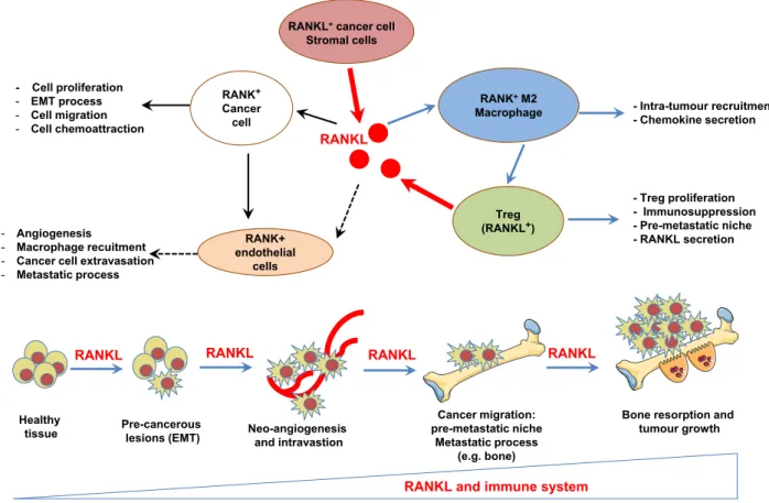
RANK+ Cancer cell RANK+M2 Macrophage Treg (RANKL+) RANK+ endothelial cells RANKL - Cell proliferation - EMT process - Cell migration - Cell chemoattraction - Intra-tumour recruitment - Chemokine secretion - Treg proliferation - Immunosuppression - Pre-metastatic niche - RANKL secretion - Angiogenesis - Macrophage recuitment - Cancer cell extravasation - Metastatic process
RANKL+cancer cell
Stromal cells
Pre-cancerous
lesions (EMT) Neo-angiogenesisand intravastion
Cancer migration: pre-metastatic niche
Metastatic process (e.g. bone)
Bone resorption and tumour growth
RANKL RANKL RANKL RANKL
RANKL and immune system
Healthy tissue
Figure 2 RANK/RANKL is involved in each stage of cancer development: from pre-cancerous lesions to the establishment of metastases
Cancer cells are direct targets for RANKL. RANKL initiates the formation of pre-cancerous lesions by facilitating the EMT process and stemness, as well as facilitating tumour growth and the metastatic process by modulating immune and vascular niches. Throughout these processes, RANKL acts as a chemoattractive factor for cancer cells and M2 macrophages. Activated macrophages facilitate both the proliferation of Treg lymphocytes, the main source of RANKL during primary tumour growth, and the initiation of the pre-metastatic niche in bone. RANKL up-regulates the angiogenic process by stimulating the proliferation and survival of endothelial cells and, in parallel, of the metastatic process by promoting the extravasation/intravasation of RANK-expressing cancer cells and their migration to distant organs. The RANKL concentration gradient drives the tumour cells to the metastatic sites.
and RANKL acts as a chemoattractive agent on cells that express
one of their receptors.
In addition to its direct effects on cancer cells, RANKL is
notably able to modulate the tumour microenvironment, in
par-ticular the formation of new blood vessels. Blood vessels are
used by cancer cells to deliver large quantities of nutriments and
are their main means of migrating so as to invade distant
or-gans. RANK expression was detected in endothelial cells, and by
interacting with this receptor, RANKL impacts the angiogenic
process by both stimulating angiogenesis through an Src and
phospholipase C-dependent mechanism [
135
,
136
], and
increas-ing cell survival in a PI3k/Akt-dependent manner [
137
]. RANKL
also induced the proliferation of endothelial cell precursors and
the neoformation of vascular tubes [
138
]. This phenomenon is
exacerbated by VEGF, which is frequently secreted by cancer
cells and which up-regulates the RANKL response of endothelial
cells by an up-regulation of RANK expression and an increase in
vascular permeability [
139
]. These works strengthen the role of
RANK/RANKL axis plays in the metastatic process by regulating
cancer cell migration and the neoangiogenesis.
Immune cell regulation by RANK/RANKL: setting
up fertile soil for cancer cells
RANKL influences the microenvironment of cancer cells by
act-ing on local immunity. The major role of RANKL in the
im-mune system was initially identified in RANKL-knockout mice
in which the development of secondary lymphoid organs was
impaired, especially the lymph nodes [
140
,
141
], but also at the
“central” level, where the maturation of the thymic epithelial
cells necessary for T-cell development was affected [
142
,
143
].
RANKL is also involved in modulating the immune response
by inducing T-cell proliferation [
25
] and dendritic cell survival
[
26
]. T-cells activated as a result of RANKL expression stimulate
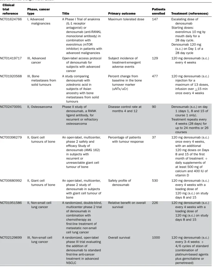
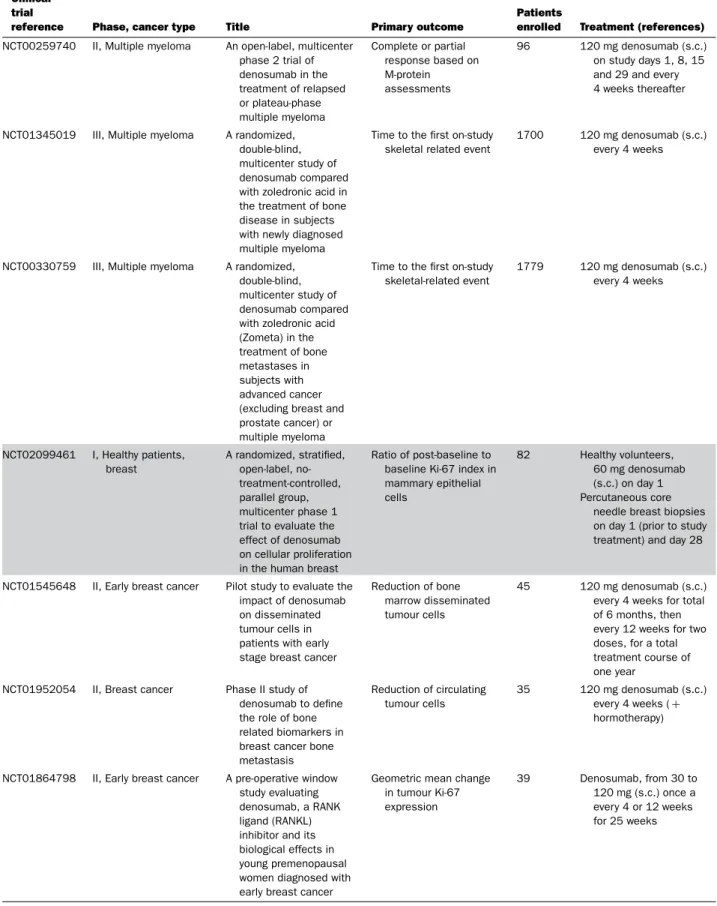
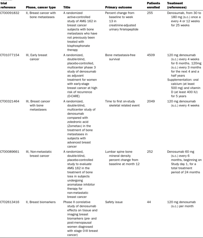
Table 2 Main clinical trials based on RANKL targeting in cancers
Source: clinical trial.gov March 2016.
Clinical trial reference
Phase, cancer
type Title Primary outcome
Patients
enrolled Treatment (references) NCT01624766 I, Advanced
malignancies
A Phase I Trial of anakinra (IL-1 receptor antagonist) or denosumab (anti-RANKL monoclonal antibody) in combination with everolimus (mTOR inhibitor) in patients with advanced malignancies
Maximum tolerated dose 147 Escalating dose of denosumab Starting doses:
everolimus 10 mg by mouth daily for a 28 day cycle. Denosumab 120 mg (s.c.) on Day 1 of a 28 day cycle NCT01419717 III, Advanced cancer
Open-label access protocol of denosumab for subjects with advanced cancer Subject incidence of treatment-emergent adverse events 129 120 mg denosumab (s.c.) every 4 weeks NCT01920568 III, Bone metastases from solid tumours A study comparing denosumab with zoledronic acid in subjects of Asian ancestry with bone metastases from solid tumours
Percent change from baseline in the bone turnover marker (uNTx/uCr)
477 120 mg denosumab (s.c.) injection for a maximum of 13 doses, infusion over>15 min once every 4 weeks
NCT02470091 II, Osteosarcoma Phase II study of denosumab, a RANK ligand antibody, for recurrent or refractory osteosarcoma
Disease control rate at months 4 and 12
90 Denosumab (s.c.) on day 1 (days 1, 8 and 15 of course 1 only). Treatment repeats every
4 weeks (28 days) for up to 24 months or 26 courses
NCT00396279 II, Giant cell tumours of bone
An open-label, multicenter, phase 2 safety and efficacy Study of denosumab (AMG 162) in subjects with recurrent or
unresectable giant cell tumour of bone
Percentage of patients with tumour response
37 120 mg denosumab (s.c.) once every 4 weeks, with an additional 120 mg doses on Days 8 and 15 of the first month of treatment+ daily supplements of at least 500 mg of calcium and 400 IU of vitamin D
NCT00680992 II, Giant cell tumours of bone
An open-label, multicenter, phase 2 study of denosumab in subjects with giant cell tumour of bone
Safety profile of denosumab
530 120 mg denosumab (s.c.) every 4 weeks with a loading dose of 120 mg (s.c.) on study days 8 and 15 NCT01951586 II, Non-small cell
lung cancer
A randomized, double-blind, multicenter phase 2 trial of denosumab in combination with chemotherapy as first-line treatment of metastatic non-small cell lung cancer
Relative benefit on overall survival
226 120 mg denosumab (s.c.) every 4 weeks with a loading dose of 120 mg (s.c.) on study days 8 and 15
NCT02129699 III, Non-small cell lung cancer
A randomized, open-label phase III trial evaluating the addition of denosumab to standard first-line anti-cancer treatment in advanced NSCLC
Overall survival 1000 120 mg denosumab (s.c.) every 3–4 weeks+ 4/6 cycles of standard (combination of platinum-based agents plus gemcitabine or pemetrexed)
Table 2 Continued
Clinical trial
reference Phase, cancer type Title Primary outcome
Patients
enrolled Treatment (references) NCT00259740 II, Multiple myeloma An open-label, multicenter
phase 2 trial of denosumab in the treatment of relapsed or plateau-phase multiple myeloma Complete or partial response based on M-protein assessments 96 120 mg denosumab (s.c.) on study days 1, 8, 15 and 29 and every 4 weeks thereafter
NCT01345019 III, Multiple myeloma A randomized, double-blind, multicenter study of denosumab compared with zoledronic acid in the treatment of bone disease in subjects with newly diagnosed multiple myeloma
Time to the first on-study skeletal related event
1700 120 mg denosumab (s.c.) every 4 weeks
NCT00330759 III, Multiple myeloma A randomized, double-blind, multicenter study of denosumab compared with zoledronic acid (Zometa) in the treatment of bone metastases in subjects with advanced cancer (excluding breast and prostate cancer) or multiple myeloma
Time to the first on-study skeletal-related event 1779 120 mg denosumab (s.c.) every 4 weeks NCT02099461 I, Healthy patients, breast A randomized, stratified, open-label, no-treatment-controlled, parallel group, multicenter phase 1 trial to evaluate the effect of denosumab on cellular proliferation in the human breast
Ratio of post-baseline to baseline Ki-67 index in mammary epithelial cells 82 Healthy volunteers, 60 mg denosumab (s.c.) on day 1 Percutaneous core
needle breast biopsies on day 1 (prior to study treatment) and day 28
NCT01545648 II, Early breast cancer Pilot study to evaluate the impact of denosumab on disseminated tumour cells in patients with early stage breast cancer
Reduction of bone marrow disseminated tumour cells
45 120 mg denosumab (s.c.) every 4 weeks for total of 6 months, then every 12 weeks for two doses, for a total treatment course of one year
NCT01952054 II, Breast cancer Phase II study of denosumab to define the role of bone related biomarkers in breast cancer bone metastasis Reduction of circulating tumour cells 35 120 mg denosumab (s.c.) every 4 weeks (+ hormotherapy)
NCT01864798 II, Early breast cancer A pre-operative window study evaluating denosumab, a RANK ligand (RANKL) inhibitor and its biological effects in young premenopausal women diagnosed with early breast cancer
Geometric mean change in tumour Ki-67 expression 39 Denosumab, from 30 to 120 mg (s.c.) once a every 4 or 12 weeks for 25 weeks
Table 2 Continued
Clinical trial
reference Phase, cancer type Title Primary outcome
Patients enrolled
Treatment (references) NCT00091832 II, Breast cancer with
bone metastases
A randomized active-controlled study of AMG 162 in breast cancer subjects with bone metastasis who have not previously been treated with bisphosphonate therapy
Percent change from baseline to week 13 in creatinine-adjusted urinary N-telopeptide 255 Denosumab, from 30 to 180 mg (s.c.) once a every 4 or 12 weeks for 25 weeks
NCT01077154 III, Early breast cancer A randomized, double-blind, placebo-controlled, multicenter phase 3 study of denosumab as adjuvant treatment for women with early-stage breast cancer at high risk of recurrence (D-CARE) Bone metastasis-free survival 4509 120 mg denosumab (s.c.) every 4 weeks for 6 months. 120mg (s.c.) every 3 months for the next 4 and a half years Supplementation: oral
calcium (at least 500 mg) and vitamin D (at least 400 IU) for 5 years NCT00321464 III, Breast cancer
with bone metastases A randomized, double-blind, multicenter study of denosumab compared with zoledronic acid (Zometa®) in the treatment of bone metastases in subjects with advanced breast cancer
Time to first on-study skeletal related event
2049 120 mg denosumab (s.c.) every 4 weeks NCT00089661 III, Non-metastatic breast cancer A randomized, double-blind, placebo-controlled study to evaluate AMG 162 in the treatment of bone loss in subjects undergoing aromatase inhibitor therapy for non-metastatic breast cancer
Lumbar spine bone mineral density percent change from baseline at month 12
252 Denosumab 60 mg (s.c.) every 6 months, beginning on Study day 1, for a total treatment period of 24 months
NCT02613416 II, Breast biomarkers Phase II correlative study of denosumab effects on tissue and imaging breast biomarkers (pre- and post-menopausal women diagnosed with stage 0-III breast cancer)
Safety issue 44 120 mg denosumab (s.c.) per month
Table 2 Continued
Clinical trial
reference Phase, cancer type Title Primary outcome
Patients
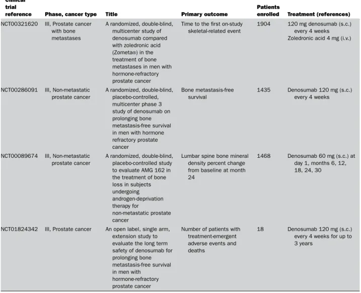
enrolled Treatment (references) NCT00321620 III, Prostate cancer
with bone metastases
A randomized, double-blind, multicenter study of denosumab compared with zoledronic acid (Zometa®) in the treatment of bone metastases in men with hormone-refractory prostate cancer
Time to the first on-study skeletal-related event
1904 120 mg denosumab (s.c.) every 4 weeks Zoledronic acid 4 mg (i.v.)
NCT00286091 III, Non-metastatic prostate cancer A randomized, double-blind, placebo-controlled, multicenter phase 3 study of denosumab on prolonging bone metastasis-free survival in men with hormone refractory prostate cancer Bone metastasis-free survival 1435 Denosumab 120 mg (s.c.) every 4 weeks NCT00089674 III, Non-metastatic prostate cancer A randomized, double-blind, placebo-controlled study to evaluate AMG 162 in the treatment of bone loss in subjects undergoing androgen-deprivation therapy for non-metastatic prostate cancer
Lumbar spine bone mineral density percent change from baseline at month 24
1468 Denosumab 60 mg (s.c.) at day 1, months 6, 12, 18, 24, 30
NCT01824342 III, Prostate cancer An open label, single arm, extension study to evaluate the long term safety of denosumab for prolonging bone metastasis-free survival in men with
hormone-refractory prostate cancer
Number of patients with treatment-emergent adverse events and deaths
18 Denosumab 120 mg (s.c.) every 4 weeks for up to 3 years
dendritic cells, expressing RANK, to enhance their survival and
thereby increase the T-cell memory response [
25
]. More recently,
Khan et al. [
144
] demonstrated that RANKL blockade can rescue
melanoma-specific T-cells from thymic deletion, and increases
the anti-tumour immune response as shown in melanoma.
Tumour-associated macrophages (TAMs) accumulate in the
tumour microenvironment and, depending on their M2 or M1
phenotype, play a part in tumour growth, angiogenesis and
meta-stasis [
145
]. RANK is present at the cell membrane of
mono-cytes/macrophages and RANKL acts as a chemoattractant factor
for these cells [
146
]. The M2-macrophages which mainly
ex-press RANK is strongly associated with the angiogenic process
[
147
]. RANK/RANKL signalling in the M2-macrophages
mod-ulates the production of chemokines, promoting the proliferation
of Treg lymphocytes in favour of an immunosuppressive
envir-onment [
148
]. In breast carcinoma, RANKL is mainly produced
by Treg lymphocytes (CD4
+CD25
+T-lymphocytes expressing
Foxp3). In this context, a vicious cycle is established between
TAMs, Treg and tumour cells resulting in tumour growth, the
spread of cancer cells and amplification of the metastatic process
[
149
]. In fact, T-lymphocytes appear to be the principal source
of RANKL in tumorigenesis. Whether RANKL-producing
T-lymphocytes are involved in the initial step of metastatic process
or not, T-lymphocytes induce a permissive environment initiating
the pre-metastastic niche [
150
].
RANK/RANKL and bone niche: ongoing clinical
trials
When proliferative tumour cells are located in the bone
environ-ment (primary bone tumours or bone metastases), they
dysregu-late the balance between bone apposition and bone resorption in
order to create a favourable microenvironment for their growth
[
151
]. In this way, this bone microenvironment becomes a source
of therapeutic targets, RANKL being one of them [
152
]. OPG-Fc
was the first generation of drug targeting RANKL to be assessed
in potsmenopausal women [
152
]. Nevertheless, due to its ability
to bind to multiple ligands, and particularly to TRAIL, OPG-Fc
based clinical trials have been suspended until the development
of a monoclonal antibody targeting RANKL [
153
]. Denosumab,
a fully-humanized antibody targeting RANKL and blocking its
binding to RANK, has been developed to bypass this risk [
51
]. In
osteoporotic patients, Denosumab was well-tolerated and a single
s.c. dose resulted in a prolonged decrease in bone turnover [
154
].
The value of blocking RANKL activities has been also
demon-strated by the inhibition bone resorption in numerous pre-clinical
models of primary bone tumours (Ewing sarcoma [
155
],
osteosar-coma [
156
,
157
]), bone metastases (breast [
158
], prostate [
159
],
non-small cell lung cancer [
160
]) and in myeloma [
161
]) and in
numerous phase II and III clinical trials (
Table 2
). In breast and
prostate carcinoma patients, bone turnover markers were reduced
in a way similar to that in the osteoporosis context and, in
addi-tion, delayed the onset of the first skeletal-related event and the
risk of multiple SRE [
162
]. A comparison with bisphosphonate
therapy demonstrated the superiority of Denosumab concerning
the two previous parameters even if the overall survival rate was
similar with both drugs. Additional clinical trials in metastatic
diseases are currently in progress and their results will be very
informative with regard to the clinical extension of Denosumab
in oncology.
CONCLUSIONS
Since their initial discovery in 1997, RANK/RANKL became
key actors in first bone remodelling and then more recently in
oncology. This molecular axis is clearly involved in all stages
of tumorigenesis, including tumour hyperplasia, pre-neoplasia
foci formation, cancer cell migration, neo-angiogenesis, immune
cell chemoattraction and the establishment of an
immunosup-pressive environment and initiation of a pre-metastatic niche.
In one decade, RANK/RANKL has not only transformed our
vision of bone biology but has also strengthened the notion of
“seed and soil”, conventionally used to explain the metastatic
process. Targeting RANK/RANKL signalling has already shown
its therapeutic efficacy in osteoporotic patients and its clinical
advantages in the management of bone metastases from breast
and prostate carcinomas. Current ongoing clinical trials will be
crucial for better defining its potential side effects after long term
use.
ACKNOWLEDGEMENTS
Nathalie Renema is currently employed by the Laboratoire Affilo-gic (Nantes, France) and is preparing her PhD at the University of Nantes (INSERM UMR957). Benjamin Navet received a PhD fellow-ship from the French Ministry of Research (2013–2016).
FUNDING
This work was supported by the French Cancer League (Equipe Labellis´ee Ligue 2012).
REFERENCES
1 Bissell, M.J. and Hines, W.C. (2011) Why don’t we get more cancer? A proposed role of the microenvironment in restraining cancer progression. Nat. Med. 17, 320–329CrossRef PubMed
2 Molon, B., Cali, B. and Viola, A. (2016) T cells and cancer: how metabolism shapes immunity. Front. Immunol. 7, 20
CrossRef PubMed
3 Paget, S. (1889) The distribution of secondary growths in cancer of the breast. Lancet 133, 571–573CrossRef
4 Plaks, V., Kong, N. and Werb, Z. (2015) The cancer stem cell niche: how essential is the niche in regulating stemness of tumor cells? Cell Stem Cell 16, 225–238CrossRef PubMed
5 Ord´o˜nez-Mor´an, P. and Huelsken, J. (2014) Complex metastatic niches: already a target for therapy? Curr. Opin. Cell Biol. 31, 29–38CrossRef PubMed
6 Molofsky, A.V., Pardal, R. and Morrison, S.J. (2004) Diverse mechanisms regulate stem cell self-renewal. Curr. Opin. Cell Biol. 16, 700–707CrossRef PubMed
7 Wan, L., Pantel, K. and Kang, Y. (2013) Tumor metastasis: moving new biological insights into the clinic. Nat. Med. 19, 1450–1464CrossRef PubMed
8 Shiozawa, Y., Berry, J.E., Eber, M.R., Jung, Y., Yumoto, K., Cackowski, F.C., Yoon, H.J., Parsana, P., Mehra, R., Wang, J. et al. (2016), The marrow niche controls the cancer stem cell phenotype of disseminated prostate cancer. Oncotarget, doi: 10.18632/oncotarget.9251
9 Weidle, U.H., Birzele, F., Kollmorgen, G. and R¨uger, R. (2016) Molecular mechanisms of bone metastasis. Cancer Genomics Proteomics 13, 1–12 PubMed
10 Lu, X., Mu, E., Wei, Y., Riethdorf, S., Yang, Q., Yuan, M., Yan, J., Hua, Y., Tiede, B.J., Lu, X. et al. (2011) VCAM-1 promotes osteolytic expansion of indolent bone micrometastasis of breast cancer by engagingα4β1-positive osteoclast progenitors. Cancer Cell 20, 701–714CrossRef PubMed
11 Wang, N., Docherty, F.E., Brown, H.K., Reeves, K.J., Fowles, A.C., Ottewell, P.D., Dear, T.N., Holen, I., Croucher, P.I. and Eaton, C.L. (2014) Prostate cancer cells preferentially home to
osteoblast-rich areas in the early stages of bone metastasis: evidence from in vivo models. J. Bone Miner. Res. 29, 2688–2696CrossRef PubMed
12 Spill, F., Reynolds, D.S., Kamm, R.D. and Zaman, M.H. (2016) Impact of the physical microenvironment on tumor progression and metastasis. Curr. Opin. Biotechnol. 40, 41–48
CrossRef PubMed
13 Meads, M.B., Hazlehurst, L.A. and Dalton, W.S. (2008) The bone marrow microenvironment as a tumor sanctuary and contributor to drug resistance. Clin. Cancer Res. 14, 2519–2526
CrossRef PubMed
14 David, E., Blanchard, F., Heymann, M.F., De Pinieux, G., Gouin, F., R´edini, F. and Heymann, D. (2011) The bone niche of
chondrosarcoma: a sanctuary for drug resistance, tumour growth and also a source of new therapeutic targets sarcoma. Sarcoma 2011, 932451CrossRef PubMed
15 Jones, V.S., Huang, R.Y., Chen, L.P., Chen, Z.S., Fu, L. and Huang, R.P. (2016) Cytokines in cancer drug resistance: cues to new therapeutic strategies. Biochim. Biophys. Acta 1865, 255–265
PubMed
16 Landskron, G., De la Fuente, M., Thuwajit, P. and Hermoso, M.A. (2014) Chronic inflammation and cytokines in the tumor microenvironment. J. Immunol. Res. 2014, 149185
CrossRef PubMed
17 Dinarello, C.A. (2006) The paradox of pro-inflammatory cytokines in cancer. Cancer Metastasis Rev. 25, 307–313
CrossRef PubMed
18 Dranoff, G. (2004) Cytokines in cancer pathogenesis and cancer therapy. Nat. Rev. Cancer 4, 11–22CrossRef PubMed
19 Grivennikov, S.I. and Karin, M. (2011) Inflammatory cytokines in cancer: tumour necrosis factor and interleukin 6 take the stage. Ann. Rheum. Dis. 70, 104–108CrossRef PubMed
20 Walsh, M.C. and Choi, Y. (2014) Biology of the
RANKL–RANK–OPG system in immunity, bone, and beyond. Front. Immunol. 5, 511CrossRef PubMed
21 Theoleyre, S., Wittrant, Y., KwanTat, S., Fortun, Y., Redini, F. and Heymann, D. (2004) The molecular triad OPG/RANK/RANKL: involvement in the orchestration of pathophysiological bone remodeling. Cytokine Growth Factor Rev. 15, 457–475
CrossRef PubMed
22 Wittrant, Y., Th´eoleyre, S., Chipoy, C., Padrines, M., Blanchard, F., Heymann, D. and R´edini, F. (2004) RANKL/RANK/OPG: new therapeutic targets in bone tumours and associated osteolysis. Biochim. Biophys. Acta 1704, 49–57 PubMed
23 Yasuda, H. (2013) RANKL, a necessary chance for clinical application to osteoporosis and cancer-related bone diseases. World J. Orthop. 4, 207–217CrossRef PubMed
24 Li, J., Yin, Q. and Wu, H. (2013) Structural basis of signal transduction in the TNF receptor superfamily. Adv. Immunol. 119, 135–153CrossRef PubMed
25 Anderson, D.M., Maraskovsky, E., Billingsley, W.L., Dougall, W.C., Tometsko, M.E., Roux, E.R., Teepe, M.C., DuBose, R.F., Cosman, D. and Galibert, L. (1997) A homologue of the TNF receptor and its ligand enhance T cell growth and dendritic cell function. Nature 390, 175–179CrossRef PubMed
26 Wong, B.R., Josien, R., Lee, S.Y., Sauter, B., Li, H.L., Steinman, R.M. and Choi, Y. (1997) TRANCE (tumor necrosis factor [TNF]-related activation-induced cytokine), a New TNF family member predominantly expressed in T cells, is a dendritic cell specific survival factor. J. Exp. Med. 186, 2075–2080
CrossRef PubMed
27 Lacey, D.L, Timms, E., Tan, H.L., Kelley, M.J., Dunstan, C.R., Burgess, T., Elliot, R., Colombero, A., Elliott, G., Scully, S. et al. (1998) Osteoprotegerin ligand is a cytokine that regulates osteoclast differentiation and activation. Cell 93, 165–176
CrossRef PubMed
28 Kong, Y.Y., Boyle, W.J. and Penninger, J.M. (1999) Osteoprotegerin ligand: a common link between
osteoclastogenesis, lymph node formation and lymphocyte development. Immunol. Cell Biol. 77, 188–193CrossRef PubMed
29 Kodaira, K., Kodaira, K., Mizuno, A., Yasuda, H., Shima, N., Murakami, A., Ueda, M. and Higashio, K. (1999) Cloning and characterization of the gene encoding mouse osteoclast differentiation factor. Gene 230, 121–127 PubMed
30 Yasuda, H., Shima, N., Nakagawa, N., Mochizuki, S.I., Yano, K., Fujise, N., Sato, Y., Goto, M., Yamaguchi, K., Kuriyama, M. et al. (1998) Osteoclast differentiation factor is a ligand for
osteoprotegerin/osteoclastogenesis-inhibitory factor and is identical to TRANCE/RANKL. Proc. Natl. Acad. Sci. U.S.A. 95, 3597–3602CrossRef PubMed
31 Ikeda, T., Kasai, M., Utsuyama, M. and Hirokawa, K. (2001) Determination of three isoforms of the Receptor activator of nuclear factor-kappa B ligand and their differential expression in bone and thymus. Endocrinology 142, 1419–1426 PubMed
32 Lum, L., Wong, B.R., Josien, R., Becherer, J.D.,
Erdjument-Bromage, H., Schlondorff, J., Temps, P., Choi, Y. and Blodel, C.P. (1999) Evidence for a role of a tumor necrosis factor alpha (TNF-alpha)-converting enzyme-like protease in shedding of TRANCE, a TNF family member involved in osteoclastogenesis and dendritic cell survival. J. Biol. Chem. 274, 13613–13618
CrossRef PubMed
33 Hikita, A., Yana, I., Wakeyama, H., Nakamura, M., Kadono, Y., Oshima, Y., Nakamura, K., Seiki, M. and Tanaka, S. (2006) Negative regulation of osteoclastogenesis by ectodomain shedding of receptor activator of NF-kappaB ligand. J. Biol. Chem. 281, 36846–36855CrossRef PubMed
34 Georges, S., Ruiz Velasco, C., Trichet, V., Fortun, Y., Heymann, D. and Padrines, M. (2009) Proteases and bone remodelling. Cytokine Growth Factor Rev. 20, 29–41
CrossRef PubMed
35 Kartsogiannis, V., Zhou, H., Horwood, N.J., Thomas, R.J., Hards, D.K., Quinn, J.M., Niforas, P., Ng, K.W., Martin, T.J. and Gillespie, M.T. (1999) Localization of RANKL (receptor activator of NF kappa B ligand) mRNA and protein in skeletal and extraskeletal tissues. Bone 25, 525–534CrossRef PubMed
36 Collin-Osdoby, P., Rothe, L., Anderson, F., Nelson, M., Maloney, W. and Osdoby, P. (2001) Receptor activator of NF-kappa B and osteoprotegerin expression by human microvascular endothelial cells, regulation by inflammatory cytokines, and role in human osteoclastogenesis. J. Biol. Chem. 276, 20659–20672
CrossRef PubMed
37 Matsuzaki, K., Udagawa, N., Takahashi, N., Yamaguchi, K., Yasuda, H., Shima, N., Morinaga, T., Toyama, Y., Yabe, Y., Higashio, K. and Suda, T. (1998) Osteoclast differentiation factor (ODF) induces osteoclast-like cell formation in human peripheral blood mononuclear cell cultures. Biochem. Biophys. Res. Commun. 246, 199–204
CrossRef PubMed
38 Quinn, J.M., Elliott, J., Gillespie, M.T. and Martin, T.J. (1998) A combination of osteoclast differentiation factor and
macrophage-colony stimulating factor is sufficient for both human and mouse osteoclast formation in vitro. Endocrinology 139, 4424–4427CrossRef PubMed
39 Xiong, J., Piemontese, M., Onal, M., Campbell, J., Goellner, J.J., Dusevich, V., Bonewald, L., Manolagas, S.C. and O’Brien, C.A. (2015) Osteocytes not osteoblasts or lining cells are the main source of the RANKL required for osteoclast formation in remodeling bone. PLoS One 10, e0138189
CrossRef PubMed
40 Hsu, H., Lacey, D.L., Dunstan, C.R., Solovyev, I., Colombero, A., Timms, E., Tan, H.L., Elliott, G., Kelley, M.J., Sarosi, I. et al. (1999) Tumor necrosis factor receptor family member RANK mediates osteoclast differentiation and activation induced by osteoprotegerin ligand. Proc. Natl. Acad. Sci. U.S.A. 96, 3540–3545CrossRef PubMed
41 Kong, Y.Y., Feige, U., Sarosi, I., Bolon, B., Tafuri, A., Morony, S., Capprelli, C., Li, J., Elliott, R., McCabe, S. et al. (1999) Activated T cells regulate bone loss and joint destruction in adjuvant arthritis through osteoprotegerin ligand. Nature 402, 304–309
CrossRef PubMed
42 Takayanagi, H. (2007) Osteoimmunology: shared mechanisms and crosstalk between the immune and bone systems. Nat. Rev. Immunol. 7, 292–304CrossRef PubMed
43 Wong, B.R., Josien, R., Lee, S.Y., Vologodskaia, M., Steinman, R.M. and Choi, Y. (1998) The TRAF family of signal transducers mediates NF-kappaB Activation by the TRANCE receptor. J. Biol. Chem. 273, 28355–28359CrossRef PubMed
44 Kanazawa, K. and Kudo, A. (2005) Self-assembled RANK induces osteoclastogenesis ligand-independently. J. Bone Miner. Res. 20, 2053–2060CrossRef PubMed
45 T´el´etch´ea, S., Stresing, V., Hervouet, S., Baud’huin, M., Heymann, M.F., Bertho, G., Charrier, C., Ando, K. and Heymann, D. (2014) Novel RANK antagonists for the treatment of bone resorptive disease: theoretical predictions and experimental validation. J. Bone Miner. Res. 29, 1466–1477
CrossRef PubMed
46 Arai, F., Miyamoto, T., Ohneda, O., Inada, T., Sudo, T., Brasel, K., Miyata, T., Anderson, D.M. and Suda, T. (1999) Commitment and differentiation of osteoclast precursor cells by the sequential expression of c-Fms and receptor activator of nuclear factor kappaB (RANK) receptors. J. Exp. Med. 190, 1741–1754
47 Nakagawa, N., Kinosaki, M., Yamaguchi, K., Shima, N., Yasuda, H., Yano, K., Morinaga, T. and Higashio, K. (1998) RANK is essential signalling receptor for osteoclast differentiation factor in osteoclastogenesis. Biochem. Biophys. Res. Commun. 253, 396–400CrossRef
48 Li, J., Sarosi, I., Yan, X.Q., Morony, S., Capparelli, C., Tan, H.L., McCabe, S., Elliott, R., Scully, S., Van, G. et al. (2000) RANK is the intrinsic hematopoietic cell surface receptor that controls osteoclastogenesis and regulation of bone mass and calcium metabolism. Proc. Natl. Acad. Sci. U.S.A. 97, 1566–1571
CrossRef PubMed
49 Dougall, W.C., Glaccum, M., Charrier, K., Rohrbach, K., Brasel, K., De Smedt, T., Daro, E., Smith, J., Tometsko, M.E.,
Maliszewski, C.R. et al. (1999) RANK is essential for osteoclast and lymph node development. Genes Dev. 13, 2412–2424
CrossRef PubMed
50 Baud’huin, M., Lamoureux, F., Duplomb, L., R´edini, F. and Heymann, D. (2007) RANKL, RANK, osteoprotegerin: key partners of osteoimmunology and vascular diseases. Cell. Mol. Life Sci. 64, 2334–2350CrossRef PubMed
51 Santini, D., Perrone, G., Roato, I., Godio, L., Pantano, F., Grasso, D., Russo, A., Vincenzi, B., Fratto, M.E., Sabbatini, R. et al. (2011) Expression pattern of receptor activator of NFkB (RANK) in a series of primary solid tumors and related metastases. J. Cell Physiol. 226, 780–784
CrossRef PubMed
52 Santini, D., Schiavon, G., Vincenzi, B., Gaeta, L., Pantano, F., Russo, A., Ortega, C., Porta, C., Galluzzo, S., Armento, G. et al. (2011) Receptor activator of NF-kB (RANK) expression in primary tumors associates with bone metastasis occurrence in breast cancer patients. PLoS One 6, e19234
CrossRef PubMed
53 Bhatia, P., Sanders, M.M. and Hansen, M.F. (2005) Expression of receptor activator of nuclear factor-kappaB is inversely correlated with metastatic phenotype in breast carcinoma. Clin. Cancer Res. 11, 162–165 PubMed
54 Park, H.S., Lee, A., Chae, B.J., Bae, J.S., Song, B.J. and Jung, S.S. (2014) Expression of receptor activator of nuclear factor kappa-B as a poor prognostic marker in breast cancer. J. Surg. Oncol. 110, 807–812
CrossRef PubMed
55 Pfitzner, B.M., Branstetter, D., Loibl, S., Denkert, C., Lederer, B., Schmitt, W.D., Dombrowski, F., Werner, M., R¨udiger, T., Dougall, W.C. and von Minckwitz, G. (2014) RANK expression as a prognostic and predictive marker in breast cancer. Breast Cancer Res. Treat. 145, 307–315
CrossRef PubMed
56 Jones, D.H., Nakashima, T., Sanchez, O.H., Kozieradzki, I., Komarova, S.V., Sarosi, I., Morony, S., Rubin, E., Sarao, R., Hojilla, C.V. et al. (2006) Regulation of cancer cell migration and bone metastasis by RANKL. Nature 440, 692–696
CrossRef PubMed
57 Owen, S., Ye, L., Sanders, A.J., Mason, M.D. and Jiang, W.G. (2013) Expression profile of receptor activator of nuclear-κB (RANK), RANK ligand (RANKL) and osteoprotegerin (OPG) in breast cancer. Anticancer Res. 33, 199–206
PubMed
58 Van Poznak, C., Cross, S.S., Saggese, M., Hudis, C., Panageas, K.S., Norton, L., Coleman, R.E. and Holen, I. (2006) Expression of osteoprotegerin (OPG), TNF related apoptosis inducing ligand (TRAIL), and receptor activator of nuclear factor kappaB ligand (RANKL) in human breast tumours. J. Clin. Pathol. 59, 56–63
CrossRef PubMed
59 Azim, Jr, H.A., Peccatori, F.A., Broh´ee, S., Branstetter, D., Loi, S., Viale, G., Piccart, M., Dougall, W.C., Pruneri, G. and Sotiriou, C. (2015) RANK-ligand (RANKL) expression in young breast cancer patients and during pregnancy. Breast Cancer Res. 17, 24
CrossRef PubMed
60 Hu, H., Wang, J., Gupta, A., Shidfar, A., Branstetter, D., Lee, O., Ivancic, D., Sullivan, M., Chatterton, Jr, R.T., Dougall, W.C. and Khan, S.A. (2014) RANKL expression in normal and malignant breast tissue responds to progesterone and is up-regulated during the luteal phase. Breast Cancer Res. Treat. 146, 515–523
CrossRef PubMed
61 Shang, W.Q., Li, H., Liu, L.B., Chang, K.K., Yu, J.J., Xie, F., Li, M.Q. and Yu, J.J. (2015) RANKL/RANK interaction promotes the growth of cervical cancer cells by strengthening the dialogue between cervical cancer cells and regulation of IL-8 secretion. Oncol. Rep. 34, 3007–3016 PubMed
62 Hsu, C.J., Lin, T.Y., Kuo, C.C., Tsai, C.H., Lin, M.Z., Hsu, H.C., Fong, Y.C. and Tang, C.H. (2010) Involvement of integrin up-regulation in RANKL/RANK pathway of chondrosarcomas migration. J. Cell Biochem. 111, 138–147CrossRef PubMed
63 Grimaud, E., Soubigou, L., Couillaud, S., Coipeau, P., Moreau, A., Passuti, N., Gouin, F., Redini, F. and Heymann, D. (2003) Receptor activator of nuclear factor kappaB ligand (RANKL)/osteoprotegerin (OPG) ratio is increased in severe osteolysis. Am. J. Pathol. 163, 2021–2031CrossRef PubMed
64 Yin, J., Wang, L., Tang, W., Wang, X., Lv, L., Shao, A., Shi, Y., Ding, G., Chen, S. and Gu, H. (2014) RANK rs1805034 T>C polymorphism is associated with susceptibility of esophageal cancer in a Chinese population. PLoS One 9, e101705
CrossRef PubMed
65 Atkins, G.J., Kostakis, P., Vincent, C., Farrugia, A.N., Houchins, J.P., Findlay, D.M., Evdokiou, A. and Zannettino, A.C. (2006) RANK expression as a cell surface marker of human osteoclast precursors in peripheral blood, bone marrow, and giant cell tumors of bone. J. Bone Miner. Res. 21, 1339–1349
CrossRef PubMed
66 Branstetter, D.G., Nelson, S.D., Manivel, J.C., Blay, J.Y., Chawla, S., Thomas, D.M., Jun, S. and Jacobs, I. (2012) Denosumab induces tumor reduction and bone formation in patients with giant-cell tumor of bone. Clin. Cancer Res. 18, 4415–4424
CrossRef PubMed
67 Song, F.N., Duan, M., Liu, L.Z., Wang, Z.C., Shi, J.Y., Yang, L.X., Zhou, J., Fan, J., Gao, Q. and Wang, X.Y. (2014) RANKL promotes migration and invasion of hepatocellular carcinoma cells via NF-κB-mediated epithelial–mesenchymal transition. PLoS One 9, e108507CrossRef PubMed
68 Sasaki, A., Ishikawa, K., Haraguchi, N., Inoue, H., Ishio, T., Shibata, K., Ohta, M., Kitano, S. and Mori, M. (2007) Receptor activator of nuclear factor-kappaB ligand (RANKL) expression in hepatocellular carcinoma with bone metastasis. Ann. Surg. Oncol. 14, 1191–1199CrossRef PubMed
69 Peng, X., Guo, W., Ren, T., Lou, Z., Lu, X., Zhang, S., Lu, Q. and Sun, Y. (2013) Differential expression of the RANKL/RANK/OPG system is associated with bone metastasis in human non-small cell lung cancer. PLoS One 8, e58361CrossRef PubMed
70 Fiumara, P., Snell, V., Li, Y., Mukhopadhyay, A., Younes, M., Gillenwater, A.M., Cabanillas, F., Aggarwal, B.B. and Younes, A. (2001) Functional expression of receptor activator of nuclear factor kappaB in Hodgkin disease cell lines. Blood 98, 2784–2790CrossRef PubMed
71 Nosaka, K., Miyamoto, T., Sakai, T., Mitsuya, H., Suda, T. and Matsuoka, M. (2002) Mechanism of hypercalcemia in adult T-cell leukemia: overexpression of receptor activator of nuclear factor kappaB ligand on adult T-cell leukemia cells. Blood 99, 634–640
CrossRef PubMed
72 Barcala, V., Ruybal, P., Garcia Rivello, H., Waldner, C., Ascione, A. and Mongini, C. (2003) RANKL expression in a case of follicular lymphoma. Eur. J. Haematol. 70, 417–419CrossRef PubMed
73 Jones, D.H., Nakashima, T., Sanchez, O.H., Kozieradzki, I., Komarova, S.V., Sarosi, I., Morony, S., Rubin, E., Sarao, R., Hojilla, C.V. et al. (2006) Regulation of cancer cell migration and bone metastasis by RANKL. Nature 440, 692–696