HAL Id: inserm-00166907
https://www.hal.inserm.fr/inserm-00166907
Submitted on 13 Aug 2007HAL is a multi-disciplinary open access
archive for the deposit and dissemination of sci-entific research documents, whether they are pub-lished or not. The documents may come from teaching and research institutions in France or abroad, or from public or private research centers.
L’archive ouverte pluridisciplinaire HAL, est destinée au dépôt et à la diffusion de documents scientifiques de niveau recherche, publiés ou non, émanant des établissements d’enseignement et de recherche français ou étrangers, des laboratoires publics ou privés.
patient with 7q11 duplication.
Christel Depienne, Delphine Héron, Catalina Betancur, Baya Benyahia,
Oriane Trouillard, Delphine Bouteiller, Alain Verloes, Eric Leguern, Marion
Leboyer, Alexis Brice
To cite this version:
Christel Depienne, Delphine Héron, Catalina Betancur, Baya Benyahia, Oriane Trouillard, et al.. Autism, language delay and mental retardation in a patient with 7q11 duplication.. Journal of Med-ical Genetics, BMJ Publishing Group, 2007, 44 (7), pp.452-8. �10.1136/jmg.2006.047092�. �inserm-00166907�
Autism, language delay and mental retardation in a patient with
7q11 duplication
C Depienne, D Heron, C Betancur, B Benyahia, O Trouillard, D Bouteiller, A Verloes, E LeGuern, M Leboyer and A Brice
Authors’ affiliations
C Depienne, D Bouteiller, E LeGuern, A Brice, INSERM U679 (formerly U289), Groupe
Hospitalier Pitié-Salpêtrière, Paris, France
C Depienne, D Heron, B Benyahia, O Trouillard, E LeGuern, A Brice, Département de Génétique,
Cytogénétique et Embryologie, AP-HP, Groupe Hospitalier Pitié-Salpêtrière, Paris, France
C Depienne, E LeGuern, A Brice, Université Pierre et Marie Curie - Paris VI, Paris, France C Betancur, M Leboyer, INSERM U513, Université Paris XII, Créteil, France
A Verloes, Département de Génétique, Hôpital Robert Debré, AP-HP, Paris, France
Correspondence to: Prof. A Brice, INSERM U679, Groupe Hospitalier Pitié-Salpêtrière, 47 boulevard de l’hôpital, 75013 Paris, France; brice@ccr.jussieu.fr
Running title: Autism and 7q11 duplication
Keywords: Autism, mental retardation, language delay, 7q11, duplication Key points
• In total, 206 patients with autism were screened for rearrangements involving the Williams-Beuren Critical Region (WBCR) in 7q11.23.
• The case of a patient with autism, language delay and mental retardation who has a de novo duplication of the WBCR from paternal origin is reported.
• The clinical picture resulting from the WBCR duplication is discussed.
ABSTRACT
Background: Chromosomal rearrangements, arising from unequal recombination between repeated
sequences, are found in a subset of patients with autism. Duplications involving loci associated with behavioural disturbances especially constitute a good candidate mechanism. The Williams-Beuren Critical Region (WBCR), located in 7q11.23, is commonly deleted in the Williams-Beuren microdeletion syndrome (WBS). However, only four patients with a duplication of the WBCR have been reported so far, one with severe language delay and the three others with variable developmental, psychomotor and language delay.
Objective and Methods: In this study, we screened 206 patients with autism spectrum disorders for
the WBCR duplication by quantitative microsatellite analysis and multiple ligation-dependent probe amplification (MLPA).
Results: We have identified one male patient with a de novo interstitial duplication of the entire
WBCR of paternal origin. The patient had autistic disorder, severe language delay and mental retardation, with very mild dysmorphic features.
Conclusion: We report the first patient with autistic disorder who has a WBCR duplication. This
observation indicates that the 7q11.23 duplication could be involved in complex clinical phenotypes, ranging from developmental or language delay to mental retardation and autism, and extends the phenotype initially reported. These findings also support the existence of one or several genes in 7q11.23 sensitive to gene dosage and involved in the development of language and social interaction.
Chromosomal micro-rearrangements have been increasingly identified as a cause of human developmental disorders. Most often, they are the consequence of recombination between highly homologous (>95%) sequences called duplicons (spanning >10 kb) that flank a relatively small region (usually <5 Mb), leading to deletion or duplication of the region.1-3 Deletions and duplications alter the normal dosage of several adjacent genes and are responsible for contiguous gene syndromes or single gene disorders. One of these disorders, the Williams-Beuren syndrome (WBS), is caused by a microdeletion in the pericentromeric 7q11.23 region. This genomic disorder (1 in 7500 births)4 is characterized by typical dysmorphic features, mental retardation, developmental and language delay, congenital heart disease and hypersensitivity to sounds. Individuals with WBS also exhibit hypersociability and a characteristic neurocognitive profile, with poor visuospatial skills but relatively preserved ability to communicate.5 Although WBS has sometimes been described as the opposite of
autism, recent studies indicate that they share many features, including pragmatic language impairments, poor social relationships and unusual or restricted interests.6 Furthermore, the hypersensitivity to sound often described in patients with WBS7 is reminiscent of the heightened
response to auditory stimuli observed in autism.8 Even though autism had been described in
association with WBS only in a small number of patients,9-11 a recent study of 128 4-to-16 year old patients with WBS identified nine children with autism spectrum disorders,12 indicating that the
coexistence of the two disorders is not coincidental.
Autism is a complex developmental disorder, characterized by impairment in social interactions and communication and repetitive interests and behaviours. It generally occurs sporadically, but in ~5% of the cases, multiple affected subjects are found in families. Twin and family studies have shown that autism has a strong heritable component; the genetic mechanisms are likely to be complex
and heterogeneous, as suggested by the imbalanced sex ratio (4 males to 1 female) and its frequent association with known genetic diseases (e.g., fragile X, tuberous sclerosis, neurofibromatosis)13 or
chromosomal rearrangements (e.g., 15q11-q13 duplication, 22q13.3 deletion). 14,15
Although unequal crossing over between duplicons is expected to lead to reciprocal deletions and duplications of the Williams-Beuren Critical Region (WBCR) with a similar frequency, the first case of a duplication of the WBCR was described only in 2005, in a patient with severe language delay.16
More recently, three additional patients were reported: one had trigonocephalic synostosis, mild developmental delay and moderate language disability17; a second had severe learning and speech
disabilities, mild dysmophism, moderate mental retardation and poor social skills18 and a third had
severe speech impairment associated with epilepsy and cortical dysplasia of the left temporal lobe.19 No clear phenotype has been attributed to the WBCR duplication, making clinically-oriented screening by FISH impractical. In the current study, we screened 206 patients with autism spectrum disorders from the PARIS (Paris Autism Research International Sibpair) study cohort for deletion/ duplication of the 7q11.23 region by quantitative microsatellite analysis and multiple ligation-dependent probe amplification (MLPA). We report the identification of a new patient with a de novo duplication of the WBCR, who had autism, severe language impairment, and mental retardation.
MATERIALS AND METHODS Patients
A total of 206 patients with an autism spectrum disorder were studied. They were recruited at specialized clinical centres in France as part of the PARIS study. The patients included 160 males and 46 females; 156 were sporadic cases and 50 subjects belonged to 27 families with two or more affected siblings. Diagnoses were based on clinical evaluation by experienced psychiatrists, DSM-IV criteria20 and the Autism Diagnostic Interview-Revised (ADI-R).21 There were 199 patients with
autism, 4 with pervasive developmental disorder-not otherwise specified and 3 with Asperger syndrome. Among the patients studied, 182 had mental retardation and 114 had very limited or no language. Most were Caucasian (n=172), 13 Black, 3 South Asian and 18 of mixed ethnicity. Laboratory tests included karyotyping, fragile X testing and metabolic screening; brain imaging and EEG were performed when possible. Patients with known genetic disorders were excluded. The local research ethics board approved the study. Informed consent was obtained from all participating families.
Quantitative microsatellite analysis
Two microsatellite markers (D7S809 and D7S3315) were used to perform quantitative allele dosage at the 7q11 locus (Figure 1). D7S3315 was developed using the Tandem Repeat Finder algorithm (http://tandem.bu.edu/trf/trf.html) and registered in the GDB Human Genome Database. Nine other microsatellite markers located inside (AFMA217XH1, D7S2479, D7S2476, D7S613, and D7S1870) or outside (D7S645, D7S672, D7S2490, and D7S2518) the WBCR were used to analyze the haplotypes transmitted at the 7q11.23 locus. Primers and experimental conditions used are indicated in supplementary table S1, available online. Amplification was performed in 25 µl containing 0.5 U AmpliTaq DNA Polymerase (Applied Biosystems, Foster City, California, USA), 2.5 µl 10X buffer (containing 15 mM MgCl2), 0.1 µl dNTP (25 µmol/l), 0.4 µl of 6-FAM fluorochrome 5’-labeled
forward primer (20 µmol/l), 0.4 µl of reverse primer and 100 ng of genomic DNA. PCR reactions were carried out in a thermocycler (Primus 96 Plus; MWG-Biotech, Highpoint, North Carolina, USA). PCR products were separated and quantified on an automated sequencer (ABI 3730; Applied Biosystems).
Multiplex ligation-dependent probe amplification (MLPA)
A commercial kit (Williams-Beuren Syndrome MLPA kit, P029, designed to search for 7q11.23/ELN deletions or duplications), was used according to the manufacturer’s instructions (MRC-Holland, Amsterdam, The Netherlands). Two other MLPA kits (the mental retardation P019/P020 kit for subtelomeric regions and P064, MR1) were used to exclude the presence of rearrangements in other candidate regions. Electrophoresis of PCR products was performed using an automated sequencer (ABI 3730; Applied Biosystems). Samples were run in triplicate. MLPA data were analysed using the GeneMapper 4.0 software (Applied Biosystems). Relative ratios were calculated for each peak using the formula r = mean (peak area patient/mean control area patient)/(peak area control individual/mean control area control individual).
Real-time quantitative PCR
Real-time quantitative PCR has been previously used to assess the copy number in the 7q11.23 region.22,23 To determine the allele copy numbers at the boundaries of the WBCR, we designed four primer pairs using Primer Express 1.5 software (Applied Biosystems). Two primer pairs were located inside the WBCR (in the FKBP6 and GTF2I genes), the other two flanked the region (next to the AFMA217XH1 marker and in the HIP1 gene, fig 1). Primer sequences are indicated in E-Table S2. Real-time amplifications were performed using 50 ng of genomic DNA, 0.4 µM of each primer and 12.5 µl of Sybr Green PCR master mix (Applied Biosystems) in a total volume of 25 µl. RNAse P (RNAse P control assay, Applied Biosystems) was used as the reference amplimer. Samples were run in triplicate on an automated dDetection system (ABI Prism 7700; Applied Biosystems) using standard conditions: 2 min at 50°C; 10 min at 95°C; 40 cycles of 15 s at 95°C, and a final step of 1 min at 60°C. Relative ratios were calculated using the formula r = mean 2–∆∆Ct, where ∆∆Ct = (Ct Mutation - Ct RNAseP) ind tested – (Ct Mutation – Ct RNAseP) ind ref.
Fluorescence in situ hybridization
Metaphase chromosome spreads and interphase nuclei preparations were obtained from lymphoblastoid cell lines from the patient using standard high resolution-banding methods. A specific DNA probe (D7S2476-D7S1465) for the WBCR, covering the ELN, LIMK1 and CYLN2 genes, was obtained from Q-BIOgene (MP Biomedicals, Illkirch, France). The specific probe was labelled with rhodamine and the 7q22 control probe with fluorescein isothiocyanate (FITC). FISH was carried out according to the supplier's protocol. Briefly, the slides were washed in 2 x SSC/0.5% NP40 at 37°C for 30 min, dehydrated in 70%, 80% and 95% ethanol at room temperature for 2 min each, co-denatured with the probe at 75°C for 5 min and incubated overnight at 37°C in a humidified chamber for hybridization. The slides were then washed and counterstained with 4,6-diamino-2-phenylindole (DAPI) for chromosome identification. Metaphase and interphase cells were examined under a motorized reflected fluorescence microscope (BX61; Olympus, Center valley, Pennsylvania, USA) with filters for separate detection of DAPI, FITC and rhodamine, as well as a triple bandpass filter to
detect signals simultaneously. In total, 50 metaphase and 50 interphase cells were examined to detect cells with >2 specific signals.
RESULTS
Screening and characterization of 7q11 duplication
In total, 206 patients with autistic spectrum disorder were screened for 7q11 rearrangements using quantitative microsatellite analysis with the D7S809 and D7S3315 markers and MLPA (Fig 1A). In the presence of a duplication, quantitative microsatellite analysis can detect either more than two peaks (when the alleles are all different) or an imbalance between the peaks (when two alleles are identical and the third is different).24 One proband had three different alleles for each marker, which suggested
that the region could be duplicated (Fig 1B). The duplication was confirmed by the presence of three alleles for D7S613 and D7S2476 as well. Two additional markers located inside the minimal region near the boundaries (D7S2479 and D7S1870) were uninformative. Four markers located outside the WBCR, either on the proximal (D7S672 and D7S645) or distal (D7S2490 and D7S2518) part, were found to be normal (Fig 1C). Analysis of the parents showed that the duplication was de novo and of paternal origin, since the child had the two alleles from his father (Fig 1B and C). MLPA analysis of the 7q11 region confirmed that the FKBP6, FZD9, TBL2, STX1A, ELN, LIMK1, RFC2 and CYLN2 genes, all comprised within the WBCR, were duplicated (Fig 1D). To further delimit the duplicated region, we developed four real-time PCR assays (Q-PCR-1 to 4) to quantify allele copy number near the boundaries of the region. These assays confirmed the presence of three alleles (1.5 ratio compared to the control) for the FKBP6 and GTF2I genes and only two copies for the two assays with probes flanking the WBCR region, in the 7q11.22 distal region and in the HIP1 gene, respectively (Fig 1E). Finally, the interstitial 7q11 duplication in the proband was confirmed by FISH. Interphase nuclei showed three specific 7q11-probe signals (Fig 2A). FISH analysis of metaphase chromosomes revealed two hybridization spots, one brighter than the other (Fig 2B), confirming that the third copy was in 7q11. Taken together, these results demonstrate that the chromosomal rearrangement detected in the proband is a tandem 7q11.23 duplication that corresponds exactly to the WBCR.
The co-occurrence of clinical features and de novo 7q11.23 duplication strongly argues in favour of a causative role of the rearrangement detected in the patient’s phenotype. However, we additionally screened 300 control individuals for WBCR duplication by quantitative microsatellite analysis to ensure that this duplication was not present in a control population. No rearrangement was identified, indicating that the 7q11.23 duplication is either pathological or very rare in healthy individuals.
Finally, additional genetic analyses in the patient using MLPA and/or quantitative PCR excluded chromosomal abnormalities involving the 15q11.2, 22q11.2, 17p11.2, 22q13, 1p36, 17p13.3, 20p12.2, 5q35.3 as well as all telomeric regions. Karyotype, fragile X testing, plasma and urine amino acids were normal.
Clinical features of the patient with the 7q11 duplication
The proband was the second child of non-consanguineous parents. His father and sister were healthy; his mother possibly had obsessive-compulsive disorder. Pregnancy was unremarkable. He was born at term by caesarean section for umbilical cord strangulation, but the Apgar score was normal. His birth weight was 2870 g (-1.2 SD) and his length 50 cm (50th percentile). He was a hypotonic baby and
made no eye contact. His early development was delayed; he sat at 15 months and walked at 26 months. He also had markedly delayed language and pronounced his first words at 3 years. An auditory evaluation performed at 18 months was normal. He was diagnosed as having autistic disorder and mental retardation at 30 months according to DSM-III criteria. He attained bladder and bowel continence at night at 4 years and partially during the day at 11 years. At 9 years, he was able to bathe, dress and feed himself. Interestingly, he showed hypersensitivity to noise. He had stereotypic behaviour, with numerous routines and difficulty tolerating changes in daily habits. He was markedly hyperactive and exhibited inappropriate behaviour towards strangers, rushing up to people to hug them or hit them. He showed a particular interest in food that he hid in his room. He also had hyperphagia. Furthermore, he showed numerous stereotypies: head nodding, repetitive jaw movements, and mannerisms of the hands and arms. He was unable to express interest.
When last examined at the age of 12 years 5 months, he was still incontinent during the day. He had aggressive behaviour with severe outbursts of anger in response to frustration. He also had paroxystic episodes triggered by intense laughter and lasting ~3 min, when he stared into space and became pale, falling down without hurting himself, occurring about three times per month. His head circumference was 54.5 cm (50th percentile), his height was 145 cm and weight was 36 kg, both –0.5 SD. He had mildly dysmorphic features, with retrognathia and incomplete folding of the helix of both ears. The rest of the physical examination was normal, a was the neurological examination.
The patient fulfilled ADI-R criteria for autism21 and scored 46 on the Childhood Autism Rating Scale25, indicating severe autism. His developmental age, evaluated with the Psycho-Educational
Profile-Revised, was between 2 and 3.5 years. In the Vineland Adaptive Behavior Scales he achieved a level of 1.9 years for communication, 3.1 years for daily skills, and 2.7 years for socialization. Psychomotor evaluation showed that he walked awkwardly and had delayed motor acquisitions; he went down stairs one step at a time and was unable to hop on one foot. He was impulsive when performing tasks requiring fine motor coordination (such as cutting-out figures), but performed better when prompted. He could place four pieces in a puzzle, and was able to copy horizontal, vertical and diagonal lines as well as a circle, but failed to reproduce a cross and an X. Language evaluation by a speech pathologist showed that he was capable of non-verbal communication, but visual contact was rare and inappropriate. He understood simple commands and answered simple questions using images or gestures. Assessment with the Reynell Developmental Language Scales gave a score of 29 for verbal comprehension, equivalent to a developmental age of 2.6 years. He failed when spatial figures or colours were involved, or when two instructions were combined. Lexical comprehension was better, indicating a level equivalent to 4-5 years. He partly understood sorting instructions, obtaining an age equivalence of 4 years. Expression was reduced to single words, with numerous phonetic alterations that made his speech unintelligible out of context. Pronunciation of words was incomplete, and repetition did little to improve the initial utterance. He persevered and asked stereotyped questions. He used several idiosyncratic words, but in general language was used in an appropriate way, facilitating understanding by others. The results on the expressive language scale showed an age equivalence <18 months.
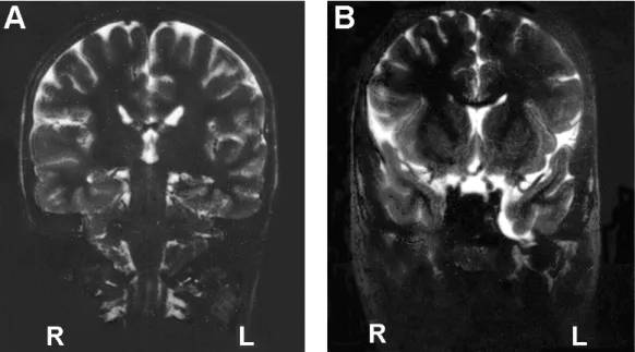
An EEG recording was inconclusive; there were unstructured rhythms but no synchronic activity. A brain MRI showed mild dilatation of the left temporal horn and a small arachnoid cyst in the temporal fossa (Fig 3). He was treated with fluoxetine and thioridazine.
DISCUSSION
We report a male patient with a de novo interstitial duplication of the WBCR of paternal origin. The patient had autistic disorder, severe speech impairment and mental retardation. In the first report of a WBCR duplication, the proband had developmental dysphasia, with a severe delay in the acquisition of expressive language, mild mental deficiency and attention deficit-hyperactivity disorder (ADHD).16
However, the developmental delay and ADHD were common to other family members who did not carry the duplication. The three additional patients with 7q11.23 duplication all had moderate to severe language disability, mild to moderate developmental delay, mild to severe mental retardation, and mild dysmorphic features (Table 1).17-19 These observations, combined with our case report, suggest that the duplication of the WBCR accounts for a highly variable and complex clinical pictures, encompassing speech and language delay, autism spectrum disorders and variable degrees of mental retardation. The only exception is the father of one patient, who also carried the duplication but was reportedly normal, except for syndactyly of the hands and feet, also present in other members of his family without the duplication. 16 Since the duplication was shown to be absent from the paternal grand-parents, the
duplication could be mosaic in the father, which would explain the absence of clinical signs. However, this hypothesis needs further investigations and we cannot completely exclude the possibility that some 7q11.23 duplications remain clinically unnoticeable.
So far, language impairment appears to be the most constant features in patients with WBCR duplication, together with a mild developmental delay and mild dysmorphic facies. Speech disability in patients with WBCR duplication contrasts with the relatively spared verbal abilities observed in WBS patients with the reciprocal deletion (Table 1). These findings support the existence of one or several genes in 7q11.23 that are sensitive to alterations in dosage and involved in the development of human language. Genotype-phenotype correlations performed in WBS patients with atypical deletions and studies of animals models have suggested that the genes located in the telomeric part of the 7q11.23 region (CYLN2 to GTF2I) are the main candidates sensitive to dosage (i.e. presence of a phenotype when hemizygous) and involved in visuo-spatial and behavioural defects as well as mental disabilities.26-34 However, over-expression of genes, such as LIMK1, which do not seem to play a main role in the phenotype resulting from the deletion could participate in the clinical features due to the duplication. Finally, genes located outside the WBCR but that lie near the breakpoints could also have their expression altered by the rearrangement and participate to the phenotype although they do not vary in copy number.35 The precise role of the genes comprised in the 7q11.23 region in language
development needs further investigation.
Interestingly, a second patient with 7q11.23 duplication also had impaired social skills, evocative of Asperger syndrome.18 This suggests that the autism in our patient is indeed part of the 7q11.23
duplication phenotype. In WBS, the WBCR deletion is associated both with features opposite to autism such as hypersociability but also with features found in autistic patients (poor social relationships, abnormal use of non-verbal behaviours including eye-to-eye gaze, facial expression and gestures to regulate social interaction).5 In addition, autism sometimes co-exists with WBS.9-12 These
observations strongly suggest that the WBCR region or nearby regions also contain one or several genes sensitive to dosage and associated with impairment in social interactions. However, we cannot completely exclude that the autistic features in our patient could be partly due to unknown additional genetic or non-genetic factor(s).
The clinical variability observed in the five patients with a WBCR duplication reported so far does not seem to result from a difference in the length of the duplicated region although the precise extent of the duplication has as only been reported for one case.16 However, the variability could result from
differences in the precise breakpoints and their effect on genes located nearby, especially the members of the GTF2 family of transcription factors (or TFII-I family, including GTF2I, GTF2IRD1 and GTF2IRD2), which have already been proposed as candidate genes for autism in a patient with an atypical 7q11 deletion.36 GTF2I hemizygozity has also been proposed to be a major cause of mental retardation found in WBD patients.26,28,33,37,38 Alternatively, this phenotypic variability, comparable to
that observed in many other disorders arising from chromosomal rearrangements, could result from the interaction with other genetic, environmental or stochastic factors.
Two distinct mechanisms could be involved in rearrangements mediated by duplicons: unequal interchromatid exchange during crossing-over and intrachromatid recombination. The first mechanism should theoretically lead to an incidence of duplications equivalent to that of deletions, whereas intrachromosomal events should mostly lead to deletions. The majority of the interstitial deletions of the 7q11.23 region have been shown to result from interchromosomal rearrangements during meiosis.1,3,39,40 There are several non-exclusive explanations for the apparent excess of cases with deletions: excessive early lethality of the duplication, underascertainement resulting from the absence of a “typical” phenotype that would prompt specific FISH diagnosis, or simply reduced penetrance/expressivity of the duplication, making most of the carriers asymptomatic. The observation that the WBCR duplication arose de novo in 3 out of 4 patients in whom the origin of the rearrangement was investigated and the absence of WBCR duplication in 300 control individuals, however, argues in favour of a deleterious role of the WBCR duplication, making therefore the last hypothesis unlikely. Our study, performed in a cohort of patients with autism spectrum disorders, indicates that the WBCR duplication is a rare cause of autism. The incidence of WBCR duplication in populations with expressive language delay, mental retardation, or autism spectrum disorders remains to be systematically evaluated.
ACKNOWLEDGMENTS
We are grateful to all the families that participated in this research. We thank Merle Ruberg for critical reading of the manuscript, the DNA and cell bank of the INSERM U679 (IFR des Neurosciences, Hôpital Pitié-Salpêtrière) and the Centre d'Investigations Cliniques, Hôpital Robert Debré, for processing the blood samples. This research was supported by Fondation de France, Fondation pour la Recherche Médicale, Fondation France Télécom, INSERM and Assistance Publique-Hôpitaux de Paris.
References
1. Thomas, N. S., Durkie, M., Potts, G., Sandford, R., Van Zyl, B., Youings, S., Dennis, N. R., and Jacobs, P. A. Parental and chromosomal origins of microdeletion and duplication syndromes involving 7q11.23, 15q11-q13 and 22q11. Eur J Hum Genet 2006;14(7):831-837.
2. Potocki, L., Chen, K. S., Park, S. S., Osterholm, D. E., Withers, M. A., Kimonis, V., Summers, A. M., Meschino, W. S., Anyane-Yeboa, K., Kashork, C. D., Shaffer, L. G., and Lupski, J. R. Molecular mechanism for duplication 17p11.2- the homologous
recombination reciprocal of the Smith-Magenis microdeletion. Nat Genet 2000;24(1):84-87.
3. Baumer, A., Dutly, F., Balmer, D., Riegel, M., Tukel, T., Krajewska-Walasek, M., and Schinzel, A. A. High level of unequal meiotic crossovers at the origin of the 22q11. 2 and 7q11.23 deletions. Hum Mol Genet 1998;7(5):887-894.
4. Stromme, P., Bjornstad, P. G., and Ramstad, K. Prevalence estimation of Williams syndrome. J Child Neurol 2002;17(4):269-271.
5. Meyer-Lindenberg, A., Mervis, C. B., and Faith, Berman K. Neural mechanisms in Williams syndrome: a unique window to genetic influences on cognition and behaviour. Nat Rev Neurosci 2006;7(5):380-393.
6. Laws, G. and Bishop, D. Pragmatic language impairment and social deficits in Williams syndrome: a comparison with Down's syndrome and specific language impairment. Int J Lang Commun Disord 2004;39(1):45-64.
7. Levitin, D. J., Cole, K., Lincoln, A., and Bellugi, U. Aversion, awareness, and attraction: investigating claims of hyperacusis in the Williams syndrome phenotype. J Child Psychol Psychiatry 2005;46(5):514-523.
8. Rosenhall, U., Nordin, V., Sandstrom, M., Ahlsen, G., and Gillberg, C. Autism and hearing loss. J Autism Dev Disord 1999;29(5):349-357.
9. Reiss, A. L., Feinstein, C., Rosenbaum, K. N., Borengasser-Caruso, M. A., and Goldsmith, B. M. Autism associated with Williams syndrome. J Pediatr 1985;106(2):247-249.
10. Gillberg, C. and Rasmussen, P. Brief report: four case histories and a literature review of Williams syndrome and autistic behavior. J Autism Dev Disord 1994;24(3):381-393. 11. Herguner, S. and Motavalli, Mukaddes N. Autism and Williams syndrome: A case
report. World J Biol Psychiatry 2006;7(3):186-188.
12. Leyfer, O. T., Woodruff-Borden, J., Klein-Tasman, B. P., Fricke, J. S., and Mervis, C. B. Prevalence of psychiatric disorders in 4 to 16-year-olds with Williams syndrome. Am J Med Genet B Neuropsychiatr Genet 2006;
13. Folstein, S. E. and Rosen-Sheidley, B. Genetics of autism: complex aetiology for a heterogeneous disorder. Nat Rev Genet 2001;2(12):943-955.
14. Jacquemont, M. L., Sanlaville, D., Redon, R., Raoul, O., Cormier-Daire, V., Lyonnet, S., Amiel, J., Le Merrer, M., Heron, D., de Blois, M. C., Prieur, M., Vekemans, M., Carter, N. P., Munnich, A., Colleaux, L., and Philippe, A. Array-based comparative genomic hybridisation identifies high frequency of cryptic chromosomal rearrangements in patients with syndromic autism spectrum disorders. J Med Genet 2006;43(11):843-849.
15. Vorstman, J. A., Staal, W. G., van Daalen, E., van Engeland, H., Hochstenbach, P. F., and Franke, L. Identification of novel autism candidate regions through analysis of reported cytogenetic abnormalities associated with autism. Mol Psychiatry 2006;11(1):1, 18-1, 28.
16. Somerville, M. J., Mervis, C. B., Young, E. J., Seo, E. J., del Campo, M., Bamforth, S., Peregrine, E., Loo, W., Lilley, M., Perez-Jurado, L. A., Morris, C. A., Scherer, S. W., and Osborne, L. R. Severe expressive-language delay related to duplication of the Williams-Beuren locus. N Engl J Med 2005;353(16):1694-1701.
17. Kriek, M., White, S. J., Szuhai, K., Knijnenburg, J., van Ommen, G. J., den Dunnen, J. T., and Breuning, M. H. Copy number variation in regions flanked (or unflanked) by duplicons among patients with developmental delay and/or congenital malformations;
detection of reciprocal and partial Williams-Beuren duplications. Eur J Hum Genet 2006;14(2):180-189.
18. Kirchhoff, M., Bisgaard, A. M., Bryndorf, T., and Gerdes, T. MLPA analysis for a panel of syndromes with mental retardation reveals imbalances in 5.8% of patients with mental retardation and dysmorphic features, including duplications of the Sotos syndrome and Williams-Beuren syndrome regions. Eur J Med Genet 2006;
19. Torniero, C., Bernardina, B. D., Novara, F., Vetro, A., Ricca, I., Darra, F., Pramparo, T., Guerrini, R., and Zuffardi, O. Cortical dysplasia of the left temporal lobe might explain severe expressive-language delay in patients with duplication of the Williams-Beuren locus. Eur J Hum Genet 2007;15(1):62-67.
20. American Psychiatric Association: Arlington, VA. American Psychiatric Association. Diagnostic and statistical manual of mental disorders, 4th ed. American Psychiatric Association. 1994;65-78.
21. Lord, C., Rutter, M., and Le Couteur, A. Autism Diagnostic Interview-Revised: a revised version of a diagnostic interview for caregivers of individuals with possible pervasive developmental disorders. J Autism Dev Disord 1994;24(5):659-685.
22. Howald, C., Merla, G., Digilio, M. C., Amenta, S., Lyle, R., Deutsch, S., Choudhury, U., Bottani, A., Antonarakis, S. E., Fryssira, H., Dallapiccola, B., and Reymond, A. Two high throughput technologies to detect segmental aneuploidies identify new Williams-Beuren syndrome patients with atypical deletions. J Med Genet 2006;43(3):266-273.
23. Schubert, C. and Laccone, F. Williams-Beuren syndrome: determination of deletion size using quantitative real-time PCR. Int J Mol Med 2006;18(5):799-806.
24. Bernard, R., Boyer, A., Negre, P., Malzac, P., Latour, P., Vandenberghe, A., Philip, N., and Levy, N. Prenatal detection of the 17p11.2 duplication in Charcot-Marie-Tooth disease type 1A: necessity of a multidisciplinary approach for heterogeneous disorders. Eur J Hum Genet 2002;10(5):297-302.
25. Schopler E, Reichler RJ Renner BR. Childhood Autism Rating Scale. Los Angeles: Western Psychological Services 1986;
26. Morris, C. A., Mervis, C. B., Hobart, H. H., Gregg, R. G., Bertrand, J., Ensing, G. J., Sommer, A., Moore, C. A., Hopkin, R. J., Spallone, P. A., Keating, M. T., Osborne, L., Kimberley, K. W., and Stock, A. D. GTF2I hemizygosity implicated in mental retardation in Williams syndrome: genotype-phenotype analysis of five families with deletions in the Williams syndrome region. Am J Med Genet A 2003;123(1):45-59. 27. Hoogenraad, C. C., Koekkoek, B., Akhmanova, A., Krugers, H., Dortland, B., Miedema,
M., van Alphen, A., Kistler, W. M., Jaegle, M., Koutsourakis, M., Van Camp, N., Verhoye, M., van der, Linden A., Kaverina, I., Grosveld, F., De Zeeuw, C. I., and Galjart, N. Targeted mutation of Cyln2 in the Williams syndrome critical region links CLIP-115 haploinsufficiency to neurodevelopmental abnormalities in mice. Nat Genet 2002;32(1):116-127.
28. Tassabehji, M., Hammond, P., Karmiloff-Smith, A., Thompson, P., Thorgeirsson, S. S., Durkin, M. E., Popescu, N. C., Hutton, T., Metcalfe, K., Rucka, A., Stewart, H., Read, A. P., Maconochie, M., and Donnai, D. GTF2IRD1 in craniofacial development of humans and mice. Science 2005;310(5751):1184-1187.
29. Tassabehji, M., Metcalfe, K., Karmiloff-Smith, A., Carette, M. J., Grant, J., Dennis, N., Reardon, W., Splitt, M., Read, A. P., and Donnai, D. Williams syndrome: use of
chromosomal microdeletions as a tool to dissect cognitive and physical phenotypes. Am J Hum Genet 1999;64(1):118-125.
30. Tassabehji, M. Williams-Beuren syndrome: a challenge for genotype-phenotype correlations. Hum Mol Genet 2003;12 Spec No 2R229-R237.
31. Botta, A., Novelli, G., Mari, A., Novelli, A., Sabani, M., Korenberg, J., Osborne, L. R., Digilio, M. C., Giannotti, A., and Dallapiccola, B. Detection of an atypical 7q11.23 deletion in Williams syndrome patients which does not include the STX1A and FZD3 genes. J Med Genet 1999;36(6):478-480.
32. Heller, R., Rauch, A., Luttgen, S., Schroder, B., and Winterpacht, A. Partial deletion of the critical 1.5 Mb interval in Williams-Beuren syndrome. J Med Genet 2003;40(8):e99- 33. Karmiloff-Smith, A., Grant, J., Ewing, S., Carette, M. J., Metcalfe, K., Donnai, D., Read, A. P., and Tassabehji, M. Using case study comparisons to explore genotype-phenotype correlations in Williams-Beuren syndrome. J Med Genet 2003;40(2):136-140.
34. Hirota, H., Matsuoka, R., Chen, X. N., Salandanan, L. S., Lincoln, A., Rose, F. E., Sunahara, M., Osawa, M., Bellugi, U., and Korenberg, J. R. Williams syndrome deficits in visual spatial processing linked to GTF2IRD1 and GTF2I on chromosome 7q11.23. Genet Med 2003;5(4):311-321.
35. Merla, G., Howald, C., Henrichsen, C. N., Lyle, R., Wyss, C., Zabot, M. T., Antonarakis, S. E., and Reymond, A. Submicroscopic deletion in patients with Williams-Beuren syndrome influences expression levels of the nonhemizygous flanking genes. Am J Hum Genet 2006;79(2):332-341.
36. Edelmann, L., Prosnitz, A., Pardo, S., Bhatt, J., Cohen, N., Lauriat, T., Ouchanov, L., Jimenez, Gonzalez P., Manghi, E. R., Bondy, P., Esquivel, M., Monge, S., Fallas, M., Splendore, A., Francke, U., Burton, B. K., and McInnes, L. A. An atypical deletion of the Williams-Beuren Syndrome interval implicates genes associated with defective visuospatial processing and autism. J Med Genet 2006;
37. Gagliardi, C., Bonaglia, M. C., Selicorni, A., Borgatti, R., and Giorda, R. Unusual cognitive and behavioural profile in a Williams syndrome patient with atypical 7q11.23 deletion. J Med Genet 2003;40(7):526-530.
38. Perez Jurado, L. A., Wang, Y. K., Peoples, R., Coloma, A., Cruces, J., and Francke, U. A duplicated gene in the breakpoint regions of the 7q11.23 Williams-Beuren syndrome deletion encodes the initiator binding protein TFII-I and BAP-135, a phosphorylation target of BTK. Hum Mol Genet 1998;7(3):325-334.
39. Dutly, F. and Schinzel, A. Unequal interchromosomal rearrangements may result in elastin gene deletions causing the Williams-Beuren syndrome. Hum Mol Genet 1996;5(12):1893-1898.
40. Bayes, M., Magano, L. F., Rivera, N., Flores, R., and Perez Jurado, L. A. Mutational mechanisms of Williams-Beuren syndrome deletions. Am J Hum Genet 2003;73(1):131-151..
Patients with 7q11.23 duplication Patients with WBS Patient 1 Patient 2 Patient 3 Patient 4 Patient 5
Reference Somerville et al., 2005 Kriek et al., 2006 Kirchloff et al., 2006 Torniero et al., 2007 This study
Language impairment Severe Moderate Severe Severe Severe Relatively spared language
Visuo-spatial skills Good NA NA Poor Poor Poor
Mental retardation Mild Mild Severe Moderate Severe Varies from mild to severe
Social skills and
psychiatric features Good social skills Attention deficit-hyperactivity disorder
NA Poor social skills / Asperger syndrome suspected
Good social skills Autism Hyperactivity, Aggressive behaviour
Good social skills (hypersociability) Attention deficit disorder
Developmental delay + + (Mild) + + + +
Dysmorphic features Mild: Retrognathia; Short philtrum; Dolichocephaly; High & narrow forehead; High & broad nose; High-arched palate ; Dental malocclusion ; Facial asymmetry
Mild: Mild aberrant skull shape
Mild: Low & broad forehead;
Narrow palperal fissures; Protruding right ear with dysplastic lobe
Mild: Low-set ears with mild posterior rotation; Round face; Short philtrum; Thin lips;Asymmetric opening of the mouth; Stocky short neck
Mild:Retrognathia; Incomplete folding of the helix of both ears
Characteristic: “Elfin face”;Prominent lips, wide mouth, periorbital fullness, Short nose with bulbous nasal tip, Long philtrum Mild micrognathia Dental malformation Neurological
examination
Mild dysmetria and mild difficulty with tandem gait
NA NA Mild dysmetria with tandem
gait and unipedal stance
Normal but walks awkwardly
Usually normal
Brain MRI NA NA NA Cortical dysplasia of the left
temporal lobe
Mild dilatation of the left temporal horn
Parieto-occipital abnormalities
Perinatal hypotonia + NA NA - + +
Cardiac anomaly None None None None None Supravalvar aortic stenosis
Other features Unspecified sleep disorder Diagnosed with a trigonocephalic
synostosis of the metopic ridge in the perinatal period
Talipes equino-varus and hip dysplasia at birth; dorsal kyphosis, truncal obesity, macrocephaly, astigmatism, diffuse hirsutism, stocky fingers, club feet;Intractable partial seizures
Abnormal EEG (sharp waves on the left temporal region) Hyperacusis Hyperphagia Paroxystic episodes Hyperacusis; Infantile hypercalcemia; Common feeding difficulty; Short stature
Familial history Attention and academic difficulties in parents, ADHD in sister
Syndactyly (hand & feet) in the father and several other family members
NA Healthy parents Mother with possible obsessive-compulsive disorder Origin of the 7q11 duplication De novo (Maternal origin)
Inherited from the father (absent from paternal grand-parents)
Absent from the mother, father unavailable De novo (Maternal origin) De novo (Paternal origin) Mostly de novo
NA: not available.
Figure 1 Molecular analysis of the proband. A) Schematic representation of the WBCR and location of the
markers used in this study. Genes are shown above and markers are indicated below with arrows. Solid arrows indicate microsatellite markers and dotted arrows indicate the real-time PCR assays (Q-PCR-1 to 4). The markers used for quantitative microsatellite analysis are underlined. B) Pedigree and electropherograms for microsatellite markers D73315 and D7S809 showing three different alleles which are indicative of a 7q11 duplication in the proband. C) Haplotype analysis of the family showing the paternal origin of the 7q11 duplication. Five microsatellite markers inside the minimal critical region (D7S2479, D7S2476, D7S809, D7S613, D7S3315) and two microsatellite markers outside the region (D7S672, D7S645) were genotyped at the 7q11 locus for the three family members. D) Fine analysis of the duplication detected in the proband by MLPA (7q11.23/ELN), showing a duplication of the FKBP6, FZD9, TBL2, STX1A, ELN, LIMK1, RFC2 and CYLN2 genes. The relative ratios indicate the relative area of the peak in the patient compared to that of a control. E) Delineation of the duplication. Four real-time PCR assays were used to quantify the allele copy number at the boundaries of the 7q11 minimal region.
Figure 2 FISH analysis of the duplication, showing three specific 7q11-probe sites on interphase cells (A) and
two hybridization spots on metaphase chromosomes (B). The normal signal is indicated by arrowheads and the duplication by arrows. Magnification x1000.
Figure 3 Brain MRI of the patient with the WBCR duplication showing mild dilatation of the left temporal
horn (A) and a small arachnoid cyst in the temporal fossa (B).