HAL Id: hal-01295150
https://hal.sorbonne-universite.fr/hal-01295150
Submitted on 30 Mar 2016
HAL is a multi-disciplinary open access
archive for the deposit and dissemination of
sci-entific research documents, whether they are
pub-lished or not. The documents may come from
teaching and research institutions in France or
abroad, or from public or private research centers.
L’archive ouverte pluridisciplinaire HAL, est
destinée au dépôt et à la diffusion de documents
scientifiques de niveau recherche, publiés ou non,
émanant des établissements d’enseignement et de
recherche français ou étrangers, des laboratoires
publics ou privés.
Distributed under a Creative Commons Attribution| 4.0 International License
Erik Schoenmakers, Bradley Carlson, Maura Agostini, Carla Moran, Odelia
Rajanayagam, Elena Bochukova, Ryuta Tobe, Rachel Peat, Evelien Gevers,
Francesco Muntoni, et al.
To cite this version:
Erik Schoenmakers, Bradley Carlson, Maura Agostini, Carla Moran, Odelia Rajanayagam, et al..
Mutation in human selenocysteine transfer RNA selectively disrupts selenoprotein synthesis.
Jour-nal of Clinical Investigation, American Society for Clinical Investigation, 2016, 126 (3), pp.992-996.
�10.1172/JCI84747�. �hal-01295150�
The Journal of Clinical Investigation
B r i e f r e p o r t
Introduction
The 21st proteinogenic amino acid, selenocysteine (Sec), is a con-stituent of more than 25 human selenoproteins. Sec incorporation requires UGA codons in selenoprotein mRNAs to be decoded as Sec, preventing their usual interpretation as termination codons targeting many mRNAs for nonsense-mediated decay (NMD) (1, 2). This is achieved via a unique, highly conserved, Sec-insertion machinery comprising trans-acting factors (e.g., SECIS binding protein 2 [SECISBP2], Sec transfer RNA–specific [tRNA-specific] eukaryotic elongation factor [EEFSEC], tRNA[Ser]Sec) interacting with cis-acting Sec-insertion sequence (SECIS) elements, located in the 3′ UTR of most eukaryotic selenoprotein mRNAs (Figure 1). Mutations in SECISBP2 cause a multisystem disorder with myo-pathic features due to selenoprotein N (SEPN1) deficiency, increased ROS attributable to lack of antioxidant selenoenzymes — glutathione peroxidases (GPxs) and thioredoxin reductases (TrxRs) — and thyroid dysfunction secondary to loss of selenopro-tein deiodinases (3, 4); O-phosphoseryl-tRNA:Sec tRNA synthase (SEPSECS) defects cause progressive cerebellocerebral atrophy, likely reflecting global disruption of selenoprotein synthesis (5, 6).
Results and Discussion
An 8-year-old male (the proband), investigated for symptoms including abdominal pain, fatigue, and muscle weakness,
exhib-ited thyroid dysfunction (raised T4, normal T3, raised reverse T3) suggestive of impaired deiodinase activity in combination with low plasma selenium levels, reflecting deficiencies of circulating red cell GPx, plasma GPx, and selenoprotein P (SEPP1) (Table 1 and Fig-ure 2B), as seen in cases with SECISBP2 mutations. Muscle imaging showed mild signal intensity change (Supplemental Figure 1A; sup-plemental material available online with this article; doi:10.1172/ JCI84747DS1), with negligible SEPN1 expression in his dermal fibroblasts (Figure 2B). Endogenous H2O2 levels in fibroblasts from the proband and a SECISBP2-deficient patient were comparably elevated, consistent with impaired antioxidant defence in both contexts (Supplemental Figure 1B). However, comparison of sele-noprotein expression profiles revealed significant differences, with preservation of housekeeping selenoproteins (e.g., TrxRs, Figure 2, A and B; GPx4, Figure 2B) in cells from the proband compared with SECISBP2 deficiency cases. In contrast, expression of stress-related selenoproteins (e.g., GPx1, GPx3, SEPW1) was similarly reduced in both contexts. Furthermore, comparison of mRNA expression pat-terns indicated markedly reduced transcript levels for some sele-noproteins in SECISBP2 deficiency cases, consistent with known propensity to NMD-mediated mRNA instability in this context (1, 4), whereas selenoprotein mRNA levels in the proband were either normal or slightly increased (Figure 2C). These findings, together with normal SECISBP2 protein expression in the proband (Figure 2B) and failure to identify abnormalities with SECISBP2 sequenc-ing, suggested a defect elsewhere in the Sec-insertion pathway.
Homozygosity mapping in this family with known parental consanguinity identified a single interval in the proband, encom-Selenium is a trace element that is essential for human health and is incorporated into more than 25 human
selenocysteine-containing (Sec-selenocysteine-containing) proteins via unique Sec-insertion machinery that includes a specific, nuclear genome–encoded, transfer RNA (tRNA[Ser]Sec). Here, we have identified a human tRNA[Ser]Sec mutation in a proband who presented with a variety of symptoms, including abdominal pain, fatigue, muscle weakness, and low plasma levels of selenium. This mutation resulted in a marked reduction in expression of stress-related, but not housekeeping, selenoproteins. Evaluation of primary cells from the homozygous proband and a heterozygous parent indicated that the observed deficit in stress-related selenoprotein production is likely mediated by reduced expression and diminished 2′-O-methylribosylation at uridine 34 in mutant tRNA[Ser]Sec. Moreover, this methylribosylation defect was restored by cellular complementation with normal tRNA[Ser]Sec. This study identifies a tRNA mutation that selectively impairs synthesis of stress-related selenoproteins and demonstrates the importance of tRNA modification for normal selenoprotein synthesis.
Mutation in human selenocysteine transfer RNA
selectively disrupts selenoprotein synthesis
Erik Schoenmakers,1 Bradley Carlson,2 Maura Agostini,1 Carla Moran,1 Odelia Rajanayagam,1 Elena Bochukova,1 Ryuta Tobe,2
Rachel Peat,3 Evelien Gevers,4 Francesco Muntoni,5 Pascale Guicheney,3 Nadia Schoenmakers,1 Sadaf Farooqi,1 Greta Lyons,1
Dolph Hatfield,2 and Krishna Chatterjee1
1Wellcome Trust–MRC Institute of Metabolic Science, University of Cambridge, Cambridge, United Kingdom. 2Molecular Biology of Selenium Section, Mouse Cancer Genetics Program, Center for Cancer
Research, NIH, Bethesda, Maryland, USA. 3INSERM, UMR S1166, Institute of Cardiometabolism and Nutrition (ICAN), Sorbonne Universités, Université Pierre et Marie Curie, University of Paris 06,
Paris, France. 4Department of Paediatric Endocrinology, Royal London Hospital, London, United Kingdom. 5Dubowitz Neuromuscular Centre, University College London Institute of Child Health,
London, United Kingdom.
Conflict of interest: The authors have declared that no conflict of interest exists. Submitted: September 18, 2015; Accepted: December 18, 2015.
Reference information: J Clin Invest. 2016;126(3):992–996. doi:10.1172/JCI84747.
levels (Supplemental Figure 3). Following transcription, tRNA[Ser]Sec undergoes maturation with sequential base modifications, yield-ing 2 major tRNA[Ser]Sec isoforms containing either 5-methoxycar-bonylmethyluridine (mcm5U) or 5-methoxycarbonylmethyl-2′-O-methyluridine (mcm5Um) at position 34, situated in the anticodon loop (Figure 3B), possibly affecting codon-anticodon interaction in the ribosomal complex. The relative preponderance of these isoforms is influenced by systemic selenium status (7–9), and each subtype has a differing role in selenoprotein synthesis. Synthe-sis of housekeeping selenoproteins (e.g., TrxR1, TrxR3, GPx4) is dependent on the mcm5U isoform, whereas production of stress-related selenoproteins (e.g., GPx1, GPx3, SEPW1) is directed by the mcm5Um species (1, 8). These 2 tRNA[Ser]Sec populations were quantitated and resolved chromatographically in patient-derived passing the chromosomal locus of only 1 gene (TRU-TCA1-1) in
the Sec-incorporation pathway, encoding tRNA[Ser]Sec (Figure 3A). Sequencing of this single-copy gene (Supplemental Table 1) in the proband indicated homozygosity for a single nucleotide change (C65G) (Figure 3B); the nucleotide change (not found in 60,000 exome [Exome Aggregation Consortium (ExAc)] and other published data sets) segregated with phenotype, with unaffected parents and 1 sibling being heterozygous (Table 1). Additional nucleotide changes outside the coding region corre-sponded to recognized SNPs that did not segregate with pheno-type (Supplemental Figure 2).
Quantitative analyses in primary cells from the homozygous proband and heterozygous parent indicated generation of both WT and C65G mutant tRNA[Ser]Sec, but reduced mutant transcript
Figure 1. Pathways of Sec synthesis and incorporation into selenoproteins. The synthesis of Sec (upper pathway) occurs on its tRNA, with initial
attachment of serine to tRNA[Ser]Sec by seryl-tRNA synthetase (SARS), phosphorylation of this residue by phosphoseryl-tRNA kinase (PSTK), subsequent
conversion of this phosphoserine (PSer) to an intermediate by SEPSECS, and acceptance of selenophosphate to generate Sec-tRNA[Ser]Sec mcm5U.
Sele-nophosphate is synthesized from selenide and ATP by seleSele-nophosphate synthetase 2 (SEPHS2) (2); Sec-tRNA[Ser]Sec mcm5U is partially methylated by an
unidentified Um34 methylase, and the level of methylation is dependent on selenium status (7, 9). A multiprotein complex containing tRNA selenocys-teine 1 associated protein 1 (TRNAU1AP), EEFSEC, and Sec-tRNA[Ser]Sec isoform by SECISBP2, wherein SECISBP2 is bound to the SECIS stem loop element
located in the 3′ UTR of mammalian selenoprotein mRNAs decoding UGA Sec codons at the ribosomal acceptor site to mediate Sec incorporation into nascent polypeptide. Other factors, such as ribosomal protein L30 (RPL30), eukaryotic translation initiation factor 4A3 (EIF4A3), and nucleolin (NCL), also regulate the Sec insertion process (1, 2, 7).
The Journal of Clinical Investigation
B r i e f r e p o r t
multisystem clinical phenotype and selectively impaired biosyn-thesis of stress-related, but not housekeeping, selenoproteins. Our results indicate that early steps (aminoacylation, Sec generation) in tRNA[Ser]Sec synthesis are unperturbed (Supplemental Figure 5). In the heterozygous father, the tRNA[Ser]Sec population was reduced by approximately 40% and comprised approximately 74% WT and 26% mutant tRNAs; in the proband, mutant tRNA[Ser]Sec levels were reduced approximately 70% (Figure 3C and Supplemental Figure 3). The basis for such reduction in mutant tRNA[Ser]Sec levels is unclear, but possibilities include anomalous transcription and greater mutant transcript turnover or transcript degradation sec-ondary to defective mutant tRNA[Ser]Sec maturation.
Preserved selenoprotein levels in heterozygous parents (Figure 2A) suggest that moderate reduction in tRNA[Ser]Sec levels is not rate limiting for selenoprotein biosynthesis, as has been documented in other contexts (8). However, in the proband, much lower lev-els of tRNA[Ser]Sec were sufficient for effective synthesis of some housekeeping (e.g., TrxRs), but not stress-related, selenoproteins. Interestingly, comparing the proband with SECISBP2-defect cases, synthesis of the essential selenoprotein GPx4 was preserved, but severely impaired (Figure 2B). Mice lacking the mcm5Um iso-form exhibit reduced GPx4 levels, suggesting that both tRNA[Ser]Sec isoforms are required for its synthesis (2); we suggest that 5-fold decreased tRNA[Ser]Sec mcm5Um in the proband (Figure 3C and Sup-plemental Figure 4) also contributed to reduced GPx4 in his case. primary cells. Compared with the control, mcm5U levels (peak I)
were virtually unchanged, with some reduction of mcm5Um (peak II) in the heterozygous parent; in the proband, disproportionately greater (5-fold) diminution in tRNA[Ser]Sec mcm5Um levels was observed (Figure 3C).
The final step in tRNA[Ser]Sec maturation, with methylation of mcm5U at position U34, requires other prior base modifications (e.g., pseudouridine [pψ] and 1-methyladenosine [pm1A] at posi-tions 55 and 58, respectively) (7), intact secondary and tertiary structures (10), and aminoacylation of tRNA[Ser]Sec (11). To inves-tigate base modifications, radiolabeled tRNA[Ser]Sec transcripts were injected into Xenopus oocytes and analyzed chromato-graphically, indicating significantly attenuated modifications (mcm5Um at position U34 [peak III in Supplemental Figure 4B] and i6A at position 37 [peak III in Supplemental Figure 4A]) in C65G tRNA[Ser]Sec compared with WT, confirming that matura-tion of mutant tRNA[Ser]Sec is indeed impaired. Additional stud-ies showed that the C65G tRNA[Ser]Sec mutation does not perturb initial tRNA[Ser]Sec aminoacylation with serine or Sec synthesis and may even enhance binding to SEPSECS (Supplemental Fig-ure 5, A–C, and see FigFig-ure 1); thus, major structural differences between mutant and WT tRNA[Ser]Sec, which would have abrogat-ed these early steps in tRNA[Ser]Sec generation, are unlikely.
In complementation studies, expression of WT tRNA[Ser]Sec (Figure 3D, middle panel, lane 2), but not SECISBP2 (Figure 3D, middle panel, lane 3), in dermal
fibroblasts from the proband restored selenoprotein synthe-sis. Conversely, introduction of SECISBP2 corrected diminished selenoprotein levels in cells from a SECISBP2-deficient patient (4) (Figure 3D, right panel, lane 3), whereas WT tRNA[Ser]Sec expression was ineffective in this context (Figure 3D, right panel, lane 2).
We have described what we believe is the first mutation in a nuclear genome–encoded, human tRNA associated with a
Figure 2. Disorder with distinctively abnormal selenoprotein profile. (A) Autoradiograph of 75Se-radiolabeled peripheral blood mononuclear cells from
proband (P), father (F), mother (M), and controls (C1, C2, C3), with position of relevant selenoproteins marked by arrowheads. A representative experiment, performed at least twice, from samples taken on 1 occasion. (B) Western blots of selenoproteins and SECISBP2 in dermal fibroblasts or plasma (GPx3,
SEPP1 lanes) from controls, proband, and patients with known SECISBP2 mutations (S1, S2). (C) Heat map comparing selenoprotein mRNA expression
levels in fibroblasts from control, uniformly designated yellow, with proband and SECISBP2-deficient patients. A representative experiment, performed at least twice, from 1 sample set.
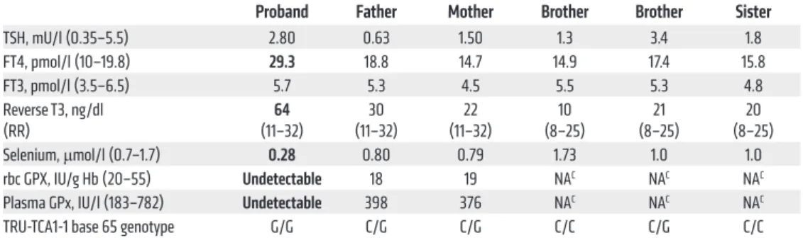
Table 1. Biochemical measurements and TRU-TCA1-1 genotypes in the family
Proband Father Mother Brother Brother Sister
TSH, mU/l (0.35–5.5) 2.80 0.63 1.50 1.3 3.4 1.8 FT4, pmol/l (10–19.8) 29.3 18.8 14.7 14.9 17.4 15.8 FT3, pmol/l (3.5–6.5) 5.7 5.3 4.5 5.5 5.3 4.8 Reverse T3, ng/dl (RR) (11–32)64 (11–32)30 (11–32)22 (8–25)10 (8–25)21 (8–25)20 Selenium, μmol/l (0.7–1.7) 0.28 0.80 0.79 1.73 1.0 1.0 rbc GPX, IU/g Hb (20–55) Undetectable 18 19 NAC NAC NAC
Plasma GPx, IU/l (183–782) Undetectable 398 376 NAC NAC NAC
TRU-TCA1-1 base 65 genotype G/G C/G C/G C/C C/G C/C
Abnormal results are shown in bold. RR, reference range.
Methods
Further methods are detailed in Supplemental Methods.
Statistics. In studies in which statistical analyses were performed, a 2-tailed Student’s t test was used to generate P values. P values less than or equal to 0.05 were considered significant.
Study approval. All investigations were part of an ethically approved protocol (Cambridgeshire LREC 98/154) and/or were clini-cally indicated and undertaken with prior informed patient consent.
Author contributions
ES, DH, and KC designed the study. CM, EG, FM, NS, and GL recruited and clinically characterized patients. EB and SF under-took SNP genotyping and bioinformatic analysis of data. OR per-formed Sanger sequence analysis and interpretation of data. ES, MA, RP, and PG quantified selenoproteins. ES, BC, and RT ana-In patients with global selenoprotein deficiency due to
defective SECISBP2, many features (e.g., photosensitivity, age-dependent hearing loss) are attributed to ROS-mediated dam-age, secondary to loss of antioxidant defence (4). It remains to be seen whether such phenotypes are ameliorated in our mutant tRNA[Ser]Sec patient, in whom synthesis of some antioxi-dant selenoenzymes (e.g., TrxRs, GPx4) is preserved. It is well recognized that systemic selenium status influences relative proportions of mutant tRNA[Ser]Sec isoforms, where mcm5Um is enriched in the Se-replete state and poorly expressed in Se defi-ciency (7–9). Should careful studies altering the selenium status of patient-derived cells in vitro verify that selective selenopro-tein deficits can indeed be corrected without undue toxicity, intervention with selenium supplementation in vivo could be a therapeutic possibility.
Figure 3. Identification, functional analysis, and complementation of tRNA[Ser]Sec mutation. (A) Representation of SNP mapping in proband visualizes runs of homozygosity (ROH) (red peaks) across the genome, with the locations of selenoprotein biosynthetic pathway genes overlaid (TRU-TCA1-1: transfer RNA-Sec [TCA] 1-1). A single ROH overlaps TRU-TCA1-1, and the inset tabulates SNP genotypes in family members (siblings: S1, S2, S3) encompassing this gene locus. (B)
Clover leaf model of Sec-tRNA[Ser]Sec with the mutated base (ringed) and uridine (boxed) that is modified to Sec-tRNA[Ser]Sec mcm5U and Sec-tRNA[Ser]Sec mcm5Um
depicted. (C) Chromatographic elution profiles and quantitation of total tRNA[Ser]Sec as well as mcm5U (peak I) and mcm5Um (peak II) isoforms in cells from
con-trol, father, or proband. (D) Autoradiograph of 75Se-labeled primary skin fibroblasts from control, proband, or SECISBP2-deficient subjects transfected with either
The Journal of Clinical Investigation
B r i e f r e p o r t
Great Ormond Street (to F. Muntoni) and the Intramural Research Program of the Center for Cancer Research, National Cancer Institute, NIH (to D.L. Hatfield).
Address correspondence to: Krishna Chatterjee, University of Cambridge, Metabolic Research Laboratories, Institute of Meta-bolic Science, Level 4, Box 289, Addenbrooke’s Hospital, Cam-bridge, CB2 0QQ, United Kingdom. Phone: 44.1223.336842; E-mail: kkc1@medschl.cam.ac.uk.
lyzed tRNA[Ser]Sec function. ES, DH, and KC prepared the draft manuscript. All authors contributed to discussion of the results and edited and approved the final version.
Acknowledgments
Our research is supported by funding from the Wellcome Trust (100585/Z/12/Z to N. Schoenmakers, 095564/Z/11/Z to K. Chat-terjee), the NIH Research Biomedical Research Centre Cam-bridge (to C. Moran, N. Schoenmakers, and K. Chatterjee) or
1. Shetty SP, Copeland PR. Selenocysteine incorpo-ration: A trump card in the game of mRNA decay.
Biochemie. 2015;114:97–101.
2. Labunskyy VM, Hatfield DL, Gladyshev VN. Selenoproteins: molecular pathways and physi-ological roles. Physiol Rev. 2014;9(3):739–777. 3. Dumitrescu AM, et al. Mutations in SECISBP2
result in abnormal thyroid hormone metabolism.
Nat Genet. 2005;37(11):1247–1252.
4. Schoenmakers E, et al. Mutations in the sele-nocysteine insertion sequence-binding protein 2 gene lead to a multisystem selenoprotein deficiency disorder in humans. J Clin Invest. 2010;120(12):4220–4235.
5. Agamy O, et al. Mutations disrupting selenocyste-ine formation cause progressive cerebello-cerebral atrophy. Am J Hum Genet. 2010;87(4):538–544. 6. Anttonen AK, et al. Selenoprotein biosynthesis
defect causes progressive encephalopathy with elevated lactate. Neurology. 2015;85(4):306–315. 7. Diamond AM, et al. Dietary selenium affects
methylation of the wobble nucleoside in the anti-codon of selenocysteine tRNA([Ser]Sec). J Biol
Chem. 1993;268(19):14215–14223.
8. Carlson BA, Yoo M, Tsuji PA, Gladyshev VM, Hat-field DL. Mouse models targeting selenocysteine tRNA expression for elucidating the role of sele-noproteins in health and development. Molecules.
2009;14(9):3509–3527.
9. Hatfield D, Lee BJ, Hampton L, Diamond AM. Selenium induces changes in the selenocysteine tRNA[Ser]Sec population in mammalian cells.
Nucleic Acids Res. 1991;19(4):939–943.
10. Kim LK, et al. Methylation of the ribosyl moiety at position 34 of selenocysteine tRNA[Ser]Sec is governed by both primary and tertiary structure.
RNA. 2000;6(9):1306–1315.
11. Kim JY, et al. Inhibition of selenocysteine tRNA[Ser]Sec aminoacylation provides evidence that aminoacylation is required for regulatory methylation of this tRNA. Biochem Biophys Res
Commun. 2011;409(4):814–819.
![Figure 1. Pathways of Sec synthesis and incorporation into selenoproteins. The synthesis of Sec (upper pathway) occurs on its tRNA, with initial attachment of serine to tRNA [Ser]Sec by seryl-tRNA synthetase (SARS), phosphorylation of this residue by pho](https://thumb-eu.123doks.com/thumbv2/123doknet/14633136.548439/3.877.107.774.77.663/pathways-synthesis-incorporation-selenoproteins-synthesis-attachment-synthetase-phosphorylation.webp)
![Figure 3. Identification, functional analysis, and complementation of tRNA [Ser]Sec mutation](https://thumb-eu.123doks.com/thumbv2/123doknet/14633136.548439/5.877.71.810.72.689/figure-identification-functional-analysis-complementation-trna-ser-mutation.webp)
