HAL Id: tel-03085778
https://tel.archives-ouvertes.fr/tel-03085778
Submitted on 22 Dec 2020HAL is a multi-disciplinary open access
archive for the deposit and dissemination of sci-entific research documents, whether they are pub-lished or not. The documents may come from teaching and research institutions in France or abroad, or from public or private research centers.
L’archive ouverte pluridisciplinaire HAL, est destinée au dépôt et à la diffusion de documents scientifiques de niveau recherche, publiés ou non, émanant des établissements d’enseignement et de recherche français ou étrangers, des laboratoires publics ou privés.
Structural characterization of bacterial defense complex
Marko Nedeljković
To cite this version:
Marko Nedeljković. Structural characterization of bacterial defense complex. Biomolecules [q-bio.BM]. Université Grenoble Alpes, 2017. English. �NNT : 2017GREAV067�. �tel-03085778�
THÈSE
Pour obtenir le grade de
DOCTEUR DE LA COMMUNAUTE UNIVERSITE
GRENOBLE ALPES
Spécialité : Biologie Structurale et Nanobiologie Arrêté ministériel : 25 mai 2016
Présentée par
Marko NEDELJKOVIĆ
Thèse dirigée par Andréa DESSEN
préparée au sein du Laboratoire Institut de Biologie Structurale dans l'École Doctorale Chimie et Sciences du Vivant
Caractérisation structurale d'un
complexe de défense bactérienne
Structural characterization of a
bacterial defense complex
Thèse soutenue publiquement le 21 décembre 2017, devant le jury composé de :
Monsieur Herman VAN TILBEURGH Professeur, Université Paris Sud, Rapporteur Monsieur Laurent TERRADOT
Directeur de Recherche, Institut de Biologie et Chimie des Protéines, Rapporteur
Monsieur Patrice GOUET Professeur, Université Lyon 1, Président Madame Montserrat SOLER-LOPEZ
Chargé de Recherche, European Synchrotron Radiation Facility, Examinateur
Madame Andréa DESSEN
Directeur de Recherche, Institut de Biologie Structurale , Directeur de These
3
Contents
ABBREVIATIONS ... 5 Summary ... 7 Résumé ... 9 Acknowledgements ... 11 INTRODUCTION ... 131.1. Host-pathogen interaction and virulence factors ... 15
1.2. Proteases as virulence factors ... 16
1.3. Protease inhibitors ... 18
1.4. The α2M subfamily ... 19
1.5. Structure of human α-2-macroglobulin ... 23
1.6. Bacterial α2Ms ... 24
1.7. The bacterial cell wall ... 27
1.8. Peptidoglycan precursor synthesis ... 28
1.9. Penicillin-Binding Proteins... 31
1.10. Glycan chain polymerization ... 32
1.11. Cross-linking ... 33
1.12. Class A PBPs ... 35
1.13. Penicillin-binding protein 1c ... 37
AIMS OF THE PROJECT ... 38
MATERIALS AND METHODS ... 41
2.1. PBP1c sequence analysis ... 43
2.2. Cloning ... 43
2.3. Protein expression and purification ... 44
2.4. Electrophoresis ... 47
2.5. Western blotting ... 49
2.6. Mass spectrometry ... 49
4
2.8. Thermal shift assay ... 53
2.9. The PBP1c activity test ... 55
2.10. Small-Angle X-ray Scattering (SAXS) ... 56
2.11. Electron microscopy ... 57
2.12. Crystallization trials ... 58
RESULTS ... 59
3.1. Expression and purification of ECAM ... 61
3.2. Purification and characterization of PBP1c ... 63
3.2.1. Amino acid sequence analysis ... 63
3.2.2. Expression and solubilization ... 66
3.2.3. Purification of PBP1c ... 69
3.2.4. SEC-MALS analysis of PBP1c ... 72
3.2.5. Cell-free expression and purification of PBP1c ... 74
3.2.6. Mass spectrometry of PBP1c ... 75
3.2.7. Thermal stability analysis ... 76
3.3. Complex reconstitution and characterization ... 79
3.3.1. Complex formation ... 79
3.3.2. AUC analysis ... 80
3.3.3. Peptidoglycan synthesis assay ... 83
3.3.4. Crystallization trials of PBP1c in apo form and in the presence of moenomycin ... 87
3.3.5. Negative staining electron microscopy ... 89
3.3.6. SAXS ... 92
DISCUSSION AND PERSPECTIVES ... 103
References ... 110
INDEX ... 122
5
ABBREVIATIONS
α2M α-2-macroglobulin
AMP Adenosine monophosphate
ATP Adenosine triphosphate
AUC Analytical ultracentrifugation
C55-PP Undecaprenyl pyrophosphate
CMC Critical micellar concentration
CTP Cytosine triphosphate
DDM n-dodecyl-β-D-maltopyranoside
DM n-decyl-β-D-maltopyranoside
DNA Deoxyribonucleic acid
DTT Dithiothreitol
ECAM E. coli α-2-macroglobulin EM Electron microscopy
GlcNAc N-acetylglucosamine
GT, GTase Glucosyltransferase
GTP Guanosine triphosphate
HPLC High-performance liquid chromatography
LAPAO 3-laurylamido-N,N’-dimethylpropyl amine oxide
MALDI-TOF Matrix-assisted Laser Desorption/Ionization Time of Flight
meso-A2pm Meso-diaminopimelic acid
MG Macroglobulin-like
MurNAc N-acetylmuramic acid
NAG N-acetylglucosamine
NAM N-acetylmuramic acid
PBP Penicillin-binding protein
PZP Pregnancy zone protein
RBD Receptor-binding domain
SAXS Small angle X-ray scattering
SEC-MALS Size exclusion chromatography with multi-angle light scattering
SDS-PAGE Sodium dodecyl sulfate polyacrylamide gel electrophoresis
6 TED Thioester-binding domain
TEM Transmission electron microscopy
TEP Thioester-containing proteins
TP, TPase Transpeptidase
UB2H UvrB Domain 2 homolog
UDP Uridine diphosphate
7
Summary
The widespread resistance to antibiotics developed by bacterial pathogens calls for the characterization of original, yet unexplored potential targets in bacteria. Alpha2-macroglobulins (α2Ms) are broad-spectrum protease inhibitors that play key roles in eukaryotic immunity. They are multi-domain molecules that carry approximately 1,800 residues and harbor a central amino acid sequence, the ‘bait site’, which is recognized and cleaved by a large number of proteases. Upon cleavage, the resulting conformational change exposes a buried thioester bond between a cysteine and a glutamine, which is readily hydrolyzed, allowing the resulting glutamate to associate covalently to the target protease, trapping it within the α2M cage-like structure. Recently, α2M homologs from pathogenic and colonizing bacteria have also started to be characterized. These findings suggest that bacteria possess a rudimentary immune system that mimics initial key steps of the eukaryotic immune pathway and that could represent a yet unexplored target in pathogen biology.
The genes for two types of α2M are present in bacterial genomes: type 1, which contains the thioester bond, and type 2 that does not harbor it. Type 1 bacterial α2Ma persistently co-occur within the same operon with a gene that encodes a cell wall biosynthesis enzyme, Penicillin-Binding Protein 1c (PBP1c). This suggests that the association between the two proteins could be highly advantageous for the cell during infection/colonization, when the outer cell wall is targeted by host defenses. In this situation, α2M and PBP1c could exert the role of ‘guardians of the periplasm’, with PBP1c repairing damaged peptidoglycan, and α2M trapping invading proteases.
The aim of this work was to demonstrate the existence of such complex and characterize this interaction structurally and functionally. For this purpose, α2M and PBP1c from E. coli were studied. The proteins were expressed and purified separately. α2M (also called ECAM in
E. coli) is a highly soluble, monomeric protein with a mass of 182 kDa, monodisperse and
stable during the course of time. PBP1c is a membrane-bound, 87 kDa protein, predominantly present as a dimer. The complex, reconstituted in vitro by mixing and incubating the proteins for 2 hours, resulted in formation of a complex, demonstrated by appearance of a new peak in size exclusion chromatography. This result was further confirmed by SDS-PAGE, analytical centrifugation and small-angle x-ray scattering (SAXS) experiments. ECAM and PBP1c associated with 2:2 and 4:4 stoichiometries. The activity test confirmed that PBP1c performs polymerization of glycan chains and that its activity is enhanced in the presence of ECAM.
8
Crystallization trials yielded crystals of PBP1c in several conditions, while the study of ECAM by electron microscopy proved that this technique could be used for structural studies of the complex. Both approaches are under optimization, and combined, they could be employed for structural characterization of the ECAM-PBP1c complex
9
Résumé
De nos jours, la résistance aux antibiotiques développée par des pathogènes bactériens est de plus en plus répandue, et en conséquent il devient primordial de caractériser de nouvelles cibles bactériennes potentielles. Les alpha2-macroglobulines (α2Ms) sont des inhibiteurs de protéases à large spectre jouant un rôle clé dans l’immunité eucaryote. Ce sont des molécules multi domaines d’environ 1800 résidus qui arborent une séquence en acide nucléique très spécifique appelée « bait site » qui est reconnu et coupé par un très grand nombre de protéases. Durant ce clivage, la nouvelle conformation de la protéine entraine l’exposition de la liaison thioester entre une cystéine et une glutamine, qui est alors hydrolysée, permettant ainsi au glutamate résultant de s’associer de façon covalente à la protéase cible, et d’être piégé dans la structure en forme de cage de α2M. Des homologues de 2M provenant de bactéries colonisant et pathogéniques ont récemment été caractérisés. Ces nouvelles recherches suggèrent que la bactérie dispose d’un système immunitaire capable de mimer les étapes clés initiales du système immunitaire eucaryote et que celui-ci pourrait constituer une cible encore inexploitée en biologie bactérienne.
On retrouve dans le génome bactérien deux types de gènes pour 2M : celui de type 1 qui contient la liaison thioester et celui de type 2 qui n’en dispose pas. De façon intéressante, le gene 2M de type 1 est situé dans le même opéron que le gène codant pour une enzyme de
biosynthèse de la paroi cellulaire appelée Penicillin-Binding Protein 1c (PBP1c). Ceci suggère qu’une association étroite entre ces deux protéines pourrait être très avantageuse pour la cellule, en particulier durant l’infection et/ou la colonisation durant laquelle la membrane cellulaire externe est ciblée par des agents de défense de l’hôte. Dans cette situation, 2M et PBP1c pourraient exercer les rôles de « gardiens du périplasme » avec PBP1c réparant les dommages causés au niveau du peptidoglycane et 2M traquant les protéases invasives.
Le but de ce travail était donc de démontrer l’existence de ce complexe PBP1C/A2M et de caractériser cette interaction de façon structurale et fonctionnelle. Dans ce sens, les deux protéines provenant d’E. coli ont été étudiées, ainsi qu’exprimées et purifiées séparément. La protéine 2M (également appelé ECAM dans E. coli) est très soluble, exprimée de façon monomérique à une taille de 182 kDa, monodisperse et stable dans l’échelle de temps. PBP1c est une protéine se fixant à la membrane, d’une taille de 87 kDa et présente de façon prédominante en tant que dimère. La reconstitution du complexe in vitro par incubation et mélange des deux protéines pendant 2 heures a permis d’obtenir un complexe PBP1C/ECAM,
10
dont la présence a été prouvée par un nouveau pic caractéristique sur chromatographie par exclusion de taille. Ces résultats ont également été appuyés par SDS-PAGE, centrifugation analytique et par diffusion des rayons X aux petits angles (SAXS). ECAM et PBP1c s’associent à des ratios stœchiométriques de 2:2 et 4:4. Enfin, un test d’activité a permis de confirmer tout d’abord le rôle de PBP1c dans la polymérisation les chaines glycanes mais aussi que cette activité est amplifiée en présence d’ECAM.
Les essais cristallographiques ont permis d’obtenir des cristaux de PBP1c dans différentes conditions. De plus l'étude d’ECAM par microscopie électronique a prouvé que la technique était applicable pour des études structurales du complexe. De ce fait, après optimisation, ces deux approches pourraient être la solution envisagée pour la caractérisation structurale du complexe ECAM/PBP1c.
11
Acknowledgements
When I arrived in Grenoble three years ago, I didn’t have a clear idea what I was getting into and whether I would succeed. Thankfully, I was entering a community that is very welcoming, knows how to deal with rookies like me and has a guy for everything. All these people helped me to make this project work and get me where I am today. The end of a cycle inevitably calls for a short retrospection and acknowledging each one of them.
Firstly, I would like to express my gratitude to my supervisor Andrea Dessen for giving me the opportunity to experience what science actually is. With a background in computational biology and overly tanned from Barcelonan sun in the middle of my internship, many would doubt if I was a suitable choice for a project starting from the scratch. Yet she didn’t, and that may be the first lesson I learned, that sometimes you have to risk with people and projects in order to push limits in science. I also thank her for all the effort she invested to teach me how to think like a scientist and for the endless positive energy she has, no matter how bad the things are.
I would like to say huge thanks to my first two tutors, Alexandre Martins for all the patience he had while training me in protein purification and not killing me during the Covalab order crisis, and Anne-Emmanuelle Foucher for the lessons in molecular biology and proper lab behavior. I would like to thank Charlotte Lombardi for all the advice and answers to so many unnecessary questions (“C'est la tête d'un Serbe qui vient poser une question dont il connaît déjà la réponse”) during my first year in the lab, and apologize for judging her eating habits during pregnancy. Further, I would like to thank Ioulia Nikolaidis for teaching me how to work with membrane proteins and giving me a shelter for three days when I locked myself out of my apartment; Vivian Job for advice on mutagenesis and all the microbiology work on my project she is currently developing which, I hope, we’ll present very soon. Even though I didn’t succeed in solving any crystal structures, I’m very grateful to Carlos Contreras for the advice on how to proceed and what to try next, and above all, for his bitter Chilean humor that I enjoy so much.
In my work I extensively used the services of different platforms and collaborated with many people. I would like to express my gratitude to Lionel Imbert for the initial tests of the cell-free expression he did, Aline Leroy and Christine Ebel from the AUC platform at the IBS for performing my AUC experiments, Caroline Mas from the Biophysical platform for the SEC-MALS analyses and Elisabetta Boeri from the Mass Spectrometry platform for teaching
12
me how to do MALDI/TOF experiments and helping me with the interpretation of results. I would like to thank Daphna Fenel for all the electron microscopy work she did and say sorry for emotionally manipulating her to put me on the schedule when there were no time slots available. I am very grateful to Gabriele Giachin for teaching me how to process SAXS data and Orso Subrini for his help in interpreting the results from Prometheus experiments. I would also like to thank Victor Hernandez Rocamora from Dr Waldemar Vollmer’s lab at Newcastle University for the work he did testing the activity of PBP1c.
During my studies I had a had two thesis advisory committee meetings during which I discussed my work with Dr Carlo Petosa and Dr Ina Atree. They had greatly contributed to my work with their recommendations, for which I’m very grateful.
The work on this manuscript was long and exhausting, and occasionally needed a fresh insight of a third party. Special thanks to my copy editor Simon Harris and my rédactrice pour la langue française Laura Lemel, for the invaluable help in their respective native languages. I promise I’ll return the favor if ever you choose to write your theses in Serbian.
Two additional thanks go to my labmate and flatmate Quentin Bertrand for all the quiches and other French food he made during the course of writing, because cooking was definitely last thing on my mind (and I hope I wasn’t a bad flatmate), and Federica Laddomada, a labmate with whom I have been sharing the same fate for the last three months, for frequently being my therapist. We made it.
I would like to thank the Université Grenoble Alpes for providing funds for the duration of my fellowship.
I am also grateful to my family for being emotional support from the beginning and making it clear that no matter what happens, there’s always a place to come back.
Pour finir, j’aimerais également mentionner des personnes qui n’ont pas été directement impliquées dans le travail de thèse, mais qui ont entièrement fait partie de ma vie quotidienne à Grenoble. Merci tout d’abord à Julien qui m’a hébergé pendant deux semaines lors de mon arrivée à Grenoble. J’ai été naïf de croire que je trouverai un appartement en trois jours. Et merci à Charles, Muge, Silvia, Tomaš, Charline,Joyce, Vilius, Sonja, Albert et tous les autres pour toutes les fêtes, les apéros, les week-ends et voyages qu’on a pu faire ensemble. Cela va être très difficile de partir, mais surtout de vous quitter.
13
I
15
1.1. Host-pathogen interaction and virulence factors
The number of existing prokaryotic species on Earth is estimated to be between 10 and 1,000 million, although it is hard to determine precisely (Curtis et al. 2002; Dykhuizen 2005). This is partly due to the lack of consensus among scientists in what defines species in microorganisms (Cohan 2002), but also because of their presence in virtually every type of environment and the fact that less than 1% of bacteria can be successfully cultivated in laboratory conditions (Pham & Kim 2012). They inhabit soil, fresh and marine waters, as well as specific habitats such as plant leaves and roots, and skin and gastrointestinal tract of mammals (Floyd et al. 2005).
The human microbiome project calculated that more than 10,000 microbial species live in a healthy human individual (NIH 2012). The majority of them are bacteria, although protozoa, archaea, bacteriophages, wormlike helminth parasites and yeast are widely present. In normal conditions they are only found on the skin and mucosal surfaces. Their penetration into the body is prevented by numerous mechanisms that comprise nonspecific and specific immune system of the host (Peterson 1996). However, there are pathogenic species that are able to evade innate immunity and disseminate inside the host, causing diseases (Wilson et al. 2002). Their capacity to cause damage in a host is called virulence (Casadevall & Pirofski 2003). There are around 1,400 human pathogens, which is far less than 1% of the total microbial species, making pathogenicity a rare exception (Diard 2017; Taylor et al. 2001).
Comparisons between commensal and pathogenic species revealed that the increased virulence of pathogens is due to the presence of specific molecules called virulence factors which aid evasion of the host immune system (Diard 2017). Virulence factors are encoded on their chromosomal DNA, plasmids, bacteriophage DNA and transposons (Peterson 1996). They have diverse functions in host-pathogen interaction, such us breaching physical barriers, shielding from host’s immune defense and acquiring nutrients during the infection (Diard 2017).
There are several types of virulence factors, depending on the role they exhibit: adhesins, invasins, capsules, endo- and exotoxins and siderophores (Peterson 1996).
16
1.2. Proteases as virulence factors
One class of virulence factors that are found in most thoroughly-studied pathogens are proteases. Proteases are enzymes that digest proteins by hydrolyzing peptide bonds (López-Otín & Bond 2008). Proteases are globally divided in two major groups: exopeptidases, which hydrolyze peptide bonds proximal to the amino or carboxy termini of the substrate, and endopeptidases which cleave internal peptide bonds (Rani et al. 2012). Furthermore, depending on the structure of their active site, they are classified as serine, cysteine, threonine, aspartic, glutamic and metalloproteases (López-Otín & Bond 2008). The first three classes utilize serine, cysteine and threonine respectively for the nucleophilic attack of the peptide bond, while aspartic, glutamic and metalloproteases utilize an activated water molecule.
Pathogen proteases belong to any of the six classes of proteases and they can be secreted or surface-bound (Armstrong 2006). They are efficient virulence factors employed in many stages of infection. The first line of defense in vertebrates is skin and mucosal surfaces of gastrointestinal and respiratory tracts. Epithelial cells produce antimicrobial peptides (AMPs), a diverse group of pore-forming molecules that act against microorganisms by increasing cell membrane permeation, leading to the leakage of cellular content (Diamond et al. 2009; Zasloff 2002). Cationic peptide LL-37 from the cathelicidin family of AMPs has a prominent role in eliminating bacteria in the stage of colonization (Henzler-Wildman et al. 2004). Bacteria, meanwhile, secrete proteases that cleave LL-37, thus evading its lethal effect. Examples include metalloprotease aureolysin (Aur) from Staphylococcus aureus, LasB (elastase) of Pseudomonas aeruginosa, Enteroccocus faecalis gelatinase and Staphylococcus
epidermidis SepA (Schmidtchen et al. 2002; Sieprawska-Lupa et al. 2004).
Keratinocytes from the skin are tightly connected by desmosomes, membrane structures that provide strong cell-to-cell contact and adhesion (Kowalczyk & Green 2013). S.
aureus is able to rupture the skin using two serine proteases, exfoliative toxins A (ETA) and B
(ETB) that digest desmoglein 1, one of the components of desmosomes (Hanakawa et al. 2004). Extracellular matrix proteins collagen, elastin, fibronectin and laminin are also substrates for various pathogen-produced proteases. LasB from P. aeruginosa cleaves human type III and IV collagens, and together with alkaline protease rapidly degrades laminin (Heck, K Morihara, et al. 1986; Heck, K. Morihara, et al. 1986). LasB and Ecp of S. epidermidis target fibronectin and elastin (Oleksy et al. 2004; Schmidtchen et al. 2003). Elastin is also proteolysed by SspB of S. aureus (Kantyka et al. 2011).
17
Once pathogens breach the primary protective layers of the host surface, they face the danger of being phagocytized by macrophages and neutrophils. The complement system and antibodies engage in process called opsonization by binding to the surface of the pathogen, marking it for digestion by phagocytes (Janeway et al. 2001). In order to avoid opsonization, pathogens employ different strategies, one of them being proteolytic cleavage of the proteins involved in this process. Complement component 3 (C3) is completely degraded by S. pyogens SpeB while S. aureus aureolysin cleaves C3 protein far from the bacterial surface converting it to active C3b that is rapidly processed by host proteases (Laarman et al. 2011; Terao et al. 2008). On the other hand, the cysteine protease IdeS from S pyogenes, as well as the serratial 56-kDa protease are involved in cleavage of immunoglobulin G (Molla et al. 1988; Su et al. 2011).
Coagulation is a process that activates upon rupture of blood vessels creating a clot that closes the opening, maintaining hemostasis and preventing pathogen infiltration (Levi et al. 2004). It is a cascade reaction that activates the host protease thrombin to convert fibrinogen to fibrin. Fibrin polymerizes into a network, creating a fibrin-based clot. Blood clotting can also be activated independently of the vessel injury, triggered by the presence of pathogens in the blood (Wang et al. 2010). In that manner, fibrin clots immobilize pathogens in an attempt to prevent their dissemination (Loof et al. 2011). Nevertheless, pathogen proteases can trigger the destruction of the fibrin clot by converting host protease plasminogen to its active form, plasmin, dissolving the clot. That is the case with Pla protease from Yersinia pestis, OmpT of
E. coli and PgtE of Salmonella enterica (Kukkonen & Korhonen 2004; Sodeinde et al. 1992).
On the other hand, CpaA of Acetinobacter baumanii interferes with clot formation by cleaving factor V (that is part of thrombin activation complex) and fibrinogen (Tilley et al. 2014).
Proteases are also engaged in fulfilling nutritional requirements of pathogens during infection. Blood parasites, such as Plasmodium falciparum and Schistosoma mansoni use hemoglobin as an amino acid and energy source. Aspartic and cysteine proteases plasmepsins and falcipain of P. falciparum and cathepsins L1 and L2 (SmCL1 and 2) and D (SmCD) of S.
mansoni digest hemoglobin (Caffrey et al. 2004; Francis et al. 1997). Moreover, there are
species like P. gingivalis that digest host proteins in order to release sequestered iron required for their growth (Carlsson et al. 1984).
18
1.3. Protease inhibitors
Pathogen proteases represent a major weapon during the infection process. Host organisms, on the other hand, responded to this threat by developing different types of protease inhibitors in order to neutralize damaging effects caused by the pathogen proteases. These inhibitors are important components of the vertebrate and arthropod plasma making up 5% of its total content (Laskowski & Kato 1980). The reason for this presumably lies in the fact that blood and hemolymph easily reach all parts of the body and have direct contact with tissues. The mechanism they use to inactivate the target protease classifies them in two groups: active-site inhibitors and alpha-2-macroglobulins (α2Ms). The first group inactivates proteases by binding to their active site and completely preventing them from performing further hydrolysis (Armstrong 2006). The second group, the α2Ms, consists of proteins that entrap the target protease into a molecular cage (figure 1.1). The members of this group possess a surface-exposed loop recognized by proteases as a substrate and which is cleaved, inducing a conformational rearrangement of the inhibitor in a form of cage around the protease. This cage sterically hinders the protease from the protein substrate. Nonetheless, the protease active site is not affected and it remains active against small substrate that can easily access the cavity inside the cage (Nielsen & Sottrup-Jensen 1993; Quigley et al. 1991).
A.
B.
C.
Figure 1.1. A and B – Early EM images showing tetrameric structure of native human α2M (hα2M) with a cavity visible as dense zone in the middle (A) and a hα2M-trypsin complex (B). C – Model representing possible protease binding mode in induced hα2M based on the crystal structure. (A and B taken from (Tapon-Bretaudiére et al. 1985), C from (Marrero et al. 2012)).
19
Alpha-2-macroglobulins belong to the family of thioester-containing proteins (TEPs) that are abundant in human plasma and are engaged in different stages of the innate immune response (Sottrup-Jensen 1989) (Law & Dodds 1997). Based on phylogenetic analyses, this family comprises three subfamilies: 1 – α2Ms, with members such as α2M, muroglobulin, α1I3
of rats, human pregnancy zone proteins (PZPs) and ovostatin from eggs, 2 – complement factors C3, C4 and C5 and 3 – a recently described subfamily of insect TEPs (Armstrong & Quigleya 1999)(Blandin & Levashina 2004). While α2Ms act as protease inhibitors, members of the complement subfamily are part of the cascade reaction that results in marking of foreign agents for elimination, either by the innate or the adaptive immune system. Even though insect TEPs show a greater overall similarity with α2M, they act as opsonins and in that aspect they are closer to complement system proteins (Shokal & Eleftherianos 2017). Amino acid sequence comparisons and evolutionary studies show that TEPs arose from a common ancestor after gene duplication of an α2M-like protein, predating the divergence of proteostomes and deutoerostomes (Levashina et al. 2002; Zhu et al. 2005).
All TEPs have several features common features. They are large (over 100 kDa), glycosylated proteins that undergo substantial conformational changes upon proteolytic cleavage of a highly variable, so-called bait region, resulting in transformation to their active forms (figure 1.2) (Williams & Baxter 2014). Most of them also contain a highly reactive thioester bond between a thiol group of a cysteine and a γ-carbonyl group of a glutamine which is readily hydrolyzed by water molecules (Law & Dodds 1997). In the native state, the thioester bond is buried inside the protein where it is protected from water. However, small nucleophiles like methylamine, hydrazine and ammonia can still diffuse inside and inactive it. Sensitivity of the thioester bond towards nucleophiles is, however, slightly different between proteins since α2M can be inhibited by methylamine, while the thioester of C3 can be efficiently inhibited by both methylamine and glycerol (Rehman et al. 2013).
1.4. The α2M subfamily
As already mentioned, the α2M subfamily contains various protease-binding proteins, including a canonical α2M widely present in animals from nematodes to mammals. Additionally, α2M homologs such as muroglobulin of mice, alpha-1-macroglobulin (α1M) and alpha-1-inhibitor III (α1I3) of rats, α2M-like protein 1 (α2ML1) in skin, human pregnancy zone
20
A.
B.
C.
D.
E.
Figure 1.2. The conformational change of thioester-containing proteins upon the cleavage of the bait region illustrated on the example of complement protein C3. A – In native C3, TED, CUB and MG8 domains are in close contact (PDB: 2A73); Upon induction by a protease (B), TED moves farther away, exposing previously hidden thioester bond (PDB: 2I07); C, D and E – translational and rotational movements of TED, CUB and MG8 domains, respectively.
21
protein (PZP), which is highly expressed during pregnancy, and ovostatin of avian and reptile eggs also belong to this subfamily (Armstrong & Quigleya 1999).
The size of monomeric forms of these proteins is between 180 and 200 kDa and they are found in different oligomerization states. Human and snail α2Ms, for example, form homotetramers, while α2Ms from arthropods and PZP are found as homodimers (Bender & Bayne 1996; Sottrup-Jensen 1989; Wyatt et al. 2016). Several of these proteins function as monomers, like α2ML1 (Galliano et al. 2006) or murinoglobulin (Overbergh et al. 1994). The sequences of the bait regions of different α2Ms are variable suggesting that, despite what appears to be a universally-cleavable sequence, a degree of specialization towards certain proteases exist, depending on the environment in which individual α2Ms are expressed (Sottrup-Jensen et al. 1989).
The principle by which α2Ms work involves sequestration of the protease in a molecular cage, making it physically inaccessible to their substrate. Each of the subunits in an oligomer has a surface-exposed bait region, that is readily accessible and recognized by a wide range of proteases. The activation of α2M occurs upon cleavage of the bait region, triggering a large conformational change that encloses the prey protease into a chamber in a way that resembles the Venus flytrap mechanism. This conformational change uncovers a highly reactive thioester bond that had been previously buried inside the molecule. Now exposed, the bond is hydrolyzed and the γ-carbonyl group of the glutamine forms an isopeptide bond with the ϵ-amino group of a lysine on the protease surface (Sottrup-Jensen et al. 1990). Covalent binding to the target protease is, however, not mandatory for efficient inhibitory function. In
Limulus polyphemus α2M for example, a thiol esterified glutamine crosslinks to a lysine
residue of the opposite chain of α2M dimer rather than to a protease, making the inhibition essentially a non-covalent reaction. Furthermore, there are forms of α2Ms like chicken ovomacroglobulin that do not harbor a thioester motif, yet are still able to inhibit proteases by confining them within a molecular cage (Nielsen & Sottrup-Jensen 1993).
Activation of α2M by proteases converts it to a more compact form, an effect that is easily observable on SDS-PAGE (Barrett et al. 1979). At the same time, a conformational rearrangement causes the exposure of the receptor binding domains on each monomer, leading the α2M-protease complex to bind to the low-density lipoprotein related protein (LRP/α2M-R) receptor, resulting in its subsequent clearing (Holtet et al. 1994; Sottrup-Jensen et al. 1986) (Kristensen et al. 1990). The LRP/α2M-R receptor is a 600 kDa heterodimer and belongs to the family of low-density lipoproteins (LDLs) found on hepatocytes, fibroblasts, macrophages, syncytiotrophoblasts, adipocytes and astrocytes (Krieger & Herz 1994).
22 A.
B.
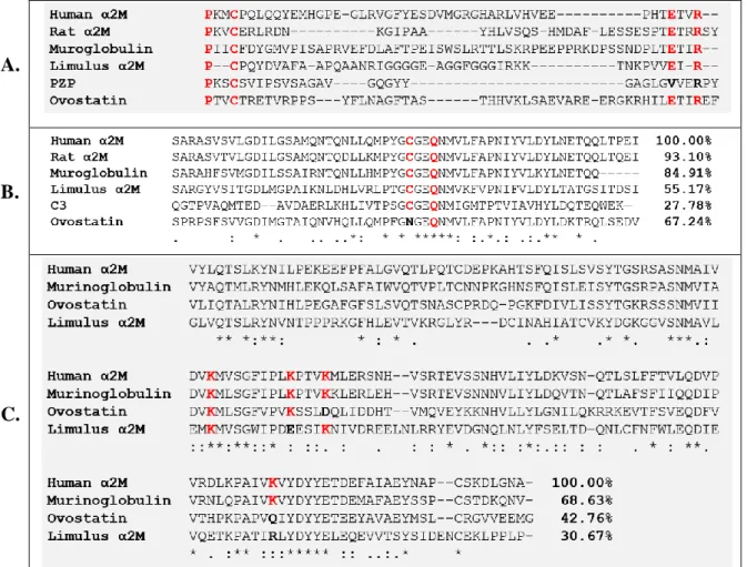
C.
Figure 1.3. Sequence alignment of three domains of thioester-containing proteins (TEPs). A – Bait region is highly variable among TEPs, with PxxC and ExxR motifs found at its N- and C- termini; B – Thioester domain (TED) with CGEQ motif; C – Receptor binding domain (RBD). Lysine residues in red are important for the binding of RBD to the receptor (Sottrup-Jensen et al. 1986). The percentages in B and C show the identity of amino acid sequence of α2Ms compared to the human form.
Apart from acting like a protease inhibitor, α2M has wider role in the innate immune system by binding and promoting clearance of many molecules. So far it is well established that α2M binds number of cytokines and growth factors, such as transforming growth factor-β (TGF-β), tumor necrosis factor-α (TNF-α), interleukin 1β (IL-1β), interleukin 8 (IL-8), platelet-derived growth factor-BB (PDGF-BB), nerve growth factor-β (NGF-β) and vascular endothelial growth factor (VEGF) (LaMarre et al. 1991). In these cases, α2M is not converted to the activated form and the mode of interaction with these molecules is independent of its protease-inhibiting function.
23
1.5. Structure of human α-2-macroglobulin
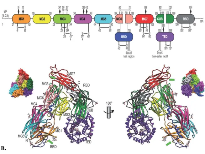
The first low-resolution crystal structure of any α2M was that of human methylamine-induced α2M (hα2M) reported in 1995 (Andersen et al. 1995). Subsequently, the 4.3 Å resolution structure of hα2M became available in 2012 (Marrero et al. 2012). Human α2M is a 1491 residue molecule made of 11 domains, with 13 disulfide bridges, 11 of them intramolecular and 2 additional ones that participate in the formation of a dimer (Marrero et al. 2012). The first seven domains are termed macroglobulin-like domains (MG) and they are approximately 110-residue antiparallel β-sandwiches of three- and four-stranded β-sheets (figure 1.4). Other regions of note include the thioester domain (TED) which harbors the CxEQ sequence and maintains it at the interior of the molecule to protect it from hydrolysis, and the bait region, 39-residue sequence that is recognized by a large number of proteases (Sottrup-Jensen et al. 1981)(Marrero et al. 2012).
A.
B.
Figure 1.4. Structure of the human α2M monomer. A – schematic representation of domain organization; B – domain arrangement; (Marrero et al. 2012)
24
Human α2M circulates in the bloodstream as tetramer of approximately 720 kDa in which two subunits (monomers) interact covalently through two disulfide bridges, leading to the association of two dimers through non-covalent interactions. In this way they build a large central cavity at the center of the tetramer, named the prey chamber, which can be large enough to capture up to two proteases of 20-30 kDa (Marrero et al. 2012). This corresponds to the previously determined stoichiometry of 1:2 (Sottrup-Jensen 1989).
1.6. Bacterial α2Ms
Protein sequences related to α2M were found in all metazoan groups and for a long time it was believed that α2Ms are an exclusively animal characteristic. In 2004, Budd et al. reported that sequences homologous to eukaryotic α2M were widely present in the genomes of different bacterial species (Budd et al. 2004). Moreover, α2M homologs that did not carry the thioester motif were also identified. In E. coli for example, there are two α2M homologs: yfhM, that has thioester motif, and yfaS, a highly divergent homolog that does not carry it. Using E. coli as a reference, they classified bacterial α2Ms into two types, with the main characteristic being the presence or absence of the thioester. The most plausible explanation of the origin of α2M-like genes in bacterial genomes is horizontal gene transfer.
Figure 1.4. Genetic localization of α2M in some bacterial species.
Notably, Budd et al. noted that these genes were present only in Gram-negative bacteria with colonizing and parasitic lifestyles, while Gram-positive bacteria and archaea lacked α2Ms
25
(Budd et al. 2004). This suggests that α2M may provide Gram-negative bacteria with an advantage during colonization and infection events.
Finally, the genetic context in which the two types of bacterial α2Ms were found was intriguing. YfhM was always found juxtaposed or in an operon with the gene encoding Penicillin-Binding Protein 1c (pbpc), whose protein product, PBP1c, is involved in cell wall formation, while yfaS was present in an operon with five other genes whose function has not yet been fully elucidated.
YfhM is a 1653-residue polypeptide containing a characteristic consensus lipobox sequence (LAGC) suggesting that it is a membrane-bound protein (Budd et al. 2004)(Kovacs-Simon et al. 2011). The lipobox cysteine is followed by an aspartic acid from an inner-membrane sorting signal, localizing YfhM in the periplasm (Fukuda et al. 2002). On the other hand, YfaS does not have a lipobox motif indicating that the protein may be secreted (Budd et al. 2004). Biochemical characterization of YfhM (named ECAM, for E. coli α-2-macroglobulin) by Doan and Gettings confirmed that it is expressed as a 180 kDa monomer that is only found in the inner membrane fraction (Doan & Gettins 2008). They also validated the presence of an intact thiol ester between cysteine and glutamine residues within the CxEQ motif that is hydrolyzed by small nucleophiles like methylamine. In addition, they showed that proteases can cleave ECAM in the predicted bait region. Their data also showed that, as in the case of human α2M, ECAM becomes more compacted upon incubation with proteases, resulting in increased electrophoretic mobility in SDS-PAGE as compared to the native “slow” form. Structural analysesusing small-angle x-ray scattering (SAXS) and electron microscopy confirmed that the induced form of ECAM is more compact than the native one and that the conformational rearrangement that takes place after the interaction with proteases resembles the changes that occur in the transition of eukaryotic C3 from a preactivated to an activated state (Neves et al. 2012).
The first crystal structure of a bacterial α2M was obtained by our group in 2014 (Wong & Dessen 2014). This is a 2.95 Å resolution structure of α2M from Salmonella enterica (Sa-A2M) (PDB-ID: 4U48). Sa-A2M is a 1644-residue protein that shares 82% identity with ECAM. The structure shows high overall similarity with the human form. It consists of 13 domains, 11 of them folding as β-sandwich-like MG domains, as in the case of human α2M. Compared to the human form, Sa-A2M has two additional domains, MG1 and MG2, spanning residues 57 to 281. MG1 is believed to associate to the inner membrane by a flexible loop and together they appear to play a role of a linker (Wong & Dessen 2014). From the MG3 domain on, they follow a similar pattern to the domain arrangement in human α2M including a highly
26
flexible bait site from residues 925 to 950 and a helical TED domain with the thioester motif. The polypeptide chain terminates with the MG10 which structurally resembles the receptor-binding domain in human α2M; however, its role remains unclear.
Figure 1.5. Structure of Salmonella enterica α2M. MG 1-10 – macroglobulin-like domains; BDR – bait region domain; TED – thioester domain. From (Wong & Dessen 2014)
Garcia-Ferrer et al. subsequently structurally characterized ECAM in both the presence and absence of a protease, and concluded that it underwent a major conformational change upon protease binding. However, they were not able to trace a trustworthy model for the protease in the complex, and thus the mechanism of this interaction still remains obscure (Garcia-Ferrer et al. 2015).
27
1.7. The bacterial cell wall
The bacterial cell wall is a robust protective layer that ensures integrity and protection from environmental stress (Scheffers & Pinho 2005). The main component of the bacterial cell wall is peptidoglycan (murein), a mesh-like sacculus surrounding the cytoplasmic membrane (Typas et al. 2011). The most important function of the peptidoglycan is to resist internal osmotic pressure and any inhibition of its synthesis or enzymatic degradation leads to cell lysis (Mengin-Lecreulx & Lemaitre 2005). It also maintains the shape of the cells and serves as a scaffold to which numerous proteins and teichoic acids are attached (Dramsi et al. 2008)(Neuhaus & Baddiley 2003).
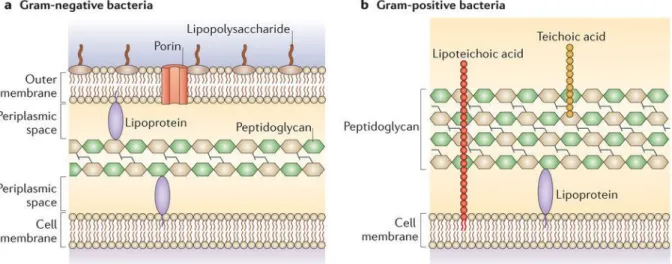
Gram-positive bacteria have a thick peptidoglycan layer that can constitute up to 95% of their cell wall, with teichoic and lipoteichoic acids embedded within, the latter directly connected to the plasma membrane lipids. On the surface of peptidoglycan there is often a layer of identical proteins attached to it called S-layer, and a capsule of polysaccharides (figure 1.6b). On the other hand, Gram-negative bacteria have inner and outer membranes that enclose a compartment called periplasm in which thin layer of peptidoglycan is located (figure 1.6a) (Vollmer et al. 2008)(Malanovic & Lohner 2016).
Figure 1.6. Differences between the cell walls of Gram-negative and Gram-positive bacteria. Taken from (Brown et al. 2015).
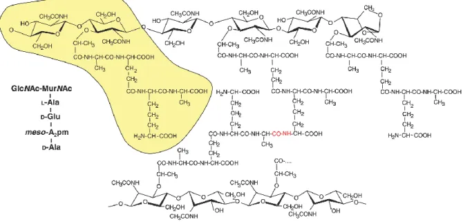
Peptidoglycan is a complex polymer consisting of linear glycan chains cross-linked by short peptides (Rogers et al. 1980). The glycan chains are composed of two alternating amino sugars, N-acetylglucosamine (GlcNAc) and N-acetylmuramic acid (MurNAc) that are linked by a β-(1,4) glycosidic bond (figure 1.7) (Vollmer et al. 2008). The pentapeptide stem is
28
attached to the lactoyl group of MurNAc. The composition of the pentapeptide varies, though generally it consists of L-Ala--D-Glu-meso-A2pm (or L-Lys)-D-Ala-D-Ala, A2pm being
2,6-diaminopimelic acid. The last D-Ala is trimmed from the mature molecule. The stem peptides are cross-linked between the carboxyl group of D-Ala and the amino group of neighboring diamino acid, either directly or through a peptide bridge (ex. pentaglycine) (Vollmer et al. 2008).
Figure 1.7. Structure of the E. coli peptidoglycan. The basic building block marked in yellow consists of N-acetylglucosamine (GlcNAc) and N-acetylmuramic acid (MurNAc) connected by a β-(1,4) glycosidic bond, with a L-Ala--D-Glu-meso-A2pm-D-Ala-D-Ala pentapeptide
attached to MurNAc. Glycan chains are cross-linked directly between a meso-A2pm residue of
one and a D-Ala residue at position 4 of another disaccharide (red). Taken from (Mengin-Lecreulx & Lemaitre 2005)
1.8. Peptidoglycan precursor synthesis
Peptidoglycan synthesis involves around 20 cascade reactions that can be grouped in three phases. It starts with the synthesis of precursors in the cytosol, followed by their linkage to an undecaprenyl phosphate group on the membrane to form lipid II. Lipid II is subsequently flipped across the membrane to the periplasm where it will be acted upon by the glycosyltransferase and transpeptidase action of Penicillin-Binding Proteins (PBPs) (Typas et al. 2011).
The first stage involves the synthesis of the soluble nucleotide-bound individual components that will later form the basic building block of peptidoglycan (figure 1.8).
29
Formation of GlcNAc starts with amination of fructose-6-phosphate to produce glucosamine-6-phosphate (GlcN-6-P), followed by its conversion to GlcN-1-P, addition of the acetyl group and linking to the UDP to create UDP-GlcNAc (Barreteau et al. 2008). The newly created UDP-GlcNAc is a substrate for formation of the second sugar component by transferring enolpyruvate from phosphoenolpyruvate (PEP) and reduction to yield UDP-MurNAc. The final stage is the synthesis of pentapeptide associated to the UDP-MurNAc in a series of reactions catalyzed by a family of enzymes called Mur ligases. This is done in a stepwise fashion where MurC, MurD and MurE add L-Ala, D-Glu and diamino acid respectively, while MurF completes the chain by adding the D-Ala-D-Ala dipeptide. These enzymes are specialized for different substrates but they share the same reaction mechanism and have overall structural similarities (El Zoeiby et al. 2003) (Barreteau et al. 2008).
Subsequent steps of peptidoglycan synthesis are localized on the cytoplasmic side of the cell membrane. Individual components, UDP-GlcNAc and MurNAc-pentapetide, must be linked, forming a basic disaccharide-pentapeptide building block of peptidoglycan, which is then translocated through the hydrophobic environment of the cellular membrane on the periplasmic side. The key role in the processes is assigned to undecaprenyl pyrophosphate (C55
-PP), also known as bactoprenol, a lipidic molecule embedded in the plasma membrane (Bouhss et al. 2007)(Manat et al. 2014). This molecule is also involved in the biogenesis of other bacterial cell wall carbohydrate polymers such as lipopolysaccharides, the enterobacterial common antigen, capsule polysaccharides and teichoic acids (Touzé & Mengin-Lecreulx 2008).
The first membrane-associated step involves anchoring of MurNAc-peptide to C55-P. It
is a transfer reaction performed by MraY, an integral membrane protein, resulting in C55
-PP-MurNAc-pentapeptide with the release of UMP. The newly created C55
-PP-MurNAc-pentapeptide is called Lipid I, an essential intermediate in peptidoglycan biosynthesis (Bouhss et al. 2007).
Formation of Lipid I provides the substrate for MurG, a membrane-associated enzyme that attaches GlcNAc to Lipid I producing Lipid II. This molecule is translocated to the outer side of the plasma membrane where is serves as a substrate for PBPs (Heijenoort 2007). The proteins involved in this translocation are called flippases. The first flippases shown to be involved in this process were FtsW and its homolog RodA, but recently other proteins have also been implicated, such as MurJ and AmiJ (Mohammadi et al. 2011) (Meeske et al. 2015; Sham et al. 2014).
30
Figure 1.8. Synthesis of peptidoglycan precursors in the cytosol. NAG – N-acetylglucosamine; NAM – N-acetylmuramic acid; C – cytosol; IM – inner membrane; PG – peptidoglycan. From (Laddomada et al. 2016).
31
1.9. Penicillin-Binding Proteins
Penicillin-binding proteins (PBPs) are peptidoglycan synthases located in the periplasm where they catalyze the final steps of peptidoglycan synthesis, i. e. glycan chain polymerization and cross-linking of stem peptides (Macheboeuf et al. 2006). PBPs were discovered in the mid-1970s due to their ability to covalently bind β-lactam antibiotics, most notably penicillin and its derivatives. After it was initially established that β-lactams interfere with cell wall formation, leading to the bursting of cells and lysis, it was suspected that they could inhibit enzymes in the late stage of peptidoglycan synthesis by interfering with the transpeptidation reaction (Blumberg & Strominger 1974). Thanks to the different effects of specific β-lactams, numerous Penicillin-Binding Proteins were identified that were capable of catalyzing transpeptidation, carboxypeptidation and endopeptidation reactions (Blumberg & Strominger 1974; Spratt 1975). Initially, 6 PBPs were identified in E. coli, named PBP1-6 based on their descending molecular weights on denaturing polyacrylamide gels. Moreover, based on their susceptibilities to different antibiotics, it was demonstrated that they are involved in different processes of peptidoglycan maintenance during the cell cycle, such as shape preservation, elongation or cell division (Spratt 1975). This gradually started to uncover the complexity of cell wall synthesis.
Eventually, 12 PBPs were identified in E. coli, 8 of which are anchored to the inner membrane (Sauvage et al. 2008). They are classified in 3 classes based on their size and activity (Goffin & Ghuysen 1998). Class A contains three members of bifunctional enzymes PBP1a, 1b and 1c having both glycosyltransferase (GTase) and transpeptidase (TPase) domains, while class B consists of monofunctional transpeptidases PBP2 and PBP3. Classes A and B are further grouped together as high molecular weight (HMW) PBPs. All of the HMW members are multimodular proteins docked to the inner membrane by an N-terminal transmembrane helix. Lack of any of them results in the formation of aberrant peptidoglycan structure (Denome et al. 1999). Class C comprises low molecular weight (LMW) proteins with carboxypeptidases PBP5, PBP6 and PBP6b, carboxypeptidase/endopeptidase PBP4 and endopeptidases PBP4b, PBP7 and AmpH. With the exception of PBP5, the deletion of any of the class C PBPs has no consequence on the cell phenotype (Denome et al. 1999). PBP5, 6 and 6b are membrane-anchored through a C-terminal amphipathic helix which in case of PBP5 is essential for its function (Nelson et al. 2002). Even though the role of the class C PBPs in peptidoglycan maintenance is not clear and their roles overlap, the reason for their redundancy may be
32
connected with the activity in different environmental conditions, like the recently demonstrated impact of pH on the function of PBP5 and PBP6b (Peters et al. 2016).
1.10. Glycan chain polymerization
Polymerization of glycan chains in bacterial cell wall is performed mainly by the GTase activity of bifunctional class A PBPs. GTases are a very large group of enzymes that catalyze the transfer of a saccharide moiety from a nucleotide-activated or lipid-linked sugar donor to a nucleophilic acceptor (Rini et al. 2009). Bacteria also have monofunctional GTases, which account for approximately 20% of the total number of GTases, and the role of which is still not completely explained (Sauvage et al. 2008).
Bacterial GTases belong to the GT51 family. To date, several structures of bifunctional bacterial GTases have been solved, such as PBP2 from S. aureus (Lovering et al. 2007), the GTase domain of PBP1a from Aquifex aeolicus (Yuan et al. 2008), PBP1b from E. coli (King et al. 2017; Sung et al. 2009) and one monofunctional Mgt from S. aureus (Huang et al. 2012). Unlike other GTases that have variants of Rossmann domains, bacterial GTases fold into a structure that resembles that of phage λ-lysozyme (Lairson et al. 2008; Lovering et al. 2007). Consisting almost entirely of α-helices, they form two lobes, a small flexible jaw and a globular head with the cleft in between that contains the active site.
Current evidence shows that the GTase reaction is performed in a processive manner, that is the growing glycan strand remains within the enzyme active site until it is fully polymerized and released (Egan et al. 2015). In order to initiate the reaction, two Lipid II molecules are required to be placed in the active site of the GTase, one in the donor and one in the acceptor position. The reaction is initiated by formation of β-(1,4) glycosidic bond between the donor MurNAc and GlcNAc of the acceptor, yielding a tetrasaccharide (Lipid IV) in the acceptor site with the release of undecaprenyl pyrophosphate from the donor site that will be reused for the formation of the new Lipid II (Terrak et al. 2008)(Egan et al. 2015). Lipid IV now moves to the donor site, while new Lipid II arrives on the acceptor position and another transfer occurs yielding a hexasaccharide. The reactions that follow are faster than the initiation step (Egan et al. 2015).
When it comes to the length of the glycan chains, this varies greatly between different species (Vollmer et al. 2008). The average length of the glycan chains can span from between 3 and 10 disaccharide units for S. aureus up to 135 for Bacillus licheniformis (Boneca et al.
33
2000; Ward 1973). The average length of E. coli strands is 21 disaccharide units (Harz et al. 1990).
Figure 1.9. Schematic representation of the glycosyltransferase reaction. Initially, two Lipid II molecules are placed at a donor and an acceptor site in a GTase domain of Class A PBPs. Formation of a β-(1,4)-glycosidic bond between MurNAc of the donor Lipid II and GlcNAc of the acceptor Lipid II is aided by a catalytic glutamate from the active site, forming Lipid IV with a release of undecaprenyl pyrophosphate. Lipid IV then moves to the donor site and engages in another GTase reaction with a new Lipid II. From (Derouaux et al. 2013).
1.11. Cross-linking
Polymerization of glycan chains is coupled with transpeptidation reactions in which peptide moieties of two disaccharide units become cross-linked with the concomitant release of D-alanine (Goffin & Ghuysen 1998). Transpeptidation is catalyzed by the transpeptidase (TPase) domain of the bifunctional class A PBPs, or by monofunctional TPases of the class B PBPs. The reaction is performed in two steps: acylation and deacylation (Ghuysen 1991; Goffin & Ghuysen 1998). Once the donor pentapeptide reaches the active site of TPase, a catalytic serine residue attacks the carbonyl carbon atom of the D-Ala-D-Ala peptide bond forming an acyl-enzyme complex, releasing terminal D-Ala. In the next step, nucleophilic attack of an acceptor peptide will resolve the acyl intermediate creating a peptide bond between donor and acceptor strands. If the acceptor is a water molecule, the ester bond of the intermediate is hydrolyzed and the enzyme releases the shortened peptide. This reaction is called carboxypeptidation and it is catalyzed by various class C PBPs and, under specific conditions, it can be performed by class A PBPs (Egan et al. 2015).
34
From a structural aspect, TPase domains are made of two subdomains, one entirely α-helical, and another consisting of β-sheet covered by 3 α-helices (Sauvage et al. 2008). The active site is located at the interface of the two subdomains, carrying 3 motifs that are highly conserved among PBPs. Motif 1, SxxK, consists of an active serine that forms an acyl-intermediate with donor pentapeptide, followed by a lysine that may play an important role in serine deprotonation. The second highly conserved motif, SxN, contains a serine that is believed to be responsible for the deacylation step. The third motif is composed of KTG(T/S), the lysine of which activates the second serine (Goffin & Ghuysen 1998).
A. B.
Figure 1.10. Transpeptidase reaction in E. coli. In the acylation step (A) a catalytic serine of PBPs attacks the bond between two D-Ala residues, forming a covalent acyl-enzyme intermediate, with a release of terminal D-Ala. In the deacylation step (B), the intermediate is resolved by forming a peptide bond between D-Ala of the donor peptide and m-A2pm of an
acceptor peptide.
In bifunctional PBP1a and PBP1b of E. coli, glycan chain polymerization and transpeptidation are coupled although they differ slightly in the time-course. In PBP1b GTase and TPase activities occur almost simultaneously, while PBP1a initially shows only GTase activity with transpeptidation starting to occur after 15 minutes. This suggests that PBP1a may require pre-oligomerized acceptor (Bertsche et al. 2005; Born et al. 2006).
The most common type of cross-linking is the 3-4 cross-link which occurs between the amino group of diamino acid residue at position 3 of the acceptor peptide and the carboxyl group of D-alanine at position 4 of the donor peptide. In most Gram-negative bacteria this occurs directly, whereas in Gram-positive bacteria, this occurs mostly through a peptide bridge. Additionally, 2-3 and 3-3 crosslinks have also been observed (Vollmer et al. 2008).
35
1.12. Class A PBPs
Class A PBPs are multidomain, high molecular weight Penicillin-Binding Proteins that have both glycosyltransferase and transpeptidase activities. In E. coli, it was initially thought that class A PBPs were in fact one protein in four isoforms, but it was later discovered that the uppermost band on the SDS-PAGE was a product of separate gene, ponA, and its protein product was eventually designated as PBP1a (Tamaki et al. 1977). Three other bands were recognized to be the isoforms α, β and γ of the same polypeptide chain, PBP1b, encoded by the
ponB gene. While β-PBP1b arises from the proteolytic cleavage of the α form, γ-PBP1b is the
product of an alternative ribosome-binding site (Nakagawa & Matsuhashi 1982). The third member of the family, PBP1c was not discovered until a few years later since it was not visible in the standard penicillin-binding assays (Schwarz et al. 1981).
PBP1a and PBP1b share several common features. They are both anchored to the inner membrane of E. coli through a short transmembrane helix (Egan et al. 2015). In the periplasm they appear as monomers and homodimers (Charpentier et al. 2002). Dimers are catalytically more efficient and more tightly bound to the peptidoglycan compared to monomers (Zijderveld et al. 1995). Their double activities as GTases and TPases were identified early on, together with the fact that their functions are largely interchangeable, meaning that the absence of one does not endanger the formation of fully protective peptidoglycan (Nakagawa et al. 1979)(Ishino et al. 1980)(Suzuki et al. 1978). Furthermore, the presence of at least one of them is essential for cell viability, as a double knock-out is deleterious for the cell, suggesting that their functions are partially overlapping (Denome et al. 1999). However, these proteins are not completely redundant. Inactivation of PBP1a does not result in the same phenotype like inactivation of PBP1b, indicating that PBP1a is involved largely in the cell elongation phase while PBP1b preferentially acts in the formation of the septum during the cell division (Del Portillo & Pedro 1990). PBP1b-knockout cells are more sensitive to β-lactam antibiotics than mutants without PBP1a (Yousif et al. 1985) and, unlike ΔPBP1a, ΔPBP1b mutants lose cell integrity upon inactivation of PBP2, PBP3, or the cell division protein FtsQ (Del Portillo & Pedro 1990).
PBP1a and PBP1b are part of multi-protein complexes referred to as elongasome and divisome respectively, involved in cell elongation and cell division (Typas et al. 2011). Each of them works closely in tandem with one class B PBP, PBP1a with PBP2 and PBP1b with PBP3. The presence of PBP2 increases Lipid II consumption by PBP1a, decreases the length of glycan chains and almost doubles the insertion of newly synthesized peptidoglycan into the
36
existing saccule (Banzhaf et al. 2012). On the other hand, the mid-cell localization of PBP1b and its GTase activity are dependent on the presence of PBP3 (Bertsche et al. 2006; Leclercq et al. 2017). Both PBP1a and PBP1b require additional stimulation from outer-membrane anchored proteins LpoA and LpoB (Paradis-Bleau et al. 2010; Typas et al. 2010). These proteins, except for being attached to the outer membrane, are not related in terms of amino acid sequence. LpoA exhibits elongated structure with dimensions that allow it to span through periplasmic space and penetrate through peptidoglycan pores, coming in direct contact with its cognate PBP1a (Jean et al. 2014). The mechanisms by which they act on PBP1a and PBP1b remain unclear.
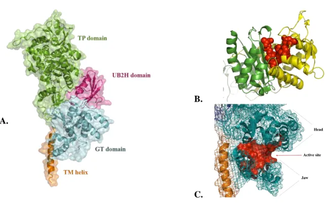
PPB1a and PBP1b essentially have four domains: a single-helix transmembrane domain (TM), a glycosyltransferase domain (GT), a transpeptidase domain (TP) and an additional domain that serves for the interaction with other periplasmic proteins (Sung et al. 2009) (Typas et al. 2010).
The first structure of PBP1b solved to 2.16 Å was reported in 2009 by Sung et al. (Sung et al. 2009) and recently complemented by more detailed model of GT domain by King et al. in which previously missing loop region was traced (figure 1.11a) (King et al. 2017). The protein is anchored to the inner membrane by a 30 residue TM domain (residues 66-96). Residues 83-88 are predominantly hydrophobic and situated close to the GT domain, possibly interacting with it.
The GT domain is highly similar to previously reported GT domain structures of S.
aureus and Aquifex aeolicus (Lovering et al. 2007; Yuan et al. 2008). It is predominantly
α-helical and composed of a globular “head” region and smaller “jaw” positioned below the head, in very close contact with the membrane (figure 1.11c). The active site is located in the shallow groove between these two regions. The TP domain has a typical penicilloyl-serine transferase fold of two subdomains, one five-stranded antiparallel β-sheet flanked by three α-helices on one side and another completely α-helical. The active site is located at the interface of these two subdomains with three conserved motifs (figure 1.11b) (SxxK, SxN and KTGT) (Sauvage et al. 2008).
Between GT and TP domains there is a smaller, PBP1b-specific domain folded into five-stranded β-sheet with one α-helix, in close contact with the TP domain. Structurally it is homologous to a domain from UvrB, a component of the nucleotide excision repair (NER) system in DNA damage repairs, which is why it was named UvrB Domain 2 homolog (UB2H). It was later determined that PBP1b and its activator LpoB are in contact through the UB2H domain of PBP1b (Typas et al. 2010).
37
B.
C.
Figure 1.11. Structure of PBP1b (PDB: 5HLB). A – Domain organization; B – Catalytic residues of the TP domain (red spheres) are at the interface of the two subdomains (in green and yellow). TP domain. C – Active site of the GT domain is in the cleft between “head” and “jaw” regions.
1.13. Penicillin-binding protein 1c
As already mentioned, PBP1c is the third member of the class A PBPs and very little information on its role is available. It was discovered later than the other HMW PBPs as it required special penicillin derivatives to be labeled. Only after application of 125I-labeled
latamoxef or 125I-labeled Bolton and Hunter derivative of ampicillin, PBP1c was observed as
a band between PBP1b and PBP2 (Schwarz et al. 1981)(Labia et al. 1985). This protein, encoded by the pbpc gene, is 770- residues long, with 85 kDa, and it is anchored to the inner membrane. In 1999, it shortly drew attention of Holtje et al. after they discovered that PBP1c was specifically eluted from a MltA-Sepharose column together with PBP1b, PBP2 and PBP3 from the crude cell extract (Vollmer et al. 1999). Initial biochemical characterization showed that PBP1c has high glycosyltransferase activity, with no transpeptidase activity reported (Schiffer & Höltje 1999). Deletion of the pbpc gene did not show any physiological effect or altered peptidoglycan structure. In addition, in the strains with inactivated PBP1a and PBP1b, overexpression of PBP1c could not compensate for the lack of the other two, eventually leading to cell death.
38
AIMS OF THE PROJECT
The discovery of α2M in bacteria was a surprising fact that brought plenty of questions. The fact that bacterial genomes conserved the sequence of such a large protein that would be energetically expensive to produce indicates that it provides advantage to bacteria during their life cycle. As a protease inhibitor localized in periplasm, it is reasonable to believe that it protects the periplasmic space from host proteases during infection. Finding the α2M gene in an operon juxtaposed to the gene of the poorly characterized, efficient transglycosylase PBP1c led to hypothesis that these two proteins form an uncharacterized defense complex that could protect the integrity of periplasm. During infection or during microbial competition events, the outer membrane could be breached, allowing the molecules of the host defense system to penetrate into the periplasm and damage the peptidoglycan. In such scenario, α2M and PBP1c could function as complex in which PBP1c could rapidly repair peptidoglycan, while α2M could protect PBP1c and other periplasmic proteins from proteolysis.
The main goal of this project was to demonstrate the existence of such complex and characterize this interaction structurally and functionally. A deeper understanding of the interaction between α2M and PBP1c could provide the basis for developing novel strategies to combat bacterial infections.
For this purpose, we chose to work with α2M and PBP1c from E. coli. Firstly, that required the biophysical characterization of individual proteins. While the protocols for α2M studies have largely been established, the study of PBP1c required extensive optimization in the phases of cloning, expression and purification. Once the protocols were set up, the aim was to reconstitute the complex in vitro and determine its stoichiometry.
In collaboration with Dr. Waldemar Vollmer’s team from Newcastle University, we analyzed the peptidoglycan synthase activity of PBP1c and determined the effects that α2M has on its activity.
Finally, for the structural analysis of the complex we employed techniques such as small-angle x-ray scattering (SAXS), electron microscopy and crystallography that were essential for different aspects of the study. This work has allowed for a better understanding of the 2M-PBP1c partnership in E. coli, which can reflect a yet undescribed link between bacterial immunity and cell wall formation.
41