HAL Id: hal-01238302
https://hal-amu.archives-ouvertes.fr/hal-01238302
Submitted on 4 Dec 2015
HAL is a multi-disciplinary open access
archive for the deposit and dissemination of sci-entific research documents, whether they are pub-lished or not. The documents may come from teaching and research institutions in France or abroad, or from public or private research centers.
L’archive ouverte pluridisciplinaire HAL, est destinée au dépôt et à la diffusion de documents scientifiques de niveau recherche, publiés ou non, émanant des établissements d’enseignement et de recherche français ou étrangers, des laboratoires publics ou privés.
Selective Cell Vulnerability during Embryogenesis
Yannan Fan, Sylvie Richelme, Emilie Avazeri, Stéphane Audebert, Françoise
Helmbacher, Rosanna Dono, Flavio Maina
To cite this version:
Yannan Fan, Sylvie Richelme, Emilie Avazeri, Stéphane Audebert, Françoise Helmbacher, et al.. Tissue-Specific Gain of RTK Signalling Uncovers Selective Cell Vulnerability during Embryogene-sis. PLoS Genetics, Public Library of Science, 2015, 11 (e1005533 ), �10.1371/journal.pgen.1005533�. �hal-01238302�
Tissue-Specific Gain of RTK Signalling
Uncovers Selective Cell Vulnerability during
Embryogenesis
Yannan Fan1☯, Sylvie Richelme1☯, Emilie Avazeri1☯, Stéphane Audebert2,
Françoise Helmbacher1, Rosanna Dono1‡, Flavio Maina1‡*
1 Aix-Marseille Université, CNRS, IBDM UMR 7288, Parc Scientifique de Luminy, Case 907, Marseille, France, 2 Aix-Marseille Université UM 105, CNRS UMR7258, Inserm U1068, CRCM, Institut Paoli-Calmettes, Marseille, France
☯ These authors contributed equally to this work. ‡ RD and FM are joint senior authors on this work. *[email protected]
Abstract
The successive events that cells experience throughout development shape their intrinsic capacity to respond and integrate RTK inputs. Cellular responses to RTKs rely on different mechanisms of regulation that establish proper levels of RTK activation, define duration of RTK action, and exert quantitative/qualitative signalling outcomes. The extent to which cells are competent to deal with fluctuations in RTK signalling is incompletely understood. Here, we employ a genetic system to enhance RTK signalling in a tissue-specific manner. The chosen RTK is the hepatocyte growth factor (HGF) receptor Met, an appropriate model due to its pleiotropic requirement in distinct developmental events. Ubiquitously enhanced Met in Cre/loxP-based Rosa26stopMetknock-in context (Del-R26Met) reveals that most tissues are capable of buffering enhanced Met-RTK signalling thus avoiding perturbation of devel-opmental programs. Nevertheless, this ubiquitous increase of Met does compromise selected programs such as myoblast migration. Using cell-type specific Cre drivers, we genetically showed that altered myoblast migration results from ectopic Met expression in limb mesenchyme rather than in migrating myoblasts themselves. qRT-PCR analyses show that ectopic Met in limbs causes molecular changes such as downregulation in the expression levels of Notum and Syndecan4, two known regulators of morphogen gradi-ents. Molecular and functional studies revealed that ectopic Met expression in limb mesen-chyme does not alter HGF expression patterns and levels, but impairs HGF bioavailability. Together, our findings show that myoblasts, in which Met is endogenously expressed, are capable of buffering increased RTK levels, and identify mesenchymal cells as a cell type vulnerable to ectopic Met-RTK signalling. These results illustrate that embryonic cells are sensitive to alterations in the spatial distribution of RTK action, yet resilient to fluctuations in signalling levels of an RTK when occurring in its endogenous domain of activity.
OPEN ACCESS
Citation: Fan Y, Richelme S, Avazeri E, Audebert S, Helmbacher F, Dono R, et al. (2015) Tissue-Specific Gain of RTK Signalling Uncovers Selective Cell Vulnerability during Embryogenesis. PLoS Genet 11 (9): e1005533. doi:10.1371/journal.pgen.1005533 Editor: Nadia Dahmane, Perelman School of Medicine, University of Pennsylvania, UNITED STATES
Received: November 25, 2014 Accepted: August 25, 2015
Published: September 22, 2015
Copyright: © 2015 Fan et al. This is an open access article distributed under the terms of theCreative Commons Attribution License, which permits unrestricted use, distribution, and reproduction in any medium, provided the original author and source are credited.
Data Availability Statement: All relevant data are within the paper and its Supporting Information files. Funding: This work was supported by grants from INCa (Institut National du Cancer), FdF (Fondation de France), ARC (Association pour la Recherche contre le Cancer), FRM (Fondation pour la Recherche Médicale), AFM (Association Française contre les Myopathies), Fondation Bettencourt-Schueller to FM; AFM to RD. The contribution of the Region Provence Alpes Côtes d’Azur and of the Aix-Marseille Université to the IBDM animal facility, and of the France-BioImaging/PICsL infrastructure
(ANR-10-Author Summary
The need to achieve precise control of RTK activation is highlighted by human pathologies such as congenital malformations and cancers caused by aberrant RTK signalling. Identi-fying strategies to restrain RTK activity in cancer and/or to reactivate RTKs for counteract-ing degenerative processes is the focus of intense research efforts. We designed a genetic system to enhance RTK signalling during mouse embryogenesis in order to examine the competence of cells to deal with changes in RTK inputs. Our data reveal that most embry-onic cells are capable of: 1) handling moderate perturbations in Met-RTK expression lev-els, 2) imposing a threshold of intracellular signalling activation despite elevated Met-RTK inputs, and/or 3) integrating variable quantitative levels of Met-RTK signalling within bio-logical responses. Our results also establish that certain cell types, such as limb mesen-chyme, are particularly vulnerable to alterations of the spatial distribution of RTK expression. The vulnerability of limb mesenchyme to enhanced Met levels is illustrated by gene expression changes, by interference with HGF chemoattractant effects, and by loss of accessibility to incoming myoblasts, leading to limb muscle defects. These findings high-light how resilience versus vulnerability to RTK fluctuation is strictly linked to cell compe-tence and to the robustness of the developmental programs they undergo.
Introduction
Signalling by receptor tyrosine kinases (RTKs) coordinates developmental processes and ensures tissue homeostasis. Upon ligand stimulation, RTKs activate a number of intracellular signalling cascades to influence in cells identity acquisition, movement, survival, and prolifera-tion [1]. Cellular responses to RTKs rely on different mechanisms of regulation that establish proper levels of RTK activation, define timing of action, and exert quantitative and/or qualita-tive signalling outcomes. Mechanisms of RTK regulation involve transcriptional or post-tran-scriptional control, lateral membrane distribution, endocytosis, and intracellular
compartmentalisation, for example early endosomes in which RTKs activate distinct effectors [2,3]. The need to achieve precise control of RTK activation is highlighted by human patholo-gies caused by altered RTK signalling such as congenital malformations and cancer [2], and establishing strategies to restrain RTK function in disease cells is currently the focus of intense efforts [4]. Despite extensive studies, we are still far from understanding the fundamental prin-ciples of how cells perceive and integrate both quantitative and qualitative RTK signalling levels and how excess RTK signalling can perturb cellular and developmental homeostasis.
The vulnerability of cells to excess RTK signalling is conditioned by their competence to respond to an instructive RTK ligand by changing their behaviour. Such competence is influ-enced by the previous history of cells, during which their intrinsic capacity to respond to a given signal has been shaped through gene regulatory events leading to the acquisition or loss of molecular components necessary for the response (e.g. receptors, co-receptors, signalling modules, and transcription factors). Cellular responses to a given RTK are also conditioned by the simultaneous activation of other signalling inputs and by their synergistic or antagonistic nature [5,6]. Vulnerability of cells to an excess of a given RTK may also vary according to whether such excess results in an enhanced activation over endogenous levels, or whether this excess would result in ectopic activation in cells that would not normally express the RTK. Thus, cell vulnerability is most likely determined by the relative contribution of all these aspects and by the intensity of RTK signalling fluctuation.
INSB-04-01) for the Imaging facility, are also acknowledged. The funders had no role in study design, data collection and analysis, decision to publish, or preparation of the manuscript. Competing Interests: The authors have declared that no competing interests exist.
As highlighted by studies in cellular and animal models, constitutive activation of RTKs results in altered duration/intensity of intracellular signalling and/or participation of additional pathways not recruited under physiological conditions. Unsurprisingly, molecular pathways activated by aberrant RTKs are integrated within other signalling networks through crosstalk, causing signalling perturbations that in turn impact cell behaviour [7]. However, most of these studies have involved models in which enhanced activation of RTKs is achieved through strate-gies such as activating point mutations, oncogenic translocation, and gene amplification. These strategies lead to constitutive activated forms of RTKs that escape regulatory mechanisms, which normally modulate signalling intensity and duration. These strategies are therefore inap-propriate to assess whether cells are capable of: a) buffering subtle fluctuations in RTK activa-tion over physiological levels, and b) imposing a threshold of intracellular signalling activaactiva-tion despites the level of RTK input.
In order to provide insights into the capacity of cells to integrate enhanced RTK signalling above endogenous levels within developmental programs, we have engineered the first mouse model in which RTK signalling can be conditionally enhanced in a tissue-specific manner. Importantly, we opted for the wild-type version of the RTK to avoid perturbation of cellular mechanisms regulating signalling maintenance, extinction, and intracellular location. The cho-sen RTK is the hepatocyte growth factor (HGF) receptor Met, an appropriate model due to: a) its pleiotropic properties elicited in distinct cell types [8]; b) the variety of biological response regulated in different developmental programs and tissue homeostasis [9–11]; c) its impact on tumour evolution and resistance to anticancer therapies [8,12–14]. During embryogenesis, alteration of the HGF/Met system in mice causes liver, placenta, muscle, and neuronal defects [15–23]. Development of muscle in the limb represents a biological model of crosstalk between limb mesenchyme and myoblasts that migrate from the somites to colonize the limb buds. Whereas migrating myoblasts express Met, limb mesenchymal cells produce the HGF acting as a chemoattractant for incoming myoblasts [15,18,23]. Acquisition of motility by myoblast at limb levels is strictly dependent on HGF/Met, with no other redundant molecules capable of bypassing loss of Met signalling.
To achieve conditional gain of wild-type Met-RTK signalling above an endogenous level, we employed the Cre-loxP strategy [24]. In particular, we generated Rosa26LacZ-stop-Met(R26stopMet) transgenic mice that permit, upon Cre-mediated excision, to: 1) increase Met expression above endogenous levels and/or 2) impose Met signalling in cells that do not normally express Met [24]. By using the R26stopMettransgenic mice, we previously demonstrated that enhancing Met signalling in the neural lineage delays the onset of motor neuron degeneration in a murine model of amyotrophic lateral sclerosis [24]. Moreover, these mice revealed the levels of HGF required for retinal ganglion cells survival and axonal growth after optic nerve axotomy lesion [25]. In the present study, we combined R26stopMettransgenic mice with either ubiquitous or tissue-specific-Cre transgenic lines to determine which cells are vulnerable to moderately enhanced Met-RTK expression levels during development. These genetic studies revealed that exogenous Met phosphorylation is restricted to few territories in developing embryos, thus highlighting the existence of a map of competence to Met activation. Intriguingly, we found that most cells, whether relying on endogenous Met signalling or being normally devoid of Met expression, can buffer a moderate increase in RTK signalling. This correlates with a limited propagation of the enhanced Met-RTK input to its downstream signals. This demonstrates that the signalling network is resilient to intensity changes in Met-RTK input for normal devel-opment. However, one cell type, limb mesenchyme, is vulnerable to ectopic Met expression, as evidenced by a drastic impairment of muscle morphogenesis, and by molecular changes in the expression levels of some extracellular regulators of morphogens. Ectopic expression of Met in mesenchymal cells, although not interfering with early limb patterning events, prevents
myoblast migration in a non-cell autonomous manner, leading to a drastic reduction of limb muscles. In contrast, enhanced Met signalling in myogenic cells themselves does not interfere with their migration or with limb muscle development. Vulnerability of limb mesenchyme is due to altered HGF bioavailability in the limbs, as assessed by functional studies. Together, our findings show that the accomplishment of developmental programs in a genetic setting with enhanced Met levels is ensured by the robustness of cell competence to buffer increased RTK activity and to conserve functional downstream signalling. These findings also indicate that developmental programs are sensitive to alterations in the spatial distribution of RTK action.
Results
Modelling enhanced wild-type Met signalling during mouse
embryogenesis
We have previously reported the generation of the Rosa26LacZ-stop-Met(R26stopMet) mice, which allow Met levels to be enhanced in a temporal and spatial regulated manner, and have used them to demonstrate that boosting neuronal RTK signalling delays disease onset in an amyo-trophic lateral sclerosis animal model [24]. In the present study, we used the R26stopMetmice to explore genetically the robustness of developmental programs following enhanced wild-type RTK expression. To enhance Met-RTK signalling levels in all developing tissues, we crossed R26stopMetmice with Tg(CMV-cre)1Cgn/J transgenics (namely Deleter-Cre) [26] thus obtaining Deleter-Cre;R26Metmice/embryos (referred to as Del-R26Met) in order to excise the LacZ-stop cassette in every cells and allow ubiquitous expression of the Met transgene (Mettg;S1A Fig). β-galactosidase staining on whole mount or section of E10.5 embryos, used as readout of absence of recombination, showed a spectrum of recombination in Del-R26Metembryos ranging from cases with only residual staining compared to R26stopMetcontrols (defined as fully recombined Del-R26Metembryos) to cases with a mosaic and more pronounced staining (defined as par-tially recombined Del-R26Metembryos;S1B–S1D Fig). These findings show that the Deleter-Cre line permits excision of the LacZ-stop cassette in all embryonic cell types, although with a variable efficiency. This mosaicism is in part due to the random X-chromosome inactivation in females, as the Cre transgene is X-linked [26]. In the present studies we only used fully recom-bined Del-R26Metembryos. Whole mount in situ hybridization (ISH) with a human Met probe, distinguishing the Mettgfrom the endogenous mouse Met (mMet), showed ubiquitous expres-sion of Mettgin fully recombined Del-R26Metembryos compared to controls (S2 Fig). To fur-ther validate that this approach leads to enhanced Met signalling, we investigated the extent of Met activation in developmental tissues by following its phosphorylation state using anti-phos-pho-Met and anti-human Met antibodies in immunohistochemistry (IHC). Whereas Mettg appeared to be uniformly expressed in most organs in Del-R26Metembryos, phospho-Met staining was detected in restricted cell types (S3 Fig). These findings indicate the existence of a restricted“competence map” for Met activation.
Highly recombined Del-R26Metneonates were identifiable at birth because of their hyper-flexed forelimbs and weak hindlimbs (Fig 1A). Forelimb sections stained with MF20 antibod-ies, which recognise the myosin heavy chain II, revealed that Del-R26Metlimbs were almost devoid of extensor muscles on their dorsal side, and exhibited a strong reduction of flexor mus-cles in the ventral side, compared to controls (Fig 1A). This unbalance between antagonist muscles leads to a pronounced wrist flexure. Whole mount ISH with a MyoD probe at E12.5 revealed a strong reduction of dorsal limb forming muscles in Del-R26Metmutants compared to controls, whereas other sites of MyoD expression were normal (Fig 1B). Next, we crossed Del-R26Metmice with the Mlc3f-2E-nlacZ transgenic mouse line, in which reporter gene expression is driven by the murine Mlc3f promoter and by the Mlc1f/3f-3’ skeletal muscle
Fig 1. Ubiquitously excess wild-type Met in developing embryos results into hyperflexed forelimbs. (A) Top: Del-R26Metand control P0 mice showing hyperflexed limbs in mutants. Bottom: anti-myosin heavy chain II IHC using MF20 antibodies on forelimb transversal sections of P0 Del-R26Metand control mice at the
enhancer [27]. In this mouse line, nLacZ (encoding nuclearβ-galactosidase) is expressed in all skeletal muscles throughout development, permitting visualisation of muscle formation in fully recombined Del-R26Metembryos that no longer express the LacZ-stop cassette. Whole mount staining showed thatβ-galactosidase activity was drastically reduced in Del-R26Metdeveloping limbs compared to controls (Figs1CandS4).
Intriguingly, the limb phenotype of the Del-R26Metmice is reminiscent of the phenotype we reported in Metgrb2/grb2, Met2P/2P, and Met2S/2Sspecificity-switch signalling mutants [18,23,
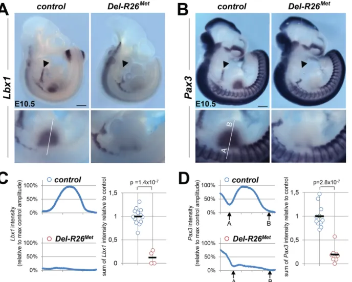
28]. In these Met signalling mutants, Met-dependent migration of myoblasts is severely com-promised, leading to a reduction of limb muscles more pronounced in the dorsal than in the ventral limb compartment [18,23,28]. We therefore assessed whether the development of migrating myoblasts was also compromised in Del-R26Metmice. We addressed this issue using distinct markers of the myogenic program [29]. Whole mount ISH on E10.5 embryos using Lbx1 and Pax3 probes revealed a drastic reduction of migrating myoblasts from the somites towards the limbs, with only few dispersed migrating cells left in Del-R26Metembryos com-pared to controls (Fig 2A and 2B). Quantification of the ISH signal throughout the limbs revealed approximately 90% reduction of migrating myoblasts in Del-R26Metforelimbs com-pared to controls (Fig 2C and 2D). Impaired migration of myoblasts towards the tongue was also found in Del-R26Metembryos (Fig 2A and 2B), indicating that ubiquitous expression of the Mettgalso compromises the developmental program of these migrating myoblasts. Together, these findings show that ubiquitously enhancing wild-type Met-RTK specifically interferes with the limb muscle developmental program by perturbing myoblast migration.
Enhanced wild-type Met expression is permissive in migrating
myoblasts, but not in limb mesenchyme
The intriguing similarity of the limb muscle phenotype in gain-of-function Del-R26Metand in loss-of-function Met specificity-switch signalling mutants [18,23,28] could be interpreted in two ways. One scenario could be that in myoblasts, the amount of Met signalling must be quali-tatively and/or quantiquali-tatively maintained within a narrow range. If the signalling level is reduced (as in Met specificity-switch signalling mutants) or enhanced (as in Del-R26Met mutants), the migration program may not occur. Alternatively, exogenous Met expression might act in limb mesenchymal cells by altering their permissiveness to invading myoblasts. To discriminate between these two possibilities, we selectively enhanced Met expression in either migrating myoblasts or in limb mesenchyme, using the Pax3-Cre knock-in or the Prx1-Cre transgenic lines, respectively [30,31]. Consistently, whole mount ISH in Prx1-R26Metand Pax3-R26Metmutants confirmed that expression of the Mettgis adequately targeted to Prx1 and Pax3 expression domains, respectively (Fig 3A and 3B). Whole mount ISH with Lbx1 and Pax3 probes unequivocally revealed a significant impairment in myoblast migration in Prx1-R26Met, but not in Pax3-R26Metmutants (Figs3C,S5A and S5B). Quantification analyses revealed approximately 50% reduction of limb myoblasts in Prx1-R26Metmutants compared to Pax3-R26Metand control embryos (Figs3D,S5C and S5D). At E12.5, limb-forming muscles were reduced in Prx1-R26Met, but not in Pax3-R26Metembryos, as revealed by MyoD ISH (S5E Fig), even though the severity of this phenotype appeared less pronounced compared to earlier stages. Finally, Prx1-R26Metmice showed hyperflexed forelimbs and weak hindlimbs at birth (arrowhead) muscle mass in mutants. (B, C) Whole mount ISH with MyoD probe of E12.5 embryos (B) and β-galactosidase staining of E11.5 embryos (C) showing that developing appendicular muscles are reduced in Del-R26Metembryos (limbs are outlined in panels). The arrowhead in bottom panel B indicates developing ventral limb muscles (flexor). Scale: 500μm.
(S5E Fig; 90%, n = 16), whereas Pax3-R26Metmutants were indistinguishable from controls. Together, these results disqualify myoblasts and identify limb mesenchymal cells as the cell type in which enhanced Met signalling acts to disrupt myoblast migration. This suggests that myoblast migration towards limb mesenchyme is impaired when exogenous Met expression overlaps with the endogenous source of HGF.
The myoblast migration defect in Prx1-R26Metmutants underlines the importance of spatial restriction of Met expression during development. We therefore asked whether alteration of the regionalised distribution of Met also interferes with the molecular program regulating limb patterning and skeletal morphogenesis. We followed expression of Shh and Fgf8, two major regulators of early limb patterning and growth, and found no obvious defects in Del-R26Met mutants compared to controls at E10.5 (S6A Fig). Furthermore, skeletal staining analyses did not reveal any major defects in bone and cartilage formation in Del-R26Metforelimbs at birth Fig 2. Myoblast migration is impaired in Del-R26Metmutants. (A, B) Whole mount ISH of E10.5 embryos with Lbx1 (A) and Pax3 (B) probes showing drastic reduction of migrating myoblasts towards the developing tongue (arrowhead), fore and hind limbs. Bottom panel reports an enlargement at forelimb levels. (C, D) Quantification analyses of Lbx1 (C) and Pax3 (D) positive domains in forelimbs. Left panels: each plot represents the average signal distribution along the white line in forelimbs. Right panels: quantifications and statistical analyses of the sum of signal intensity based on intensity plots in left panels. Numbers of samples for Lbx1: control, n = 13; Del-R26Met, n = 4; for Pax3: control, n = 11; Del-R26Met, n = 8. The sum of Pax3 signal intensity was calculated between point A and B: A indicating a fixed position between the somites and the limb whereas B being placed at a fixed distance from A. Note almost lack of signal in Del-R26Metmutants. Scale: 500μm. Mann-Whitney and Student-t test.
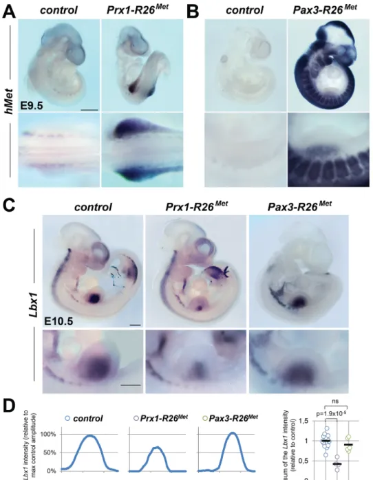
Fig 3. Ectopic Met in limb mesenchyme drastically reduces limb colonization by migrating myoblasts. (A, B) Whole mount ISH using the human Met probe showing the domain with Cre recombinase activity in Prx1-R26Met(A) and Pax3-R26Met(B) embryos. Note that: a) in Prx1-R26Metembryos, the expression of the
Mettg(detected by hMet probe) is restricted in forelimbs; b) in Pax3-R26Metembryos, Mettgexpression is found in Pax3-positive territories. (C) Whole mount ISH of E10.5 embryos with the Lbx1 probe showing drastic reduction of migrating myoblasts in the forelimbs of Prx1-R26Met, but not of Pax3-R26Metmutants.
Note that intact migration of myoblasts towards the forming tongue (arrowhead) in Prx1-R26Metmutants
correlates with the restricted expression of enhanced Met in limb mesenchyme. Bottom panel reports an enlargement at forelimb levels. (D) Quantification analyses of Lbx1 positive domain in forelimbs. Left panels: each plot represents the average signal distribution along the white line in forelimbs. Right panel:
quantifications and statistical analyses of the sum of signal intensity based on intensity plots in left panels. Numbers of samples for Lbx1: control, n = 13; Prx1-R26Met, n = 4; Pax3-R26Met, n = 5. Note the reduced Lbx1
level in Prx1-R26Metmutants. Mann-Whitney and Student-t test. doi:10.1371/journal.pgen.1005533.g003
(S6B Fig). Together, these findings highlight the robustness of limb skeletal patterning pro-gram, ensured by the intercalation of multiple signalling components, which does not permit interference by ectopic Met.
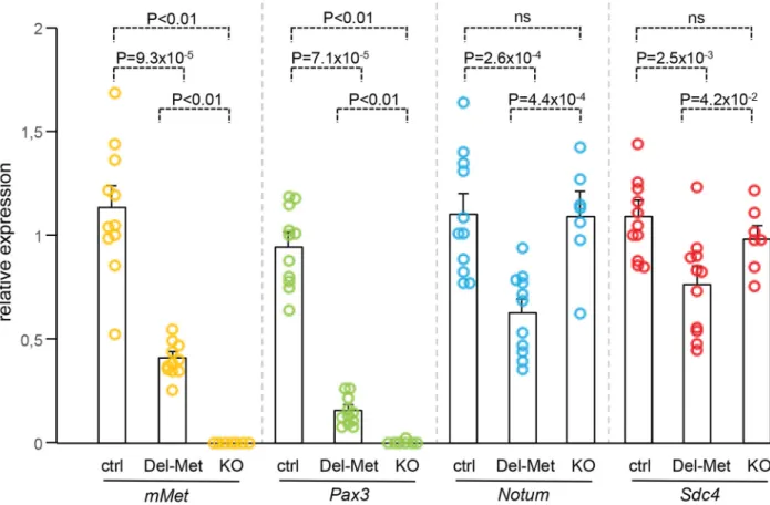
We next analysed whether alteration of the regionalised distribution of Met leads to molecu-lar changes in limb buds by following mainly the expression levels of cell surface proteins such as heparan-sulfate proteoglycans (HSPGs), glypicans [32–34] and syndecans [35], which act as gatekeepers of cellular responses by modulating extracellular signal distribution and perception by targeted cells. We also analysed the expression levels of HSPG modifiers, such as Notum, Hst2st1, and Hst3st1 [36–38]. Among these candidates, some have been previously reported to also modulate HGF signalling [39–41] or to be regulated by HGF/Met [42–44]. We also ana-lysed Sdf1 levels for its cooperative function with Met signalling to control myoblast migration [45]. A total of 12 genes were screened: the 6 Glypicans, Syndecan3 and 4, Hst2st1, Hst3st1, Notum, and Sdf1. To analyse the expression levels of these genes, E10.5 forelimbs dissected from Del-R26Metmutants and controls were used for qRT-PCR. Studies were restricted to embryos that showed low levels ofβ-galactosidase activity in the whole body as a read-out of efficient activation of the Mettg(seeS1 Fig). We first assessed the sensitivity of this approach in detecting molecular changes possibly occurring in a limited number of cells within the limb. In particular, we quantified transcript levels of Pax3 and mouse Met, both expressed in migrating myoblasts, and consistently found a drastic reduction in their mRNA levels in Del-R26Met limbs compared to controls (Fig 4). Second, we confirmed a switch in LacZ versus Mettg expression in control and Del-R26Metlimbs (S7A Fig). We next screened the 12 selected candi-dates by comparing their expression levels in control versus Del-R26Metlimbs. No significant changes were observed in the expression levels of all Glypicans, of Syndecan3, Hst2st1, Hst3st1, and Sdf1 (S7B and S7C Fig). In contrast, we found that expression levels of Notum and Synde-can4 were significantly reduced in Del-R26Metversus control limbs (Fig 4). We next asked whether such molecular differences in Del-R26Metmutants resulted from the depletion of limb myoblasts, or reflected expression changes in limb mesenchyme. To address this question, we took advantage of Met loss-of-function mutants (MetLacZ/d(neo)), in which limbs are also devoid of migrating myoblasts (S8 Fig), but where no specific changes in limb mesenchyme can be expected [18,22]. Consistently, Pax3 and mouse Met myoblast-specific transcripts were also absent from MetLacZ/d(neo)mutant limbs (Fig 4). In contrast, the expression levels of Notum and Syndecan4 (changed in Del-R26Metlimbs) were similar in MetLacZ/d(neo)and control limbs (Fig 4). Altogether, these results exclude that downregulation of Notum and Syndecan4 in Del-R26Metlimbs is a consequence of lack of migrating myoblasts, and identify them as molecular changes caused by Met expression in limb mesenchyme.
Met-expressing myoblasts are capable of buffering enhanced wild-type
Met levels
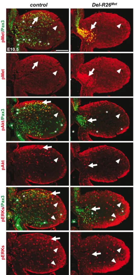
The intact myoblast migration in Pax3-R26Metmutants could reflect either the robustness of the migration response to increased activation levels of Met intracellular effectors or an intrin-sic capability of cells to restrain Met-RTK input. By performing IHC on embryo sections, we followed Met signalling levels in control and mutant forelimbs at E10.5, when myoblast migra-tion occurs. In myoblasts, levels of pTyr1234–1235-Met were enhanced in Del-R26Met
(non-migrating cells blocked at the base of the limb) and Pax3-R26Met(migrating cells progressing within the limb) compared to controls, whereas no major changes were observed in
Prx1-R26Met(where non-migrating myoblasts are also blocked in a proximal position in the limb) (Figs5and6A). By exploring the activation levels of two Met effectors, we found that the phosphorylation levels of Akt and ERKs were comparable in control, Del-R26Met, Pax3-R26Met,
and Prx1-R26Metmyoblasts, despite high levels of phospho-Met in Del-R26Metand Pax3-R26Met(arrows inFig 5andFig 6B) Together, these results show that myoblasts have the competence to buffer enhanced Met levels. In contrast, in limb mesenchymal cells levels of phospho-Met were slightly increased in Del-R26Metand Prx1-R26Metwhen compared to con-trol and Pax3-R26Metembryos (Figs5and6A). This is best seen by comparing levels in areas devoid of myoblasts even in control embryos (arrowheads inFig 5and“distal mes” boxes in
Fig 6A). This was accompanied by an increase in the phosphorylation levels of Akt and more moderately of ERKs (Figs5and6B). These findings indicate that limb mesenchymal cells lacks the capacity to buffer an ectopic Met activation, which in turn causes defects in myoblast migration.
Restriction of enhanced Met signalling occurs at distinct levels as
revealed by biochemical studies in embryonic hepatocytes
We next explored how buffering of enhanced Met signalling occurs in cells. Because of techni-cal difficulties in isolating and establishing primary myoblast cultures from E10.5 embryos for quantitative western blot studies, we addressed this issue using primary embryonic hepato-cytes in which endogenous Met is expressed and required for their survival, as shown through our earlier studies of Met signalling mutants [18,28,46–48]. Primary embryonic hepatocyte Fig 4. Ectopic Met in limb mesenchyme down-regulates the expression levels of Notum and Syndecan4. qRT-PCR analysis of transcript levels of mouse Met (mMet), Pax3, Notum, and Syndecan4 (Sdc4) in controls (ctrl; n = 11), Del-R26Met(Del-Met; n = 11), MetLacZ/d(neo)(KO; n = 7). Each dots
corresponds to transcript levels in forelimbs of E10.5 individual embryos (done in triplicate). Columns correspond to the average value, expressed as mean± s. e.m. Note: downregulation of mMet and Pax3 in Del-R26Metand MetLacZ/d(neo)mutants compared to control, consistent with lack of migrating myoblasts; downregulation of Notum and Syndecan4 in Del-R26Metmutants compared to control, whereas no significant changes were found in MetLacZ/d(neo)mutants. Mann-Whitney and Student-t test.
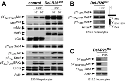
Fig 5. Enhanced Met expression levels in Del-R26Metmyoblasts does not perturb activation of
cultures were established from E15.5 R26stopMetand Del-R26Metembryos and used to follow biochemical changes in the Met signalling cascade. As expected, Del-R26Metembryonic hepato-cytes express the Mettg, but not theβ-galactosidase (in the stop cassette;Fig 7A). Total Met pro-tein levels in R26stopMetand Del-R26Metprimary embryonic hepatocytes were assessed by using the Met25H2antibodies generated against a synthetic peptide containing the amino acids sur-rounding the Tyr1234–1235within the kinase domain (recognising human and mouse Met).
Quantification of the Met25H2levels revealed an approximately 4-fold increase of total Met pro-tein amount in Del-R26Metversus R26stopMetcontrols. Increased levels of Met proteins correlate with a concomitant increase in Met phosphorylation levels on Tyr1234–1235(critical for Met
kinase activation; visualized with an antibody recognising mouse and human phosphorylated Met), which are not further enhanced upon HGF stimulation (Fig 7A). We found two intrigu-ing aspects of enhanced Met expression on downstream components of the Met signallintrigu-ing cas-cade. Concerning Gab1, a cytoplasmic protein directly recruited by Met and functioning as a platform for Met effectors, we found that: a) it was already phosphorylated in Del-R26Metcells to a level comparable to that of control cells upon HGF stimulation, b) its phosphorylation lev-els in Del-R26Metcells was not further enhanced upon HGF stimulation (Fig 7A). Concerning further downstream Met effectors such as Akt, Stat3, and ERKs, we found that their phosphor-ylation levels increased upon HGF stimulation with comparable kinetic profiles in Del-R26Met and control cells (Fig 7A). These findings indicate that the constitutive enhancement of Met expression and phosphorylation on Tyr1234–1235in hepatocytes does not result in a constitutive
activation of downstream signalling components.
To get insights into how enhanced Met signalling is restricted in embryonic cells, we ana-lysed the phosphorylation levels of two other tyrosine residues implicated in receptor signalling and endocytosis. Intriguingly, we found basal levels of Met phosphorylation on Tyr1003(critical
for Met protein ubiquitination and degradation) and on Tyr1349(one of the two
multifunc-tional docking sites required for Met signalling), which were enhanced upon HGF stimulation (Fig 7B). Consistently, Met and Gab1 phosphorylation was impaired in the presence of the Met inhibitor PHA665752 (Fig 7C). Thus, basal levels of Met phosphorylation in mutant embry-onic hepatocytes appear sufficient to initiate signalling activation (e.g. Gab1 phosphorylation on Tyr627), but not to propagate signalling activation to downstream pathways (e.g. Akt, Stat3,
ERKs). Together, these findings indicate that the Mettgachieves its full signalling competence following a burst of HGF stimulation.
Gain of wild-type Met expression in limb mesenchyme does not perturb
expression pattern and levels of its ligand HGF, but its bioavailability
As the domain of Prx1-cre activity in the limb encompasses the source of HGF exerting che-moattraction on migrating myoblasts, we next asked whether ectopic Met in these cells alters the bioavailability of HGF to migrating cells. ISH and IHC analyses revealed no significant dif-ferences on HGF expression patterns or levels between Del-R26Metand control embryos (Fig 8A, 8B and 8C), ruling out the possibility that ectopic Met alters Hgf gene expression and HGF protein production in limb mesenchyme. Moreover, we found comparable protein levels of uncleaved HGF in E10.5 Del-R26Metand control forelimbs (Fig 8D) as well as unchanged showing the distribution of phospho-Met (on Tyr1234–1235), phospho-Akt, phospho-ERKs (red) and of Pax3
protein (green) in myoblasts. Note ectopic phospho-Met in limb mesenchyme (arrowheads) and in non-migrating myoblasts (arrows) in Del-R26Metmutants. Asterisks indicate non-specific staining in blood cells. Scale: 100μm.
Fig 6. Immunohistochemical analysis showing that myoblasts possess a buffering competence to enhanced Met levels. (A) Limb transverse sections of E10.5 control, Prx1-R26Met, and Pax3-R26Met
embryos showing the phospho-Met (red) and Pax3-positive myoblasts (green). Note ectopic phospho-Met in limb mesenchyme of Prx1-R26Metand in migrating myoblasts of Pax3-R26Metmutants. Few migrating
myoblasts are present in limb mesenchyme of Prx1-R26Metmutants. White boxes indicate the position of
enlargements shown in panel B. Yellow boxes indicate distal mesenchyme (distal mes) area devoid of myoblasts where enhanced phospho-Met signal is detected in Prx1-R26Metmutants. (B) High magnification of limb transverse sections showing phosphorylation levels of Met, Akt, and ERKs (red) in migrating
myoblasts (Pax3-positive; green) of E10.5 control, Prx1-R26Met, and Pax3-R26Metembryos. Note that despite
the high phospho-Met levels in Pax3-R26Metmutants, no major changes are observed in phospho-Akt and
phospho-ERKs when compared to controls and Prx1-R26Metmutants. For Prx1-R26Metmutants, two areas are shown: square 1 indicates few migrating myoblasts; square 2 indicates mesenchyme devoid of myoblasts. Asterisks indicate non-specific staining in blood cells. Scale: 100μm.
mRNA levels of urokinase-type plasminogen activator (Plau), an enzyme involved in HGF pro-cessing (Fig 8E).
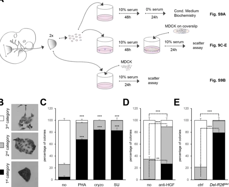
Next, we investigated whether Met expression in mesenchymal territories would alter HGF bioavailability. For this purpose, we established primary embryonic mesenchymal cell cultures from E10.5 forelimbs and biochemically assessed their competence to respond to HGF stimula-tion. As expected, control limb mesenchymal cells did not respond to HGF stimulation, consis-tent with the fact that these cells do not express the receptor (Fig 8F). In contrast, we found high levels of Met phosphorylation on Tyr1234–1235in Del-R26Metlimb mesenchymal cells (Fig
8F). HGF stimulation induced Gab1 phosphorylation and enhanced the basal levels of Akt and ERKs phosphorylation (Fig 8F). Furthermore, treatment with anti-HGF blocking antibodies drastically reduced the phosphorylation levels of Met in Del-R26Metlimb mesenchymal cells, thus showing that HGF is indeed produced by these cells and that basal Met phosphorylation is due to endogenous HGF in these culture conditions (Fig 8F). Finally, we biochemically ana-lysed the levels of HGF in conditioned media of Del-R26Metlimb mesenchymal cells and found that levels of uncleaved HGF were comparable to those of control cells (S9A Fig).
To explore whether the HGF released by control and Del-R26Metcultures was bioactive, we co-cultured MDCK cells with E10.5 dissociated limb mesenchymal cells as a source of HGF Fig 7. Biochemical analyses in embryonic hepatocytes show restriction of enhanced Met signalling at distinct levels. (A) Western blot analyses of total protein extracts of E15.5 primary embryonic hepatocytes derived from R26stopMet(containing the LacZ-stop cassette therefore expressing the
β-galactosidase) and from Del-R26Met(after deletion of the LacZ-stop cassette, therefore expressing the transgenic Met detected by anti-human Met antibodies) embryos. Analyses was performed before and after HGF stimulation (50ng/ml). Note: a) similar levels of mouse Met in R26stopMetand Del-R26Metcells; b) high levels of Met
phosphorylation on Tyr1234–1235in Del-R26Metcells independently of HGF stimulation; c) comparable Gab1,
Akt, Stat3, and ERK phosphorylation levels upon HGF stimulation in R26stopMetand Del-R26Metcells; d) despite Gab1 phosphorylation in untreated Del-R26Metcells in contrast to control R26stopMetcells, the phosphorylation levels of Akt, Stat3, and ERK is unchanged. Actin protein levels were used as loading controls in all western blot analyses. (B) Left: western blot analyses of total protein extracts of E15.5 primary embryonic hepatocytes derived from Del-R26Metembryos. Note basal levels of Met phosphorylation on Tyr1003and on Tyr1349; phosphorylation levels are further increase upon HGF stimulation (50ng/ml). Right:
schematic representation of Met indicating the different tyrosine residues analysed by western blots. (C) Western blot analysis of total protein extracts of E15.5 primary embryonic hepatocytes derived from Del-R26Metembryos showing impaired phosphorylation of Met and Gab1 in the presence of the Met inhibitor PHA665752 (PHA; 1μM).
(Fig 9A). MDCK cells acquire a“scattered phenotype” upon HGF stimulation [49,50], thus providing an excellent readout of HGF bioavailability in conditioned media by limb mesenchy-mal cells. To evaluate HGF bioavailability, we established an experimental setting in which MDCK cells were co-cultured either with dissociated limb mesenchymal cells or with dissected limbs (Fig 9A). HGF activity was quantified by scoring the percentage of MDCK clones exhib-iting a dense phenotype (category I), an expanded phenotype (category II), or a scattered phe-notype with some or several cells detached from the clone (category III;Fig 9B). A condition with mesenchymal cells from two control forelimbs consistently triggered an efficient MDCK Fig 8. Ectopic Met does not change the expression profile and levels of HGF in limb mesenchyme. (A) Whole mount ISH of E10.5 control and Del-R26Metembryos with Hgf probe showing comparable expression
domain in developing forelimbs. (B) Quantification analyses of Hgf positive domains in forelimbs. Left panels: each plot represents the average signal distribution along the white line in forelimbs. Right panels:
quantifications and statistical analyses of the sum of signal intensity based on intensity plots in left panels. Numbers of samples: control, n = 6; Del-R26Met, n = 6. Note no significant changes of signal in controls and
Del-R26Metmutants. (C) Immunofluorescence analysis of HGF (red) and Pax3 (green) proteins in transversal
sections of E10.5 control and Del-R26Metembryos at forelimb levels. Note the impaired myoblast migration (Pax3-positive cells) in Del-R26Metmutants, whereas no significant changes are detected in HGF protein levels. Asterisks indicate non-specific staining in blood cells. Scale: 100μm. (D) Western blot analyses of total protein extracts of E10.5 control and Del-R26Metdissected forelimbs. Note comparable levels of uncleaved
HGF in Del-R26Metmutants despite the ectopic expression of Mettgand the enhanced phosphorylation levels of Met and Gab1. (E) qRT-PCR analysis showing comparable Plau transcript levels in E10.5 dissected forelimbs of control (ctrl; n = 11) and Del-R26Met(Del-Met; n = 11) embryos. (F) Western blot analysis of total
protein extracts of E10.5 forelimb mesenchymal cell cultures showing phosphorylation levels of Met, Gab1, Akt, and ERKs in control and Del-R26Metcultures, in cells untreated, stimulated with HGF (H) or in the presence of anti-HGF blocking antibodies (αH).
scattering response (Fig 9C). MDCK scattering response triggered by conditioned media from control limb mesenchymal cells was drastically reduced in the presence of Met inhibitors (PHA665752, cryzotinib, or SU11274: 1μM;Fig 9C) or of anti-HGF blocking antibodies (anti-HGF: 30μg/ml;Fig 9D). In contrast, we found a drastic reduction in the percentage of MDCK colonies with a scattered phenotype using mesenchymal cells from two Del-R26Metforelimbs (Fig 9E). Similar results were obtained using MDCK cells directly co-cultured with E10.5 Fig 9. Ectopic Met in limb mesenchyme alters HGF bioavailability. (A) Scheme illustrating the experimental procedure employed for evaluating through MDCK cell scattering the bioavailability of HGF from control and Del-R26Metmutant limb mesenchymal cells or from dissected forelimbs. The scheme indicates the experimental procedure applied for collecting media conditioned by limb mesenchymal cells for biochemical analysis (top; shown inS9A Fig), for MDCK scattering assays using co-cultures with limb mesenchymal cells (middle; shown in Fig 9C, 9D and 9E) or with dissected limbs (bottom; shown in
S9B Fig). (B) Pictures of MDCK colonies showing the three categories that were defined to determine the extent of cell contact and spreading for quantification studies of scattering response. (C) Quantitative analysis of MDCK cell scattering in co-cultures with control limb mesenchymal cells in the absence (no) and in the presence of the Met inhibitor PHA665752 (PHA; 1μM), cryzotinib (Cryzo; 1μM), or SU11274 (SU; 1μM). (D) Quantitative analysis of MDCK cell scattering in co-cultures with control limb mesenchymal cells in the absence (no) and in the presence of the anti-HGF blocking antibodies (anti-HGF; 30μg/ml). (E) Quantitative analysis of MDCK cell scattering in co-cultures with control or Del-R26Metlimb mesenchymal cells. Note a drastic reduction in the scattering response when MDCK cells are co-cultured with Del-R26Metmutant cells (control: n = 4; Del-R26Met: n = 3). Mann-Whitney and Student-t test.
forelimb explants (S9B Fig). Together, these findings indicate that although ectopic Met expression in limb mesenchyme does not alter HGF expression pattern or levels, it impairs bio-availability of HGF from mesenchymal cells.
Discussion
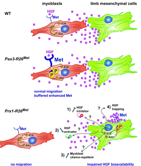
The events that cells experience overtime shape progressively their intrinsic capability to respond and to integrate RTK inputs. Our data illustrate that enhanced wild-type RTK signal-ling is differentially interpreted by cells according to their competence and the robustness of developmental programs they undergo. Most developing cell types appear insensitive to changes in Met expression either because enhanced Met protein levels is not followed by its activation (e.g. in the absence of ligand), or because it is neutralized by the action of other sig-nals acting on cells. This is not the case of migrating myoblasts that, although insensitive them-selves to enhanced Met expression, failed to migrate towards the limb buds as a result of ectopic Met expression in mesenchymal cells. Ectopic Met in the mesenchyme perturbs tran-scriptional regulation of some genes and interferes with HGF bioavailability, thus rendering the mesenchyme non accessible to myoblasts (Fig 10). Our results demonstrate that whereas most cell types during development are capable of handling moderate perturbations in RTK signalling levels, certain cell types are vulnerable to alterations in changes of spatial RTK action.
Cell competence to Met activation
The strategy of using the wild-type Met-RTK, rather than constitutive active forms, to generate Del-R26Metmice has enabled expression of the receptor at more physiological levels. A moder-ate increase in Met levels during development drastically affects limb muscle formation by per-turbing myoblast migration. Although we cannot exclude subtle phenotypes in other
developing organs, these results highlight the restricted sensitivity of the myoblast migration process to ectopic Met in limbs. We genetically demonstrate that myoblasts themselves are not susceptible to enhanced Met signalling levels for their migration, when gain of expression is achieved in the myogenic lineage with Pax3-Cre. This context contrasts with a genetic setting in which Pax3 gain-of-function triggers ectopic myoblast delamination and migration at non-limb somite levels, caused by Met over-expression [51]. It is therefore tempting to propose the existence of a threshold level of sensitivity to enhanced Met signalling for migrating myoblasts: Met signalling is enhanced below this level in Del-R26Met, thus failing to elicit any qualitative change in myoblast behaviour, whereas Met signalling is enhanced above this threshold in Pax3 gain-of-function mutants, thus triggering ectopic delamination. However, it is also possi-ble that the effects of Pax3 gain-of-function mutants involve missregulation of other Pax3 tar-get genes. The Met specificity-switch mutants have previously revealed that myoblast
migration is impaired by lowering qualitative and/or quantitative Met signalling levels [18,28]. Thus, findings from the present studies together with those discussed here highlight the exis-tence of a precise window of Met signalling compatible with proper myoblast migration: within this window, cells can buffer fluctuations in RTK signalling input to avoid changes in qualita-tive output. This also implies that myoblasts are competent to read Met signalling for proper migration within a defined quantitative range.
Modulating HGF bioavailability in limb mesenchyme for muscle
patterning
Our studies also uncover another intriguing aspect linked to myoblasts migration: their suscep-tibility to alterations in the spatial distribution of Met expression. The limb mesenchyme is a
local source of HGF exerting a chemoattractant effect on myoblasts that migrate from the somites. In a wild-type context, migrating myoblasts experience a territory devoid of Met-posi-tive cells along their path. Our results ruled out that ectopic Met would act by altering the HGF mRNA and protein expression levels in limb mesenchyme. Instead, they provide evidence that Fig 10. Schematic representation summarizing the different molecular and phenotypic effects of enhanced Met expression in myoblasts and limb mesenchymal cells. In a wild-type context (top), limb mesenchymal cells secrete HGF required for migration of myoblasts towards the limb buds. Enhanced expression of Met in myoblasts (as assessed in Pax3-R26Metembryos; middle) does not alter their migration due to a buffering event: the activation levels of signalling effectors such as ERKs and Akt are restrained despite enhanced Met phosphorylation. The size of each signal is representative of their
phosphorylation levels. Limb mesenchymal cells are vulnerable to ectopic Met expression (as assessed in Prx1-R26Metembryos; bottom), illustrated by
changes in gene expression, by failure of HGF bioavailability, and by myoblast migration defects. Alteration of HGF bioavailability can be due to: 1) upregulation of a negative interactor that would interfere with the capacity of HGF to bind/activate Met (indicated as“HGF inhibitor”), 2) downregulation of a HGF interactor acting as enhancer of its bioactivity (indicated as“HGF activator”), 3) expression of a chemorepellent factor that renders limb mesenchyme inaccessible to migrating myoblasts, or 4) HGF titration by ectopic Met in mesenchymal cells (indicated as“HGF trapping”).
limb mesenchyme must be devoid of Met expressing cells to ensure normal HGF bioavailabil-ity. Although we cannot fully exclude that ectopic Met acts by titrating the HGF secreted by limb mesenchyme, a series of experimental arguments make this possibility unlikely. Compara-ble levels of HGF protein were found in conditioned media from control and Del-R26Metlimb mesenchymal cells, in spite of reduced scattering-induced activity, indicating that HGF secre-tion and release is not impaired by the occurrence of autocrine HGF/Met binding. Moreover, an event of HGF trapping by Met should recapitulate all developmental defects observed in Met loss-of-function mutants. Although the molecular mechanism remains to be identified, it is tempting to speculate that defects in HGF bioavailability could be linked to transcriptional and/or posttranscriptional changes caused by ectopic Met in limbs. This possibility is sup-ported by our data showing signalling (e.g. phospho-Met, phospho-Gab, and phospho-ERKs upregulation) and transcriptional (e.g. Notum and Syndecan4 downregulation) changes occur-ring in mutant limbs compared to controls. Thus, ectopic Met expression and activation in limb mesenchyme triggers a number of qualitative responses that cannot be buffered by cells. Our co-culture studies highlight a difference in HGF bioavailability, which could result from a change in expression levels of a modulator of HGF, such as either the downregulation of a HGF interactor acting as enhancer of HGF bioactivity, or the upregulation of a negative inter-actor that would interfere with the capacity of HGF to bind/activate Met (Fig 10). Among posi-tive modulators, it has been shown that Glypican1 regulates HGF/Met-triggered migration of C2C12myoblasts [52], Syndecan4 is expressed together with Met in adult satellite cells where it
contributes to muscle regeneration [53,54], and Glypican4 sustains HGF-mediated branching morphogenesis [39]. Alternatively, mutant cells could express a chemorepellent factor that would render the limb mesenchyme inaccessible to migrating myoblasts (Fig 10). The identifi-cation and the functional validation of the molecular mechanism(s) responsible for the non-cell autonomous defect of myoblast migration in Del-R26Metmutants is highly challenging, as: 1) a putative HGF modulator could be altered at the level of transcription, post-transcription, and/or secretion; 2) modulators of chemoattractants do not exert an“on or off” action, but rather fine tune signalling mechanisms, and may act redundantly with one another; 3) distinct mechanisms could cooperate to cause this qualitative phenotype, each of them participating although with a variable quantitative extent.
Robustness versus sensitiveness of developmental programs to
fluctuation on RTK signalling levels
By using motor neurons as a cellular paradigm, we have provided examples of robustness ver-sus sensitivity to RTK signalling in developmental outcomes [22,23]. We have shown that a specific motor neuron pool requires HGF/Met for survival, hence for proper maintenance of muscle innervation. In contrast, neighbouring Met-expressing motor neuron pools do not depend on the action of HGF/Met for their survival, in spite of their early dependence for motor neuron subtype specification and axon growth [22]. Furthermore, we have shown that whereas HGF/Met-dependent motor neuron survival and fate specification is permissive to substitutions of downstream signalling routes, nerve growth patterning is strictly dependent on a selective, non-substitutable, pathway [23]. Intriguingly, signalling requirements for cell sur-vival in motor neurons is different than those in hepatocytes [46–48], demonstrating how sig-nalling requirements for RTK-triggered biological outcomes is refined by the type of cells. Altogether, these results are consistent with the model whereby the degree of equivalence between signalling cascades available to RTK-mobilisation within a developmental program for a given biological outcome is refined in a cell type-dependent manner, thus determining the degree of robustness versus sensitiveness to RTK signals according to cell competence.
The present study further expands our knowledge on robustness versus sensitiveness of cells in developmental processes with respect to subtle increases in RTK signalling. Besides the sen-sitivity of myoblasts to changes in Met signalling in limb mesenchyme, most other develop-mental processes appear robust and capable of buffering excess Met-RTK signalling (Fig 10). How can most cells deal with RTK signalling perturbation? For cells with endogenous Met, one possibility is that they can buffer the additional burst of RTK signalling by“considering” it as a negligible quantity with respect to the overall signalling network operating in cells. Such buffer-ing competence—or resilience—would be possible as long as this additional burst of RTK occurs within a defined quantitative window. Our biochemical studies show that effectiveness of the enhanced Met expression and activation is limited at different intracellular levels. A first point of restriction occurs at the level of the receptor itself: although Mettgis fully phosphory-lated on Tyr1234–1235that are critical for its kinase activation, it only becomes fully signalling
competent after HGF stimulation, as shown by HGF-induced increased phosphorylation levels on Tyr1003and Tyr1349implicated in receptor signalling and endocytosis and by activation of
Akt, STAT3, and ERKs. Thus, in Del-R26Metcells, HGF switches Met from a subthreshold sig-nalling status into a fully sigsig-nalling competent form. These results also show that the R26stopMet mice represent a valuable genetic model to explore the consequences of moderate perturbations of Met as its full signalling competence still depends on ligand stimulation. A second point of restriction occurs at the levels of Gab1, which is phosphorylated at comparable levels in control and Del-R26Metcells upon HGF stimulation. Notably, in the absence of HGF stimulation Gab1 is phosphorylated in Del-R26Methepatocytes (also expressing endogenous Met), but not in Del-R26Metmesenchymal cells (not expressing endogenous Met), indicating that the competence of cells to restrict signalling of enhanced Met is different according to the cell type, and that this competence can be influenced by factors such as expression levels of the receptor. Another restriction occurs further downstream, as revealed by the comparable phosphorylation levels of Akt, STAT3, and ERKs observed in control and Del-R26Methepatocytes (by biochemical stud-ies in culture) and myoblasts (by IHC in vivo). Such restrictions imply that different cell types possess distinct mechanisms that sense and calibrate the level of activation required for and compatible with biological programs. Overall, our studies provide additional insights into the existence of an exquisite monitoring of signalling levels within cells to attenuate enhanced RTK levels.
For cells that do not express endogenous Met, the issue of buffering an ectopic Met is only relevant provided that cells possess the adequate co-factors and downstream effectors. When they do, buffering may occur through mechanisms similar to those in cells with endogenous Met. Whereas most territories/cell types with ectopic Met appear capable of resilience, the limb mesenchyme is vulnerable to this signalling perturbation, illustrated by changes in gene expres-sion, by failure of HGF bioavailability, and by resulting myoblast migration defects.
Conditional R26
stopMetmouse model to enhance wild-type Met signalling
in skeletal muscle
The conditional R26stopMetmouse model represents a valuable tool for future studies on whether and how enhanced Met signalling impacts muscle development at later time points and during regeneration [55]. This issue is consistent with the persistence of Met expression in migratory muscles even after completion of the migration process, as shown by MetLacZ/+ embryos at E13.5 (S8 Fig). The migration defect we report here may be clinically relevant as failure of subsets of myoblasts to migrate properly might represent a developmental dysfunc-tion at the root of some muscle pathologies. This link is nicely illustrated by our recent findings showing that alterations of Fat1 are associated with Facioscapulohumeral dystrophy (FSHD)
[56]. Loss of Fat1 in mice causes muscle shape defects resulting from altered migration polarity of a selective group of muscles matching those affected in FSHD [56]. Fat1 expression is also regulated by HGF/Met, by Pax3-FKHR, and by Lbx1 [42,45,57]. Alteration of Met signalling can also impact muscle homeostasis by causing atrophy [58] and tumour formation. For exam-ple, Met is overexpressed in rhabdomyosarcoma, where it contributes to invasive growth [57], and Met gene amplification is frequently associated with sarcoma susceptibility in muscular dystrophy mouse models [51]. Therefore, R26stopMetmice can be instrumental for future evalu-ation of how Met signalling impacts on muscle physiology, regenerevalu-ation, and/or pathologies.
Conclusion
The R26stopMetmice modelling conditional gain of RTK signalling exemplify robustness versus sensitiveness of cells to signalling perturbations in order to ensure reproducible developmental outcomes. The genetic approach we employed permits subtle changes in RTK signalling, in contrast to others where constitutive RTK activation, either by point mutations or ligand over-expression, leads to dramatic biological consequences. It is therefore not surprising that the Del-R26Metmice do not recapitulate defects reported in other transgenics with an over-activa-tion of the HGF/Met system. It is the case of mice in which HGF over-expression causes aber-rant myoblast and neural crest migration, leading to ectopic muscle formation and melanosis in the central nervous system [59,60]. A number of transgenic mice have been instrumental to demonstrate the capability of oncogenic forms of RTKs to trigger neoplasia. However, these mice do not permit assessment of how cells perceive and handle moderate changes in RTK sig-nalling overtime. The R26stopMetgenetic setting represents therefore a suitable system to explore the in vivo robustness of cells to subtle increase of RTKs signalling during tissue homeostasis as well as during development and may disclose unexpected switches in cell sensitivity from development to adulthood.
Materials and Methods
Ethics statement
All procedures involving the use of animals were performed in accordance with the European Community Council Directive of 24 November 1986 on the protection of animals used for experimental purposes (86/609/EEC). The experimental protocols were carried out in compli-ance with institutional Ethical Committee guidelines for animal research (comité d’éthique pour l’expérimentation animale–Comité d’éthique de Marseille; agreement number D13-055-21 by the Direction départementale des services vétérinaires–Préfecture des Bouches du Rhône).
Transgenic lines and genotype analysis
The generation of the R26stopMetmice (international nomenclature Gt(ROSA)26Sortm1(Actb-Met)Fmai) carrying a conditional mouse–human chimeric Met transgene into the Rosa26 locus was previ-ously reported [24,25]. The mouse lines expressing Cre recombinase under Prx1 [31] and Pax3 [30] promoters, the Deleter-Cre [26], Mlc3F2E-LacZ transgenic [27], the Metd(neo/+)[18] and MetLacZ/+[22] mouse lines were previously described. Mice and embryos were genotyped by PCR analysis of genomic DNA as reported in the above studies. For embryo collection, embryonic days were counted considering midday post-coitum as E0.5. All procedures involv-ing the use of animals were performed in accordance with the European Community Council Directive of 24 November 1986 on the protection of animals used for experimental purposes (86/609/EEC). The experimental protocols were carried out in compliance with institutional
Ethical Committee guidelines for animal research (comité d’éthique pour l’expérimentation animale–Comité d’éthique de Marseille; agreement number D13-055-21 by the Direction départementale des services vétérinaires–Préfecture des Bouches du Rhône).
RNA in situ hybridisation and histological analysis
For ISH, embryos were collected in phosphate buffered Saline (PBS) and fixed in 4% parafor-maldehyde (PFA) overnight. Whole mount RNA ISH done by using the relevant digoxigenin-labeled RNA probes. X-Gal and Salmon-Gal staining were performed as previously described [23,24,61]. For IHC of P0 limbs, newborns were embedded in cold glycol methacrylate (Tech-novit 8100) for cryosections. Limbs were cut transversally (16μm) and processed for antibodies staining as described [24]. For IHC at E10.5, embryos were embedded in paraffin, cut transver-sally (10μm), and processed for antibodies staining as described [62].
MDCK scattering assays
Procedures were performed as described [49,50]. Briefly, MDCK cells (ATCC, Rockville, MD) were seeded in 96-well plates (for forelimb co-cultures; Corning, Acton, MA) or on coverslips (for forelimb mesenchymal cells co-cultures) at 1250 cells/cm2in DMEM containing 10% (v/v) fetal bovine serum (FBS), 100U/mL penicillin, 100μg/mL streptomycin, 4mM L-glutamine, and 1mM of sodium pyruvate. Cells were first incubated for 24 h at 37°C, in 5% CO2to allow
attachment and colony formation. For forelimb co-cultures, plates were processed as followed. In the first group, the media was changed with fresh media containing increasing concentra-tions of recombinant human HGF (0.1, 0.3, 1, 3, 10, 30ng/ml; R&D) to estimate the bioactivity dose of HGF released by wild-type limbs (comparable to 5ng/ml of recombinant human HGF). In the second group, media was replaced and added together with freshly dissected 2 forelimbs derived from E10.5 embryos of different genotypes (done by PCR using remaining tissue). Twenty-four hours later, the MDCK cells were fixed with 4% PFA in PBS, then cells were stained with Crystal violet. For quantification, at least 10 images with several cell colonies were analysed. Three different categories were defined: category 1 corresponds to not-scattered colo-nies; category 2 corresponds to colonies in which cells start losing their contact; category 3 cor-responds to colonies with visible scattered cells.
Limb mesenchymal cell cultures
Forelimbs from E10.5 embryos were dissected in HBSS supplemented with 100U/mL penicil-lin, 100μg/mL streptomycin, and 7mM Hepes (Life Technologies) and incubated for 5 min in 2% trypsin (Sigma). Limbs from individual embryos were processed separately and a piece of the body was used for genotyping. Then, trypsin was inactivated with DMEM supplemented with 10% (v/v) FBS, 100U/mL penicillin, 100μg/mL streptomycin, 4mM L-glutamine, 1mM of sodium pyruvate, and non-essential amino acids (defined as complete media). Cells were disso-ciated by pipetting, then spin down for 5 min, resuspended in DMEM complete media, and plated in 4 well plates (2 forelimbs per well). Cells were grown at 37°C in a humidified atmo-sphere of 5% CO2. For biochemical analysis of conditioned media, after 48 hrs cells were
washed twice then cultured in DMEM complete media without serum for 24 hrs. For scattering assays, after 48 hrs a coverslip with previously plated MDCK cells was placed upside down in order to have MDCK cells in contact with media pre-conditioned by limb mesenchymal cells. In order to avoid MDCK and limb mesenchymal cell contact, a spacer was added between the plate and the coverslip. For HGF/Met inhibition, Met chemical inhibitors (cryzotinib, PHA665752, and SU11274; 1μM; Selleckchem) or blocking anti-HGF antibody (30μg/ml;
R&D) where added prior to co-culture with MDCK cells on coverslips. After 24 hrs co-cultures, coverslips were fixed and processed as described above.
Western blots
Culture of primary embryonic hepatocytes were performed as previously described [28,46– 48]. Protein extracts from embryonic hepatocytes and limb mesenchymal cells were prepared and western blot (WB) analysis was performed as previously described [63,64]. For biochemi-cal analysis, conditioned media was collected, spin down to remove cellular debris, and then incubated with either heparin-beads or lectin-beads (Amersham).
Antibodies
Antibodies used were from Cell Signaling: anti-Met 25H2 (1:2000 for WB), anti-phospho-Tyr1234/1235-Met (1:2000 for WB; 1:50 for IHC), anti-phospho-Tyr1003-Met (1:2000 for WB),
anti-phospho-Tyr1349-Met (1:1000 for WB), anti-phospho-Tyr627-Gab1 (1:2000 for WB),
anti-phospho-Ser473-Akt (1:2000 for WB; 1:20 for IHC), anti-phospho-Ser727-Stat3 (1:2000 for
WB), anti-phospho-Thr202-Tyr204-ERKs (#9106; 1:10000 for WB), anti-phospho-Thr202
-Tyr204-ERKs (#4376; 1:150 for IHC), anti-ERKs (1:10000 for WB); from Santa-Cruz
Biotech-nology: anti-mouse Met (1:200 for WB), anti-human Met (1:1000 for WB), anti-HGF (1:500 for WB); from Abcam: anti-β-galactosidase (1:2000 for WB); from R&D: anti-mouseHGF (1:50 for IHC); from Sigma-Aldrich: actin (1:12000 for WB); from Assay Designs: anti-human Met (1:500 for IHC); from hybridoma bank: Pax3 (1:10 for IHC), anti-myosin heavy chain II (MF20; 1:50 for IHC); from Jackson: anti-rabbit peroxidase or anti-mouse IgG-peroxidase (1:4000 for WB), anti-mouse fluorescent-coupled secondary antibodies (1:400 for IHC), anti-mouse or rabbit biotin-coupled secondary antibodies (1:500 for IHC).
Quantitative RT-PCR analysis
Total RNA was isolated from embryos using the RNeasy Mini Kit (Qiagen, Valencia, CA) according to manufacture instruction. cDNA was generated using the Reverse Transcription Kit (Biorad). cDNA (30ng) was amplified by real time PCR using 4μL SYBR Green qPCR SuperMix-UDG with Rox (Biorad) and 2μL of forward and reverse primers (0.1 μM). The anal-ysis was performed on each sample in triplicates using Applied Biosystems (Foster City, CA). Relative transcript levels were calculated using the comparative Ct method and normalized to the housekeeping gene GAPDH. Primer sequences are listed inS1 Table.
Skeletal staining
New-born mice were scarified and placed in water overnight at 4°C, then eviscerated, and skin was removed. Samples were fixed in 96% ethanol overnight. Cartilage staining was performed using alcian blue solution for 24 hours (0,15mg/ml alcian blue from Sigma in 1:4 volumes of acetic acid glacial and 96% ethanol). Samples were then rinsed in ethanol 96% for 1 hour and clearing was done for 6 hours in 2% KOH. Bone staining was performed using alizarin red solution for 24 hours (0,07mg/ml alizarin red from Sigma in KOH 1%). Samples were treated with 1% KOH/20% glycerol, then stored in glycerol/ethanol (1:1 volume).
Image processing and analyses
For whole mount ISH, quantification of signal intensity in limbs was performed using the Ima-geJ software. Briefly, images were first converted to a grey scale and then inverted to a negative scale (with the highest signal intensity matching being white and the lowest black). Signal
intensity was measured along a horizontal line of a given pixel length (matching the forelimb). After background and threshold subtraction, the values were averaged between several samples of each genotype to generate an average signal distribution plot (considering left and right limbs separately). The total signal intensity was also calculated for each sample and plotted individually.
For Met25H2and HGF protein levels, quantification of signal intensity was done on western blots using the image J software. Images were process as described above. Values were averaged between different samples.
Statistical analysis
Results were expressed as the mean ±SEM. Statistical significant differences were estimated by applying unpaired t-Student test for data showing normal distribution and by Mann-Whitney test otherwise. P values are indicated in Figs.
Supporting Information
S1 Fig. Strategy to ubiquitously enhance wild-type Met in developing embryos.(A) Sche-matic representation of transgenic mice carrying the LacZ-stop cassette followed by chimeric Met before (Rosa26LacZ-stop-Met, namely R26stopMet) and after Cre-mediated recombination (tis-sue-specific-R26Met). (B, C) Whole mount (B) or transverse section (C)β-galactosidase staining of E10.5 R26stopMetand Del-R26Metembryos. Note that different degree of Cre-mediated recombination results in Del-R26Metmutants with high (right) or low (middle)β-galactosidase activity. hb: hindbrain; ov: optic vesicles; sc: spinal cord; di: diencephalon. (D) Genotype analy-sis of embryos showing the mutant allele before and after Cre recombination. Note that the efficiency of Cre-mediated recombination results into Del-R26Metembryos with total (right) or partial (middle) deletion of the LacZ-stop cassette. Scale: 500μm.
(TIF)
S2 Fig. Expression pattern of humanMet and mouse Met in control and Del-R26Met embryos.Whole mount ISH with human Met (hMet) and mouse Met (mMet) probes in E10.5 control and Del-R26Metembryos. Scale: 500μm.
(TIF)
S3 Fig. Immunohistochemical analysis of phospho-Met and transgenic Met in E16.5 con-trol andDel-R26Metembryos.(A) In Del-R26Metembryos, whereas Mettg(detected by human Met antibodies; hMet) is expressed by most cell types in all tissues, phospho-Met is present only in a restricted number of cell types. Middle and bottom panels show enlargement of top panels at the levels of different organs. (B) Schematic representation of E16.5 embryos showing the level of sections reported in panel A and C. (C) Immunohistochemical analysis of phos-pho-Met and human Met (hMet) in control embryos showing background levels. (D) Table summarizing organs positive or not for phospho-Met. Scale: 200μm.
(TIF)
S4 Fig. Genetic analysis of muscle development using theMLC-LacZ transgenics. (A) Whole mountβ-galactosidase staining showing reduced developing appendicular muscles in E13.5 Del-R26Metembryos compared to controls. Note residual cytoplasmicβ-galactosidase staining in the R26stopMetline due to a small proportion of cells in which the LacZ-stop cassette was not completely deleted. (B) Schematic representation of transgenic mice carrying the MLC-LacZ transgene alone (controls) or together with the transgenic Met in Del-R26Met embryos (limbs are outlined in bottom panels). Scale: 500μm.