HAL Id: hal-03160976
https://hal.sorbonne-universite.fr/hal-03160976
Submitted on 5 Mar 2021
HAL is a multi-disciplinary open access archive for the deposit and dissemination of sci-entific research documents, whether they are pub-lished or not. The documents may come from teaching and research institutions in France or abroad, or from public or private research centers.
L’archive ouverte pluridisciplinaire HAL, est destinée au dépôt et à la diffusion de documents scientifiques de niveau recherche, publiés ou non, émanant des établissements d’enseignement et de recherche français ou étrangers, des laboratoires publics ou privés.
autophagy and nucleoplasmic reticulum formation
concentrating the RNA binding proteins UNR/CSDE1
and P62/SQSTM1
Wencan He, Lamia Azzi-Martin, Valérie Velasco, Philippe Lehours, Pierre
Dubus, Mojgan Djavaheri-Mergny, Armelle Ménard
To cite this version:
Wencan He, Lamia Azzi-Martin, Valérie Velasco, Philippe Lehours, Pierre Dubus, et al.. The CDT of Helicobacter hepaticus induces pro-survival autophagy and nucleoplasmic reticulum formation concen-trating the RNA binding proteins UNR/CSDE1 and P62/SQSTM1. PLoS Pathogens, Public Library of Science, 2021, 17 (3), pp.e1009320. �10.1371/journal.ppat.1009320�. �hal-03160976�
survival autophagy and nucleoplasmic
reticulum formation concentrating the RNA
binding proteins UNR/CSDE1 and P62/
SQSTM1
Wencan HeID1☯, Lamia Azzi-MartinID1,2☯, Vale´rie Velasco3, Philippe LehoursID1,4, Pierre Dubus1,2,5, Mojgan Djavaheri-MergnyID6,7, Armelle Me´nardID1*
1 Univ. Bordeaux, INSERM, UMR1053 Bordeaux Research in Translational Oncology, BaRITOn, Bordeaux,
France, 2 Univ. Bordeaux, UFR des Sciences Me´dicales, Bordeaux, France, 3 De´partement de
BioPathologie, Service d’Anatomie Pathologique, Plateforme de Pathologie Expe´rimentale, Institut Bergonie´, Bordeaux, France, 4 CHU de Bordeaux, Laboratoire de Bacte´riologie, Centre National de Re´fe´rence des Campylobacters et He´licobacters, Bordeaux, France, 5 CHU de Bordeaux, Institut de Pathologie et de Biologie du Cancer, Bordeaux, France, 6 Centre de Recherche des Cordeliers (CRC), Universite´ de Paris, Sorbonne Universite´ , INSERM, Institut Universitaire de France, Paris, France, 7 Metabolomics and Cell Biology Platforms, Institut Gustave Roussy, Villejuif, France
☯These authors contributed equally to this work. *armelle.menard@u-bordeaux.fr
Abstract
Humans are frequently exposed to bacterial genotoxins of the gut microbiota, such as coli-bactin and cytolethal distending toxin (CDT). In the present study, whole genome microar-ray-based identification of differentially expressed genes was performed in vitro on HT29 intestinal cells while following the ectopic expression of the active CdtB subunit of Helico-bacter hepaticus CDT. Microarray data showed a CdtB-dependent upregulation of tran-scripts involved in positive regulation of autophagy concomitant with the downregulation of transcripts involved in negative regulation of autophagy. CdtB promotes the activation of autophagy in intestinal and hepatic cell lines. Experiments with cells lacking autophagy related genes, ATG5 and ATG7 infected with CDT- and colibactin-producing bacteria revealed that autophagy protects cells against the genotoxin-induced apoptotic cell death. Autophagy induction is associated with nucleoplasmic reticulum (NR) formation following DNA damage induced by these bacterial genotoxins. In addition, both genotoxins promote the accumulation of the autophagic receptor P62/SQSTM1 aggregates, which colocalized with foci concentrating the RNA binding protein UNR/CSDE1. Some of these aggregates were deeply invaginated in NR in distended nuclei together or in the vicinity of UNR-rich foci. Interestingly, micronuclei-like structures and some vesicles containing chromatin and
γH2AX foci were found surrounded with P62/SQSTM1 and/or the autophagosome marker LC3. This study suggests that autophagy and P62/SQSTM1 regulate the abundance of micronuclei-like structures and are involved in cell survival following the DNA damage induced by CDT and colibactin. Similar effects were observed in response to DNA damaging
a1111111111 a1111111111 a1111111111 a1111111111 a1111111111 OPEN ACCESS
Citation: He W, Azzi-Martin L, Velasco V, Lehours
P, Dubus P, Djavaheri-Mergny M, et al. (2021) The CDT of Helicobacter hepaticus induces pro-survival autophagy and nucleoplasmic reticulum formation concentrating the RNA binding proteins UNR/ CSDE1 and P62/SQSTM1. PLoS Pathog 17(3): e1009320.https://doi.org/10.1371/journal. ppat.1009320
Editor: Steffen Backert, Friedrich-Alexander
Universita¨t Erlangen, GERMANY
Received: August 31, 2020 Accepted: January 18, 2021 Published: March 4, 2021
Peer Review History: PLOS recognizes the
benefits of transparency in the peer review process; therefore, we enable the publication of all of the content of peer review and author responses alongside final, published articles. The editorial history of this article is available here:
https://doi.org/10.1371/journal.ppat.1009320
Copyright:© 2021 He et al. This is an open access article distributed under the terms of theCreative Commons Attribution License, which permits unrestricted use, distribution, and reproduction in any medium, provided the original author and source are credited.
chemotherapeutic agents, offering new insights into the context of resistance of cancer cells to therapies inducing DNA damage.
Author summary
The mucosal epithelium is a common target of damage induced by chronic bacterial infec-tions and their toxins. Cytolethal Distending Toxin-secreting bacteria and colibactin-pro-ducing bacteria are frequently found in the human digestive microbiota and their toxins trigger potent DNA damage. Here, we showed the activation of autophagy following the genotoxic stress induced by these toxins. Autophagy led to selective removal of geno-toxin-induced micronuclei-like structures and protected the cells against the genotoxin-induced apoptotic cell death. Under these conditions, the induction of autophagy could also be associated with the formation of transient messenger-rich ribonucleoprotein parti-cles concentrating the autophagic receptor P62/SQSTM1 clustered in the genotoxin-induced nucleoplasmic reticulum. P62/SQSTM1 plays a central role in this pro-survival autophagy, which deserves to be investigated. Similar responses were observed with some DNA damaging agents used during chemotherapies, suggesting that autophagy together with nucleoplasmic reticulum formation following DNA damage may contribute to the resistance of cancer cells to therapies inducing DNA damage.
Introduction
Bacterial genotoxins, cytolethal distending toxin (CDT) and colibactin are frequently
identi-fied in bacteria associated with digestive pathologies. CDT is an AB2toxin composed of 3
sub-units (A: CdtB; B2: CdtA; and CdtC) of which the binding moiety comprised of CdtA and
CdtC allows the internalization of CdtB which is the active subunit and the most conserved of
the subunits among CDT-secreting bacteria [1]. Colibactin is a complex secondary metabolite
produced mainly by some genotoxicEscherichia coli strains from the phylogenetic group B2
containing a polyketide synthase machinery (pks genomic island, pks+E. coli) [1]. Both
geno-toxins induce DNA damage, activating the checkpoint responses and leading to cell cycle
arrest to allow DNA repair and cell survival [2]. If improperly repaired, DNA damage leads to
genomic instability and deregulation of cellular functions, potentially leading to cancer. If the damage is beyond repair, cells undergo either apoptosis or senescence.
Upon DNA damage, autophagy and the DNA damage response are activated. Both pro-cesses are essential for cellular homeostasis and survival. In this context, CDT-induced DNA
damage promotes autophagyin vitro, as evidenced by an increased level of autophagosome
marker LC3 [3,4], the accumulation of autophagosomes and the stimulation of the autophagic
flux [4]. All of these effects are absent using a CDT mutant strain unable to trigger DNA
dou-ble-strand breaks (DSBs), suggesting the requirement of DSBs. In line with that, it was recently shown that colonization of non-tumoral colonic mucosa from patients with colorectal cancer
with colibactin-producingE. coli is associated with high autophagy-related messenger RNA
(mRNA) levels [5].In vitro, colibactin also promotes autophagy in human colon cancer cells
that is necessary for DNA damage repair (DDR) [5]. In theApcMin/+mice model,
colibactin-induced autophagy is required to prevent colorectal tumorigenesis [5]. Recent evidence
sup-ports the idea that autophagy induced by colibactin is not only involved in bacterial
Data Availability Statement: All relevant data are
within the manuscript and itsSupporting Informationfiles.
Funding: WH was the recipient of a pre-doctoral
fellowship from the China Scholarship Council Scholarships. This work was supported in part by the University of Bordeaux and INSERM. The funders had no role in study design, data collection and analysis, decision to publish, or preparation of the manuscript.
Competing interests: The authors have declared
degradation but also in genotoxic damage repair [5]. However, little work has focused on the role of autophagy in regulating cell death induced during genotoxin-secreting bacterial infection.
The nuclear remodeling resulting from the DNA damage induced by CDT and colibactin promotes the formation of nucleoplasmic reticulum (NR), deeply invaginated in the
nucleo-plasm of giant nuclei in surviving cells [6]. CDT-induced NR formation was observed bothin
vivo and in vitro. The core of these NR concentrate protein production machinery of the cell, as well as controlling elements of protein turnover. Indeed, NR are active sites of mRNA trans-lation and they concentrate ribosomes, polyadenylated RNA, proteins involved in mRNA translation (eIF4F complex), and the main components of the complex mCRD involved in
mRNA turnover [6]. The CDT-intoxicated giant polyploid cells accumulate NR, survive and
keep proliferating with an apparent NR resorption and return to normal size a few days after intoxication, suggesting that insulation and concentration of these transient and reversible adaptive ribonucleoprotein particles within the nucleus are highly dynamic and allow the cell to pause and repair the DNA damage caused by bacterial genotoxins in order to maintain cell survival. In this context, pro-survival autophagy may occur in response to CDT and may play a role in genotoxin-induced NR formation in surviving cells.
The present study aimed to define the role of autophagy in regulating the cell death/survival balance in the context of CDT intoxication. Microarray-based identification of differentially expressed genes involved in autophagy was performed following the lentiviral expression sys-tem of the CdtB subunit ofH. hepaticus in intestinal epithelial cells [7]. Then, autophagy
mark-ers were evaluated using a 2-way original system previously validated [7] comprised of (1)
coculture experiments with anH. hepaticus strain and its corresponding ΔCDT isogenic
mutant strain to examine non-CDT bacterial factors in the effects observed and (2) direct
cel-lular ectopic expression ofH. hepaticus CdtB and its corresponding mutated CdtB (H265L)
lacking catalytic activity to examine the effects specifically related to the CdtB. The effects of CDT/CdtB on autophagy were assessed on human intestinal and hepatic epithelial cell lines, as H. hepaticus colonizes primarily the intestine and the liver. CDT-induced NR formation in the context of autophagy was also investigated. Cells were treated with two pharmacological inhib-itors of autophagy (bafilomycin A1 and chloroquine); ATG5 and ATG7 silencing was per-formed using the CRISPR-Cas9 System; then the consequences of autophagy inhibition were evaluated on bacterial genotoxin effects.
Results
Autophagy-associated genes are regulated in response to the CdtB of
Helicobacter hepaticus
The global expression of human genes was quantified in transduced epithelial intestinal HT29 cells using whole genome microarrays following ectopic expression of the active CdtB subunit
of the CDT ofH. hepaticus versus the control tdTomato fluorescent protein (TFP) [7]. This
lentiviral approach was previously validated [8] since the well-known cytopathogenic effects
associated with the CdtB were observed,i.e. actin cytoskeleton remodeling, cellular and
nuclear distension, vinculin delocalization and formation of cortical actin-rich large
lamellipo-dia [8–10]. Among autophagy-related genes from the Human Autophagy Database (raw data
presented inS1 Table, Sheet 1) and included in those microarrays (Fig 1andS2 Table),
numer-ous genes involved in positive regulation of autophagy were significantly upregulated upon CdtB expression, except for ATG16L2, PARK2, STK11 and ULK2 genes whose expression was decreased. CdtB-upregulated transcripts encode proteins involved in different events during
(induction); PIK3C3/Vps34 and PIK3R4/Vps15 (class III phosphatidylinositol 3-kinase com-plex I); BECN1/Vps30/ATG6, SH3GBL1/BIF1, KIAA0226/RUBCN and AMBRA1 (nucle-ation), WIPI1, ATG5, ATG12, ATG16L1, MAP1LC3A/B, GABARAPL1/2 (expansion and conjugation); RAB7A and RAB24 (maturation); and P62/SQSTM1 (cargo degradation/recy-cling). Other genes related to autophagy/lysosome processes were also regulated by CdtB, such as ZFYVE1/DFCP1 (omegasome, the site where phagophores form) or LAMP1 (lysosome bio-genesis). As expected, RPTOR (Regulatory associated Protein of mTOR complex 1) and MLST8 (target of the rapamycin complex subunit LST8) mRNA levels were downregulated, as their respective encoded proteins are involved in negative regulation of autophagy.
Autophagy and apoptosis are intimately interconnected and many ATGs are recognized and cleaved by caspases [12]. It is thus not surprising that the CdtB ofH. hepaticus also
regu-lated certain genes involved in the ‘apoptosis and autophagy’ pathway (S1A FigandS3 Table).
As expected, CdtB intoxication led to an increase in the mRNA level of apoptosis-regulator proteins, BID and caspase-3, as well as those from inflammatory caspase-1 and -4 involved in
pyroptosis during host-pathogen interaction [13]. CdtB upregulation of Caspase 1 protein was
also confirmed using xenograft mouse models (S1B Fig). Raw data of inflammation-related
genes (TFP and CdtB) are presented inS1 Table(Sheet 2).
CdtB also upregulates some transcripts encoding proteins involved in the positive regula-tion of autophagy, such as those of BIRC5/Survivin, the autophagy-induced DNA damage
sup-pressor [14]; cathepsin D (CTSD) that can function as an anti-apoptotic mediator by inducing
autophagy under cellular stress [15]; cathepsin L1 (CTSL1), a key member of the lysosomal
protease family that facilitates autophagy and proteasomal protein processing [16]; and
PEA-15 (proliferation and apoptosis adaptor protein PEA-15), an inducer of autophagy associated with cell survival [17].
Taken together, the upregulation of the expression of numerous genes involved in positive regulation of autophagy concomitant with the downregulation of RPTOR and MLST8 mRNA
Fig 1. Microarray-based identification of differentially expressed autophagy-related genes in response toHelicobacter hepaticus CdtB in intestinal epithelial cells. The expression of genes was determined in HT29 intestinal cells using the Human GE 4x44K v2 Microarray Kit (Agilent Technologies) after
a 72 h transduction with lentiviral particles expressing the CdtB ofH. hepaticus strain 3B1 versus the tdTomato fluorescent protein (TFP) as previously described [7]. The relative expression of genes in response to CdtB is reported as a fold changeversus the value for cells cultured with lentiviral particles expressing the TFP. Results are presented as the mean of 4 replicates as 4 independent transduction experiments were performed. The data presented for ATG5, HIF1A and NPC1 are the results of 40 replicates as 10 probes for each mRNA of these genes were included on the Microarray Kit. The list of genes related in autophagy to be checked in the Microarray data was first extracted from the Human Autophagy Database (HADb,http://autophagy.lu/clustering/
). Then a subsequent selection based on their autophagic status annotation available in The Human Gene Database was applied in order to select the major genes involved in autophagy. The discontinuous line shows the basal rate in cells expressing TFP. Asterisks denote significant results. P1 and P2 represent the 2 probe names (S2 Table) used for mRNA quantification. Details are presented inS2 Table(name and sequence of the probes, the corresponding gene name, the genbank accession number, the locus and the transcript variant). Abbreviations: AMBRA1, Autophagy and Beclin-1 Regulator 1; ATG, Autophagy Related Gene; ATG16L, Autophagy Related 16 Like Gene; BAG3, BAG Cochaperone 3;BECN1/Vps30/ATG6, Beclin 1; CALCOCO2, Calcium Binding And Coiled-Coil Domain 2; DRAM1, DNA-damage Regulated Autophagy Modulator 1; eiF2AK3, Eukaryotic Translation Initiation Factor 2-Alpha Kinase 3; FOXO1, Forkhead Box O1; GABARAPL, GABA(A) Receptor-Associated Protein Like; HIF1A, Hypoxia Inducible Factor 1 Subunit Alpha; ITPR1, Inositol 1,4,5-Triphosphate Receptor, type 1; KIAA0226/RUBCN, Rubicon Autophagy Regulator; LAMP1, Lysosomal Associated Membrane Protein 1; LAMP2, Lysosomal Associated Membrane Protein 2; MAP1LC3A, Microtubule Associated Protein 1 Light Chain 3 Alpha; MAP1LC3B, Microtubule Associated Protein 1 Light Chain 3 Beta; MLST8, MTOR (Mechanistic Target of Rapamycin Kinase) Associated Protein, LST8 Homolog (mammalian lethal with Sec13 protein); MTMR14, MyoTubularin Related Protein 14; NPC1, NPC Intracellular Cholesterol Transporter 1 (Niemann-Pick disease, type C1); PARK2/PARKN, Parkin RBR E3 Ubiquitin Protein Ligase; PEX3, Peroxisomal Biogenesis Factor 3; PIK3C3//Vps34, Phosphoinositide-3-Kinase, class 3; PIK3R4/Vps15, Phosphoinositide-3-Kinase, Regulatory subunit 4; PINK1; Phosphatase and Tensin Homolog (PTEN) Induced Kinase 1; RAB1A, Member RAS Oncogene Family; RAB7A, Member RAS Oncogene Family; RAB24, member RAS oncogene family; RB1CC1/FIP200, RB1 (RetinoBlastoma Transcriptional Corepressor 1) Inducible Coiled-Coil 1; RGS19, Regulator of G-protein Signaling 19; RPS6KB1, Ribosomal Protein S6 Kinase B1; RPTOR, Regulatory Associated Protein Of MTOR Complex 1; SESN2, Sestrin 2; SH3GLB1/BIF1, SH3 Domain Containing Growth Factor Receptor Bound Protein 2 (GRB2) Like, Endophilin B1; SIRT2, Sirtuin 2; SPNS1, Sphingolipid Transporter 1 (Putative); P62/SQSTM1, Sequestosome 1; STK11; Serine/Threonine Kinase 11; TMEM49/VMP1, Vacuole Membrane Protein 1; TP53INP2, Tumor Protein P53 Inducible Nuclear Protein 2; ULK, unc-51-like kinase; WD, tryptophan-aspartic acid; WDR45/WIPI4, WD (tryptophan-aspartic acid) Repeat Domain 45; WIPI, WD Repeat Domain, Phosphoinositide Interacting; ZFYVE1/DFCP1, Zinc Finger FYVE-Type Containing 1.
involved in negative regulation of autophagy suggest an activation of the autophagy pathway in response to CdtB intoxication.
CDT,
via its active CdtB subunit, induces autophagy
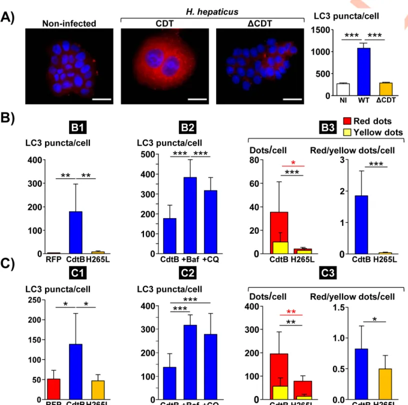
Upon autophagy, the microtubule-associated protein 1A/1B light chain 3 (LC3) is conjugated to phosphatidylethanolamine to yield LC3-II on the surface of nascent autophagosomes. Autophagosome numbers are widely scored by evaluation of either LC3-II puncta or LC3-II expression levels. Autophagosome numbers were thus assessed by quantifying endogenous LC3 puncta numbers through immunofluorescence microscopy with a specific LC3 antibody
during coculture experiments withH. hepaticus strain and its corresponding CDT-knockout
(ΔCDT) mutant strain (Fig 2A). Compared to non-infected cells, an increase in LC3 puncta
was measured in Hep3B cells infected withH. hepaticus, while the increase in LC3 puncta was
almost absent in cells infected with theΔCDT mutant strain (Fig 2A), suggesting that the CDT
is most likely the main virulence factor associated with LC3 puncta increase. No CDT effects
were observed uponH. hepaticus infection of HT29 cells. This cell line had already been
reported to be resistant to CDT effects afterHelicobacter pullorum infection but became
sus-ceptible to CdtB by using direct expression of the toxin in the cell [8].
Similarly, LC3 puncta were thus quantified in epithelial intestinal HT29 and hepatic Hep3B
cells expressing the red fluorescent protein (RFP), the CdtB subunit of the CDT ofH. hepaticus
(CdtB) or the CdtB ofH. hepaticus with the H265L mutation lacking catalytic activity. In both
cell lines (Figs2B1 and 2C1andS2A), the level of LC3 puncta was strongly increased upon
expression ofH. hepaticus CdtB versus control RFP. No significant increase in LC3 puncta was
observed in response to the mutant form ofH. hepaticus CdtB harboring a His!Leu mutation
at residue 265 (H265L) that is crucial for CdtB catalytic activity [7], indicating that LC3 puncta increase is attributed to the CdtB. Time-course western blot analysis of the protein level of LC3
also confirmed the accumulation of LC3-II upon expression ofH. hepaticus CdtB (Fig 3A).
In order to understand the role of autophagy in response to CdtB intoxication, cells were treated with two well-known pharmacological inhibitors of autophagy flux, bafilomycin A1 and chloroquine, and LC3 puncta were subsequently quantified. Both compounds significantly
increased LC3 puncta upon CdtB expression in HT29 and Hep3B cell lines (Figs2B2 and 2C2
andS2B) again confirming the activation of autophagy flux upon CdtB expression.
Autophagic flux is defined as a measure of the amount of cellular material degraded by the autophagy process. The exogeneous fluorescent tandem tagged mCherry-GFP-LC3 mono-meric protein was used to monitor autophagic flux with subsequent determination of the ratio
between the number of red (mCherry+/GFP-, autophagolysosomes) and yellow (mCherry+/
GFP+, autophagosomes) dots [18]. The basis of this method lies in higher sensitivity of the GFP signal to the acidic pH environment of the lysosome compared to the mCherry signal [18]. As transgenic cells expressing the RFP could not be evaluated using this assay, transgenic
cells expressing the CdtBversus CdtB-H265L were used. In both HT29 and Hep3B cell lines,
CdtB strongly stimulated the appearance of red LC3 puncta, compared to CdtB-H265L,
indi-cating activation of the whole autophagy flux in response to CdtB (Figs2B3 and 2C3andS2C).
Taken together, these data showed that CDT,via its active CdtB subunit, promotes autophagy
activation in cells. CdtB also activates AMP-activated protein kinase (AMPK), a key sensor of
autophagy as evidenced by an increase in the phosphorylation of AMPK (on Thr172,Fig 3B).
CdtB upregulates the expression of P62/SQSTM1
P62/SQSTM1 is an autophagy substrate that is involved in the selective transport of cargo to the autophagosomes. It is also used as a reporter of selective autophagy activity. The level of
Fig 2. Effects ofHelicobacter hepaticus cytolethal distending toxin on LC3 expression in human intestinal and hepatic epithelial cells. A) Analysis of LC3 puncta in
hepatic Hep3B cells infected for 3 days withH. hepaticus and its corresponding ΔCDT mutant strain. These cells were processed for fluorescent labeling of LC3 (red) and a counterstaining with DAPI (blue). Fluorescent staining was observed using wide field fluorescence imaging. Transgenic HT29 (B) and Hep3B (C) cells were cultivated with doxycycline for 72 h to induce the expression of the control Red Fluorescent Protein (RFP), the CdtB ofH. hepaticus strain 3B1 or the CdtB of H. hepaticus strain 3B1 with the H265L mutation which has no catalytic activity [20]. These cell lines were also treated with bafilomycin A1 (30 nM) or chloroquine (30μM) 48 h and 24 h after doxycycline induction for a duration of 24 h and 48 h, respectively. Then, cells were processed for fluorescent staining with primary antibodies generated against LC3 associated with fluorescent labeled-secondary antibodies (green) and DAPI to counterstain the nuclei (blue). Autophagic flux was also measured in those cells expressing the tandem-tagged mCherry-GFP-LC3 protein with subsequent yellow (mCherry+/GFP+) and red (mCherry+/GFP-) dot/puncta counting (yellow dots) [18]. The number of fluorescent LC3 puncta was quantified using the "Find Maxima" function of ImageJ. The results are presented as the mean in one representative experiment (performed in triplicate) out of three. A minimum of 500 cells were measured. B) Analysis of LC3 puncta in transgenic HT29 cells. B1) RFP-, CdtB- and H265L-expressing cells. B2)
P62/SQSTM1 mRNA was upregulated upon CdtB intoxication (Fig 1). CdtB also led to an increase in the expression of P62/SQSTM1 protein, as well as its phosphorylated (Ser403) form (Fig 3A and 3B). The increase in P62/SQSTM1 upon CdtB intoxication was also confirmed by
CdtB-expressing cells treated with bafilomycin A1 or chloroquine. B3) autophagic flux measured in CdtB- and H265L-expressing cells. C) Analysis of LC3 puncta in transgenic Hep3B cells. C1) RFP-, CdtB- and H265L-expressing cells. C2) CdtB-expressing cells treated with bafilomycin A1 or chloroquine. C3) autophagic flux measured in CdtB- and H265L-expressing cells.�p<0.05,��p< 0.01,���p< 0.001. Abbreviations: Baf., bafilomycin A1; CdtB, CdtB ofH. hepaticus strain 3B1; CQ,
chloroquine; DAPI, 40, 60-diamidino-2-phenylindol;ΔCDT, CDT isogenic mutant of H. hepaticus strain 3B1; H265L, H. hepaticus CdtB with the mutation His!Leu at
residue 265 involved in catalytic activity; NI, non-infected; RFP, Red fluorescent protein; WT,H. hepaticus strain 3B1 = wild type strain.
https://doi.org/10.1371/journal.ppat.1009320.g002
Fig 3. Effects ofHelicobacter hepaticus cytolethal distending toxin on LC3, P62/SQSTM1 and AMPK expression in human epithelial cells. Transgenic HT29 and
Hep3B cells were cultivated with doxycycline for 72 h to induce the expression of the control Red Fluorescent Protein (RFP), the CdtB ofH. hepaticus strain 3B1 or the CdtB ofH. hepaticus strain 3B1 with the H265L mutation which has no catalytic activity [20]. Then, cells were processed for western blot analysis or fluorescent staining with primary antibodies generated against P62/SQSTM1 associated with fluorescent labeled-secondary antibodies (green) and DAPI to counterstain the nuclei (blue). The number of fluorescent P62/SQSTM1 bodies was quantified using the "Find Maxima" function of ImageJ. The results are presented as the mean in one representative experiment (performed in triplicate) out of three. A minimum of 500 cells were measured. A) Time-course western blot analysis of the protein expression level of LC3 and P62/SQSTM1 in response to the CdtB ofH. hepaticus in HT29 cells. B) The protein expression level and phosphorylation status of P62/SQSTM1, AMPK and P70/ ribosomal protein S6 kinase beta-1 (RPS6KB1) in response to RFP, CdtB and H265L were analyzed by western blot in transgenic HT29 cells. (C) Quantification of P62/ SQSTM1 bodies in transgenic HT29 cells. (D) Quantification of P62/SQSTM1 bodies in transgenic Hep3B cells. E) Confocal image of Hep3B transgenic cells expressing the CdtB ofH. hepaticus strain 3B1 (72 h) processed for P62/SQSTM1 fluorescent staining (green) and DAPI (blue). Scale bar, 10 μm.�p<0.05,��p< 0.01,�� �p< 0.001.
Abbreviations: AMPK, AMP-activated protein kinase; CdtB, CdtB ofH. hepaticus strain 3B1; DAPI, 40, 60-diamidino-2-phenylindol; H265L,H. hepaticus CdtB with the
mutation His!Leu at residue 265 involved in catalytic activity; P-AMPK, phosphorylated AMP-activated protein kinase; P62, P62/SQSTM1; P-P62, phosphorylated P62/SQSTM1; RFP, Red fluorescent protein; Tub., tubulin.
immunofluorescence microscopy with subsequent quantification (Fig 3C and 3D). In addi-tion, confocal analysis revealed P62/SQSTM1 bodies in the giant nuclei of Hep3B cells upon
CdtB expression (Fig 3E), supporting the idea that P62/SQSTM1 may shuttle to the nucleus in
response to CdtB intoxication. Indeed, P62/SQSTM1 can shuttle between the nucleus and cytoplasm to bind with ubiquitinated cargos and facilitate nuclear and cytosolic protein quality control [19,20]. To exclude the possibility that this observation could be a side effect of
2-dimensionnal cell culture,in vivo models were used to investigate the presence of nuclear
P62/SQSTM1. Xenograft mouse models were used [21]. HT29 and Hep3B CdtB-derived
tumors were compared to control RFP-derived tumors. LC3 staining was first performed and quantification revealed the increase in LC3 in engrafted HT29- and Hep3B-CdtB-derived
mice, compared to RFP-derived mice (Fig 4A). P62/SQSTM1 staining revealed an increase in
Fig 4. Effect ofHelicobacter hepaticus CdtB expression on LC3 and P62 expression in engrafted cells. HT29- and Hep3B-transgenic cell lines were engrafted into
immunodeficient mice as previously reported [21]. Threeμm-tissue sections of the xenograft-derived tumors were prepared from formalin-fixed paraffin-embedded tissues and submitted to standard hematoxylin staining and immunostaining raised against LC3 (A) and P62/SQSTM1 (B). Boxes correspond to enlargement. The yellow, black, green and red arrows represent the murine infiltrates, the giant cells, the mucins (HT29) and the LC3 puncta or P62/SQSTM1 bodies, respectively. Quantification was performed by using the ‘Threshold’ function of ImageJ (v. 1.52n). Scale bar, 50μm.��p< 0.01. Scale bar, 50μm. Abbreviations: CdtB of H. hepaticus strain 3B1; P62,
P62/SQSTM1; RFP, Red fluorescent protein.
diffuse cytoplasmic P62/SQSTM1 staining, as well as the presence of P62/SQSTM1 bodies in
the cytoplasm (Fig 4B) in both cell lines. Furthermore, the labeling of P62/SQSTM1 from
CdtB-Hep3B-derived tumor suggests the presence of nuclear P62/SQSTM1 bodies (magnified area in the box ofFig 4B).
Cytoplasmic P62/SQSTM1 aggregates are invaginated in distended nuclei
in response to CdtB
To further confirm the presence of nuclear P62/SQSTM1, nuclear lamina and P62/SQSTM1 immunofluorescent co-staining was performed upon CdtB intoxication in Hep3B cells. As
shown inFig 5, CdtB promotes the accumulation of P62/SQSTM1 bodies in the cytosol. In
addition, some P62/SQSTM1 bodies were found outside of the nuclei and seemed to be engulfed in the nuclear lamina, suggesting that P62/SQSTM1 bodies are not nuclear but invag-inated in nucleosome of CdtB-giant cells (Fig 5).
It was shown that CDT induced-nuclear remodeling can be associated with the formation of deep cytoplasmic invaginations in the nuclei of giant cells. These invaginations also known
Fig 5. Effect ofHelicobacter hepaticus CdtB expression on nucleus remodeling. Wide field image of Hep3B transgenic cells expressing the CdtB of H. hepaticus strain
3B1 processed for the nuclear lamina (red) and P62/SQSTM1 fluorescent staining (green), and DAPI to counterstain the nuclei (blue). Yellow arrows indicate DAPI-lacking nucleoplasmic reticulum surrounding P62/SQSTM1 bodies. Boxes correspond to enlargement. Scale bar, 10μm. Abbreviations: DAPI, 40, 60
-diamidino-2-phenylindol; P62, P62/SQSTM1.
as nucleoplasmic reticulum (NR) were observed bothin vivo and in hepatic Hep3B and intesti-nal SW480 cell lines while tiny NR were more rarely observed in other cell lines as HT29 [6]. These phenotypes observed in Hep3B and HT29 cell lines are reminiscent of those observed for P62/SQSTM1 in the present study, suggesting that P62/SQSTM1 bodies could be a compo-nent of CDT-induced NR. NR invaginations originate from the nuclear envelope and are
reversible and dynamic structures shown to be resorbed back into the envelope [22]. Their
identification is thus carried out in tissues and cell culture by immuno-staining combined
with microscopy [6]. The core of CDT-induced NR concentrates some RNA binding proteins
of eIF4F complex and the subunits of the major coding-region determinant
(mCRD)-medi-ated mRNA instability complex [6]. Thus, UNR/CSDE1 (upstream of N-Ras/Cold shock
domain-containing protein E1), a subunit of the mCRD complex, was used to monitor the
for-mation of NR using immunofluorescence microscopy [6] with a concomitant staining of P62/
SQSTM1 and nuclei.
For this study, Hep3B and SW480 intestinal cells (S2D Fig) were used because NR
forma-tion is easily detected in these cell lines [6]. Both UNR protein and P62/SQSTM1 bodies were
found randomly distributed in the cytosol upon CdtB intoxication. As reported, the nuclear remodeling induced by CdtB was associated with the formation of NR lacking DAPI staining and concentrating UNR-rich foci in giant nuclei. P62/SQSTM1 bodies were also invaginated
in those CdtB-induced NR together or in the vicinity of UNR-rich foci (Fig 6A and 6B). In
another cell line (SK-Hep-1), P62/SQSTM1 bodies were present in the cytosol colocalized with
UNR-rich foci (S3A1 Fig) and in NR (S3A2 and S3A3 Fig). These effects were also observedin
vivo. Indeed, immunofluorescent staining of the liver of mice infected with H. hepaticus for 14
months [23] confirmed the nuclear remodeling of hepatocytes in association with the
forma-tion of NR concentrating UNR-rich foci in giant nuclei, as previously reported [6]. P62/
SQSTM1 bodies were also found invaginated in CdtB-induced NR of large hepatocytes (S3B
Fig).
Whether there is CDT-induced NR formation or not, the size and location of P62/SQSTM1 varied in response to CDT/CdtB. Overall, all P62/SQSTM1 large bodies or aggregates were
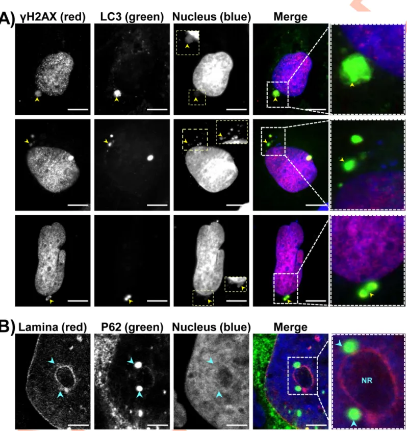
found near UNR-rich foci (Figs6andS3). Micronuclei-like structures surrounded by P62/
SQSTM1 intense staining were observed (Fig 7A and 7B). Moreover, large P62/SQSTM1
aggregates were found far from the distended nuclei (Fig 7C). These structures, which
proba-bly do not correspond to micronuclei, were strongly positive for phosphorylated histone
(γH2AX) foci and seemed to be surrounded with P62/SQSTM1 and/or LC3 (Figs7Cand8A).
Some P62/SQSTM1 large bodies/aggregates were also found tightly connected to the lamina of
the nuclear membrane, and the nuclear lamina seemed to engulf these aggregates (Fig 8B).
Some other structures with low DAPI staining were devoid of intense P62/SQSTM1 staining. All of these effects were not observed in non-infected cells and in the cells infected withH. hepaticus CdtB-H265L, lacking CdtB activity.
CDT-associated cell survival involves autophagy and nucleoplasmic
reticulum formation
CdtB-expressing cells were also treated with bafilomycin A1 and chloroquine with subsequent
quantification of NR (Fig 9A). The number of CdtB-induced NR decreased significantly in
presence of autophagy inhibitors, as compared to untreated cells, suggesting the involvement of autophagy in the formation of NR upon CdtB intoxication.
In order to better understand the effects of the inhibition of autophagy on the formation of NR, we studied the effect of silencing two key autophagy genes, ATG5 and ATG7. For this study, we used lentiviruses that express Cas9 along with single guide (sg) RNAs that target
Fig 6. Effects ofHelicobacter hepaticus CdtB on P62/SQSTM1 localization. As previously demonstrated, NR formation is primarily
observed in response to CDT intoxication,via its active CdtB subunit [6]. Thus, images of non-infected cells are not presented below. (A) Hep3B and (B) SW480 Transgenic cells were cultivated with doxycycline for 72 h to induce the expression of the CdtB ofH. hepaticus strain 3B1 [21]. Then cells were processed for staining with primary and fluorescent secondary antibodies: P62/SQSTM1 (red), UNR (green) and DAPI to counterstain the nuclei (blue). Subsequent quantification of P62/SQSTM1 in nucleoplasm, cytoplasm and foci were performed using capture of fluorescent staining (confocal imaging) by measuring the pixel intensity with the “Plot Profile” function of ImageJ (v. 1.52n) [54], each count being performed on 100 NRs. The relative expression rate of P62/SQSTM1 in NR in response to the CdtB was reported as fold increaseversus the expression in the cytosol. Scale bar, 20 μm.���p<0.0001. Abbreviations: Cyto., cytoplasm; DAPI, 40, 60
-diamidino-2-phenylindol; NR, nucleoplasmic reticulum; Nuc., nucleus; P62, P62/SQSTM1.
ATG5 or ATG7 or a non-specific sg RNA control (Mock) and then selected cells using
puro-mycin [24]. RFP-, CdtB- and H265L-transgenic cell lines were previously selected using
puro-mycin [21], so it was therefore not possible to use these cells. We thus performed this study on non-transgenic Hep3B cells.
A time-course coculture experiment was performed with Mock-, ATG5- and ATG7-KO
Hep3B cells infected withH. hepaticus and its CDT corresponding ΔCDT strain and the effects
of the infection following gene extinction were analyzed. As NR are transient and dynamic structures invaginated in the nucleus, their formation was analyzed using immunofluorescence microscopy. It should be noted that during co-culture experiments, not all cells are infected. Moreover, it was not possible to determine the percentage of infected cells, since no antibody
targeting specificallyH. hepaticus is available for immunofluorescence analysis. With regard to
Mock-, ATG5- and ATG7-KO cells infected with theΔCDT mutant strain, H. hepaticus
infec-tion led to profound nuclear remodeling with enlarged nuclei in associainfec-tion with an increase
inγH2AX foci, a surrogate marker for double-stranded DNA breaks (Figs9B and 9Cand10).
CDT-induced nuclear reorganization was associated with the formation of UNR-rich
cyto-plasmic foci invaginated in the nucleus of giant cells (Figs9Dand10). As previously reported
[6], the strongerγH2AX signal correlated with the bigger nuclei.
These effects were associated with a decrease in cell number and an increase in the large fragment of caspase-3 resulting from cleavage adjacent to Asp175 indicative of caspase-3 acti-vation, a feature of apoptotic cell death (Figs9E and 9Fand10). All of these effects were
con-comitant with the increase in P62/SQSTM1 (Figs9Gand10). With the exception of the cell
proliferation curve (Fig 9E), the overall CDT-induced effects observed on Mock-Hep3B cells
started to gradually increase until the third day afterH. hepaticus infection to reach a maxi-mum effect between the third and fifth day after infection; then a concomitant resumption of these effects started and the effects gradually decreased until the eighth day. None of these
effects were observed in the Mock-KO cells infected with theΔCDT mutant strain, showing
that the CDT is the main virulence factor associated with these phenotypes. When compared
to the Mock-KO cells infected withH. hepaticus, H. hepaticus infection of ATG5- and
ATG7-KO cells led to narrower nuclear remodeling with less-distended nuclei, lessγH2AX
foci, and this was associated with the formation of less UNR-NR (Figs9B–9Dand10). The
decrease in these effects was concomitant with an important decrease in cell proliferation and
an increase in apoptotic cells (Figs9E and 9Fand10). With regards to P62/SQSTM1, a strong
increase in the protein was observed in ATG5- and ATG7-KO infected cells in response toH.
hepaticus infection, compared to the Mock-KO cells infected with H. hepaticus (Figs9Gand
10). P62/SQSTM1 increase was already present in ATG5- and ATG7-KO cells at the basal level
due to the inhibition of autophagy flux.
The formation of NR was also observed in response to colibactin-induced DNA damage [6]. Similar experiments were thus carried out with those cell lines infected with
colibactin-Fig 7. Effects of bacterial genotoxins on P62/SQSTM1 bodies localization andγH2AX in intestinal and hepatic cell lines. HT29 and Hep3B cells were infected for 3 days with CDT-secretingH. hepaticus or colibactin-secreting extra-intestinal pathogenicE. coli. Then, cells were processed for fluorescent staining with primary antibodies generated against γH2AX (red), P62/SQSTM1 (green), associated with fluorescent-labeled secondary antibodies (green) and DAPI to counterstain the nuclei (blue). Fluorescent staining was observed using wide field fluorescence imaging. (A) Images of HT29 cells: P62/SQSTM1 (green), DAPI (blue). (B) Images of HT29 cells:γH2AX (red), P62/SQSTM1 (green), DAPI (blue). (C) Images of Hep3B cells:γH2AX (red), P62/SQSTM1 (green), DAPI (blue). Scale bars, 20 μm. Yellow arrowheads indicate extra-nuclear structures containing chromatin and P62/SQSTM1 aggregates. Yellow and pink boxes correspond to enlargement in which the DAPI andγH2AX were overexposed, respectively, in order to see the micronucleus-like structures. White boxes on the right correspond to enlargement of the merge. Abbreviations: DAPI, 40, 60
-diamidino-2-phenylindol.
Fig 8. Effects of bacterial genotoxins on P62/SQSTM1 bodies localization and LC3 in hepatic cell lines. Hep3B cells were processed as inFig 7. Fluorescent staining was observed using wide field fluorescence imaging. (A) Images of Hep3B cells:γH2AX (red), LC3 (green), DAPI (blue). (B) Images of Hep3B cells: nuclear lamina (red), P62/SQSTM1 (green), DAPI (blue). Scale bars, 20μm. Yellow arrowheads indicate extra-nuclear structures containing chromatin. Blue arrowheads indicate P62/ SQSTM1 aggregates tightly connected all along the nuclear membrane, respectively. Yellow boxes correspond to enlargement of micronucleus-like structures with DAPI. White boxes on the right correspond to enlargement of the merge. Abbreviations: DAPI, 40, 60-diamidino-2-phenylindol; NR, nucleoplasmic reticulum.
secreting extra-intestinal pathogenicE. coli (pks+E. coli). As expected, a profound nuclear remodeling was observed in response to colibactin in Mock-KO cells, compared to non-infected cells or cells non-infected with pks-E. coli (Fig 11A and 11C). This effect was associated
with an increase in both LC3 puncta,γH2AX foci, NR formation, P62/SQSTM1 bodies,
increased cell death and caspase-3 activity (Figs11B and 11Dand12A–12D). Disruption of
autophagy by silencing ATG5 and ATG7 inhibited the enlargement of the nuclei induced by colibactin while enhancing the caspase-3 activity and decreasing cell number, suggesting that autophagy protects cells against colibactin-induced apoptotic cell death. Disruption of autop-hagy led to an increase in colibactin-induced micronuclei-like structures. This increase was
significant for ATG7-KO cells (Fig 12E) suggesting that micronuclei-like structures were not
removed upon disruption of ATG7.These latest results suggest an autophagic-based selective removal of these micronuclei, also called nucleophagy. A decrease in cells presenting
coloca-lizedγH2AX foci and P62/SQSTM1 bodies was also observed in response to colibactin (Fig
12F) in ATG5- and ATG7-KO cells. This decrease likely reflects the global decrease inγH2AX
foci upon autophagy disruption.
Taken together, all of these results showed that CDT and colibactin enhance autophagy leading to selective removal of genotoxin-induced micronuclei-like structures and protecting the cells against the genotoxin-induced apoptotic cell death. Pro-survival autophagy can also be associated with the formation of NR concentrating the autophagic receptor P62/SQSTM1 clustered in the genotoxin-induced nucleoplasmic reticulum.
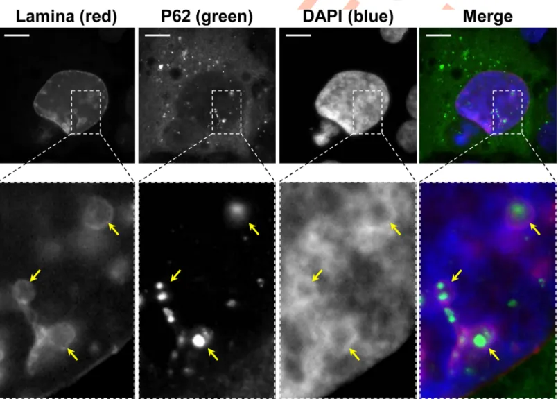
DNA damaging agents induce nuclear remodeling and autophagy
Both CDT and colibactin trigger DNA double-strand breaks. Colibactin was shown to alkylate
DNA [25]. We therefore evaluated DNA damaging chemotherapeutic agents with different
mechanisms of action: etoposide, a topoisomerase II enzyme inhibitor, and streptozotocin, a
glucosamine-nitrosourea alkylating compound (Fig 13). Both compounds trigger NR
forma-tion enclosing UNR and P62/SQSTM1 bodies as well as autophagy activaforma-tion. Moreover,
DAPI-lacking large aggregates positive for LC3 andγH2AX foci were also found. Thus, NR
formation and autophagy are not restricted to CDT and colibactin, but occur also in response to other non-alkylating and alkylating DNA damaging agents.
Fig 9. Time-course analysis of the effects of ATG5 and ATG7 silencing onHelicobacter hepaticus CDT-induced effects. A)
CdtB-transgenic Hep3B cell line cultivated with doxycycline for 72 h to induce the expression of the CdtB ofH. hepaticus strain 3B1. Cells were also treated with bafilomycin A1 (30 nM) or chloroquine (30μM) 48 h and 24 h after doxycycline induction for a duration of 24 h and 48 h, respectively. Then cells were processed for fluorescent staining with primary antibodies generated against UNR (red) and P62/SQSTM1 associated with fluorescent labeled-secondary antibodies (green) and DAPI to counterstain the nuclei (blue). Quantification of UNR-NR positive cells (%) was performed on a minimum of 500 nuclei. The results are presented as the mean in one representative experiment (performed in triplicate) out of three. B) to G) Mock-KO, ATG5-KO and ATG7-KO Hep3B cells were infected for 3 days withH. hepaticus and its corresponding ΔCDT mutant strain. Then, the medium was removed, new medium was added and incubation continued until 8 days. These cells were processed daily for fluorescent staining with DAPI to detect the nucleus and fluorescent primary and secondary antibodies targetingγH2AX, UNR, cleaved caspase-3, and P62/SQSTM1. Fluorescent staining was observed using wide field fluorescence imaging. The results are presented as the mean in one representative experiment (performed in triplicate) out of three. A minimum of 500 cells were measured. (B) Nucleus surface (area) was quantified by isolating the DAPI fluorescence for each nucleus by using the ‘Threshold’ function of ImageJ (v. 1.52n). Nucleus size was measured in viable and early apoptotic cells. (C)γH2AX foci quantification was performed in viable and early apoptotic cells by measuring the pixel intensity with the “Integrated density” measure function of ImageJ (v. 1.52n) [29]. (D) The percentage of cells presenting UNR-NR was determined by manually counting the number of nuclei displaying UNR spots in the nucleoplasm. (E) Cell number quantification was performed manually. (F) Caspase-3-positive cells were quantified by counting the caspase 3 positive cells on 10 fields.�p<0.05,��p<0.01,���p<0.001. Abbreviations: AU, arbitrary
unit; Baf., bafilomycin A1; CQ, chloroquine;ΔCDT, CDT isogenic mutant of H. hepaticus strain 3B1; DAPI, 40, 60
-diamidino-2-phenylindol; KO, Knock-Out, NR, nucleoplasmic reticulum; P62, P62/SQSTM1.
Discussion
In the present study, we showed that treatment of cells with the bacterial genotoxin CDT/CdtB induced autophagy. Whole genome microarray data performed on intestinal cells pointed to numerous mRNA upregulated in response to CdtB intoxication. Little data relative to the
regu-lation of autophagy-related genes in response to CDT are known [3]. However, it was shown
that patient colonic mucosa colonized with colibactin-producingE. coli presented higher levels
of transcript encoding proteins involved in autophagy than colonic mucosa colonized withE.
coli that do not carry the pks island [5]. Most of these transcripts were also found upregulated by CdtB in the present study (ATG5, ATG12, ATG13, ATG16L1, BECN1, MAP1LC3A/B, WIPI1).
Fig 10. Images of the effects of ATG5 and ATG7 silencing on Helicobacter hepaticus CDT-induced effects. (Fig 9continued) Mock-KO, ATG5-KO and ATG7-KO Hep3B cells were processed as inFig 9B–9G. Cells were stained for nuclei (DAPI), as well as forγH2AX, UNR, cleaved caspase-3, and P62/SQSTM1 (red), along with DAPI to counterstain the nuclei (blue). Wide field images of cocultures experiment at days 4 are presented. Scale bars: 50μm for cell number (DAPI only), 100 μm for γH2AX and DAPI double staining, and 20 μm for other double staining (UNR, cleaved caspase-3, and P62/SQSTM1 with DAPI).�p<0.05,��p<0.01,���p<0.001.
Abbreviations:ΔCDT, CDT isogenic mutant of H. hepaticus strain 3B1; DAPI, 40, 60-diamidino-2-phenylindol; KO, Knock-Out, P62, P62/SQSTM1.
We found that the autophagy flux is activated in cells subjected to CdtB, as previously
reported [3]. Seiwertet al. also showed that CDT-induced DSBs trigger pro-survival
autop-hagy, as demonstrated during experiments with chloroquine [3]. Similarly, pharmacological
inhibitors of autophagy (bafilomycin A1, chloroquine) and CRISPR-Cas9 silencing of ATG5 and ATG7 genes increased colibactin- and CDT-induced apoptotic cell death, which was not
observed during transient silencing of ATG5 [3]. In the present study,H. hepaticus CDT and
E. coli colibactin are most likely the main virulence factors associated with the induction of autophagy since the corresponding mutants (ΔCDT and pks-) play a negligible role in this effect, in agreement with previous data [4,5]. Autophagy induced by bacteria degrades inter-nalized pathogens in addition to the infected cell and reduces the spread of infection. For
CDT-secretingH. hepaticus and colibactin-secreting E. coli, their genotoxin would be the
main virulence factor responsible for the induction of autophagy and bacterial clearance. In
addition,H. hepaticus CdtB also regulated some genes involved in the ‘apoptosis and
autop-hagy’ pathway and the increase in the mRNA level of apoptosis-regulator proteins and
inflam-matory caspases suggests CdtB-induced pyroptosis duringH. hepaticus infection. Both
autophagy and apoptosis are thus induced following CDT and colibactin intoxication. This is in agreement with some studies, which showed that the regulation of apoptosis and autophagy is intimately connected and that both of these supposedly different processes, can be
stimu-lated by the same stresses [26]. Under certain circumstances, autophagy can protect cells from
various apoptotic stimuli by preventing them from undergoing apoptosis, whereas in other cel-lular settings, autophagy is an alternative cell-death pathway [26]. Accordingly, we can assume that autophagy constitutes an adaptation to the genotoxic stress induced by CDT and colibac-tin, which counteracts the genotoxin-induced apoptosis to maintain cell survival during DDR. However, for cells whose DNA damage is beyond repair, autophagy might not be sufficient to
maintain cell survival leading to apoptotic cell death. In the present study,γH2AX foci and
nucleus size were measured in viable and early apoptotic cells; thus, their global decrease in response to CDT and colibactin upon autophagy disruption did not reflect their overall cellular quantification but likely reflects the enhanced apoptotic cell death. Moreover, it is also possible that autophagy disruption conducted to activation of alternative DDR pathways (instead of the
canonical pathway) [27], which functioned as backup and processed the genotoxin-induced
DNA breaks, resulting inγH2AX foci and nucleus size decrease. Further investigation is
needed to explore the DDR molecular regulation in autophagy-deficient cells in the context of bacterial genotoxin response. Preliminary results showed that autophagy disruption by ATG7
silencing led to increased IL8 secretion and nuclear factorκB translocation following bacterial
genotoxin intoxication (S4A and S4B Fig), suggesting that autophagy would protect
intoxi-cated cells by reducing inflammatory responses. However, these latest results deserve further study.
We found that CDT/CdtB increased the expression levels of both P62/SQSTM1 mRNA and
protein, in contrast to previous experiments with purified CDT [4]. The difference between
Fig 11. Analysis of the effects of ATG5 and ATG7 silencing onEscherichia coli colibactin-induced effects. Mock-KO, ATG5-KO and ATG7-KO
Hep3B cells were infected for 4 hours with colibactin-secreting extra-intestinal pathogenicE. coli and its corresponding isogenic mutant and cultivated for 3 days in a bacteria free medium. Then, cells were processed for fluorescent staining with primary antibodies generated against LC3 andγH2AX associated with fluorescent labeled-secondary antibodies (green) and DAPI to counterstain the nuclei (blue). (A) Wide field images of Hep3B cells: LC3 (green), DAPI (blue). Scale bar, 20μm. (B) The number of fluorescent LC3 puncta was quantified using the "Find Maxima" function of ImageJ. (C) Nucleus surface (area) was quantified in viable and early apoptotic cells by isolating the DAPI fluorescence for each nucleus by using the ‘Threshold’ function of ImageJ (v. 1.52n). (D)γH2AX foci quantification was performed in viable and early apoptotic cells by measuring the pixel intensity with the “Integrated density” measure function of ImageJ (v. 1.52n) [29]. A minimum of 500 cells were measured.�p<0.05,��p<0.01,���p<0.001. Abbreviations:
AU, arbitrary unit; DAPI, 40, 60-diamidino-2-phenylindol; KO, Knock-Out, pks-, bacterial artificial chromosome vector; pks+, bacterial artificial
chromosome vector with pks island encoding colibactin.
P62/SQSTM1 expression most likely reflects the amount of internalized CdtB in the cell. The expression of the P62/SQSTM1 protein level cannot be used as an indicator of the autophagy
activity as SQSTM1 mRNA level is often upregulated under stressful conditions [28]. Thus, the
important increase in P62/SQSTM1 mRNA by CdtB, most likely hides the degradation of P62/
SQSTM1 protein by autophagy [29,30]. The significant increase in the P62/SQSTM1
phos-phorylated (Ser403) isoform induced by CdtB suggests selective autophagic clearance of
ubi-quitinated proteins and protein aggregates that are poorly degraded by proteasomes [23].
Activation of prosurvival autophagy requires clusterin expression and this cytoprotective chaperone facilitates stress-induced lipidation of LC3 and induces autophagosome biogenesis
[31]. Clusterin protects against genotoxic stress and suppresses DNA damage-induced cell
death [32]. Clusterin-enhanced cell survival to DNA damage occurs thereforevia autophagy.
We previously reported that clusterin mRNA is highly upregulated (~8-fold increase) in response toH. hepaticus CdtB [33]. In line with that, clusterin may be involved in pro-survival autophagy following DNA damage triggered by CdtB.
DNA damage following exposure to bacterial toxins induces cell death (necrosis, apoptosis) and cellular senescence [21,34,35]. Senescent cells following DNA damage display multiple morphological aspects such as cytoskeleton and organelle remodeling and nuclear size enlarge-ment. Selective degradation of the entire nucleus or nuclear components occur in mammals in order to maintain nuclear integrity. Exposure to CDT and colibactin induces the formation of
nucleoplasmic bridges and micronuclei [34,36]. Micronuclei contain damaged chromosome
fragments enclosed by the nuclear membrane that can be sequestered and degraded by
autop-hagy [37]. Indeed, 2 to 5% of micronuclei induced by cell cycle blocker compounds 1) show a
decreased intensity in DAPI staining consistent with chromatin degradation in these
compart-ments, 2) exhibitγH2AX-positive DNA damage foci, 3) colocalize to LC3-positive vesicles and
4) contain P62/SQSTM1 [37]. In the context of bacterial genotoxin, micronuclei-like
struc-tures with DAPI,γH2AX, LC3 and P62/SQSTM1 were observed, suggesting micronuclear
autophagy. Regardless of the P53 status of the line used, similar results were observed. Indeed, TP53 gene is mutated in HT29 and Hep3B [38,39] resulting in a high level of constitutive P53
protein expression in HT29 [21] and a truncated protein in Hep3B while no mutations
occurred in SK-Hep-1.
Senescent cells secrete the senescence-associated secretory phenotype (SASP) and upregu-late the formation and release of small extracellular vesicles (exosomes, microvesicles, nucleo-somes and apoptotic bodies) from epithelial cells [40]. Small extracellular vesicles and autophagy cooperatively prevent apoptotic cell death by removing cytoplasmic DNA frag-ments derived from chromosomal DNA or bacterial infections in order to protect cells from
excessive inflammatory responses [40]. P62/SQSTM1 is present within these vesicles [41].
P62/SQSTM1 bodies induced by CDT vary greatly in size and suggest the presence of different structures or agglomerates. Because inhibition of autophagy increases apoptotic cell death
Fig 12. Analysis of the effects of ATG5 and ATG7 silencing onEscherichia coli colibactin-induced effects (Fig 11continued) Mock-KO, ATG5-KO and ATG7-KO Hep3B cells were processed as inFig 11. Then, cells were processed for fluorescent staining with primary antibodies
generated against UNR, cleaved Caspase-3 or P62/SQSTM1 orγH2AX associated with fluorescent labeled-secondary antibodies and DAPI to counterstain the nuclei. (A) The percentage of cells presenting UNR-NR was determined by manually counting the number of nuclei displaying UNR spots in the nucleoplasm. (B) Cell number quantification was performed manually. (C) Caspase-3-positive cells were quantified by counting the caspase 3 positive cells on 10 fields. (D) P62/SQSTM1 bodies were quantified using the "Find Maxima" function of ImageJ. The results are presented as the mean in one representative experiment (performed in triplicate) out of three. (E) Quantification of micronucleus-like structures in Hep3B cells was performed manually. (F) Quantification of Hep3B cells with colocalizedγH2AX-foci and P62/SQSTM1-bodies was performed manually. A minimum of 500 cells were measured.�p<0.05,��p<0.01,���p<0.001. Abbreviations: AU, arbitrary unit; DAPI, 40, 60
-diamidino-2-phenylindol; KO, Knock-Out, NR, nucleoplasmic reticulum; P62, P62/SQSTM1; pks-, bacterial artificial chromosome vector; pks+, bacterial artificial chromosome vector with pks island encoding colibactin.
Fig 13. Effects of DNA damaging agents on nuclear remodeling and autophagy. Hep3B cells were cultivated in the presence of etoposide (5μM) or steptozocin (10 mM) for a duration of 24 h. Then, the medium was removed and incubation was continued for 48 h. The cells were then stained with fluorescent primary and secondary antibodies targeting LC3 (red)/γH2AX (green), P62/SQSTM1 (red)/UNR (green) and DAPI to counterstain the nuclei (blue). Fluorescent staining was observed using wide field fluorescence imaging.γH2AX foci quantification was performed by measuring the pixel intensity with the “Integrated density” measure function of ImageJ (v. 1.52n) [29]. LC3 puncta and P62/SQSTM1 bodies were quantified using the "Find Maxima" function of ImageJ. The percentage of cells presenting UNR-NR was determined by manually counting the number of nuclei displaying UNR spots in the nucleoplasm. Quantification was performed on a minimum of 500 cells. The results are presented as the mean in one representative experiment (performed in triplicate) out of three.���p<0.001. Scale bar, 20μm. White arrow indicates DAPI-lacking
large aggregate positive for LC3 andγH2AX foci. Yellow arrow indicates DAPI-lacking nucleoplasmic reticulum enclosing UNR and P62/SQSTM1 bodies. Abbreviations: AU, arbitrary unit; DAPI, 40, 60-diamidino-2-phenylindol; ETP, etoposide; NR, nucleoplasmic reticulum; P62, P62/SQSTM1; STZ, steptozocin.
upon CDT intoxication (Figs9,10, and13), some of these large CDT-induced P62/SQSTM1 bodies found in the cytosol could correspond to P62-positive extracellular vesicles prior to their externalization.
Senescent cells process their chromatinvia the autophagy/lysosomal pathway, which
con-tributes to the proteolytic processing of histones [42]. In this context, cytoplasmic chromatin fragments are closely juxtaposed to P62/SQSTM1 bodies in senescent cells [42]. It is believed that P62/SQSTM1 bodies enhance the nucleocytoplasmic export of chromatin in the
cyto-plasm of senescent cells [42]. A cytoplasmic accessvia CDT-induced NR would facilitate
chro-matin transport to the cytoplasm for degradation by autophagy pathway.
In addition to maintaining nuclear DNA homeostasis, nuclear autophagy (also known as nucleophagy) plays an important role in the post-transcriptional RNA modification process
[43]. We previously reported that the nuclear remodeling induced by CDTvia CdtB is
associ-ated with the formation of NR whose core concentrates mRNA and RNA binding proteins involved in mRNA translation and decay, amongst which is the cytoplasmic RNA-binding
protein UNR/CSDE1 [6]. P62/SQSTM1 large bodies are also invaginated in the core of
CdtB-induced NR together or in the vicinity of UNR-rich foci. UNR/CSDE1 influences proliferation under stress conditions, is overexpressed in melanoma tumors and promotes invasion and
metastasis partly by inducing translation elongation of targeted mRNAs [44]. In melanoma,
P62/SQSTM1 was shown to 1) extend mRNA half-life of a several pro-metastatic factors by opposing mRNA decay and 2) recruit RNA-binding proteins that are enriched within P62/
SQSTM1 interactome [45]. In this context, P62/SQSTM1 would have recruited RNA-binding
proteins from eIF4F and mCRD complexes, as UNR/CSDE1, to shuttle them in the core of CDT-induced NR, protecting and translating mRNA in order to maintain cell survival. The fact that P62/SQSTM1 bodies are systematically present near UNR-rich foci in response to CDT/CdtB (with or without formation of NR structure) supports this hypothesis.
P62/SQSTM1 was recently shown to be a RNA-binding protein whose vault RNAs are the
major interacting RNA [46]. Vaults are large ribonucleoprotein particles composed of the
major vault protein (VMP), two minor vault proteins (VPARP and TEP1) and a variety of
small untranslated RNA molecules [47,48]. DNA-damaging agents directly activate the human
VMP gene [49], but no increase in MVP transcript in response to the CdtB ofH. hepaticus was observed in HT29 cells (microarray data). Given the association with the nuclear membrane and the location within the cell, vaults are thought to play roles in intracellular and nucleocyto-plasmic transport processes. If vault ribonucleoprotein particles are involved in CDT/CdtB-induced NR, they may regulate nucleocytoplasmic transport in those structures. Vault RNA1-1 directly binds P62/SQSTMRNA1-1 and inhibits P62/SQSTMRNA1-1-dependent autophagy and ubiquitin
aggregate clearance by interfering with P62/SQSTM1 multimerization [46]. CDT/CdtB
induced-NR are deeply invaginated in the nucleus and concentrate large P62/SQSTM1-rich aggregates, most likely reflecting the absence of the small non-coding vault RNA1-1 in those protected structures in which P62/SQSTM1-dependent autophagy takes place.
P62/SQSTM1 contains two nuclear localization signals and a nuclear export signal. Thus, it shuttles continuously between nuclear and cytosolic compartments at a high rate and
cyto-plasmic P62/SQSTM1 body aggregates regulate its nucleocytocyto-plasmic shuttling [19]. P62/
SQSTM1 is also involved in the assembly of proteasome-containing degradative compart-ments in the vicinity of nuclear aggregates [19]. In this hypothesis, the bacterial genotoxins induce the transport of P62/SQSTM1 protein in the nucleus; this transfer would have been very fast because overall P62/SQSTM1 has not been visualized in the nucleus of the various models used in the present study.
Following various stresses, cytoplasmic ribonucleoprotein granules,i.e. stress granules (SG)
relief. SG and GW/P-bodies were not observed upon CDT intoxication [6] but the
composi-tion of these organelles is reminiscent of that of CdtB-induced NR [6]. As SG and
GW/P-bod-ies are cleared from cells through autophagy [50], P62/SQSTM1 bodies may also participate in
the clearance of messenger ribonucleoprotein particles clustered in CDT-induced NR structures.
Thus, many observations suggest a central role for P62/SQSTM1 upon infection with geno-toxin- secreting bacteria in regulating DNA damage response. However, it should be noted that P62/SQSTM1 is not necessary for NR formation in this context, as P62/SQSTM1 silencing
(CRISPR-Cas9 System) did not affect NR formation (S5A Fig) neither UNR expression. On
the other hand, MAFB silencing [33] reduced NR formation and P62/SQSTM1 bodies
increased during bacterial cocultures withH. hepaticus (S5B Fig). This effect of MAFB is not surprising as this oncoprotein is involved in nuclear remodeling following DNA damage
induced by Helicobacter CDT [33].
Finally, cell survival following the DNA damage induced by CDT and colibactin involved prosurvival autophagy in intestinal and hepatic cells. Macroautophagy including nucleophagy occurred upon bacterial genotoxin intoxication and P62/SQSTM1 bodies may play a central
role in this pro-survival autophagy, probablyvia different mechanisms in light of the observed
disparity of P62/SQSTM1 bodies (shape, localization). Further studies are required to decipher the role of P62/SQSTM1 in the survival of cells stressed by bacterial genotoxins and more par-ticularly its role in the formation of NR in light of its RNA-binding protein function recently demonstrated [46].
It should be emphasized that similar effects were also observed with DNA damaging agents used during chemotherapies, etoposide and streptozotocin, suggesting that NR formation and increased autophagy following DNA damage are not a specific adaptation to bacterial genotox-ins but rather a survival mechanism following a genotoxic stress. Accordingly, these survival mechanisms may contribute to the resistance of cancer cells to therapies inducing DNA dam-age. In line with that, altered nuclear morphology reminiscent of NR was reported as a com-mon feature of many cancers and has an adverse prognostic significance for various cancers [51]. These latest data should open new fields of investigation.
Material & methods
Ethics statement
Animal material provided from previous studies was approved by the Ethics Committee for Animal Care and Experimentation CEEA 50 in Bordeaux (Comite´ d’Ethique en matière d’Expe´rimentation Animale agre´e´ par le ministre charge´ de la Recherche, “dossier no. Dir
13126BV2”, “saisines” no. 4808-CA-I [23] and 13126B [8], Bordeaux, France), according to
treaty no. 123 of the European Convention for the Protection of Vertebrate Animals. Animal experiments were performed in an A2 animal facility (security level 2) by trained authorized personnel only.
Transcriptomics
The global analysis of the expression of human genes in response to theH. hepaticus CdtB was
quantified using the Human GE 4x44K v2 Microarray Kit (Agilent Technologies, Les Ulis, France). Total RNA was extracted with the miRNeasy Mini Kit (Qiagen) according to the
manufacturer’s recommendations. Cyanine-3 (Cy3) labeled cRNA was prepared from 0.2μg
RNA using the One-Color Microarray-Based Gene Expression Analysis kit (Low Input Quick Amp Labeling, Agilent Technologies) according to the manufacturer’s instructions, followed by RNAeasy column purification (Qiagen). Dye incorporation and cRNA yield were checked
with a NanoDrop 2000 spectrophotometer. For each sample, a total of 1.65μg of cRNA was fragmented and hybridized overnight at 65˚C onto Human Gene Expression v2 4x44k Micro-array (Agilent Technologies). The slides were washed as recommended by the manufacturer, and scanned on an Agilent G2565CA scanner, at a 5 micron resolution and using the 20-bit scan mode (Agilent Technologies). Images were processed with Feature extraction (version 10.7). For satistical and bioinformatic analyses, data were normalized for inter-array
compari-sons and analyzed using the BRB-ArrayTools package (version 4.2.0,http://linus.nci.nih.gov/
BRB-ArrayTools.html). As recommended by Agilent Technologies, we chose the percentile method for the normalization, and adjusted the 75th percentile of all of the non-control probes to a value of 500. We identified genes that were expressed differentially among the two groups using a random-variance t-test (Class comparison between groups of arrays package, BRB-Ar-rayTools). Gene expression differences were considered statistically significant if they showed �30% difference in mean expression levels between samples from CdtB versus control condi-tions (tdTomato fluorescent protein), and if their associated p-value was <0.01.
Reagents and antibodies
Bafilomycin A1 (#B1793), chloroquine (#C6628), doxycycline hyclate (#D9891), etoposide (#E1383), gentamicin (#G1522), puromycin (#P7255) and streptozocin (#S0130) came from Sigma Aldrich (Saint-Quentin Fallavier, France).
Antibodies used for western blot analyses were: mouse monoclonal anti-LC3 (clone 4E12) (#M152-3, MBL Life science) distributed by CliniSciences (Nanterre, France); mouse mono-clonal anti-P62/SQSTM1 (Clone 3/P62 Ick ligand) (#610832) from BD Biosciences (San Jose, CA); rat monoclonal anti-phospho-P62/SQSTM1 (Ser403) (clone 4F6) (#MABC186, Merck Chimie Fontenay sous Bois) and mouse monoclonal anti-α-tubulin (clone DM1A) (#05–829) from Sigma Aldrich; rabbit polyclonal AMPKα1 (#2795) and rabbit monoclonal anti-phospho-AMPKα (Thr172) (clone 40H9) (#2535) from Cell Signaling Technology (distributed
by Ozyme, Saint-Cyr-L’E´cole, France).
Antibodies used for immunofluorescence analyses were: mouse monoclonal anti-LC3 (clone 4E12) (#M152-3) (MBL Life Sciences) and mouse monoclonal anti-P62/SQSTM1 (D-3) (#sc-28359, Santa Cruz Biotechnology, Heidelberg, Germany) (1/100). The working conditions for anti-LC3 were 1/100 in Phosphate-Buffered Saline with 1% digitonin as permeabilizing agent.
The simultaneous immunodetection of LC3 andγH2AX was performed under LC3 conditions.
Rabbit polyclonal anti-UNR/CSDE1 (#HPA018846) (1/100) came from Sigma Aldrich. Rabbit monoclonal cleaved Caspase-3 (Asp175) (clone 5A1E) (1/200) (#9664), rabbit monoclonal anti-Caspase-1 (clone D7F10) (#3866) (1/100) and rabbit monoclonal anti-phospho-histone H2A.X (γH2AX) (Ser139) (20E3) (#9718) (1/100) came from Cell Signaling Technology. The antibody used for Caspase-1 immunodetection did not work for immunocytochemistry but gave a nice immunodetection of Caspase-1 on tissue sections. Rabbit polyclonal anti-53BP1 (#NB100-304 1/ 100) came from Novus Biologicals (Centennial, CO, USA). Alexa 488 or Alexa 594-labeled sec-ondary antibodies and 4’,6’-diamidino-2-phenylindol (DAPI) were purchased from Molecular Probes (Invitrogen, Cergy Pontoise, France).
Sequential 53BP1 and
γH2AX immunostaining on tissue sections
Both anti-53BP1 and anti-γH2AX originated from rabbits. To correlate 53BP1 association
withγH2AX, sequential fluorescent labeling was performed using 3-μm tissue sections,
pre-pared from formalin-fixed paraffin-embedded human CdtB-Hep-3B xenografts. Firstly, 53BP1 immunodetection was performed, the stained tissue section was scanned, and the


![Fig 6. Effects of Helicobacter hepaticus CdtB on P62/SQSTM1 localization. As previously demonstrated, NR formation is primarily observed in response to CDT intoxication, via its active CdtB subunit [6]](https://thumb-eu.123doks.com/thumbv2/123doknet/14379786.505755/13.918.126.862.88.939/helicobacter-hepaticus-localization-previously-demonstrated-formation-primarily-intoxication.webp)