HAL Id: hal-02335509
https://hal.archives-ouvertes.fr/hal-02335509
Submitted on 21 Oct 2020
HAL is a multi-disciplinary open access
archive for the deposit and dissemination of
sci-entific research documents, whether they are
pub-lished or not. The documents may come from
teaching and research institutions in France or
abroad, or from public or private research centers.
L’archive ouverte pluridisciplinaire HAL, est
destinée au dépôt et à la diffusion de documents
scientifiques de niveau recherche, publiés ou non,
émanant des établissements d’enseignement et de
recherche français ou étrangers, des laboratoires
publics ou privés.
toxin-antitoxin systems
Valerie Guillet, Patricia Bordes, Cécile Bon, Julien Marcoux, Virginie Gervais,
Ambre Julie Sala, Suzana dos Reis, Nawel Slama, Israel Mares-Mejía,
Anne-Marie Cirinesi, et al.
To cite this version:
Valerie Guillet, Patricia Bordes, Cécile Bon, Julien Marcoux, Virginie Gervais, et al.. Structural
insights into chaperone addiction of toxin-antitoxin systems. Nature Communications, Nature
Pub-lishing Group, 2019, 10 (1), �10.1038/s41467-019-08747-4�. �hal-02335509�
ARTICLE
Structural insights into chaperone addiction
of toxin-antitoxin systems
Valérie Guillet
1
, Patricia Bordes
2
, Cécile Bon
1
, Julien Marcoux
1
, Virginie Gervais
1
, Ambre Julie Sala
2
,
Suzana Dos Reis
1
, Nawel Slama
2
, Israel Mares-Mejía
1
, Anne-Marie Cirinesi
2
, Laurent Maveyraud
1
,
Pierre Genevaux
2
& Lionel Mourey
1
SecB chaperones assist protein export by binding both unfolded proteins and the SecA motor.
Certain SecB homologs can also control toxin-antitoxin (TA) systems known to modulate
bacterial growth in response to stress. In such TA-chaperone (TAC) systems, SecB assists
the folding and prevents degradation of the antitoxin, thus facilitating toxin inhibition.
Cha-perone dependency is conferred by a C-terminal extension in the antitoxin known as
cha-perone addiction (ChAD) sequence, which makes the antitoxin aggregation-prone and
prevents toxin inhibition. Using TAC of Mycobacterium tuberculosis, we present the structure
of a SecB-like chaperone bound to its ChAD peptide. We
find differences in the binding
interfaces when compared to SecB
–SecA or SecB-preprotein complexes, and show that the
antitoxin can reach a functional form while bound to the chaperone. This work reveals how
chaperones can use discrete surface binding regions to accommodate different clients or
partners and thereby expand their substrate repertoire and functions.
https://doi.org/10.1038/s41467-019-08747-4
OPEN
1Institut de Pharmacologie et de Biologie Structurale, IPBS, Université de Toulouse, CNRS, UPS, 31077 Toulouse, France.2Laboratoire de Microbiologie et de
Génétique Moléculaires, Centre de Biologie Intégrative (CBI), Université de Toulouse, CNRS, UPS, 31062 Toulouse, France. Correspondence and requests for
materials should be addressed to P.G. (email:pierre.genevaux@ibcg.biotoul.fr) or to L.M. (email:lionel.mourey@ipbs.fr)
123456789
U
niversally conserved molecular chaperone machines
maintain cellular protein homeostasis by preventing
protein misfolding and aggregation, off-pathways in
protein biogenesis. In bacteria, the ATP-independent
homo-tetrameric chaperone SecB (69 kDa) assists protein export by
binding to presecretory proteins and delivering them
post-translationally to the Sec translocon at the inner membrane
1.
SecB binds its preprotein clients in a one to one ratio and
maintains them in nonnative translocation competent state,
protected from premature folding and recognition by cytosolic
proteases
2. Although SecB has no apparent specificity for signal
sequences, in vitro peptide scan analysis revealed preferences for
stretches of nine amino acids enriched in aromatic and basic
residues present in protein clients
3,4. SecB can accommodate long
fragments of up to 150 amino acids in length within polypeptide
substrates
5. The recently determined nuclear magnetic resonance
(NMR) structures of SecB bound to its full-length native
sub-strates maltose-binding protein (MBP) and alkaline phosphatase
(PhoA) confirmed that SecB possesses a remarkably large
substrate-binding surface (about 7600 Å
2) where the client
pro-tein wraps all around the chaperone in an unfolded
conforma-tion, occupying the entire SecB substrate-binding surface
6.
In addition to its preprotein clients, SecB specifically interacts
with SecA, the essential ATPase motor subunit of the Sec
trans-locon
7. Upon interaction, SecB passes on its client to SecA, which
subsequently promotes preprotein translocation through the
SecYEG pore. SecA-SecB interaction mainly involves the
C-terminal zinc-binding region of SecA and the negatively charged
surface formed by the
β-sheets on both sides of the SecB
tetra-mer
8. However, additional contacts between the C-terminal helix
of SecB and the N-terminal region of SecA have been found
9.
Although the substrate transfer mechanism remains unknown,
the surfaces of SecB that interact with the preprotein and with
SecA overlap significantly, thus likely facilitating substrate
transfer from the chaperone to the translocase.
Besides the generic role of SecB in protein export, SecB
homologs are frequently associated (in about 7% of cases) with
stress-responsive type II toxin–antitoxin (TA) systems, together
forming functional tripartite TA-chaperone (TAC) systems
10.
Classical type II TA systems are small genetic modules encoding
two cytosolic proteins: a poisonous toxin and a less stable and
protease-sensitive cognate antitoxin, which binds and inhibits
the toxin. Under normal growth conditions, the antitoxin-bound
toxin is inactive and the bacteria grow normally. It is believed
that under certain stress conditions the less stable antitoxin is
degraded by proteases and that the resulting free active toxin
targets essential cellular processes such as protein synthesis or
DNA replication, thus inhibiting growth until normal growth
conditions resume
11,12. TA systems are also widely distributed
on mobile genetic elements, including plasmids, where they
contribute to their stability. In this case, when the TA-containing
plasmid is lost, de novo synthesis of the antitoxin stops and the
reservoir of cytosolic antitoxin is rapidly degraded, which results
in toxin activation and cell growth inhibition
13. Control of
bacterial growth by toxins has been associated with various
cellular processes, including stabilization of genomic regions,
protection against foreign DNA, biofilm formation, control of
the stress response, bacterial virulence and persistence
14,15.
Although an involvement of TA systems in persistence was
found for several bacteria
16–19their contribution to Escherichia
coli K-12 drug persisters has not been demonstrated
20,21. In
tripartite TAC systems, the SecB chaperone encoded within the
same operon as the TA genes, directly interacts with the highly
unstable TAC antitoxin and prevents its aggregation and
degradation, thus facilitating subsequent inhibition of the
toxin
22.
The human pathogen Mycobacterium tuberculosis possesses
such a TA-associated SecB chaperone, named Mtb-SecB
TA, which
specifically controls the Mtb-HigB1 (toxin)-HigA1 (antitoxin) TA
pair
12,22. Mtb-HigB1, which likely belongs to the RelE toxin
superfamily of ribonucleases, was shown to severely inhibit E. coli,
M. smegmatis, M. marinum, and M. tuberculosis growth
12.
Noticeably, Mtb-TAC genes were shown to be significantly
induced in response to several stresses relevant for M. tuberculosis
pathogenesis. This includes DNA damage
23, heat shock
24,
nutrient
starvation
25,
hypoxia
26,
host
phagocytes
27and
antibiotic-induced persistence
28, suggesting that the TAC system
could contribute to the stress adaptive response and/or to the
virulence of this bacterium. Yet, such a role for TAC remains to
be demonstrated.
We have recently shown that in contrast with
chaperone-independent TA systems, the Mtb-HigA1 antitoxin of TAC
possesses a C-terminal extension, named ChAD (chaperone
addiction), which makes the antitoxin more aggregation-prone
and renders it chaperone-dependent. In this case, the ChAD
extension is specifically recognized by the Mtb-SecB
TAchaperone,
which protects it from both aggregation and degradation
29.
In this work, we provide biochemical and structural insights
into the mechanism of chaperone addiction. Using TAC of M.
tuberculosis as model system, we solved the crystal structure, at
high resolution, of a TA-associated SecB chaperone bound to the
ChAD peptide of its cognate antitoxin. The structure reveals
major differences in binding mode and substrate occupancy when
compared to SecB–SecA or SecB–substrate complexes. Further
extensive mutagenesis of contact surface residues revealed several
previously unidentified residues essential for TA-control by
Mtb-SecB
TAchaperone in vivo. Finally, we showed that the antitoxin
can reach a functional form while bound to the chaperone and
that ChAD peptide may efficiently destabilize the interface
between the antitoxin and the chaperone in vitro and specifically
activate the Mtb-HigB1 toxin in vivo.
Results
Three-dimensional structure of
Mtb-SecB
TAin complex with
ChAD. In contrast with the classical export chaperone SecB,
which binds and wraps unfolded presecretory protein substrates
and maintains them in an unfolded conformation compatible
with translocation through membrane
6, previous work revealed
that the Mtb-SecB
TAchaperone of TAC is capable of binding to
its aggregation-prone Mtb-HigA1 antitoxin substrate to facilitate,
and thus not prevent, its folding in the cytosolic space. This
strongly suggests a different binding mode of SecB-like
chaper-ones with their antitoxin clients. To investigate the SecB
substrate-binding property, we have solved the structure of
Mtb-SecB
TAtogether with the short C-terminal fragment of the
Mtb-HigA1 antitoxin (named ChAD), which was found as the main
site of interaction with the chaperone, both in vivo and
in vitro
29,30. The structure comprises a Mtb-SecB
TAhomo-tetramer (the subunits, termed A–D, contain 136–138 residues of
the 181 amino acids found in the Mtb-SecB
TAsequence) to which
three
ChAD
peptides
(corresponding
to
sequence
104
EVPTWHRLSSYRG
116of Mtb-HigA1 named C4 in a previous
study
29; subunits E–G), 2 dimethyl sulfoxide (DMSO) molecules,
and 3 Ca
2+ions are bound (Table
1
and Fig.
1
a). The
Mtb-SecB
TAstructure resembles that of SecB for which four structures
have been solved: the X-ray structures of Haemophilus influenzae
SecB alone
31and in complex with the C-terminal 27 residues of
SecA
8, the X-ray structure of E. coli SecB alone
32, and the NMR
structures of its complexes with alkaline phosphatase and
maltose-binding protein in their unfolded states
6(Supplementary
Fig. 1a). The three-dimensional fold of Mtb-SecB
TAconsists of a
six-residue long N-terminal helix (αN) followed by a
four-stranded antiparallel
β-sheet (β1–β4), each strand contributing
13–16 residues, and a 30-residue long C-terminal helix (α1) that
is prolonged by an extended peptide segment running parallel to
α1 (Fig.
1
b and Supplementary Fig. 1b). Missing residues are
found in the majority at the N and C termini (5–12 and 15–19
residues missing, respectively) and in the loop between
β3 and β4
(13–16 residues missing), in all subunits. The superposition of the
subunits making up the Mtb-SecB
TAtetramer led to a root mean
square deviation (rmsd) of Cα atoms ranging between 0.4
(sub-unit C vs. B) and 1.2 Å (sub(sub-unit C vs. A). This is in line with the
NMR characterization of SecB, which revealed two pairs of
spectroscopically equivalent subunits formed on the one hand by
subunits A and D and on the other hand by subunits B and C
6.
Largest deviations correspond to spatial locations lining regions
with missing residues, which in turn correlates with high
B-factors, thus indicating structural
flexibility.
The Mtb-SecB
TAtetramer consists of a dimer of dimers as
already described for SecB
31,32. Two subunits associate through
their
β1 strands to form β-dimers (A–B and C–D), resulting in an
eight-stranded antiparallel
β-sheet (Fig.
1
a). In the tetramer, the
A–B and C–D β-dimers are stacked up through their helices α1 at
an angle of ca. 45° (Fig.
1
a). The dimer and dimer–dimer
interface areas are in the same range: 739/728 Å
2for A–B/C–D vs.
820/704 Å
2for A–C/B–D (Supplementary Table 1). It is
noteworthy that the tetramer displays internal dihedral (D
2)
symmetry that includes three perpendicular twofold symmetry
axes. However, not all among the three possible superimpositions
through rotations around these axes are equivalent. Only one
permutation (DCBA) gives a rmsd value of 0.6 Å after
super-posing 522 Cα atoms to the reference tetramer (ABCD), whereas
the two others (BADC and CDAB) led to much higher rmsd
values of 3.5 Å. The DCBA permutation is generated through
rotation around the twofold axis that lies between the
dimer–dimer interface and runs nearly perpendicular to helices
α1 (Fig.
1
a). This distorted dihedral symmetry is also found in the
previously determined SecB structures (PDB entries 1FX3, 1OZB,
1QYN, and 5JTL), but has never been described before. Molecular
dynamics calculations performed on the structure suggest that
this distorted arrangement is stable over the 60-ns simulation
time, and that once trapped into such a conformation, no further
fluctuation does occur (Supplementary Fig. 2a).
It has recently been reported that diffraction-quality crystals of
Mtb-SecB
TAcould only be grown at high concentrations of
DMSO. Indeed, the use of DMSO improved initial needle-like
crystals but was not sufficient to resolve the structure
33. Adding
5–10% DMSO in the protein and/or crystallization buffer played
a role with respect to nucleation but did not markedly influence
the diffraction limit of Mtb-SecB
TA/ChAD crystals. We also
noted that the presence of calcium ions was important for the
crystallization of the complex. Three Ca
2+ions were localized
based on electronic density and geometric criteria, and calcium
binding occurs in subunits A, B, and D at the same position
(Fig.
1
a). Indeed, all three ions interact with main-chain nitrogen
atom and side-chain oxygen atom of S65, with the main-chain
oxygen atom of Y111, and with the carboxylate of D110 thereby
linking the N- and C-termini of strands
β3 and β4, respectively.
The lack of calcium binding to subunit C is due to crystal packing
constraint where loop
β4-α1 of a symmetry mate partly occupies
the site.
Comparison of
Mtb-SecB
TAand SecB structures. Pairwise
comparison of the Mtb-SecB
TAcrystallographic tertiary structure
with those of Hi-SecB and Ec-SecB gave rmsd values around 2.0 Å
for about 110 Cα atoms of residues sharing 15% identity. In
comparison, superimposition of the Hi- and Ec-SecB subunits led
to rmsd values less than 1.0 Å for ca. 130 residues sharing 59%
identity. There are three major differences between the
Mtb-SecB
TAtertiary structure and its SecB counterparts (Fig.
1
b and
Supplementary Fig. 1b): (i) Mtb-SecB
TAhas a longer N terminus
that adopts a helical fold (αN), (ii) it has a 12- to 14-residue long
insertion in the loop between
β3 and β4, which is disordered in
the structure, and (iii) its C terminus is shorter and could not be
traced beyond residue number 164. In contrast, the C-terminal
ends of Hi- and Ec-SecB are better defined in density where they
form helix
α2. We verified by mass spectrometry (MS) of
dis-solved crystals that no proteolytic cleavage, which could account
for the missing C-terminal residues, had occurred in the
crys-talline state. Structural dynamics might then be responsible for
the absence of electron density for the C-terminal residues of
Table 1 Crystallographic data collection and re
finement
statistics
Mtb-SecBTA/HigA1(104–116)
PDB code 5MTW Data collection
Diffraction source ESRF ID29
Space group P212121
Unit cell a, b, c (Å) 86.86, 89.96, 91.34
Unit cellα, β, γ (°) 90.00, 90.00, 90.00
Resolution range (Å) 64.09–1.835 (1.867–1.835)a
No. of unique reflections 63,188 (3135)
Completeness (%) 99.9 (99.6) Redundancy 6.5 (6.7) 〈I/σI〉 13.7 (2.3) Rmerge 0.093 (0.796) CC1/2 0.999 (0.338) Refinement Resolution range (Å) 64.09–1.835 (1.883–1.835)a
No. of reflections (work/test) 59,790/3397 (4325/275)
No. of molecules/AUb 1 Mtb-SecBTAtetramer+ 3 bound
Mtb-HigA1(104–116) peptides Rwork/Rfree 0.212/0.265 (0.330/0.354) Chain/no. of residues/missing residues A/138/1–8, 43–44, 82–96, 164–181 B/138/1–5, 43–46, 81–96, 164–181 C/138/1–12, 82–94, 114–116, 167–181 D/136/1–9, 81–95, 114–115, 163–181 E/12/116 F/12/116 G/12/116 No. of nonhydrogen atoms
All atoms 4660 Protein atoms 4282 Peptide atoms 318 Other ligands 8 Ions 3 Water molecules 49 Average B-factors (Å2) All atoms 34.7 Protein atoms 34.3 Peptide atoms 39.7 Other ligands 63.5 Ions 45.7 Water molecules 32.6 RMS deviations Bond lengths (Å) 0.019 Bond angles (°) 1.941 Ramachandran plot (%)
Most favored regions 93.9
Allowed/disallowed 6.1/0.0
aValues in brackets are for the highest-resolution shell. bAsymmetric unit.
Mtb-SecB
TAand the location of a C-terminal
α2 helix as found in
Hi- and Ec-SecB. Such
flexibility might be fostered by the
prox-imal location of the N-terminal helix, whose position is dictated
for some of the subunits by crystal packing constraints, and which
otherwise would induce steric clashes (Supplementary Fig. 1b).
Other differences between the Mtb-SecB
TAand SecB structures
occur in loops
β1–β2, β2–β3, and β4–α1 (Fig.
1
b and
Supple-mentary Fig. 1). Superimposing the crystal structure of
Mtb-SecB
TAtetramer with those of Ec- and Hi-SecB gave rmsd values
around 2.0 Å. In comparison, superposing the Ec- and Hi-SecB
tetramers led to rmsd values of 0.9 Å. Although the tetramers are
highly similar in structure, important variations were found when
comparing the dimer and dimer-dimer interface areas
(Supple-mentary Table 1). Whether these structural differences are
important for TA control, export function or protein stability is
unclear. The fact that all the TAC chaperones tested so far could
Chain D Chain D Chain C Chain C Chain A Chain B Chain B Chain A Chain F Chain G Chain F Chain G Chain E Ca Ca Ca Ca Ca DMS DMS β1 β1 β2 αN α1 αN α1 β3 β4 β2 β3 β4 αN αN β1 β2 β3 β4 α1 α1 αN β1 β2 β3 β4 Chain E DMS DMS
a
b
c
90° . . . . . . . . β1 β2 β3 β4 α1 α2 . . . αN β1 β2 β3 β4 α1 I I I I I I I I I I I Iefficiently replace Ec-SecB export function, regardless of the
sequence similarities
10,29, and that single-point mutations in
substrate-binding area were sufficient to redirect Ec-SecB toward
Mtb-HigBA1 without affecting its function in protein export
30,
suggest that such differences may not be critical for TA control or
export.
Mtb-SecB
TAand related Ec- and Hi-SecB are acidic proteins
that remarkably share a strictly identical theoretical pI value of
4.3. In accordance, analysis of the topography and charge
distribution revealed that they display very electronegative
molecular surfaces but differences between the SecB proteins
and Mtb-SecB
TAare noticeable (Supplementary Fig. 3). Major
differences pertain in fact to negative charge distribution. For
instance, there is a strong electronegative cluster right in the
middle of the SecB eight-stranded
β-sheet, which arises from
side-chain clustering of residues D20/D27, E24/E31, and E77/E86
(Ec-SecB/Hi-SecB numbering), which is important for interaction
with SecA C-terminal region. In Mtb-SecB
TA, these residues are
replaced by R32, A36, and D102, respectively. The fact that
Mtb-SecB
TAis less efficient than Ec-SecB when tested for
SecA-dependent function in E. coli suggests that such modified
electrostatic interface might be responsible for the reduced
complementation in vivo.
Another important difference is found in a groove delineated
by the N-terminus of strand
β1, the β1–β2 loop, helix α1, and the
extended C-terminal segment. This groove is neutral or slightly
positively charged in SecB whereas it is electronegative in
Mtb-SecB
TA, involving the D27, E143, and D147 residues and the
positively charged R151. In contrast, only one acidic residue is
found in SecB, E112/E121, which corresponds to E143 of
Mtb-SecB
TA, and is counterbalanced by R120/R129 equivalent to R151
in Mtb-SecB
TA.
Mtb-SecB
TA/ChAD interaction. In order to investigate how the
chaperone specifically recognizes the C-terminal
chaperone-addiction region of the Mtb-HigA1 antitoxin, key residues
involved in the interaction between the ChAD peptide
(Mtb-HigA1
104EVPTWHRLSSYRG
116) and the Mtb-SecB
TAchaper-one substrate-binding surface were analyzed in detail. Note that
this small peptide represents the main region of the ChAD
extension of Mtb-HigA1 that is recognized by the chaperone,
both in vivo and in vitro
29. In the structure, there are three bound
ChAD peptides (chains E–G) per tetramer (Fig.
2
a). For all three
peptides, there was no adequate electron density to build the last
residue (G116) and the side chain of R115 is also missing in
peptide G (Supplementary Fig. 4). Binding occurs on the thinner
side of the Mtb-SecB
TAchaperone tetramer. On one side, two
peptides (chains E and F) are bound in a symmetrical way
(related by a twofold axis) and adopt strictly the same extended
conformation with a rmsd of 0.6 Å based on main-chain atoms.
The third peptide (chain G) binds on the opposite side and also
adopts an extended conformation identical from residues
104–108 (rmsd of 0.5 Å on backbone atoms) but differs in
main-chain (φ,ψ) angles from residues 109–114 (rmsd of 2.1 Å). It is
noteworthy that peptide G differs in conformation from peptides
E and F to conserve the same type of interactions with the
pro-tein, and molecular dynamics (MD) calculations indicate that
such binding mode is preserved during the 60 ns of simulation
(Supplementary Fig. 2b). On the other hand, no sufficient
inter-pretable electron density could allow the building of a
symme-trical equivalent of peptide G (Supplementary Fig. 4), which
because of steric clashes due to the distorted D
2symmetry would
not be possible without structural rearrangement. Each bound
peptide makes contact with three protein subunits in a similar
way, with some variations (Fig.
1
c). A residue-per-residue
detailed description of the interactions is given in
Supplemen-tary Table 2.
In agreement with previous in vivo data
29,30, the structure
reveals that three ChAD residues play a major structural role in
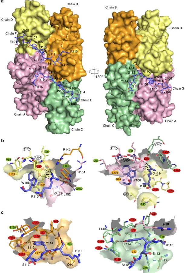
complex formation, namely W108, R110, and Y114. Mtb-HigA1
(W108) makes strong interactions with the protein (Fig.
2
b). It is
involved in hydrogen bonding with D27 located on strand
β1. In
addition, its indol ring is completely buried in a protein cavity
delineated by residues D27, R29, V68, and L108, from one
Mtb-SecB
TAsubunit, and A153 (at the C terminus of
α1) from another
Mtb-SecB
TAsubunit. It is also involved in cation-π interactions
with R29 side chain. Mtb-HigA1(R110) makes strong polar
interactions with D27 and I28 from one subunit, and with G150
(at the C terminus of
α1) from another subunit (Fig.
2
b). van der
Waals interactions with a third protein subunit also contributes to
the steady anchoring of Mtb-HigA1(R110). Mtb-HigA1(Y114) is
hydrogen bonded to the protein through interaction with P155.
In addition, its phenol group perfectly
fits a gorge delineated by
residues L47, Y49, L154, P155, P156, and L157 (Fig.
2
c).
Mutagenesis of
Mtb-SecB
TAresidues involved in ChAD
bind-ing. In order to investigate whether the interaction between
Mtb-SecB
TAand the antitoxin was relevant in vivo, all residues of
Mtb-SecB
TAinvolved in polar interactions with the ChAD peptide,
either through side-chain or main-chain atoms, and those
involved in nonbonding interactions through burying more
than 50% of their solvent accessible surface (Fig.
1
b and
Sup-plementary Table 2) were systematically mutated to alanine
residues. The resulting variants were then tested for both their
Fig. 1 Overall structure of the Mtb-SecBTA/ChAD complex and determinants of SecB ligand specificity. a The A–D subunits forming the Mtb-SecBTA
tetramer are respectively colored in green, orange, violet, and yellow whereas ChAD peptides (chains E–G) are in slate blue. Calcium ions are shown as
green spheres. DMSO molecules (labeled DMS) are shown as a stick representation. Left and right views are separated by a 90° rotation around a vertical
axis. Secondary structures have been labeled for the A–B (left) and A–C (right) dimers. b Structure-based sequence alignment. Sequence similarity is
indicated by red letters, whereas sequence identity is indicated by white letters on a red background. Aligned and unaligned residues are displayed in
uppercase and lowercase, respectively, taking Mtb-SecBTAas reference. Secondary structure elements as deduced from the crystal structures of
Mtb-SecBTA/ChAD, Ec-SecB (PDB code 1QYN), and Hi-SecB (1FX3) are indicated. Residues absent from the structures are on a green background. Residues
involved in protein–protein interface of the Mtb-SecBTA/ChAD, Ec-SecB/PhoA (5JTL), and Hi-SecB/SecAc (1OZB) complexes are indicated by symbols:
solid ellipses and triangles, residues involved in polar interactions through side-chain and main-chain atoms, respectively; stars, residues having≥50% of
their surface area buried upon van der Waals interactions. For Mtb-SecBTA, color coding of the symbols is according to the impact of alanine scanning on
the ability to maintain TA control from green (no effect) to orange (medium effect) to red (most severe effect). Residues that were not tested (i.e., Ala
residues and Y146, which was too strongly expressed) are designated by unfilled symbols. The V40A/L42A/L44A triple substitution that causes a 40-fold
reduction in the affinity of Ec-SecB for PhoA is in purple. c Sequence of the Mtb-HigA1 ChAD peptide. The rectangles below the sequence represent the
amino acids that have been changed for alanine as previously reported29. Green rectangles represent mutations without apparent phenotype with respect
to their chaperone dependence in vivo; orange, mutations with partial inactivation; red rectangles, mutations with the most severe phenotype. Interfacing
R110 W108 R142 L152 R151
b
c
W108 R110 Y114 S112 S113 R115 Q80 T149 Y114 S113 R115 S112 R26 Q80 T149 D27 I28 R29 A106 A107 L108 T158 G150 A153 L154 P155 P156 L157 T158 L47 I77 Y49 T48 V68 V68 A107 I28 D27 L154 P155 P155 P156 A106 L108 R29 A153 A40 P43 Y146 Y49 I77 L157 L47 P155 L154 P156 T48 G46 R115 Y114 S113 S112 L111 R110 H109 W108 T107 P106 V105 E104 R115 Y114 S113 S112 L111 R110 H109 W108 T107 P106 V105 E104 R115 Y114 S113 S112 L111 R110 H109 W108 T107 P106 V105 E104a
Chain D Chain C Chain A Chain B Chain F Chain E Chain G Chain D Chain C Chain A Chain B 180°Fig. 2 ChAD binding on Mtb-SecBTA.a Molecular surface of the tetramer. The three bound peptides are shown as a stick representation with carbon,
nitrogen, and oxygen atoms in slate blue, blue, and red, respectively. The protein and peptide chains as well as ChAD residues have been labeled. Left and
right views are separated by a 180° rotation around a vertical axis.b Close-up view centered on ChAD residues W108 and R110. c Close-up view centered
on ChAD residue Y114. Inb and c, Mtb-SecBTAatoms found within 5 Å of the peptides are shown as enlarged sticks and depict the ChAD-binding site
shown as a semitransparent surface. Polar interactions are represented by black dotted lines. Views on the left (resp. right) are for subunit F (resp. G).
ability to control the toxin (Fig.
3
) and to functionally replace the
canonical E. coli export chaperone SecB with respect to its generic
chaperone function at low temperature and in the presence of
novobiocin antibiotic
34,35(Supplementary Fig. 5). Steady state
expression of the Mtb-SecB
TAderivatives revealed that all
mutants are well expressed when compared to the wild type
(Supplementary Fig. 6). Mtb-SecB
TAmutations that most
sig-nificantly affect TA control were found at positions making
strong interactions with ChAD residues shown to be involved in
chaperone binding
29, namely residues D27, R29, and L108
interacting with Mtb-HigA1(W108), and residues Y49, I77, L154,
P155, P156, L157 interacting with Mtb-HigA1(Y114) (Fig.
2
b, c).
–
Mtb-SecBTA
WT
– D27AI28AR29AP41AP43AG46AL47AT48AY49AD58AD60AI64AF67AV68AI77AL108AG150AL154AP155AP156APP155-156AAPP155-156GGL157AT158A
I64 I64 D27 D27 Y49 Y49 F67 F67 I77 I77 L154 L154 P155 P155 P156 P156 L157 L157 T158 T158 R29 R29 L47 L47 D60 D60 D60 D60 L108 L108I28 I28 P41 P41 P43 P46 P48 P48 D58 D58 V68 G150 G150 V68 0.5% Ara PBAD Mtb-HigBA1 Ptrc Mtb-SecBTA E. coli ΔsecB 5 μM IPTG 50 μM IPTG 500 μM IPTG
a
b
Fig. 3 In vivo functions of TA-directed Mtb-SecBTAmutants.a Suppression of Mtb-HigBA1 toxicity by Mtb-SecBTAmutants. E. coli W3110ΔsecB
transformants containing the p29SEN-based Mtb-SecBTAchaperone mutants and pK6-Mtb-HigBA1 were grown to mid-log phase, serially diluted and
spotted on LB ampicillin kanamycin agar plates with or without IPTG (to express Mtb-SecBTA) and arabinose (to express Mtb-HigBA1) as indicated. Plates
were incubated overnight at 37 °C. Spot tests have been performed in triplicates.b Localization of the mutations on the Mtb-SecBTA/ChAD structure. The
molecular surface of the tetramer is represented in light gray. Color coding of interacting residues is the same as in Fig.1b. Residues that were not tested
It is noteworthy that Mtb-HigA1(G116), whose mutation to
alanine was also associated to a defect in vivo
29, could not be
resolved in the electron density. Moreover, residues D27 and
L154 of Mtb-SecB
TAare also involved in interactions with R110
of Mtb-HigA1 (Fig.
2
b). Two other Mtb-SecB
TAresidues, L47 and
F67, appeared to be important in vivo. Although not directly
interacting with Mtb-HigA1(Y114), L47 contributes to the pocket
that accommodates the tyrosine phenol ring (Fig.
2
c). The role
played by F67 seems to be more structural since the
phenylala-nine ring is oriented toward the interior of the protein thereby
contributing to a network of hydrophobic/aromatic interactions.
The mutation for an alanine might locally destabilize the protein
hydrophobic core and in turn the positioning of strand
β3
backbone, which is involved in ChAD interaction. Noticeably,
although spread all over the sequence, essential residues of
Mtb-SecB
TAinvolved in the interaction with ChAD define two major
binding spots when mapped on the 3D structure (Fig.
1
). The
first
spot is located on the
β-sheet of one of the four tetramer subunits,
and involves strands
β1–β3. The second spot, which comprises
the residues most severely affected by mutations, is found on the
protomer of the adjacent
β-dimer. It involves the N-terminal tip
of strand
β2 and residues that follow helix α1 and form the helix
connecting loop. Such crosslinking of ChAD peptides at the
surface of the Mtb-SecB
TAtetramer might tighten, or even lock,
the dimer–dimer interface and explain the improvement found in
both diffraction quality and resolution of the crystals of the
Mtb-SecB
TA/ChAD complex vs. those of Mtb-SecB
TA.
All the Mtb-SecB
TAmutations that most significantly impaired
TA control additionally affected the ability to replace the E. coli
export chaperone SecB in vivo at low temperature of growth, with
the exception of Y49A (Supplementary Fig. 5). Remarkably, Y49A
significantly improved Mtb-SecB
TAability to replace Ec-SecB
in vivo. Two other mutations, G46A and D58A, slightly improved
Mtb-SecB
TAchaperone generic function but, in contrast with
Y49A, without affecting TA control. These three residues are
spread along strand
β2, G46, and Y49 close to each other at the
N-terminal side and D58 at the C terminus. It is noteworthy that
Mtb-SecB
TA(Y49) corresponds to Ec-SecB(V40) whose mutation
as part of the triple amino-acid substitution V40A/L42A/L44A
(with all three residues on
β2) was shown to cause a 40-fold
reduction in the affinity of Ec-SecB for PhoA
6. Whether these
mutations affect SecB substrate-binding specificity or its
interac-tion with its SecA partner is not known.
Speci
fic affinity of ChAD for Mtb-SecB
TAchaperone. Several
approaches were used to characterize complex formation between
Mtb-SecB
TAand the ChAD peptide in solution and to further
confirm structure-function relationships deduced from the X-ray
structure and mutagenesis experiments. Isothermal titration
calorimetry (ITC), performed under both direct and reverse
conditions, and microscale thermophoresis (MST) showed that
the chaperone interacts with the ChAD peptide with moderate
affinity, with a K
ddissociation constant of 1.9 ± 0.2 and 2.6 ± 0.6
µM, respectively (Fig.
4
a, b). The ITC-derived thermodynamic
parameters indicated an enthalpy-driven mode (−50.1 kJ mol
−1)
accompanied with an entropic penalty (−TΔS = 18.7 kJ mol
−1),
likely suggesting formation of hydrogen bonds associated with
binding, which is in line with the crystal structure. On the other
hand, the stoichiometry obtained by direct and reverse ITC for
this specific association is 0.47 and 2.2, respectively, consistent
with a model preferentially involving two peptides per
Mtb-SecB
TAtetramer. Extensive small-angle X-ray scattering (SAXS)
analysis performed on different protein preparations
system-atically revealed that the scattering curves corresponding to
Mtb-SecB
TAand Mtb-SecB
TA/ChAD peptide displayed small but
significant differences (Fig.
4
c).
Both
Guinier
and
the
concentration-independent method
36were applied to SAXS data
and gave respective molecular weight of 84.5 ± 0.1, 81.7 ± 0.4 kDa
for Mtb-SecB
TAand 89.7 ± 0.1, 83.6 ± 0.3 kDa for Mtb-SecB
TA/
ChAD. The chaperone alone and its complex with ChAD form
well folded molecular species, as can be observed from normalized
Kratky plots (Fig.
4
c, inset), and Guinier analysis gave R
gvalues of
3.17 ± 0.02 and 3.02 ± 0.02 nm and maximum dimensions of the
particles (Dmax) of 9.5 ± 0.2 and 8.8 ± 0.2 nm, respectively. This
indicates that, at the resolution probed, Mtb-SecB
TAdoes not
undergo a major conformational change upon ChAD peptide
binding but rather displays a slight compaction and a less dynamic
behavior. These properties might be related to the better
dif-fracting quality of the crystals obtained in the presence of the
peptide. In addition, the scattering curve calculated from the
Mtb-SecB
TA/ChAD peptide crystal structure, where the missing loops
representing 24% of the unbuilt residues have been modeled, led
to a rather good, albeit not perfect, agreement (χ = 2.6) with
experimental data (Fig.
4
c). To further investigate the dynamic
behavior of the chaperone, we performed Hydrogen–Deuterium
eXchange MS (HDX-MS) on Mtb-SecB
TAalone and in the
pre-sence of ChAD peptide. The kinetics of exchange for Mtb-SecB
TAalone revealed some highly dynamic and/or accessible regions
(namely residues 13–18, 37–51, 59–67, and 161–181, in line with
the absence of electron density for the C-terminal residues of
Mtb-SecB
TA) compared to more protected regions (residues 68–75,
98–108, and 124–152). Interestingly, a large decrease in
deutera-tion upon binding of the ChAD peptide was observed for residues
21–27 and 153–159, which are precisely located along the
peptide-binding pocket, and juxtaposing residues 52–57 (Supplementary
Fig. 7a). These results also confirm that the chaperone is rather
stable upon complex formation with ChAD.
The deconvoluted MS spectrum obtained for Mtb-SecB
TAunder
nondenaturing conditions clearly showed the presence of a tetramer
at 81,575 Da and up to 7 additional species, separated by 1587 Da,
progressively appearing upon titration with the ChAD peptide
(Fig.
4
d and Supplementary Fig. 8a). Binding of the
first four
peptides to the tetrameric chaperone corresponds to relatively
high-affinity events, as they occur at small peptide:tetramer ratios (<4),
whereas binding of the three additional peptides only occurred at
high molar ratios (>4), probably due to nonspecific interactions
(Supplementary Fig. 8a). Plotting the relative abundance obtained
for each species at increasing molar ratios and
fitting the
corresponding curves (Supplementary Fig. 8b) confirmed this
observation, with K
dvalues in the range 6–17 µM. Incubation of the
chaperone with 8 molar equivalents of 13-mer peptides, which also
belong to the ChAD sequence but do not contain the main
chaperone-binding determinants
29, only showed limited binding to
the tetramer, confirming the higher binding specificity observed for
the ChAD peptide (Fig.
4
e). This low-abundance interaction may be
related to some non-specific binding also observed at high molar
ratios of ChAD peptide. The nonspecific adduction is a common
feature of native MS when working with substrate/ligand
concentrations greater than 50 µM
37.
Full-length
Mtb-SecB
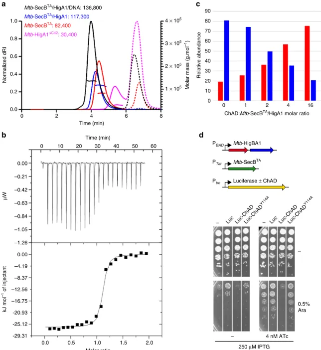
TA/HigA1 chaperone/antitoxin complex.
Multiple attempts to crystallize the full-length Mtb-SecB
TA/
HigA1 complex were unsuccessful, although such complex could
be easily purified in a stable soluble form. Size exclusion
chro-matography and multi-angle light scattering (SEC-MALS) clearly
indicated that Mtb-SecB
TAalone primarily forms tetramers while
experiments performed on Mtb-SecB
TAco-expressed with
Mtb-HigA1 revealed a single-molecular species representing a 4:2
chaperone/antitoxin stoichiometry (Fig.
5
a). These results were
further confirmed by native MS (114.9 kDa, see next paragraph).
In addition, we found that under the same experimental in vitro
condition, purified Mtb-HigA1
ΔC42mutant, which was shown to
be fully soluble and functional in vivo in the absence of
chaper-one
29, was isolated as a dimer (Fig.
5
a), as observed with known
chaperone-independent HigA antitoxins
38,39. SAXS was also used
to characterize full-length Mtb-SecB
TA/HigA1. The
correspond-ing scattercorrespond-ing curve is significantly distinct from those obtained
for Mtb-SecB
TAalone or in complex with the peptide and reflects
a well folded protein/protein complex (Fig.
4
c) of molecular
weight 114.4 ± 0.1 and 120.0 ± 0.3 kDa as obtained from Guinier
and concentration-independent methods, respectively, and with
R
gand Dmax values of respectively 3.61 ± 0.02 and 12.0 ± 0.2 nm.
Finally, we performed HDX-MS on the Mtb-SecB
TA/HigA1
complex. Strikingly, binding of the chaperone to Mtb-HigA1
client resulted in the protection of the same stretches of residues
as for Mtb-SecB
TA/ChAD with the addition of residues 48–51
and 59–66, which are located in the continuity of the
peptide-binding pocket (Supplementary Fig. 7b). This observation
sug-gests a significantly larger binding interface between the
chaper-one and full-length Mtb-HigA1 when compared to the short
ChAD peptide. We also did not
find any specific destabilization of
Mtb-SecB
TAupon complex formation with Mtb-HigA1, which is
in agreement with the Kratky analysis of the SAXS data.
As observed for most type II DNA binding antitoxins,
Mtb-HigA1 can act as a repressor of its own Mtb-higBA1 operon
40. In
order to further investigate the activity of soluble dimeric
Mtb-HigA1 bound to Mtb-SecB
TAtetramers, we have used
SEC-MALS, native MS, and ITC to monitor the interaction between
the purified Mtb-SecB
TA/HigA1 complex and dsDNA using a
38-bp palindromic sequence derived from the higB P2 promoter.
First, we found that Mtb-SecB
TAalone does not interact with the
38-bp dsDNA sequence. In contrast, SEC-MALS experiments
a
b
d
0.001 0.01 0.1 1 10 100 Ligand concentration (μM) 0 5 10 Δ Fnorm (‰)c
70 80 90 100 110 120 0.00 –0.21 –0.42 –0.63 –0.84 μ W 0.0 0.5 1.0 –50.23 –41.86 –33.49 –25.12 –16.74 –8.37 0.00 0 10 20 30 40 50 60 Time (min) Molar ratio 10 0 1 2 3 4 5 6 8 12 4 6 0 2 8 10 12 14 0.2 0.4 0.6 0.8 1.0 0 25 50 75 100 Control ChAD-8 eq C2-8 eq C3-8 eq C7-8 eqe
130 0 0 1 2 3 4 5 6 kJ mol –1 of injectant Relative intensity 100 80 60 40 20 80,000 85,000 Mass (Da) 100,000 95,000 90,000 ln ( I) Q (nm–1) (Q .R g ) 2× I( Q )/ I(0) Q.RgFig. 4 Molecular analysis of ChAD binding to Mtb-SecBTA.a ITC profile of ChAD titration into Mtb-SecBTA(upper and lower parts represent raw and
integrated binding heats, respectively).b Differences in normalizedfluorescence generated by various concentrations of ChAD added to Mtb-SecBTA
during MST analysis and calculatedfit. Error bars are as calculated with the NanoTemper analysis software based on independent experimental data.
c Experimental SAXS data for Mtb-SecBTAin the unbound state (red line), in the presence of ChAD (green line), and for the complex formed with
Mtb-HigA1 (blue line) and comparison with the theoretical scattering patterns computed from the Mtb-SecBTA/ChAD structure (black line). The inset shows
the SAXS-derived Kratky plots. d Deconvoluted native mass spectrum of Mtb-SecBTAincubated with 4 molar equivalents of ChAD peptide. The species
detected correspond to the Mtb-SecBTAtetramer (MW 81,575 Da, purple circle) and to the binding of up to four ChAD peptides with MWs of 83,157;
84,746; 86,334 and 87,922 (cyan, green, orange, and red, respectively).e Native MS-derived relative abundance of the different species obtained upon
demonstrated that Mtb-SecB
TA/HigA1 forms a single stable
molecular species in the presence of DNA, most likely
corresponding to 4:2:1 Mtb-SecB
TA/HigA1/DNA stoichiometry
(Fig.
5
a), with a shift in the hydrodynamic radius from 4.4 to 5.0
nm upon DNA binding. We verified by SEC-MALS that no
interaction occurs between Mtb-SecB
TA/HigA1 and a nonspecific
dsDNA such as a 16-bp oligonucleotide derived from the RRM1
promoter
41. We also used native MS to analyze Mtb-SecB
TA/
HigA1 in complex with its specific DNA target and observed that
they indeed form a 4:2:1 complex of molecular weight 138.25 kDa
(Supplementary Fig. 9a). In addition, ITC experiments showed
that the higB P2 promoter DNA fragment interacts with the
hexamer with a high affinity (2.2 ± 0.5 × 10
7M
−1at 25 °C)
(Fig.
5
b). The ITC-derived thermodynamic parameters indicated
Molar mass (g.mol
–1) 1 × 10⁵ 2 × 10⁵ 3 × 10⁵ 4 × 10⁵ 0.0 0.2 0.4 0.6 0.8 1.0 Normalized dRI 0 2 4 6 8 Time (min) Mtb-SecBTA/HigA1: 117,300 Mtb-SecBTA: 82,400 Mtb-HigA1ΔC42: 30,400 Mtb-SecBTA/HigA1/DNA: 136,800 Time (min) 0.0 0.5 1.0 1.5 2.0 Molar ratio –1.05 0.00 –0.21 –0.42 –0.63 –0.84 μ W -25.12 -20.93 –16.75 –12.56 –8.37 –4.19 0.00 –1.26 -29.31 Relative abundance 0 1 2 4 16 0 10 20 30 40 50 60 70 80 90 0.5% Ara – – 4 nM ATc 250 μM IPTG PBAD Mtb-HigBA1 PTet Mtb-SecBTA Ptrc Luciferase ± ChAD kJ mol –1 of injectant 0 10 20 30 40 50 60
ChAD:Mtb-SecBTA/HigA1 molar ratio
– – Luc Luc-ChADLuc-ChAD
Y114A
Luc Luc-ChADLuc-ChAD
Y114A
a
c
b
d
Fig. 5 Characterization of full-length Mtb-SecBTA/HigA1 chaperone/antitoxin complex.a SEC-MALS analysis. Continuous and dashed lines represent the
variation of refractive index against elution time for Mtb-SecBTA(red), Mtb-SecBTA/HigA1 (blue), Mtb-HigA1ΔC42(violet), and Mtb-SecBTA/HigA1/dsDNA
(black) for protein and salt contributions, respectively. The experimentally measured molar mass distribution (same color code) and deduced mean molar
mass (in g mol−1) are indicated for each elution peak. Calculated molecular masses of proteins and protein complexes used for experiments (in daltons):
Mtb-SecBTAtetramer, 81,552; Mtb-SecBTA/HigA1 hetero-hexamer, 115,068; Mtb-HigA1ΔC42dimer, 27,994; Mtb-SecBTA/HigA1/dsDNA, 138,420.b ITC
profile of 38-bp dsDNA titrated into Mtb-SecBTA/HigA1 (upper and lower parts represent raw and integrated binding heats, respectively).c Native
MS-derived relative abundance of Mtb-SecBTA/HigA1 (blue bars) and Mtb-SecBTA(red bars) obtained upon titration of Mtb-SecBTA/HigA1 with 0, 1, 2, 4, 16
molar equivalents of ChAD.d Overexpression of luciferase-ChAD chimera activates Mtb-HigB1. E. coli WPtet57 strain expressing chromosomally encoded
Mtb-SecBTAunder the control of an anhydrotetracycline inducible promoter was co-transformed with pC6-Mtb-HigBA1 and the pSE380-based vector (−),
luciferase (Luc), or the Luc-Mtb-ChAD and Luc-Mtb-ChADY114Achimeras. Transformants were grown to mid-log phase, serially diluted and spotted on LB
ampicillin chloramphenicol agar plates without or with IPTG (to express Luc and Luc chimeras), arabinose (to express Mtb-HigBA1), and
an enthalpically and entropically favored binding (ΔH = −26.1
kJ mol
−1and
−TΔS = −15.9 kJ mol
−1) suggesting association to
the major groove of DNA
42. The stoichiometry obtained by ITC
for this specific association is 1.1, consistent with a binding
involving one palindromic DNA sequence per Mtb-HigA1 dimer
present in the Mtb-SecB
TA/HigA1 hetero-hexamer. Measuring
the temperature dependence of the enthalpy gave a negative heat
capacity of binding (−1.3 kJ mol
−1K
−1) as typically reported for
the majority of sequence-specific protein/DNA associations
43.
Together these results show that the highly unstable Mtb-HigA1
antitoxin can form a functional dimer following binding to its
specific chaperone partner.
It has been proposed that disrupting the TA interfaces by small
molecules or peptides to trigger endogenous toxin activation
in vivo could represent a novel antibacterial tool to induce
bacterial death from the inside
44. The tripartite TAC system
significantly differs from classical two-component TA systems
due to the fact that the TAC antitoxin has acquired a C-terminal
ChAD segment, which efficiently triggers antitoxin aggregation
and/or degradation in the absence of chaperone, thus leading to
toxin activation. Therefore, we hypothesized that disrupting an
earlier step in the toxin inactivation pathway, namely the
interaction between the chaperone and its antitoxin client, might
lead to growth inhibition. To begin to approach this specific
point, we
first used native MS experiments to investigate whether
a stably formed Mtb-SecB
TA/HigA1 complex could be disrupted
in vitro by increasing concentrations of ChAD peptide. In the
absence of peptide, we mainly found 4:2 Mtb-SecB
TA/HigA1
hetero-hexamers together with Mtb-SecB
TAtetramers, with MW
of 114.9 and 81.6 kDa, respectively (Supplementary Fig. 9b).
Upon incubation with low amounts of ChAD, we observed
peptide binding to both tetrameric Mtb-SecB
TA(as described
above) and to the Mtb-SecB
TA/HigA1 hexamer (Supplementary
Fig. 9c, d). At higher peptide concentrations, more ChAD
peptides bound to the hexamer, leading to the progressive
dissociation of the tetramer from the complex (Fig.
5
c and
Supplementary Fig. 9e, f). These results suggest that a preformed
antitoxin/chaperone complex in vivo might also be disrupted by
the presence of a ChAD competitor, thus leading to a free active
toxin and the subsequent inhibition of bacterial growth. To test
such a hypothesis, we constructed luciferase chimeras containing
a grafted C-terminal ChAD extension, either wild type or
carrying the Y114A mutant unable to interact with the chaperone,
overexpressed them in the presence of TAC and monitored
bacterial growth in our toxicity rescue assay (Fig.
5
d).
Remark-ably, we found that expression of the luciferase chimera with
wild-type ChAD sequence could severely inhibit bacterial growth
in the presence of TAC, while the Y114A mutant or the luciferase
without ChAD could not. Together these results suggest that
Mtb-SecB
TA/HigA1 complexes, with or without Mtb-HigB1
toxin, can be disrupted both in vitro and in vivo.
Discussion
In this work, we provide biophysical and structural insights
into the mechanism of chaperone addiction. We solved the
high-resolution crystal structure of a TA-associated chaperone bound
to the ChAD region of its antitoxin client. This structure
reveals major differences in binding mode when compared to
SecB–SecA or SecB-presecretory substrate complexes (Fig.
6
)
and identifies major contact surface residues involved in the
chaperone–antitoxin interface.
The
first structural report of a complex between SecB and a
partner protein was the 2.8 Å X-ray structure of Hi-SecB with the
C-terminal 27 residues of SecA (SecAc)
8. In this structure, SecAc
adopts a totally different conformation than the one adopted by
the Mtb-HigA1 (104–116) region and binding to the Hi-SecB
chaperone occurs at a very different position with respect to the
tetramer (Fig.
6
and Supplementary Fig. 1a). In this case, there are
two bound SecAc peptides per Hi-SecB tetramer and each peptide
interacts with the acidic cluster found on the eight-stranded
β-sheet, resulting in a predominantly electrostatic interface
8(Fig.
1
b
and Supplementary Fig. 3). More recently, a comprehensive NMR
analysis of the interaction between Ec-SecB and two different
secretory protein clients (namely PhoA and MBP) in the unfolded
state has been published
6. The solution structure of the different
complexes shows how a single-protein client wraps around the
Ec-SecB tetrameric assembly and is in agreement with earlier
biochemical data showing that Ec-SecB can interact with long
stretches of unfolded protein substrates
5,45. Here, we mainly refer
to the Ec-SecB–PhoA complex, which was the most thoroughly
analyzed in the NMR study (Supplementary Fig. 1a). In this case,
the client protein interacts through long unstructured
hydro-phobic segments that mostly
fill the chaperone hydrophobic
grooves (Fig.
1
b and Supplementary Fig. 3). Remarkably, our data
show that binding of PhoA precursor to Ec-SecB shares no
apparent similarity with binding of ChAD to Mtb-SecB
TA.
Indeed, we found that the ChAD peptide binding sites on
Mtb-SecB
TArun perpendicular with respect to the different PhoA
binding sites in Ec-SecB (Supplementary Fig. 10) and that only
few residues of Ec-SecB that interact with PhoA have
counter-parts in Mtb-SecB
TAthat interact with ChAD. Finally, although
some of these residues of the contact surface spatially overlap, the
interaction modes are significantly different (Fig.
1
b). This is
especially true for the three W108, R110, and Y114 residues of
ChAD that are involved in strong interactions with Mtb-SecB
TA(Supplementary Fig. 11).
Analysis of the full-length Mtb-SecB
TA/HigA1 complex shows
that the aggregation-prone antitoxin of TAC forms a dimer when
bound to the Mtb-SecB
TAchaperone. The dimeric nature of the
chaperone-bound antitoxin is in agreement with the observation
that the chaperone-independent Mtb-HigA1 deleted of its
C-terminal ChAD region (Mtb-HigA1
ΔC42) also forms a soluble
dimer and is functional in vivo in the absence of the chaperone
(this work and ref.
29). Such data strongly suggest that the
chaperone-bound antitoxin dimer could reach a functional form.
This is further supported by the fact that chaperone-bound
Mtb-HigA1 has the ability to bind to its native promoter region
in vitro and to inhibit the toxin in vivo. Whether such a
con-formation represents the fully folded antitoxin remains to be
determined. Although SecB chaperones mainly assist protein
export by preventing the folding of presecretory protein clients,
our data show that some SecB chaperones can also assist the
folding of cytosolic substrates such as the Mtb-HigA1 antitoxin.
This property is in agreement with the observation that
over-expressed Ec-SecB can partially rescue the bacterial growth defect
of an E. coli strain lacking both major cytosolic chaperones DnaK
and Trigger Factor, and that Ec-SecB can bind cytosolic proteins
co- and/or post-translationally in vitro
46.
Our data also reveal that Mtb-SecB
TAcan potentially
accom-modate four ChAD peptides, although in this work we show that
only one Mtb-HigA1 dimer is bound to one Mtb-SecB
TAtetra-mer. Yet, the full-length ChAD region of Mtb-HigA1 contains a
second stretch of residues (
137WARHISVR
144) that is similar to
the
first motif (
108WHRLSSYR
115) present in the ChAD peptide
used in this work. Since these motifs resemble the previously
identified SecB-binding motifs
4, that they both contain at least the
key tryptophan and arginine residues involved in tight interaction
with the chaperone, and that they are separated by a stretch of
21 amino-acid residues, such additional segment might well
contribute to a secondary, lower affinity binding site. Note that in
this case, binding would require a structural rearrangement at the
chaperone level to unlock steric constraints due to its distorted
dihedral symmetry. In addition, secondary binding events could
further increase the affinity of the antitoxin for the chaperone,
when compared to the peptide alone. On the other hand, these
two binding sites might stay readily accessible to other substrates
or partners, including SecA or presecretory proteins, suggesting
that they could also contribute to the activation cycle of the TAC
system in response to certain stress, as previously proposed
1.
Methods
Bacterial strains and culture conditions. E. coli strains W3110 and W3110 secB::
CmR35have been described. Bacteria were routinely grown in LB medium
sup-plemented when necessary with kanamycin (50 µg ml−1), chloramphenicol (15 µg
ml−1), or ampicillin (50 µg ml−1). For strain W3110ΔaraBAD::PTet-Rv1957
(KanR)ΔsecB, named WPtet57, the chromosomal araBAD operon was replaced by
a cassette containing the Mtb-SecBTAunder the control of an anhydrotetracycline
inducible promoter using recombineering technology47.
Plasmid constructs. Plasmids pMPMK648, p29SEN49, p29-Mtb-SecBTA30,
pET-Duet-6HISMtb-SecBTA-HigA1, pET15b-Mtb-SecBTAand pK6-Mtb-HigBA122, and
pET15b (Novagen) have been described. The p29-Mtb-SecBTAmutant derivatives
were constructed by QuickChange mutagenesis (Stratagene) using appropriate
primers (Supplementary Table 3) and p29-Mtb-SecBTAas DNA template. The
constructs were sequenced verified using appropriate primers. To construct
pET15b-Mtb-HigA1ΔC42, Mtb-higA1ΔC42was PCR amplified with primers pBAD
FOR(5′-CGCAACTCTCTACTGTTTC-3′) and higA ΔC42 REV H/B (5′-TTTAA
GCTTGGATCCTCACGTAGGCACTTCGCGAAGCGTG-3′) and cloned as a NdeI-BamHI fragment into pET15b vector. To construct plasmids pSE-Luc and pSE-Luc-Mtb-ChAD, the EcoRI/HindIII digested fragments containing the Pho-tinus pyralis luciferase gene, either WT or fused to the Mtb-HigA1 (ChAD)
C-terminal region22, were subcloned into EcoRI/HindIII digested pSE380ΔNcoI49.
Plasmid Mtb-ChAD was used as template to construct plasmid
pSE-Luc-Mtb-ChADY114Aby QuickChange mutagenesis with higA1 ChAD codon change
TAT→GCC. To construct plasmid pC6-Mtb-higBA1, the EcoRV-SphI digested
Mtb-higBA1 fragment from pK6-Mtb-higBA1 was cloned into the chloramphenicol
resistant plasmid pBAD3350digested with the same enzymes.
In vivo TA control by chaperones. Toxin inhibition assay in vivo was performed
as described22, except that Mtb-SecBTAand its mutant derivatives were cloned on
the low-copy plasmid p29SEN49. Briefly, fresh transformants of E. coli strains
co-expressing the p29SEN-based chaperone and the pMPMK6-based Mtb-HigBA1
pair under the control of the Ptrcand PBADpromoter, respectively, were grown in
LB ampicillin kanamycin to mid-log phase, serially diluted and spotted on LB ampicillin kanamycin agar plates with or without IPTG (to induce the chaperone)
and arabinose (to induce Mtb-HigBA1) as indicated in thefigure legends. Plates
were incubated overnight at 37 °C. When strain WPtet57 was used, 4 nM of
anhydrotetracycline was added to induce expression of Mtb-SecBTA.
In vivo chaperone assays. It was previously shown that E. coli secB mutants do
not grow at low temperature and are sensitive to novobiocin antibiotic30,34,35, likely
due to the intrinsic cold sensitive property of the Sec translocon and to an impaired
outer membrane1,51. In this case, it was found that growth at low temperature and
resistance to novobiocin requires a functional interaction between SecB and SecA
in vivo30,35. Complementation of SecB chaperone activity in vivo at 18 °C was
performed as follows. Fresh transformants of W3110ΔsecB::CmR containing
p29-Mtb-SecBTAor its mutant derivatives were grown at 37 °C to mid-log phase in LB
ampicillin, serially diluted, and spotted on LB ampicillin agar plates in the absence or presence of IPTG inducer. Plates were incubated at 37 °C overnight or for 3 days
at 18 °C. For novobiocin resistance, mid-log phase cultures of strain W3110ΔsecB
containing p29-Mtb-SecBTAor its mutant derivatives in LB ampicillin were serially
diluted and spotted on LB ampicillin agar plate with 30 µg ml−1novobiocin, and
incubated overnight at 37 °C.
Protein purification. Mtb-SecBTAwith a N-terminal His6-tag was purified
fol-lowing overexpression in a BL21(λDE3) E. coli strain harboring plasmid
pET15b-Rv1957 as described22. The Mtb-SecBTA/HigA1 complex was overexpressed from
plasmid pDUET-1 transformed in BL21(λDE3) giving his-tagged Mtb-SecBTAand
untagged HigA1. For production, cells (500 ml) were grown at 30 °C to an OD600of
0.5 in LB medium containing 100 µg ml−1ampicillin, and expression of the protein
or the complex was induced with 1 mM IPTG for 3 h. Cells were harvested, washed
(SecB)4 (Preprotein)1 Unstable antitoxin ChAD C N (SecB)4 Folded (antitoxin)2 Toxin inhibition (SecB)4 (SecA)2 Sec Export SecAc Mtb-SecBTA/ChAD Hi-SecB/SecAc Ec-SecB/PhoA
a
b
c
Fig. 6 High structural plasticity of SecB proteins favors ligand avidity. Model showing the different SecB-binding modes based on NMR and X-ray data. a SecB displays robust antifolding activity when bound to presecretory protein clients as illustrated by the Ec-SecB/PhoA structure, b SecB binds to its SecA
partner to perform protein export as illustrated by the Hi-SecB/SecAc structure, andc SecB associated with TA systems binds to the chaperone addiction
region of the antitoxin (ChAD), facilitating its folding and subsequent toxin inhibition. The oligomeric states of the different proteins are indicated in parentheses