HAL Id: tel-01877874
https://tel.archives-ouvertes.fr/tel-01877874
Submitted on 20 Sep 2018HAL is a multi-disciplinary open access archive for the deposit and dissemination of sci-entific research documents, whether they are pub-lished or not. The documents may come from teaching and research institutions in France or abroad, or from public or private research centers.
L’archive ouverte pluridisciplinaire HAL, est destinée au dépôt et à la diffusion de documents scientifiques de niveau recherche, publiés ou non, émanant des établissements d’enseignement et de recherche français ou étrangers, des laboratoires publics ou privés.
Activation of phosphodiesterase type 2 to treat heart
failure
Marta Lindner
To cite this version:
Marta Lindner. Activation of phosphodiesterase type 2 to treat heart failure. Tissues and Organs [q-bio.TO]. Université Paris Saclay (COmUE), 2016. English. �NNT : 2016SACLS336�. �tel-01877874�
NNT : 2016SACL336
T
HESE DE DOCTORAT
DE
L’U
NIVERSITE
P
ARIS
-S
ACLAY
PREPAREE A
U
NIVERSITE
P
ARIS
S
UD
ECOLE DOCTORALE N° 569 ITFA
Innovation thérapeutique: du fondamental à l’appliqué
Spécialité de doctorat: Physiologie, physiopathologie
Par
Marta Lindner
Activation de la phosphodiestérase de type 2 pour traiter
l'insuffisance cardiaque
Thèse présentée et soutenue à Châtenay-Malabry, le 12 Octobre 2016
Composition du Jury :
M. Christian POÜS Professeur, UPSud Président
Mme Claire LUGNIER DR Emérite CNRS Rapporteur
Mme Catherine PAVOINE CR INSERM Rapporteur
Mme Liliana CASTRO Maître de Conférence, UPMC Examinateur M. Ali EL-ARMOUCHE Professeur, Univ. Dresden Examinateur
Table of contents
Sommaire
Introduction ______________________________________________________________ 13 I. The heart _______________________________________________________________ 14
I.1 Anatomy of the heart ________________________________________________________ 14 I.2 Cardiac excitation-contraction coupling __________________________________________ 15
I.2.1 Structures involved in cardiac ECC _____________________________________________________ 15 I.2.1.1 Sarcomere and T-tubules _______________________________________________________ 15 I.2.1.2 Sarcoplasmic reticulum ________________________________________________________ 17 I.2.1.3 Myofilaments ________________________________________________________________ 17 I.2.1.4 Mitochondria ________________________________________________________________ 18 I.2.2 Excitation _________________________________________________________________________ 19 I.2.3 Contraction _______________________________________________________________________ 21 I.2.4 Relaxation ________________________________________________________________________ 22
I.3 Regulation of ECC ____________________________________________________________ 22
I.3.1 Frank-Starling law __________________________________________________________________ 22 I.3.2 Neurohormonal regulation of heart function ____________________________________________ 23
II. Cyclic AMP signaling in the heart ___________________________________________ 24
II.1 G-protein coupled receptors (GPCRs) ___________________________________________ 24 II.2 β-adrenergic receptors _______________________________________________________ 25
II.2.1 β1-adrenergic receptor ______________________________________________________________ 27
II.2.2 β2-adrenergic receptors _____________________________________________________________ 28
II.2.3 β3-adrenergic receptors _____________________________________________________________ 29
II.2.4 Desensitization of β-adrenergic receptors ______________________________________________ 29
II.3 Adenylyl cyclases ___________________________________________________________ 31 II.4 Effectors of cAMP ___________________________________________________________ 34
II.4.1 Protein kinase A ___________________________________________________________________ 34 II.4.1.1 Structure and regulation _______________________________________________________ 34 II.4.1.2 Targets of PKA in the heart _____________________________________________________ 35 L-type Ca2+ channels (LTTCs) ____________________________________________________ 35 Ryanodine receptor ___________________________________________________________ 37 Phospholamban ______________________________________________________________ 39 Contractile proteins___________________________________________________________ 39 II.4.1.3 Subcellular targeting of PKA ____________________________________________________ 40 II.4.2 Other effectors of cAMP ____________________________________________________________ 40 II.4.2.1 Exchange factor Epac _________________________________________________________ 40 II.4.2.2 CNG and HCN channels ________________________________________________________ 42 II.4.2.3 Popeye domain containing protein family _________________________________________ 43
II.5 cAMP degradation __________________________________________________________ 43
II.5.1 Structure, localization and regulation of phosphodiesterases _______________________________ 45 II.5.1.1 Phosphodiesterase type 1 ______________________________________________________ 45 II.5.1.2 Phosphodiesterase type 2 ______________________________________________________ 47 II.5.1.3 Phosphodiesterase type 3 ______________________________________________________ 48
II.5.1.4 Phosphodiesterase type 4 ______________________________________________________ 49 II.5.2 Pharmacological inhibition of phosphodiesterases _______________________________________ 50 II.5.3 cAMP compartmentation ____________________________________________________________ 51
III. Cyclic GMP signaling in the heart ___________________________________________ 54
III.1 Cyclic GMP production _______________________________________________________ 54
III.1.1 Soluble guanylyl cyclase ____________________________________________________________ 55 III.1.2 Particulate guanylyl cyclase _________________________________________________________ 56
III.2 cGMP targets in cardiac myocytes _____________________________________________ 57
III.2.1 Protein kinase G __________________________________________________________________ 57 III.2.1.1 LTCCs ______________________________________________________________________ 59 III.2.1.2 Troponin I __________________________________________________________________ 59 III.2.1.3 Phospholamban _____________________________________________________________ 60 III.2.2 Phosphodiesterase ________________________________________________________________ 60
III.3 cGMP signaling in cardiac hypertrophy__________________________________________ 62
IV. cAMP and cGMP signaling crosstalk ________________________________________ 63 V. Heart failure ____________________________________________________________ 66
V.1 Pathophysiology of heart failure _______________________________________________ 66 V.2 Treatment of heart failure ____________________________________________________ 67
V.2.1 β-blockers ________________________________________________________________________ 67 V.2.2 Inhibitors of phosphodiesterases _____________________________________________________ 68 V.2.3 Vasodilators ______________________________________________________________________ 69 V.2.4 Ca2+ channel blockers _______________________________________________________________ 69 V.2.5 ACE inhibitors and angiotensin receptor blockers ________________________________________ 70 V.2.6 LCZ 696 __________________________________________________________________________ 70
VI. Phosphodiesterase type 2 _________________________________________________ 71
VI.1 Structure, function and subcellular localization of PDE2 ____________________________ 71 VI.2 Regulation of expression and activity of PDE2 ____________________________________ 73 VI.3 PDE2 as potential target in heart failure ________________________________________ 74
Objectives ________________________________________________________________ 76 Materials and methods _____________________________________________________ 79 I. Animal model ____________________________________________________________ 80
I.1 Transgenic mice with PDE2 overexpression _______________________________________ 80 I.2 Experimental model of hypertrophy _____________________________________________ 80
V. Sarcomere shortening and Ca2+ transient measurements ________________________ 85
V.1 Principle __________________________________________________________________ 85 V.2 Fluorescent Ca2+ indicator Fura-2-AM ___________________________________________ 85 V.3 Experimental set-up _________________________________________________________ 86 V.4 Experimental protocols ______________________________________________________ 87 V.5 Data processing _____________________________________________________________ 88 V.6 Data analysis _______________________________________________________________ 88
VI. Fluorescence resonance energy transfer (FRET) imaging ________________________ 89
VI.1 Principle __________________________________________________________________ 89 VI.2 Experimental set-up ________________________________________________________ 90 VI.3 FRET sensor – H187 _________________________________________________________ 90 VI.4 Adenoviral infection ________________________________________________________ 91 VI.5 Experimental protocol _______________________________________________________ 91 VI.6 Data analysis ______________________________________________________________ 91
VII. L type Ca2+ current measurement – the patch clamp technique __________________ 91
VII.1 Principle _________________________________________________________________ 91 VII.2 Solutions _________________________________________________________________ 92 VII.3 Experimental setup _________________________________________________________ 92 VII.4 Experimental protocol ______________________________________________________ 93
VIII. Biochemical methods ___________________________________________________ 93
VIII.1 Genotyping of WT and PDE2 TG mice __________________________________________ 93 VIII.2 Extraction of proteins from cardiac tissue ______________________________________ 94 VIII.3 Protein assay _____________________________________________________________ 94 VIII.4 Western Blot _____________________________________________________________ 94
VIII.4.1 Polyacrylamide gel electrophoresis (SDS-PAGE) ________________________________________ 95 VIII.4.2 Protein transfer onto the membrane _________________________________________________ 95 VIII.4.3 Immunostaining and revelation _____________________________________________________ 95 VIII.4.4 Analysis and quantification _________________________________________________________ 96 VIII.4.5 Antibodies ______________________________________________________________________ 96
VIII.5 cAMP and cGMP measurements by radioenzymatic assay _________________________ 97 VIII.6 Measurement of genes expression by quantitative RT-PCR ________________________ 98
VIII.6.1 Extraction of RNA ________________________________________________________________ 99 VIII.6.2 Inverse transcription ______________________________________________________________ 99 VIII.6.3 Quantitative PCR using technology « SYBR Green » ____________________________________ 100 VIII.6.4 Analysis _______________________________________________________________________ 100
IX. Statistics __________________________________________________________________ 100
I. Article: Phosphodiesterase 2 Controls Heart Rate and Protects against Catecholamine-induced Arrhythmias ______________________________________________________ 102 II. Conclusions ____________________________________________________________ 147 III. Additional results_______________________________________________________ 148
III.1 cAMP- and cGMP-hydrolytic activity of PDE2 ____________________________________ 148 III.2 Effect of PDE2 overexpression on exercise capacity ______________________________ 149 III.3 Effect of PDE2 overexpression on heart rate and developed pressure in isolated hearts _ 150 III.4 Effects of PDE2 overexpression on cardiac remodeling ____________________________ 154 III.5 Effect of PDE2 overexpression on the cardiac hypertrophy induced by a chronic
isoproterenol infusion__________________________________________________________ 157 III.6 Effect of PDE2 overexpression on expression level of other PDE isoforms _____________ 159 III.7 Effect of PDE2 overexpression on phosphorylation level of MyBP-C _________________ 161
Discussion and perspectives _________________________________________________ 163 I. Discussion ______________________________________________________________ 164 II. Perspectives ___________________________________________________________ 168 Bibliography _____________________________________________________________ 172
Abstract
Cyclic AMP (cAMP) and cyclic GMP (cGMP) are critical second messengers for the regulation of cardiac function. Intracellular cAMP concentration is regulated by the activities of at least two families of enzymes: adenylyl and guanylyl cyclases, responsible for cAMP and cGMP synthesis and cyclic nucleotide phosphodiesterases (PDEs) that mediate cAMP and cGMP hydrolysis.
Among the PDE superfamily, PDE2 is a dual substrate enzyme that hydrolyzes both cAMP and cGMP and has the unique property to be stimulated by cGMP. It was recently showed that myocardial PDE2 is increased in human and experimental heart failure (HF), while other PDEs (e.g. PDE3 and PDE4) are reduced. However, the pathophysiological consequences of enhanced PDE2 activity in the heart are unknown.
In this context, we generated a transgenic (TG) mouse with a heart specific overexpression of the PDE2A3 isoform (PDE2 TG mouse). Using immunoblotting and radioenzymatic assay we showed that total cardiac cAMP and cGMP PDE activity and specific PDE2 activity was strongly increased in PDE2 TG compared to wild type (WT) mice. Sarcomere shortening, Ca2+ transients and the whole L-type Ca2+ current (ICa,L) were recorded in adult ventricular
myocytes from WT and PDE2 TG mice and isoprenaline (ISO) was used to examine and compare the β-adrenergic (β-AR) response of these parameters. We showed that upon β-AR stimulation, cell contractility, Ca2+ transient and ICa,L were severely blunted. Accordingly,
PDE2 overexpression in cardiomyocytes reduced the cAMP levels and abolished the inotropic effect following acute β-AR stimulation. ECG telemetry in PDE2 TG mice showed a marked reduction in resting as well as in maximal heart rate, while cardiac output was completely preserved due to greater contractility. Importantly, PDE2 TG mice were resistant to triggered ventricular arrhythmias and to isoprenaline-induced arrhythmias.
In conclusion, this work demonstrates that PDE2 plays a critical role in the regulation of cardiac excitation-contraction coupling. PDE2 overexpression appears to protect the cardiomyocytes against excessive β-AR drive and reduces the risk of arrhythmias during sympathetic activation. PDE2 activation may thus represent a new subcellular anti-adrenergic and anti-arrhythmic therapeutic strategy in HF.
Résumé
L’AMP cyclique (AMPc) et le GMP cyclique (GMPc) sont des seconds messagers essentiels pour la régulation de la fonction cardiaque. La concentration de l’AMPc intracellulaire est régulée par les activités d'au moins deux familles d'enzymes: les cyclases et guanylyl cyclases, responsable de la synthèse de l'AMPc et du GMPc, et les phosphodiestérases (PDE) qui interviennent dans l’hydrolyse de l'AMPc et du GMPc.
Parmi la superfamille des PDEs, la PDE2 est une enzyme à double substrat qui hydrolyse à la fois l'AMPc et le GMPc et a la propriété unique d'être stimulée par le GMPc. Il a été récemment montré que la PDE2 du myocarde est augmentée dans l'insuffisance cardiaque humaine et expérimentale (IC), tandis que d'autres (par exemple PDE3 et PDE4) sont réduites. Cependant, les conséquences physiopathologiques de l'activité PDE2 renforcée dans le cœur sont inconnues.
Dans ce contexte, nous avons généré des souris transgénique (TG) avec une surexpression spécifique cardiaque de l’isoforme PDE2A3 (souris PDE2 TG).
Grace à l’utilisation de Western blot et de dosage radioenzymatique nous avons montré que l'activité PDE AMPc et l'activité PDE GMPc et l'activité spécifique de PDE2 sont fortement augmentées dans les PDE2 TG par rapport à des souris de type sauvage (WT).
Le raccourcissement cellulaire, les transitoires calciques et le courant calcique de type L (ICa,L) ont été enregistrés dans les myocytes ventriculaires adultes de souris WT et PDE2 TG
et l'isoprénaline (ISO) a été utilisée pour examiner et comparer la réponse β-adrénergique (β-AR) de ces paramètres. Nous avons montré que lors de la stimulation β-AR, la contractilité cellulaire, la transitoire Ca2+ et l’amplitude du courant ICa,L sont fortement diminués. En
conséquence, la surexpression de la PDE2 dans les cardiomyocytes a réduit les taux d'AMPc et abolit l'effet inotrope après une stimulation β-AR aiguë. L'ECG mesuré par télémétrie chez la souris PDE2 TG a montré une réduction marquée de la fréquence cardiaque au repos ainsi que de la fréquence cardiaque maximale, tandis que le débit cardiaque a est entièrement préservé en raison d'une contractilité plus forte. Fait important, les souris TG PDE2 sont résistantes à des arythmies ventriculaires déclenchées et à des arythmies induites par isoprénaline.
En conclusion, ce travail montre que PDE2 joue un rôle essentiel dans la régulation du couplage excitation-contraction cardiaque. La surexpression de PDE2 semble protéger les cardiomyocytes contre une stimulation excessive β-AR et réduit le risque d'arythmie lors de l'activation sympathique.
L’activation de la PDE2 peut donc représenter une nouvelle stratégie thérapeutique anti-adrénergique et anti-arythmique subcellulaire dans l’insuffisance cardiaque.
Abbreviations
AC: Adenylyl cyclase
ACE: Angiotensin-converting enzyme ACh: Acetylcholine
Ad: Adrenaline
AKAP: A-Kinase Anchoring Protein cAMP: cyclic adenosine monophosphate ANP: Atrial natriuretic peptide
AP: Action potential
ATP: Adenosine triphosphate ATPase: Adenosine triphosphatase β-AR: Beta-adrenergic receptor Bay: Bay 60-7550
BNP: Brain natriuretic peptide Ca2+: Calcium
CaM: Calmodulin
CaMKII: Ca2+/calmodulin-dependent protein kinase
Cav1.2: Voltage dependent Ca2+ channel
CFP: Cyan fluorescent protein
cGMP: cyclic guanosine monophosphate CICR: Ca2+-induced Ca2+-release
Cil: Cilostamide
CNG: Cyclic nucleotide-gated channels CNP: C-type natriuretic peptide
CRU: Ca2+ release unit
CSQ: Calsequestrin
DAD: Delayed after depolarization DAG: Diacylglycerol
DP: Developed pressure
EAD: Early after depolarization ECC: Excitation-contraction coupling
EHNA: Erythro-9-(2-hydroxyl-3-nonyl) adenine Epac: Exchange factor directly activated by cAMP FADH: Flavin adenine dinucleotide
FRET: Fluorescence resonance energy transfer GAF: cGMP, Adenylyl cyclase, Fh1A
GAPDH: Glyceraldehyde 3-phosphate dehydrogenase Gi: Inhibitory G protein
GPCR: G protein-coupled receptor GRK: G-protein associated kinase Gs: Stimulatory G protein
GTP: Guanosine triphosphate
HCN: Hyperpolarization activated Cyclic Nucleotide gated channel HF: Heart failure
HR: Heart rate
IBMX: 3-isobutyl-1-methylxanthine ICa,L: L-type Ca2+ current
IP3: Inositol trisphosphate
K+: Potassium KO: Knock-out
LTCC: L-type cCa2+ channel
mAKAP: muscle AKAP
MAPK: Mitogen-activated protein kinase MCU: Mitochondrial Ca2+ uniporter
Mg2+: Magnesium
MHC: Myosin heavy chain MLC: Myosin light chain MOI: Multiplicity of infection MyBP-C: Myosin binding protein C
NADH : Nicotinamide adenine dinucleotide NCX: Na+/Ca2+ exchanger
NEP: Neprilysin
NFAT: Nuclear factor of activated T cells NHR: N-terminal hydrophobic region NO: Nitric oxide
NOR: Noradrenaline NOS: Nitric oxide synthase NP: Natriuretic peptide
PBS: Phosphate buffered saline
PDE: Cyclic nucleotide phosphodiesterase pGC: Particulate guanylyl cyclase
PGE-R: Prostaglandin E receptor PI3K: Phosphoinositide 3 Kinase PKA: cAMP-dependent protein kinase PKC: Protein kinase C
PKG: cGMP-dependent protein kinase PKI: Protein kinase inhibitor
PLB: Phospholamban PLC: Phospholipase C POP: Popeye family PP1: Protein phosphatase 1 PP2A: Protein phosphatase 2
(m)PTP: (Mitochondrial) permeability transition pore RAAS: Renin-angiotensin-aldosterone system
Ro: Ro 20-1724
RyR2: Ryanodine receptor 2
SERCA2: Sarco-endoplasmic reticulum Ca2+ ATPase type 2
sAC: Soluble adenylyl cyclase sGC: Soluble guanylyl cyclase SNS: Sympathetic nervous system SNP: Parasympathetic nervous system
T-tubule: transverse tubule
UCR: Upstream conserved regions UP: Uniporter
WT: Wild type
YFP: Yellow fluorescent protein Zn2+: Zinc
List of figures
Figure 1: Anatomy of the heart
Figure 2: Schematic structure of a sarcomere
Figure 3: The movement of Ca2+ in ventricular myocytes with the key ion channels and pumps
Figure 4: Molecular mechanism of muscle contraction
Figure 5: Autonomic regulation of the heart
Figure 6: GPCR-mediated G-protein activation
Figure 7: β-adrenergic signaling pathway
Figure 8: Regulation of β-AR desensitization by GRK and β-arrestins
Figure 9: Schematic structure and membrane topology of transmembrane adenylyl cyclases
Figure 10: Schematic structure of PKA
Figure 11: Signal transduction mediated by PKA in ECC
Figure 12: Structure of the cardiac L-type Ca2+ channel Cav1.2
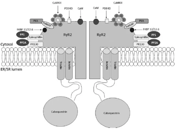
Figure 13: Structure of the RyR2 macromolecular complex
Figure 14: Structure and activation mechanism of Epac proteins
Figure 15: Schematic structure of the 11 phosphodiesterase (PDE) families
Figure 16: Distribution of PDE isoforms in cyclic nucleotide compartmentation
Figure 17: Soluble GC/NO signaling pathway
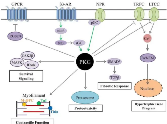
Figure 18: PKG signaling pathway in cardiac myocytes
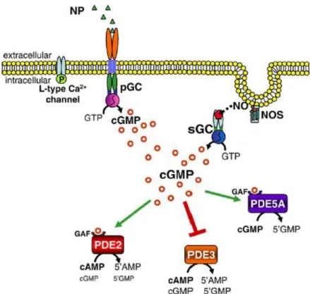
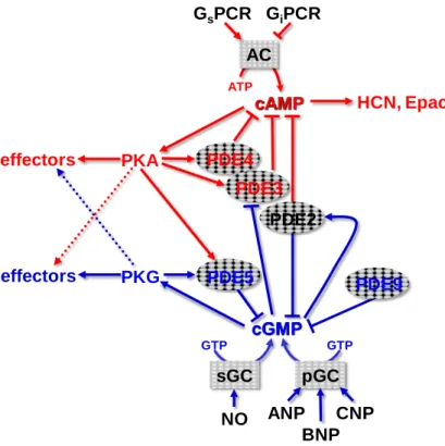
Figure 19: cGMP signaling pathway
Figure 24: Determination of amplitude and kinetic parameters calculated for Ca2+ transients and sarcomere shortening
Figure 25: Functioning of the H187 cAMP-FRET sensor
Figure 26: Determination of PDE2-cAMP-cGMP hydrolytic activity
Figure 27: PDE2 activity in heart extracts from WT and PDE2 TG mice
Figure 28: Similar training capacity of WT and PDE2 TG mice
Figure 29: The higher contractility in PDE2 TG mice is due to lower heart rate
Figure 30: Higher contractility in Langendorff perfused hearts from PDE2 TG mice
Figure 31: Lower heart rate in Langendorff perfused hearts from PDE2 TG mice
Figure 32: Lower rate pressure product in Langendorff perfused hearts from PDE2 TG mice
Figure 33: Heart weight to body weight ratio
Figure 34: ventricular interstitial fibrosis by Masson’s Trichrome Staining
Figure 35: ANP and BNP quantification
Figure 36: Lower heart rate in PDE2 TG mice chronically treated with ISO
Figure 37: Left ventricular interstitial fibrosis determined by Masson’s Trichrome Staining
Figure 38: PDE4 isoform expression in heart extracts from WT and PDE2 TG mice
Figure 39: PDE3 isoform expression level in heart extracts from WT and PDE2 TG mice
List of tables
Table 1: Classification of mammalian tmACs following their regulation by endogenous
modulators and tissue expression
Table 2: Eleven families of phosphodiesterases
Table 3: Pharmacology of PDE family-selective inhibitors
Table 4: Ligands, activators, localization and main functions of the various mammalian
particulate guanylyl cyclase receptors
Table 5: Composition of culture medium I and II used for adult mouse ventricular
cardiomyocytes
Table 6: Composition of Ringer solutions used for measurements of Ca2+ transient, cell shortening, SR Ca2+ leak and load
Table 7: Compositions of the solutions used in whole-cell patch clamp experiments in adult
I. The heart
I.1 Anatomy of the heart
The heart is an organ in human and other animals, which pumps blood through the blood vessels of the cardiovascular system. Blood provides the body with oxygen and nutrients and takes part in removal of metabolic wastes. The human heart beats about 2.5 billion times during average lifetime and pumps about 300 millions liters of blood. Depending on the individual body’s needs, the heart can vary its output between 5 and 20 liters per min. The heart is located in the middle compartment of the chest directly under the breastbone and is composed mainly of cardiac muscles attached to network of collagen fibers. The mammalian and bird heart is divided into four compartments: upper left and right atria; lower left and right ventricles (Figure 1). The right atrium and ventricle are separated from the left atrium and ventricle by a wall, called septum. Blood low in oxygen enters the right atrium from superior and inferior vena cava and passes to the right ventricle. Oxygenated blood returns to the left ventricle through the left atrium and is pumped out through the aorta and pulmonary artery. To prevent the heart from blood backflow, the heart has valves that close automatically. The valve between the right atrium and right ventricle is named the right atrioventricular valve (AV) or tricuspid valve. The valve between left atrium and left ventricle is called the mitral valve. When blood returns from the tissues, fills the atria, as atrial pressure increases above that of the ventricles, the valves open, filling the ventricles with blood. As the ventricles contract, blood is forced back against the valve, pushing them closed. The valve between the left ventricle and the aorta is named the aortic valve. During the left ventricle (LV) contraction (systole), pressure rises in the left ventricle. When the pressure in the LV rises above the pressure in the aorta, aortic valve opens and allows blood to exit the LV. The valve between the right ventricle (RV) and the pulmonary artery is named the pulmonary valve. Pulmonary valve opens in ventricular systole, when the pressure in RV rises above the pressure in the pulmonary artery. At the end of ventricular systole, the pressure in the pulmonary artery closes the pulmonary valve.
fibers. When an impulse reaches the ends of the Purkinje fibers, ventricular muscle contracts (Anon, n.d.).
.
Figure 1: Anatomy of the heart.
Section through the human heart showing the atria, ventricles, valves and circulatory system. (Modified from http://www.texasheart.org)
I.2 Cardiac excitation-contraction coupling
Cardiac excitation-contraction coupling (ECC) is the process by which electrical stimulation of the cardiomyocytes leads to the heart contraction. Ca2+ ions play an important and essential role in this process acting as second messengers, direct activators of the myofilaments during contraction.
I.2.1 Structures involved in cardiac ECC I.2.1.1 Sarcomere and T-tubules
A sarcomere is the basic unit of striated muscle (Figure 2). Highly ordered organization of striated muscle is essential for fast development of force and motion during the heart contraction. In the sarcomeres, the contractile filaments such as actin and myosin are integrated in paracrystalline order by a group of proteins called sarcomeric cytoskeleton. This group of cytoskeletal proteins plays important architectural, mechanical and signaling roles (Gautel & Djinović-Carugo, 2016).
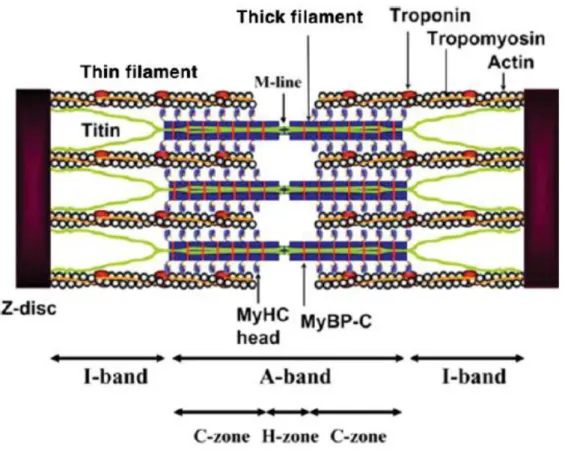
Figure 2: Schematic structure of a sarcomere.
Tropomyosin forms α-helical double strands located along the grooves of actin double strands. Troponin is linked to the specific regions of each tropomyosin in the regular intervals of 38 nm. Myosin heavy chain (MHC) is a protein containing globular head domain with actin binding and ATP hydrolytic sites. Myosin-binding protein C (MyBP-C) is a thick filament which forms 7-9 transverse stripes at a regular distance of 43 nm located in the C-zone of the sarcomere A-band. Titin is the third myofilament located in Z-disc and M-line (Morimoto, 2008).
The transverse tubules (T-tubules) are invaginations of external membrane of skeletal and cardiac muscle and allow depolarization of the membrane to quickly penetrate to the interior of the cell (Orchard & Brette, 2008). The transverse T-tubules of mammalian ventricular cardiomyocytes were first time described by Lindner in 1957 (Lindner, 1957) and the first report of axial T-tubules was done in 1971 (Sperelakis & Rubio, 1971). The transverse tubules are rich in ion channels. Most of the Ca2+ ions enter the cell via Ca2+ channels located
there is a relation between heart rate and density of T-tubule network. In large animals (e.g pigs) T-tubules density is lower than in small mammals (e.g. mice) which have higher heart rate than bigger mammals. The explanation of this occurrence is that the Ca2+ homeostasis (increase and decrease of cytosolic Ca2+) appears faster in animals with higher heart rates (Orchard & Brette, 2008).
I.2.1.2 Sarcoplasmic reticulum
The sarcoplasmic reticulum (SR) is a system of membrane-bounded tubules that surrounds muscle fibrils, first time described by Bennett and Porter and Palade (Bennett & Porter, 1953; Porter & Palade, 1957). The main role of SR is to keep balance between Ca2+ storage, release and reuptake in skeletal and cardiac muscle. This balance is achieved through three major classes of SR Ca2+-regulatory proteins:
1) luminal-Ca2+ binding proteins, e.g. calsequestrin, involved in Ca2+ storage;
2) SR Ca2+ release channels, e.g. ryanodine receptors and IP3 receptors, for Ca2+ release;
3) sarcoplasmic reticulum Ca2+-ATPases type 2 (SERCA2) for Ca2+ reuptake (Rossi & Dirksen, 2006).
Mammalian SR can be arranged into at least three structural and functional elements: longitudinal or “free” SR (fSR), the junctional SR (jSR) and corbular SR (cSR). The longitudinal network is the part of the SR membrane where Ca2+ is pumped into the SR via SERCA2. Both jSR and cSR consist of functional clusters of RyRs or ‘Ca2+ release units’ (CRUs). CRUs are localized in the proximity to LTTCs and activated by Ca2+ influx from LTCCs, causing Ca2+ sparks to produce global Ca2+ signals.
cSR is located in ventricular myocytes but is less important than in atrial myocytes. cSR can play a role as a secondary Ca2+ amplification system in response to Ca2+ diffusion from jSR release (Shiels & Galli, 2014).
I.2.1.3 Myofilaments
The sarcomere is the principal element of the muscle (Figure 2). This repeating unit dominates the anatomy of striated muscles. Each sarcomere is bound by two Z-discs (forming the Z-lines), two I-bands that are regions surrounding the Z-lines, and an A-band between the two I-bands, which is composed of one H-zone and tow C-zones. The Z-line (Zwinschenscheibe) appears as a sequence of dark lines and anchors the actin thin fiber from adjacent units. The A-band (anisotropic) is the region containing the full length of a single
thick filaments. Within the A-band is a brighter region, the zone (heller). Within the H-zone is the M-line (Mittelscheibe) formed of components of sarcomeric cytoskeleton (Hwang & Sykes, 2015). Without an external force, passive sarcomere length of cardiac myocytes is about 1.9 µm (Granzier & Labeit, 2004).
Thick filaments are composed of 300 to 400 molecules of myosin. Myosins are actin-dependent motor proteins using energy going from ATP hydrolysis to move along actin fibers. The family of myosin consists partially of 20 structurally and functionally different classes and in human genome nearly 40 myosin genes are expressed and encoding 12 classes of proteins. Most myosins are composed of three different regions: 1) NH2 head domain
binding actin and hydrolyzing ATP, 2) neck domain consisting of one or more IQ motifs binding light chains (binding e.g. calmodulin), 3) COOH-terminal tail mediating interactions with cargo molecules and/or dimerization of heavy chains (Krendel & Mooseker, 2005).
The thin filament group is composed of three molecular units: F-actin, tropomyosin and troponin (Robertson et al., 2010). Actin was discovered for the first time in 1942 by Straub (Straub, 1942). A major component of the thin filament is the fibrous form of actin, F-actin. Actin occurs as dynamic equilibrium between polymerized F-actin and monomeric G-actin. One adenosine nucleotide strongly binds to actin and polymerization that occurs during transition from G to F-actin activates ATPase (Oda et al., 2009). Troponin is a protein complex composed of troponin C (TnC), troponin I (TnI) and troponin T (TnT). Troponin C is a Ca2+-binding protein containing two E-F hand motifs in terminal domains. The C-terminal domain of cTnC binds 2 cations, Mg2+ or Ca2+, during the contraction-relaxation cycle of the heart. Troponin I interacts with F-actin and inhibits contraction. Troponin T is a scaffold protein that tethers troponin to the thin filament by association with tropomyosin and TnI (Robertson et al., 2010).
I.2.1.4 Mitochondria
Mitochondria are cytosolic double-membrane bound organelles called ‘powerhouse’ of the cell because they generate most of the cell’s energy supply. In addition, mitochondria play an
and shape the cytosolic Ca2+ signal. The mitochondrial inner membrane is rich in systems of channels and transporters for Ca2+ uptake and removal that allows to stock Ca2+ ions in the mitochondrial matrix compartment (Maack & O’Rourke, 2007).
Under physiological conditions, glucose is converted to pyruvate which enters mitochondria and is converted into acetyl-coenzyme A (CoA) by pyruvate dehydrogenase complex (PDC). Fatty acids are transformed to fatty acyl-CoA and carnitine-acyltranslocase is responsible for transporting fatty acid across the inner mitochondrial membrane. Acyl-CoA enters β-oxidation, resulting in the formation of NADH and FADH2. NADH is involved in redox
reactions in the inner mitochondrial membrane transporting electrons from one reactions to another. The respiratory chain provides the free energy for the ATP generation from ADP.
Mitochondria are important for cell contraction and relaxation because of their efficient machinery for Ca2+ transport and ability to store Ca2+. Cardiac contraction is regulated by an elevation of cytosolic Ca2+ ([Ca2+]i). The main mechanism of Ca2+ efflux in mitochondria is
Na+-dependent via mitochondrial Na+/Ca2+ exchange (mNCX). This antiporter utilizes the Na+ gradient across the inner membrane. The second way of Ca2+ extrusion is via the mitochondrial permeability transition pore (mPTP). This mechanism is based on strong increase in the permeability of the inner mitochondrial membrane to ions with molecular mass up to 1.5 kDa. mPTP is regulated by high concentration of Ca2+ in the matrix, oxidative stress and depolarization (Dedkova & Blatter, 2013).
Relaxation occurs upon removal of Ca2+ ions from cytoplasm via: 1) SERCA2, 2) sarcolemmal NCX, 3) sarcolemmal Ca-ATPases, and 4) mitochondrial Ca2+ uptake. Mitochondrial Ca2+ uptake involves the mitochondrial Ca2+ uniporter (MCU), a rapid mode of Ca2+ uptake (RaM), the mitochondrial ryanodine receptor type 1 (mRyR1) and a recently described leucine-zipper-EF-band-containing transmembrane protein 1 (LETM1). All channels involved in Ca2+ uptake are localized to the inner mitochondrial membrane. The most known mechanism of Ca2+ uptake is via MCU forced by the large electrochemical gradient. MCU is regulated by several cations, lanthanides, adenine nucleotides and ruthenium compounds (Dedkova & Blatter, 2013).
I.2.2 Excitation
Each beat of the heart is initiated by a pulse of electrical excitation in a group of pacemaker cells. This electrical pulse is initiated by the electrochemical gradient in the surface membrane of each cardiomyocyte. During rest (diastole), the cell membrane is selectively accessible to K+ ions, so the membrane potential of ventricular cardiomyocyte is about -80 mV, close to
equilibrium potential of K+ ions (-90 mV). During electrical excitation, the cell membrane starts to be permeable to Na+ ions, the opening of Na+ and Ca2+ channels brings positive charges inside the cell which leads to depolarization of the membrane potential towards positive values (40 mV or more). Each cell behaves like a dipole and the electrical membrane potential of a cell rapidly rises and falls. The resulting electrical signal is called ‘action potential’ (Marbán, 2002).
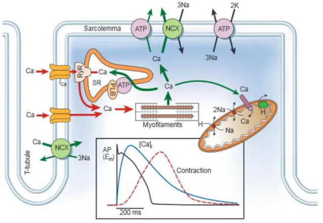
Figure 3: The movement of Ca2+ in ventricular myocytes with the key ion channels and pumps.
The graph shows the time course of an action potential, Ca2+ transient and contraction in a rabbit ventricular myocytes. Ca, Calcium; NCX, Na+/Ca2+ exchange; ATP, ATPase; PLB, phospholamban, SR, sarcoplasmic reticulum, RyR, ryanodine receptors. (From: (Bers, 2002)). During the cardiac action potential Ca2+ enters the cell through depolarization-activated L-type Ca2+ channels (LTCC) generating an inward L-type Ca2+ current (ICa,L). Ca2+ entry
I.2.3 Contraction
Molecular mechanism of contraction based on the sliding filament theory was described first time by Huxley in 1954 (Huxley & Hanson, 1954). This theory is based on cyclic interactions between myosin and actin driven by hydrolysis of ATP. Contraction of muscle appears when the thin filaments (actin) slide past the thick fibers (myosin). The crossbridge hypothesis is used to explain this process: it states that actin and myosin filaments create a protein complex, forming a sort of cross-bridge between the two filaments. The myosin head possesses an ATPase activity that allows to convert chemical energy in the form of ATP to mechanical energy, generating force and movement.
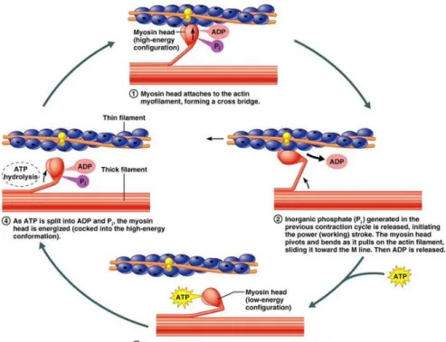
Figure 4: Molecular mechanism of muscle contraction.
In presence of ATP, ADP and Pi are located in the head of myosin. Actin binding to myosin releases ADP and Pi initiating the power stroke resulting in movement of myosin along the thin filament. The cycle is stopped in the absence of ATP. New ATP binds to myosin decreasing its affinity to the actin filament. Separation of myosin and actin results in ATP hydrolysis to ADP and Pi and new cycle starts.
The contraction cycle begins with myosin tightly bound to actin. This interaction activates the myosin ATPase and the energy generated from the hydrolysis of ATP to ADP+Pi (inorganic phosphate) is rapidly converted to mechanical force resulting in movement of myosin along the thin filament by about 5 nm. The products of hydrolysis (ADP+Pi) remain bound to the myosin head. Myosin head rebinds at a new position on the thin filament resulting in the release of ADP+Pi producing the ‘power stroke’ in which the myosin head comes back to the
initial conformation sliding the actin filament toward the M line of the sarcomere (Figure 4). The contraction of cardiac muscle occurs when Ca2+ concentration increases in the cytosol from approximately 10-7 to 10-5 M. The increasing Ca2+ concentration gives a signal for contraction via the action of two proteins: tropomyosin and troponin that bind to the actin filaments. Each tropomyosin is bound to troponin formed of troponin C, troponin I and troponin T. The complex of tropomyosin with troponins blocks the interaction of myosin with actin when the concentration of Ca2+ in the cytosol is low preventing muscle contraction. At high concentrations, Ca2+ binds to troponin C and this leads to conformational changes in the complex resulting in contraction (Cooper, 2000).
I.2.4 Relaxation
Relaxation to occur requires a reduction in cytosolic Ca2+ level, allowing Ca2+ to dissociate from troponin. This process involves Ca2+ transport out of the cytosol by the following pathways: 1) SR Ca2+-ATPase type 2 (SERCA), 2) sarcolemmal Na+/Ca2+ exchange (NCX), 3) sarcolemmal Ca2+-ATPase and 4) mitochondrial Ca2+ uniport (MCU). The sarcolemmal NCX and sarcolemmal Ca2+-ATPase transport Ca2+ out of the cell, SERCA2 allows to restore SR Ca2+ reserve and mitochondrial Ca2+ uniport allows the passage of Ca2+ from a the cytosol into the mitochondrial matrix. To keep steady state conditions in the cell, during each action potetial, Ca2+ influx must be balanced by Ca2+ efflux (Bers, 2002). However, the importance of the different pathways for Ca2+ extrusion varies among species. In rabbit ventricular cardiomyocytes, SERCA2 is responsible for removing 70% of Ca2+ ions, while NCX removes 28% and MCU only 1%. In rat and mouse ventricular myocytes, the activity of SERCA2 is higher than in rabbit cells (~92%), while the activity of NCX is lower (~7%).
I.3 Regulation of ECC
The force of cardiac contraction is regulated by intrinsic way as an interplay between length-dependent changes in cardiac function and changing myocardial contractility (Starling’s ‘law of the heart’ and ‘Family of Starling Curves’). Heart can be also regulated via the autonomic nervous system.
Increase in Ca2+ sensitivity leads to a greater number of actin-myosin crossbridges. The force that any single cardiac filament develops is proportional to the initial sarcomere length. Larger or smaller sarcomere length than the optimal values will decrease the force of the muscle contraction.
I.3.2 Neurohormonal regulation of heart function
Even if the heart possesses a special contractile protein-machinery that allows to generate mechanical force, the force and rate of contraction are regulated by the autonomic nervous system, namely the sympathetic system and parasympathetic system (Figure 5).
Figure 5: Autonomic regulation of the heart.
The diagram shows the two nervous systems that regulate heart rate and force of contraction. In green, the sympathetic nervous system; in blue, the parasympathetic nervous system. ACh, acetylcholine, Ad, adrenaline, NOR- noradrenaline (Modified from thesis of Nolwenn Merlet, http://www.sudoc.abes.fr/DB=2.1/SRCH?IKT=12&TRM=159710308 ).
The parasympathetic nervous system decreases heart rate through release of the neurotransmitter acetylcholine (ACh) that raises the threshold for spontaneous action potential generation. In contrast, the sympathetic system increases the rate and force of contraction which is mediated through release of catecholamines like adrenaline (Ad) and noradrenaline
(NOR). This leads to the sinoatrial node (SAN) stimulation and increase in firing rate of action potentials (AP) (Soni et al., 2014).
The sympathetic and parasympathetic nervous system comprises a great number of molecular components like second messengers: cAMP, cGMP, inositol trisphosphate (IP3), diacylglycerol (DAG), which leads to phosphorylation and dephosphorylation of many proteins involved in ECC: LTCCs, RyRs, PLB that regulates the activity of SERCA2, and contractile proteins: TnI and MyBP-C. These signaling pathways predominantly involve α- and β-receptors, which are activated by catecholamines that bind to extracellular sites of various isoforms of GPCRs.
II. Cyclic AMP signaling in the heart
Cyclic AMP was the first discovered second messenger. cAMP was identified by Earl Sutherland in 1958 (Rall & Sutherland, 1958) who won a Nobel Prize in Physiology and Medicine in 1971 for his discovery regarding the mechanisms of action of hormones via second messengers, mainly cAMP.
Cyclic AMP plays a very important role in the cellular response to many hormones like glucagon or adrenaline and neurotransmitters that cannot pass through the plasma membrane. Cyclic AMP is strongly involved in modulating the contractile function of cardiac muscle by activation of cyclic AMP-dependent protein kinases (PKA) which are responsible for phosphorylation of key proteins in E-C coupling.
Intracellular cAMP concentration is regulated by the activities of at least two enzymes: adenylyl cyclase (AC) responsible for cAMP synthesis and cyclic nucleotide phosphodiesterases (PDEs) responsible for cAMP degradation (Fimia & Sassone-Corsi, 2001).
seven transmembrane α-helices (7TMH) connected with intracellular and extracellular loops. The extracellular region (EC) of receptor is responsible for ligand binding and the intracellular (IC) region of receptor is a place of interactions with G-proteins, arrestins and other downstream effectors (Latek et al., 2012).
Figure 6: GPCR-mediated G-protein activation.
The diagram shows GTPase cycle in three steps: 1) The interaction between a cell surface receptor with endogenous ligand promotes the coupling of receptor with intracellular heterotrimeric G protein, 2) The receptor-G protein coupling leads to the exchange of GDP for GTP, 3) Gα–GTP subunit dissociates from Gβγ and receptor, both units are ready to modulate the activity of intracellular effectors. Termination of signal occurs when GTP is removed from Gα subunit by the GTPase activity of Gα subunit (Cabrera-Vera et al., 2003).
The cellular or physiological response mediated by a GPCR requires interactions of the receptor with heterotrimeric guanine nucleotide-binding proteins (G proteins). G-proteins are composed of three units: α, β and γ. In inactive state, GDP is bound to Gα subunits. During receptor activation GDP is released and GTP binds to α subunit. Gα-GTP dissociates from receptor and Gβγ subunit. Then, both Gα-GTP and Gβγ are ready to activate or inhibit downstream effectors of signaling pathway (Figure 6) (Cabrera-Vera et al., 2003).
II.2 β-adrenergic receptors
β-adrenergic receptors (β-ARs) belong to the superfamily of GPCRs and their stimulation by sympathetic nervous system or circulating catecholamines is implicated in metabolic regulation, peripheral blood circulation, central neural activities and muscle contraction.
In the heart, acute β-AR stimulation is the most powerful mechanism of cardiac output regulation in response to a flight-or-fight situation. Chronic β-AR stimulation is part of physiological and pathological heart remodeling. Three subtypes of receptor are expressed in cardiac myocytes: β1, β2, β3, where β1 and β2 play a dominant role in ECC (Woo & Xiao,
2012).
β1-AR is the predominant receptor subtype in the mammalian heart. In general, the ratio β1:β2
-AR is around 70%:30% in the atria and 80%:20% in the ventricles (Brodde et al., 2006). β-ARs are involved in many different signaling pathways (Figure 7). β1-AR is the main
expressed receptor in healthy cardiomyocytes. β2-ARs are overexpressed in the failing heart,
while the expression of β1-AR is decreased. Both β1- and β2-AR pathways involve the
stimulatory G proteins (GαS) leading to increase in contractility. β2-ARs can interact also with
inhibitory G-proteins (Gαi) pathway opposing the positive inotropic effects and activating a
cell survival pathway. The expression of β3-ARs is very low in the healthy heart, but β3-ARs
are upregulated in heart failure (HF) and exert both positive and negative inotropic effects in the failing hearts (Najafi et al., 2016).
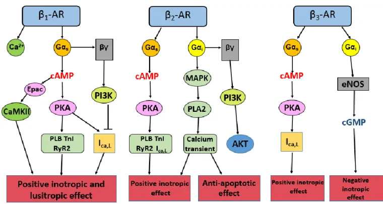
Figure 7: β-adrenergic signaling pathway.
The diagram shows pathways activated by β-adrenergic receptor subtypes and their physiological effects (inspired by (Steinberg, 1999)).
II.2.1 β1-adrenergic receptor
β1-ARs are the most studied subtype of β-ARs. β1-ARs are located on the surface of
cardiomyocytes. They are involved in the production of cAMP. cAMP activates exchange proteins directly activated by cAMP (Epac) and PKA. This leads to phosphorylation of key proteins of ECC: LTCCs, phospholamban (PLB), ryanodine receptors (RyR), Troponin T and I, resulting in positive inotropic, chronotropic and lusitropic effect (Fu & Xiang, 2015). However, previous studies showed that β1-AR stimulation increases activity of phosphatidyl
inositol 3-kinase (PI3K) via Gαs and Gβγ proteins in adult rat cardiomyocytes. Increased
PI3K activity negatively regulates β1-AR mediated contractile response. This leads to a
decrease in ICa,L and intracellular Ca2+ without phosphorylation of myofilaments involved in
EC coupling. The PI3K-dependent inhibition of β1-AR can be implicated in agonist-induced
desensitization of receptor and can lead to HF (Leblais et al., 2004).
Chronic stimulation of β1-ARs by catecholamines exerts a toxic effect on the heart. Mouse
model with overexpression of human β1-ARs showed a presence of early hypertrophy and
via PKA and Ca2+ calmodulin-dependent protein kinase (CaMKII), but the role of PKA in apoptotic pathway is not well known (Baker, 2014).
The second mouse model showing β1-AR-induced toxicity is a model lacking β1-ARs. This
study showed that β1-ARs are involved in catecholamine induced increase in contraction and
stimulation of β1-ARs activates CaMKII signaling pathway. In addition, mice leaking β1-ARs
showed preserved cardiac function after myocardial infarction (Yoo et al., 2009).
II.2.2 β2-adrenergic receptors
While β1-ARs are the predominant subtype of β-AR in the heart, β2-ARs are expressed in
many cell types: mainly in cardiac myocytes and vascular and pulmonary smooth muscle cells, where they are responsible for the regulation of inotropy, chronotropy, vasodilation and bronchodilation. In the heart, β2-ARs similar to β1-ARs promote cAMP production resulting
in positive inotropic and chronotropic effect (Taylor & Bristow, 2004).
Stimulation of β2-ARs produces different effects than β1-AR stimulation. In contrast to β1
-ARs which lead to a rather uniform distribution of cAMP (Fu et al., 2013), β2-AR-mediated
production of cAMP is highly localized (Nikolaev et al., 2006). β2-ARs are specifically
concentrated inside caveolae(Rybin et al., 2000; Xiang et al., 2002). cAMP production upon β2-AR stimulation is limited by phosphodiesterases (PDEs) (Jurevičius et al., 2003; Nikolaev
et al., 2006). The compartmentation of β-AR signaling defines the spatial activation of PKA
anchored through A kinase anchoring proteins, thus β2-ARs activate a specific pool of PKA,
which is localized in the close proximity to protein targets (Dodge-Kafka, 2006). β2-ARs form
a complex with L-type Ca2+ channels, G proteins, caveolin and AKAPs, PKA and phosphatases like calcineurin and phosphatase 2A (Davare et al., 2001). This complex provides the specificity, efficacy and rapidity of Ca2+ channels modulation (Flynn & Altier, 2013).
Study on mouse model with overexpression of the human β2-AR shows markedly elevated
myocardial relaxation and reduced PLB protein level without changes in SR Ca2+ or calsequestrin (Rockman et al., 1996). Another study shows that β2-AR overexpression does
exercise. β2 knockout mice became hypertensive; hypertension was probably associated with
peripheral vasoconstriction (Bernstein, 2002).
In addition, β2-AR stimulation, in contrast to β1-AR stimulation, has anti-apoptotic effect and
β2-ARs are important regulators during chronic activation of the adrenergic system. This
difference between β1 and β2-AR stimulation can be explained by the interactions of β2-ARs
with inhibitory G proteins (Daaka et al., 1997).
II.2.3 β3-adrenergic receptors
β3-AR mRNA was detected in the human heart but with much lower expression than β1 and
β2-ARs (Michel et al., 2010). β3-ARs were first characterised in 1989 (Emorine et al., 1989).
Most studies focused on β3-AR function in adipose tissue. In contrast to β1 and β2-ARs, the
gene coding the β3-AR has introns (Gauthier et al., 2000). The structure of β3-AR shows only
51% identity with β1-ARs and 46% with β2-ARs (Imbrogno et al., 2015). In mammalian
cardiovascular system, β3-AR is expressed in the myocardium and the coronary microarteries
(Dessy et al., 2004). In the mammalian heart, β3-ARs are more expressed in atrial cells than in
ventricular myocytes (Moniotte et al., 2001). In human ventricular myocytes, β3-ARs
stimulation produces negative inotropic effect associated with increase in intracellular cGMP after activation of nitric oxide synthase (NOS) by inhibitory G proteins (Gauthier et al., 1996, 1998). While in human atrium, β3-ARs activation is either ineffective (Christ et al., 2011) or
exerts a positive effect on ICa,L and contractility (Skeberdis et al., 2008).
In contrast to β1 and β2-ARs, the selectivity of ligands for β3-ARs is less clear. It was shown
that β3-ARs in the human heart has higher affinity for noradrenaline than adrenaline. Well
known antagonists for β1 and β2-ARs like propranolol, atenolol, pindolol, etc. bind also to β3
-ARs, but with much lower affinity (Imbrogno et al., 2015).
β3-ARs seems to be upregulated in human HF, while β1 and β2-ARs are downregulated or
desensitized. It can be a compensatory upregulation of β3-ARs to avoid further myocyte
damage. In addition, β3-ARs upregulation can act on vessel tone resulting in decrease in the
peripheral vascular resistance and the afterload of failing hearts (Gauthier et al., 2000).
II.2.4 Desensitization of β-adrenergic receptors
Desensitization is a process of loss of receptor responsiveness induced by long term exposure to catecholamines. This process can be ‘agonist-specific’ or ‘agonist non-specific’.
Agonist non-specific desensitization (Figure 8A) is mediated by phosphorylation of β-ARs by PKA, protein kinase C (PKC) and G protein-coupled receptor kinase (GRK) on the plasma membrane of the cardiomyocytes. PKA and PKC-mediated phosphorylation of β-ARs directly leads to conformational changes of receptor and prevents the interactions with G proteins and further signal transduction.
The agonist-specific desensitization of β-ARs (Figure 8B) is initiated by phosphorylation of six members of GRK. GRK family is composed of three subfamilies: 1) rhodopsin kinases (GRK1 and 7), 2) β-AR kinases (GRK2 and 3), 3) GRK4 subfamily (GRK4, 5 and 6) (Najafi
et al., 2016). GRK 2, 3 and 5 are expressed in the heart. GRK 5 is a membrane associated
form and GRK 2 and 3 are mainly localized in the cytoplasm. It has been shown that correlation between hyperactive sympathetic nervous system and β-ARs desensitization is linked mainly to elevated GRK2 expression (Huang et al., 2011). Study with GRK2 knockout mice showed that mice died early during embryonic period, because of HF and hypoplasia, suggesting that GRK2 plays a fundamental role in cardiac development (Jaber et al., 1996).
In both mechanisms of desensitization, ARs phosphorylation promotes binding to β-arrestins (Najafi et al., 2016). β-β-arrestins are small family of proteins that block interactions between β-ARs and G proteins preventing further signaling. β-arrestins use mechanism that involve PDEs which degrade the second messenger, cAMP. β-arrestins interact with PDE4 type D (Baillie et al., 2003). This interactions of β-arrestins with PDE4D decreases a local pool of cAMP and leads to loss of cAMP-induced activation of PKA resulting in reduction of downstream effects of PKA on cell contractility, motility and transcription (Najafi et al., 2016).
In contrast to β1 and β2-ARs, β3-ARs are lacking in PKA and GRK phosphorylation site
resulting in resistance to short-term agonist-specific desensitization (Rozec & Gauthier, 2006).
Figure 8: Regulation of β-AR desensitization by GRK and β-arrestins.
A) Receptor activation is followed by binding of GRK2 and Gβγ to the receptor β-AR. The activity of GRK2 can be increased by protein kinase A (PKA), protein kinase C (PKC) and c-Src. GRK2 activity can be decreased by phosphorylation by Erk1/2 or its binding to Ca2+/calmodulin (CaM), B) β-AR activates the effector, adenylyl cyclase (AC) to produce cAMP. After phosphorylation of receptor by GRK, β-arrestin binds to the receptor, translocating PDE4 with it. This places PDE4 on the membrane, reducing local pool of cAMP and prevents further signaling (modified from (Kohout & Lefkowitz, 2003)).
II.3 Adenylyl cyclases
Adenylyl cyclases (ACs) are enzymes that catalyze cAMP synthesis. In mammals, ACs exist as transmembrane adenylyl cyclases (tmACs) or soluble adenylyl cyclases (sAC) anchored at various locations within the cell forming with PDEs local cAMP signaling pool . In mammals, nine isoforms of tmAC exist and they differ in their patterns of expression and regulatory properties (Table 1) (Hanoune & Defer, 2001).
AC enzymes catalyze reaction of intramolecular cyclization of ATP to cAMP with release of pyrophosphate (Steegborn, 2014). All nine isoforms of tmACs are composed of two catalytic domains, C1 and C2, in a single protein chain. Catalytic domains are located downstream of a six membrane helices (M1 and M2) forming pseudoheterodimer, where C1 and C2 domains have low affinity to each other.
Group Isoform Activators Inhibitors Tissue Distribution
1 AC1 GSα, FSK, Ca
2+
/CaM Giα, Gβγ, CaM kinase
IV, P-site analogs
Brain, retina
AC3 GSα, FSK, Ca 2+
/CaM CaM kinase II, P-site analogs
Olfactory neurons, brain
AC8 GSα, FSK, Ca 2+
/CaM P-site analogs Brain
2 AC2 GSα, FSK, Gβγ, PKC P-site analogs Brain, olphactory bulb
AC4 GSα, FSK, Gβγ P-site analogs Kidney, brain, liver
AC7 GSα, FSK, Gβγ, PKC P-site analogs Heart>brain>kidney,
testis, liver 3 AC5 GSα, FSK, PKC, PKCζ Giα, PKA, Gβγ, Ca2+, P-site analogs Brain>heart
AC6 GSα, FSK Giα, PKA, PKC,
Ca2+, P-site analogs
Heart>brain>kidney, testis, liver
4 AC9 GSα Calcineurin, P-site
analogs
Skeletal muscle, brain, lungs, liver
Table 1: Classification of mammalian tmACs following their regulation by endogenous modulators and tissue expression (Pavan et al., 2009).
GSα: G stimulatory-protein α subunit, FSK: forskolin, Gβγ: G-protein βγ subunit, PKC:
protein kinase C, CaM: calmodulin, Giα: G inhibitory-protein α subunit, PKA: protein kinase A, PKC: protein kinase C.
Mammalian C1 and C2 domains are located a head to tail by antiparallel coupling forming two distinct pockets at the interface. One pocket binds ATP and is a place where cyclization occurs. The second pocket is non-catalytic and is the site for forskolin binding. The cyclization reaction requires four residues. Two aspartate residues are located in C1 domain and bind two Mg2+ ions, two other residues, arginine and asparagine, are located in the C2 domain (Linder & Schultz, 2008).
Mammalian sAC contains two heterologous catalytic domains (C1 and C2) located in the amino terminus of the protein. C-terminus of sAC consists of an auto inhibitory region, canonical P-loop and leucine zipper sequences. Similarly to tmACs, sAC binds two metal cations in the catalytic site, which are responsible for ATP binding and cyclization. sAC is most active in the presence of Mn2+ and in contrast to tmACs, activity of sAC is insensitive to
Figure 9: Schematic structure and membrane topology of transmembrane adenylyl cyclases.
M1 and M2 are anchors of a six membrane helices domains. C1a and C2a are the cytoplasmic catalytic domains with two active sites to bind ATP and forskolin (FSK). C1b is the cytoplasmic-linker domain and C2b is the second non-catalytic domain present only in AC1, AC2, AC3 (Pavan et al., 2009).
All isoforms of tmACs are expressed in the heart, except AC8. AC5 and AC6 are the most expressed in the heart with AC5 being dominant. AC1 is expressed only in sinoatrial node controlling pacemaker function. Isoforms 2, 3, 4, 5/6 and 7 are mainly located in cardiac fibroblasts. The most expressed isoforms in the heart are AC5 and 6 and they may serve different functional roles (Efendiev & Dessauer, 2011; Guellich et al., 2014)A mouse model with deletion of AC5 showed that basal cardiac functions remained unchanged, but left ventricle ejection fraction was decreased under isoproterenol condition. AC5 disruption leads to total loss of acetylcholine-mediated inhibition and to decrease in Ca2+-induced inhibition of cAMP production. Studies with AC5 KO models showed that deletion of AC5 can be protective against HF under stress conditions (Yan et al., 2007b). Cardioprotective effect is associated with elevated level of Bcl-2 and reduction in myocardial apoptosis (Okumura et
al., 2003; Sadana & Dessauer, 2009).
Studies with AC6 knockout mice showed that deletion of AC6 leads to decrease in cAMP level in left ventricle under basal and stress conditions, reduction in PKA and Akt activity and phospholamban phosphorylation resulting in abnormalities in Ca2+ transient (Sadana &
Dessauer, 2009). In addition, AC6 is overexpressed in fetal hearts and decreases progressively with age to obtain low level of expression in adult hearts. Expression profile of AC5 is exactly opposite to that of AC6 (Espinasse et al., 1995).
II.4 Effectors of cAMP
Generally, cardiac effects of cAMP are attributed to the activation of cAMP-dependent protein kinase A (PKA). cAMP can also act via 2 other effectors: exchange protein directly activated by cAMP (Epac) and hyperpolarization-activated cyclic nucleotide-gated channels (HCN).
II.4.1 Protein kinase A
II.4.1.1 Structure and regulation
PKA is a threonine/serine kinase and its activity depends on the cellular level of cAMP. PKA catalyzes the reaction of transfer γ-phosphate of ATP to threonine or serine residues. The PKA phosphorylation is essential for many processes including contraction, metabolism and gene expression. PKA is composed of two catalytic (C) and two regulatory (R) subunits, forming heterotetramer in its inactive state (Rababa’h et al., 2015). Each of the two regulatory subunits has two sites A and B to cooperatively bind cAMP. In the inactive state, only B site is exposed to bind cAMP. cAMP binding to B site facilitates cAMP binding to the A site. Binding of four cAMP molecules to the two regulatory domains results in conformational changes and dissociation of enzyme into R subunit dimer with four cAMP molecules and two C monomers (Figure 10). C subunit monomers are catalytically active and are able to phosphorylate serine and threonine residues of substrate proteins (Taskén & Aandahl, 2004).
PKA holoenzyme exists in 2 types, type I and type II, determined on the basis of their elution from ion exchange column. They are composed of different R subunits: RI and RII. It was also shown that there are two RI subunits, RIα and RIβ, and two RII subunits, RIIα and RIIβ (Turnham & Scott, 2016). These two classes of PKA differ in their affinity for cAMP with activation constant of 50-100 nM for PKA type I and 200-400 nM for PKA type II. RI and RII subunits show also different expression profile in cells and tissue and they are involved in different cAMP-dependent responses.
II.4.1.2 Targets of PKA in the heart
Signal transduction by PKA is involved in many cellular responses including metabolism, gene expression and contraction (Walsh & Van Patten, 1994). PKA is activated by β-AR signaling and phosphorylates many of the components involved in ECC (Figure 11) including PLB, TnI, LTCCs, MyBP-C, RyR2 and PDE4D3 (Rababa’h et al., 2015).
PKA dependent phosphorylation of these ECC components leads to increase in Ca2+ current ICa,L, SR Ca2+ uptake and release, SR Ca2+ content and the dissociation of Ca2+ from the
myofilaments allowing the heart to contract and relax (Rababa’h et al., 2015).
Figure 11: Signal transduction mediated by PKA in ECC.
Activation of β-ARs allows for the activation of adenylyl cyclase (AC), mediated by specific G proteins (Gs) and the generation of cAMP, which activates PKA. PKA phosphorylates key components of ECC: L-type Ca channels (LTCCs), ryanodine receptors (RyR2), phospholamban (PLN) (modified from (Marks, 2013)).
L-type Ca2+ channels (LTCCs) are essential in ECC. They are involved in the generation of the mechanical and electrical properties of the heart muscle. There are four types of LTCCs: Cav 1.1-1.4. The LTCC is composed of four different α1 subunits together with β, α2δ and γ
subunits. α1 subunits form a pore for ion conduction composed of four homologous domain
(I-IV), each contains six transmembrane segments (Figure 12). Cav1.2 is the primary LTCC
expressed in ventricular myocytes, while both Cav1.2 and Cav1.3 are also expressed in atrial
and sinoatrial cells (Bers & Perez-Reyes, 1999; Harvey & Hell, 2013).
Figure 12: Structure of the cardiac L-type Ca2+ channel Cav1.2.
The purple segments are responsible for channel activation and segment 6 (green) for channel inactivation. Channel is regulated by the complex Ca2+/calmodulin (CaM), by PKA, PKC, PKG and CaMKII phosphorylation and by glycosylation of the α2δ subunit (Brette et al., 2006).
PKA phosphorylation of LTCCs increases activity of LTCCs, resulting in an increase ICa,L