HAL Id: hal-00297526
https://hal.archives-ouvertes.fr/hal-00297526
Submitted on 26 Oct 2005
HAL is a multi-disciplinary open access
archive for the deposit and dissemination of
sci-entific research documents, whether they are
pub-lished or not. The documents may come from
teaching and research institutions in France or
abroad, or from public or private research centers.
L’archive ouverte pluridisciplinaire HAL, est
destinée au dépôt et à la diffusion de documents
scientifiques de niveau recherche, publiés ou non,
émanant des établissements d’enseignement et de
recherche français ou étrangers, des laboratoires
publics ou privés.
In vitro formation of Ca-oxalates and the mineral
glushinskite by fungal interaction with carbonate
substrates and seawater
K. Kolo, Ph. Claeys
To cite this version:
K. Kolo, Ph. Claeys. In vitro formation of Ca-oxalates and the mineral glushinskite by fungal
interaction with carbonate substrates and seawater. Biogeosciences, European Geosciences Union,
2005, 2 (3), pp.277-293. �hal-00297526�
Biogeosciences, 2, 277–293, 2005 www.biogeosciences.net/bg/2/277/ SRef-ID: 1726-4189/bg/2005-2-277 European Geosciences Union
Biogeosciences
In vitro formation of Ca-oxalates and the mineral glushinskite by
fungal interaction with carbonate substrates and seawater
K. Kolo and Ph. Claeys
Vrije Universiteit Brussel, Department of Geology, Pleinlaan 2, 1050 Brussels
Received: 21 February 2005 – Published in Biogeosciences Discussions: 15 April 2005 Revised: 19 August 2005 – Accepted: 13 October 2005 – Published: 26 October 2005
Abstract. This study investigates the in vitro formation of
Ca-oxalates and glushinskite through fungal interaction with carbonate substrates and seawater as a process of biolog-ically induced metal recycling and neo-mineral formation. The study also emphasizes the role of the substrates as metal donors. In the first experiment, thin sections prepared from dolomitic rock samples of Terwagne Formation (Carbonifer-ous, Vis´ean, northern France) served as substrates. The thin sections placed in Petri dishes were exposed to fungi grown from naturally existing airborne spores. In the second exper-iment, fungal growth and mineral formation was monitored using only standard seawater (SSW) as a substrate. Fungal growth media consisted of a high protein/carbohydrates and sugar diet with demineralized water for irrigation. Fungal growth process reached completion under uncontrolled lab-oratory conditions. The newly formed minerals and textu-ral changes caused by fungal attack on the carbonate sub-strates were investigated using light and scanning electron microscopy (SEM-EDX), x-ray diffraction (XRD) and Ra-man spectroscopy. The fungal interaction and attack on the dolomitic and seawater substrates resulted in the forma-tion of Ca-oxalates (weddellite CaC2O4·2(H2O), whewellite (CaC2O4·(H2O)) and glushinskite MgC2O4·2(H2O) associ-ated with the destruction of the original hard substrates and their replacement by the new minerals. Both of Ca and Mg were mobilized from the experimental substrates by fungi. This metal mobilization involved a recycling of substrate metals into newly formed minerals. The biochemical and diagenetic results of the interaction strongly marked the at-tacked substrates with a biological fingerprint. Such finger-prints are biomarkers of primitive life. The formation of glushinskite is of specific importance that is related, besides its importance as a biomineral bearing a recycled Mg, to the possibility of its transformation through diagenetic pathway
Correspondence to: K. Kolo
(kakolo@vub.ac.be)
into an Mg carbonate. This work is the first report on the in vitro formation of the mineral glushinskite through fun-gal interaction with carbonate and seawater substrates. Be-sides recording the detailed Raman signature of various crys-tal habits of Mg- and Ca-oxalates, the Raman spectroscopy proved two new crystal habits for glushinskite. The results of this work document the role of microorganisms as metal recyclers in biomineralization, neo-mineral formation, sedi-ment diagenesis, bioweathering and in the production of min-eral and diagenetic biomarkers. They also reveal the capacity of living fungi to interact with liquid substrates and precipi-tate new minerals.
1 Introduction
The formation of Ca-oxalate minerals (weddellite and whewellite) through fungal mediation in various growth en-vironments is well-documented (e.g. Silverman and Munoz, 1970; Arnott, 1982, 1995; Verrecchia et al., 1993; Garieb et al., 1998; Sayer and Gadd, 1997; Gadd, 1999, 2002; Garvie et al., 2000; Verrecchia, 2000; Burford et al., 2002; Gadd et al., 2002; Kolo et al., 2002). This is not the case for glushinskite [MgC2O4·2(H2O)], where its formation in na-ture and in vitro (this work) shows that a pre-existing Mg-rich substrate is a prerequisite. Glushinskite, a natural or-ganic magnesium oxalate dihydrate was discovered in 1960 (Wilson et al., 1980). Its natural formation is similar to other organic oxalates (e.g. Ca-oxalates) through fungally medi-ated mineralization processes (Garvie, 2003). The forma-tion of oxalates contributes to the Ca, Mg and C recycling in nature and to bio-weathering of rock substrates (Ascaso et al., 1998; Arocena et al., 2000; Sterflinger, 2000). Frost et al. (2003) considered the finding of organic oxalates (e.g. weddellite, whewellite, glushinskite) on other planets as a possible evidence of primitive life forms such as lichens and fungi. Microbial mineralization processes (whether bacterial
278 K. Kolo and Ph. Claeys: A study on substrate biomineralization and diagenesis by fungi or fungal) leave mineralogical (metal oxalates) and organic
traces on the affected rock surfaces. These traces, when re-vealed by Raman spectrometry analysis would indicate the existence of a life signature on earth (Howell et al., 2004) or on Mars (Bishop and Murad, 2004; Hochleitner et al., 2004; Howell et al., 2004).
Magnesium can form both simple and complex oxalates (Gadd, 1999). In nature, the formation of glushinskite, as a biomineral, was attributed to the reaction of oxalic acid produced by the fungal mycobiont of the lichen Lecanora
atra growing with a Mg-rich substrate of serpentinites
(Wil-son et al., 1980, 1987). In the case of a rotting saguaro cac-tus, Garvie (2003) suggested that the reaction between oxalic acid released by fungi and Mg-rich solutions had produced glushinskite. Buildings constructed with Mg-rich limestone or marble could also provide an exposed substrate to fun-gal interaction and formation of glushinskite under natural conditions. The mineral is also found in human and animal bodies as kidney or urinary tract stones (Diaz-Espineira et al., 1995). Ca-oxalates and glushinskite are considered in-dicators of biological weathering in Ca and Mg-rich milieus (Arocena et al., 2003; Wilson et al., 1987).
This experimental study reports on the biomineralization of Ca and magnesium oxalates produced through free, un-selective and un-controlled in vitro fungal growth and in-teraction with carbonate and seawater substrates that could develop on any suitable surfaces and causes important min-eralogical, petrographic alterations of the substrates. More-over, these alterations could cause permanent damage to works of art. Geologically, fungal interaction will have dia-genetic effects on carbonate substrates including mineralogy, petrography and even isotopic signature (Kolo et al., 2004). This is the first report on experimental formation of glushin-skite under laboratory conditions simulating natural fungi-rock substrate environment.
2 Experimental: materials and methods
2.1 Thin sections
Standard petrographic thin sections (30 µm) were prepared from dolomitic rock samples of “Terwagne Formation” (Vis´ean, Bochaut quarry, in northern France). Detailed pet-rographic and sedimentological analysis could be found else-where (Mamet and Preat, 2005). For two samples, duplicate thin sections were prepared to monitor fungal growth and in-teraction during the experiment. This made it possible to fol-low the evolution of the experiment without disturbing the original settings on the other thin sections. The dolomite in the thin sections provided the Mg and Ca necessary, ei-ther for the reaction with oxalic acid, the main metabolite of fungi or for possible uptake-expulsion by fungi during growth process. The experiment targeted airborne spores naturally inhabiting laboratory and outdoors space especially
the saprophytes Mucorales that are commonly referred to as sugar fungi which respond rapidly to and grow rapidly on simple sugars in artificial media besides their ability to break down starch, fats and proteins. These fungi also grow well under a wide pH range making them tolerant to possible pH fluctuations during the growth process. The growth media tailored to meet the experiment requirements consisted of ground chips (3–4 mm) of almond seeds as a source of pro-tein and carbohydrates, 10% sugar solution (dextrose and su-crose) and demineralized water for irrigation. This growth media mixture was found highly efficient for natural, unse-lective and free fungal growth that produced thick mycelia with high coverage surface area/thin section ratio. Interfer-ence with the fungal growth process was minimized to mimic fungal growth under natural outdoor conditions.
Thin sections were placed separately in plastic Petri dishes ( =90 mm) containing 20 g of the prescribed natural growth media. The solid chips of growth media surrounded the thin sections but did not touch their surfaces. Ten mL of sugar solution prepared with demineralized water were carefully added to each Petri dish, taking care not to cover the thin sections surfaces with the solution. Keeping the thin sections surfaces free of growth media and sugar solution was impor-tant in monitoring the development of the fungal mass and the extracellular polymeric substance (EPS) layer over the surfaces.
2.2 Standard seawater (liquid substrate)
Ten mL of filtered standard seawater (3.69 wt % Mg and 1.16 wt % Ca) were added to three Petri dishes containing the same growth media. The standard sea water (SSW) was intended to monitor fungal interaction with liquid substrate and possible recycling of Ca and Mg in the biomineraliza-tion process. The Petri dishes from both experiments were kept open for 24 h then were covered and placed in fume hood under continuous air current. Laboratory temperature varied between 23–25◦C. All samples were kept stationary during the course of experiment. No pH control was carried out. Throughout the experiment, the growing fungi were oc-casionally spray-irrigated with demineralized water to insure continuous humidity. Specimens of the extracellular poly-meric substance (EPS) layer formed on the dolomitic and seawater substrates were continually examined under the op-tical microscope.
2.3 Instrumentation
SEM-EDX: The coating of thin sections, slides of fungal and
EPS material, and extracted mineral species was carried out with Carbon Coater EMITECH-Carbon evaporator K450. Acquisition of data concerning biominerals morphology, dis-tribution, chemistry and relationship to fungal mass and EPS layer was carried out using scanning electron microscopy (SEM). The instrument was a JEOL-JSM-6400, working at
K. Kolo and Ph. Claeys: A study on substrate biomineralization and diagenesis by fungi 279 20 kV and 39 mm working distance, equipped with energy
dispersive system (EDX, Thermo-Noran Pioneer) with a Si-Li detector. The spectra treatment was done using a SUN workstation and the Voyager III software.
Raman spectroscopy: Crystal identification was done by
multiple Raman spectroscopy measurements on selected sin-gle crystals. The Raman device is a Dilor XY equipped with an Olympus BH2 microscope (50x objective was used si-multaneously for illumination and collection) and a liquid nitrogen cooled 1024x256 CCD-3000 detector. The excita-tion source is a Coherent Innova 70C Argon/Krypton mixed gas laser (excitation at 514.5 nm, laser power at 25 mW). In-tegration time ranged from few to 60 s per spectral domain. Baseline subtraction and smoothing were performed on the spectra to improve readability. The analyzed crystals are pin-pointed by viewing the image of the sample using a TV cam-era attached to the microscope.
X-ray diffraction: A Siemens GADDS-MDS
(MicroD-iffraction System) was used for the determination of mineral species in mineral clusters within fungal mass, EPS layer, thin section surfaces and extracted crystals.
3 Results and discussion
After 48 h, fine whitish and translucent fungal hyphae were observed on the solid growth media in all Petri dishes. Within the fourth day, the fungal hyphae displayed a darker colour and filled the entire space in Petri dishes, Extensive sporula-tion started within the first week. The growing fungi were identified as Mucor and Rhizopus. Where fungi grew in the Petri dishes, a thick layer (∼2 mm thick) consisting of mucilaginous material and hyphal mass, developed beneath the thick mycelium that overlaid both substrate types. Fungi also developed attachment rhizoids on the mineral phase and within the mucilaginous substrate. This mucilaginous mate-rial is the extracellular polysaccharide substance (EPS) pro-duced by fungal bioactivity (Jones, 1994; Gadd, 1999; Ster-flinger, 2000).
3.1 Thin section substrates
The EPS layer (∼2 mm thick) came in direct contact with the thin sections surfaces. Microscopic examination carried out every 72 h on the two thin section duplicates showed progres-sive fungal attack on the exposed surfaces. Parts of the sur-faces displayed evidence of gradual disappearance and dis-integration of the original mineral phase conferring a patchy appearance to the exposed surfaces. Newly formed miner-als appeared as fine, bright and whitish crystminer-als littering the dolomitic substrates and adhering also to the fungal hyphae that traversed the substrates. Parts of the thin sections that were not covered by fungi or by the EPS layer did not show any signs of attack or new mineral deposition. Under the optical microscope, fresh, untreated specimen from the EPS
39
In Vitro formation of Ca-oxalates and the mineral glushinskite
by fungal interaction with carbonate substrates and seawater
KOLO K
1and CLAEYS Ph
11
Vrije Universiteit Brussel, Department of Geology, Pleinlaan 2, 1050 Brussels.
E-mails:
kakolo@vub.ac.be
,
phclaeys@vub.ac.be
FIGURES
Figures 1a, b
(a)
(b)
Fig. 1. Clusters of minerals biomineralized by fungal interaction with dolomitic substrate. The minerals (white crystals) are: (a) Attached to fungal hyphae and (b) Embedded in fungal mycelium (black and grey filamentous material). The bead-like ordering of minerals is caused by alignment along hyphal sheaths, revealing a genetic relationship. Optical microscope, XPL. Bar scale=50 µm.
layer and mounted on glass slides displayed rich mineral neo-formation, crosscut by fungal network (Figs. 1a, b). Total destruction and assimilation of the exposed surfaces of the thin sections was observed by the 15th day of the experiment especially where the fungal-substrate interaction was active. Fresh substrates were formed with new minerals replacing the original dolomite or calcite. At this level of fungal attack, the experiment was considered completed. All thin sections were carefully removed from the Petri dishes, oven dried at 30◦C for 24 h and submitted to optical, SEM-EDX XRD and
Raman analyses. Fresh and untreated fungal material from the thin section-EPS layer interface was also sampled and mounted on glass slides for similar analysis. The newly formed minerals embedded in fungal mass and EPS layer were extracted by digesting the organic mass in 30% H2O2at 30◦C for 60 min in a magnetic stirrer, followed by successive settling, decantation and washings with demineralised water.
280 K. Kolo and Ph. Claeys: A study on substrate biomineralization and diagenesis by fungi
40
Figures 2a, b, c, d, e, f
(a)
(b)
(c)
(d)
(e)
(f)
Fig. 2. Scanning electron microscope (SEM) images of dolomitic thin sections attacked by fungi showing forms of mineral neoformations produced by fungal interaction with the dolomitic substrates. (a) Formation of new substrates by replacement or deposition on the original substrate. Dense precipitation of Ca-oxalates forms the new substrate. Heavily biomineralized fungal filaments are also visible. (b) Biomin-eralization engulfing fungal hyphae. The filaments appear completely encrusted by Ca-oxalates. The background shows a biomineralized new substrate. (c) “Nesting”, intra-space filling of a void produced by fungal attack on original matrix grain. The newly produced minerals (prismatic crystals) are “laid” by fungi in the “nest”. (d) Hyphal biomineralization. A broken fungal hypha shows Ca-oxalates lining the inner hyphal wall. This suggested an endogenous precipitation of Ca-oxalates. (e) A biomineralized fungal filament extracted from fungal mass. The hypha still preserves its tube shape. The detached fragments show the outer and the inner fungal wall. Both walls are highly mineralized. (f) A detail of (e) showing the Ca-oxalates lining the hyphal wall inside-out. In places, the hyphal wall has been totally replaced by Ca-oxalates.
K. Kolo and Ph. Claeys: A study on substrate biomineralization and diagenesis by fungi 281
41 (g)
(h)
Fig. 2. Continued. (g) Bridging and inter-space filling, fun-gal biomineralization bridging original attacked dolomite grains. The mineral-bridge is composed of Ca-oxalates. The empty space between the grains is filled with newly produced minerals (Ca-oxalates). (h) Honey-comb structure formed by preferential de-position of biominerals (Ca-oxalates) on original grain boundaries and void creation by fungal attack and dissolution of those grains. The image also shows the depth of the dissolution. Bar scale: a=100 µm; b, c, d, e, g, h=10 µm; f=1 µm.
The crystals residue was carefully removed from the liquid by suction and mounted on glass slides. The extracted min-eral material contained minmin-eral crystals and biominmin-eralized fungal hyphae.
Optical and SEM imagery of attacked thin sections re-vealed mineral neo-formations that formed definite strates composed entirely of new minerals. These new sub-strates covered or replaced the original dolomitic ones. In some areas of the thin sections, the mineralization was in-tense and the new substrates appeared as densely packed layer of fine-grained crystals 5–20 µm (Fig. 2a). In relation to fungal hyphae, the new minerals appear attached to
hy-42 Figure 3a Figure 3b C o unt s C o unt s
Fig. 3a. Energy dispersive x-ray (EDX) analysis of prismatic and bipyramidal crystals (inserted figure) showing high calcium con-tent with C and O. These biomineralized crystals making the fine background proved to be composed of Ca-oxalates: weddellite and whewellite. 42 Figure 3a Figure 3b C o unt s C o unt s
Fig. 3b. Energy dispersive x-ray (EDX) analysis of foliated and spindle shaped crystals (inserted figure) showing high Mg compo-sition. These crystals proved to be the mineral glushinskite.
phae as single, pairs, clusters or beads of crystals external to fungal sheaths. Where mineralization was most active, the newly produced minerals formed an envelope that engulfed the whole fungal sheaths (Figs. 1a, b and 2a, b, d, e, f). Fig-ures 2e and f show an interesting intra-hyphal wall mineral-ization where newly formed minerals formed an inner lining that extends externally through the wall. Through dissolu-tion, fungi created voids between and within the grains of the carbonate substrates, which were subsequently filled by min-eral neo-formations (Fig. 2c). Minmin-eral neo-formations also precipitated at the rim of the grains, as fringing and bridg-ing crystals (Figs. 2g, h). EDX analysis of the new minerals deposited on thin sections or attached to the fungal material revealed two different chemical compositions with distinct crystal habits.
The EDX spectrum of the first group represented by pris-matic and bipyramidal crystal habits showed a Ca, C, and O (Fig. 3a) composition. Bipyramidal crystals formed the bulk of the newly precipitated crystals on the thin section surfaces. The second group with lamellar spindle shapes that appeared under optical microscope as striations produced a composi-tion of Mg, C, and O peaks (Fig. 3b). The lamellas appear
282 K. Kolo and Ph. Claeys: A study on substrate biomineralization and diagenesis by fungi 43 Figure 4 Lin (Cps) 0 10 20 30 40 2-Theta - Scale 10 20 30 40 50 2 2 2 2 2 2 2 2 2 2 2 2 2 2 2 2 I 1 1 1 3 3 3 3 1 : Glushinskite 2 : Weddellite 3 : Whewellite
Fig. 4. XRD analysis of mineral neoformations. The identified minerals are Ca-oxalates: weddellite and whewellite as the main phase. Glushinskite appears in the background.
to be originating from a central longitudinal axis. XRD anal-ysis confirmed the Ca-oxalates (weddellite and whewellite) identity of the first group of minerals. The second group was identified as glushinskite (Fig. 4).
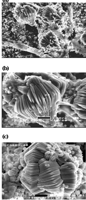
Glushinskite crystals like the Ca-oxalates also showed strong association with fungal hyphae. While the Ca-oxalates formed ordered and packed encrustation enveloping fungal hyphae (Fig. 2a, b and 2e, f); glushinskite adhered to hyphae as single large crystals (Figs. 5a, b). This asso-ciation reveals a genetic relationship with fungal hyphae for both mineral groups.
The Raman spectra (Fig. 6) of a selected single large crystal gave intense band at 1474 cm−1 and low band at 1417 cm−1corresponding to the symmetric C=O stretching vibration, which is cation–mineral dependant (Frost et al., 2003). A low band corresponding to anti-symmetric stretch-ing region appears at 1629 cm−1 and a high band in the ν (C-C) stretching region at 913 cm−1. A low band is ob-served in the C-C-O bending region at 502 cm−1. The 193, 225, 234 cm−1triplet appear in the low wavenumber region. These bands correspond to OMO ring bending mode. In the hydroxyl-stretching region, a low broad band appears at 3462 cm−1. These values are in good agreement with the pub-lished Raman data for calcium oxalate di-hydrate (Frost et al, 2003a). Whewellite (Ca-oxalate monohydrate) was not identified.
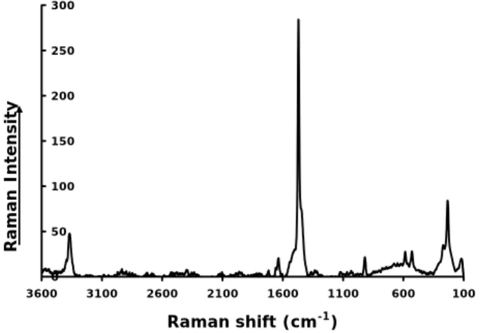
The Raman analysis of the large, foliated, spindle shaped and Mg-bearing crystals produced a different signature (Fig. 7). In the symmetric C=O stretching region appears an intense band at 1470 cm−1that is associated with a shoulder band at 1445 cm−1. Other bands appear at 3369, 1720, 1638, 920, 584, 528, 272, 232 and 124 cm−1. These bands are in good agreement with published data (Frost et al., 2003a, b, 2004a, b) and are assigned here to the magnesium oxalate mineral glushinskite. All other measurements on similar crystals produced relatively high intensities with good res-olution in the low and high wavenumber regions.
A peculiar spectrum was obtained from a prismatic crys-tal attached to fungal hyphae. The spectrum (Fig. 8) gave
44
Figures 5a, b, c
(a)
(b)
(c)
Fi 5 Fi 5Fig. 5. The peculiar forms of glushinskite crystals revealing the lamellar and foliated spindle habits. (a) Glushinskite crystals adher-ing to hyphae as sadher-ingle large (40 µm) crystals; in the background the much finer Ca-oxalates are visible. (b) and (c) Multiple growths of glushinskite crystals (40–60 µm) in twinning formations showing the same lamellar habits. This association reveals a genetic rela-tionship with fungal hyphae.
K. Kolo and Ph. Claeys: A study on substrate biomineralization and diagenesis by fungi 283 45 Figure 6 0 50 100 150 200 250 300 350 400 450 500 100 1100 2100 3100 Raman shift (cm-1) Figure 7 0 50 100 150 200 250 300 100 600 1100 1600 2100 2600 3100 3600 Raman shift (cm-1) Raman Intensi ty Raman Intensi ty
Fig. 6. Raman spectrum of prismatic bipyramidal crystals extracted from fungal mass attacking dolomitic substrate in vitro. The spec-trum obtained from single crystal analysis was cross-checked by repeated measurements on several other crystals. The spectrum is typical for weddellite.
45 Figure 6 0 50 100 150 200 250 300 350 400 450 500 100 1100 2100 3100 Raman shift (cm-1) Figure 7 0 50 100 150 200 250 300 100 600 1100 1600 2100 2600 3100 3600 Raman shift (cm-1) Raman Intensi ty Raman Intensi ty
Fig. 7. Raman spectrum of lamellar and spindle shaped crystals ex-tracted from fungal mass attacking dolomitic substrate in vitro. Ra-man analysis identified these crystals (after EDX assertion of their high Mg content) as the Mg-oxalate glushinskite. The crystals pro-duced typical glushinskite spectrum.
intense band at 1051 cm−1that could be assigned to carbon-ate bonded to a metal with wcarbon-ater coordincarbon-ated to it. The bands 749, 714 and 528 cm−1 are associated with this carbonate. The spectrum does not agree with dolomite or calcite. The other bands (low intensity bands) at high and low wavenum-bers reveal a signature of both calcium and magnesium ox-alates (weddellite and glushinskite) existing in low concen-trations in the same crystal.
3.2 Seawater substrate
The fungal interaction with the standard seawater substrate produced 7.6 mg of biominerals after extraction from the or-ganic mass (fungal and EPS layer). This quantity is quite large. It reveals considerable biomineralization in only 15
0 200 400 600 800 1000 1200 100 600 1100 1600 2100 2600 3100 3600 Raman shift (cm-1) Rama n Intensity
Fig. 8. Raman spectrum of a crystal with mixed signatures of an unidentified carbonate mineral and weddellite. The XRD analyses of the newly formed biominerals did not produce any of the usual carbonate signatures (dolomite or calcite).
days. At the same time, it is a clear manifestation of the role of fungi in modifying the substrate system by addition and subtraction of minerals.
Under optical and SEM microscopy, the specimens showed rich biomineral content forming encrustations, clus-ters or beads on fungal hyphae traversing the EPS layer (Figs. 9a, b, c). The lower parts of the hyphae embedded in the EPS layer were specifically highly biomineralized. The hyphal parts outside EPS layer did not produce biominerals encrustations, clusters or free crystals. This implies a genetic relationship combining the EPS layer and fungal hyphae in the biomineralization process. Biomineral crystals attached to fungal hyphae were mainly tetragonal bipyramidal and tetragonal prismatic-bipyramidal with well-developed {101} and {100} crystal faces. These forms are normally attributed to Ca-oxalates (Arnott, 1995; Verrecchia 2000; Pr´eat et al., 2003). The same morphologies occurred as free crystals in the fungal mass within the EPS layer (Figs. 9a, b).
Some of the Ca-oxalates crystals revealed a unique and in-teresting crystal habit (Figs. 9d–e). The new crystal habit shows a prism (20–25 µm) with two prismatic-pyramidal endings forming a “Greek Pillar”. Some of these “Greek Pillar” crystals formed clusters that radiate from a central point (Fig. 9e) Sceptre crystal habits are known for amethyst. But those have only single end crystal re-growth. The new biomineralized ca-oxalates appear to show a two-end re-growth.
The EDX analysis of the hyphal and EPS crystals revealed a composition of calcium and magnesium respectively. The magnesium bearing crystals represent more than 90% of the extracted crystals. Ca-bearing crystals formed the rest of the extract. The Ca crystals occurred mainly as encrustations on the hyphal sheath. XRD analysis of organic mass and extract revealed two mineral groups: Ca-oxalates (weddellite and
284 K. Kolo and Ph. Claeys: A study on substrate biomineralization and diagenesis by fungi 47 Figures 9a, b, c, d, e (a) Figures 9a, b, c (b) 48 (c) Fig.9.d (d) 5µm 50µm 50µm 49 (e)
Fig. 9. (a) SEM image showing fungal specimen with rich biomineral contents forming highly ordered and packed crystal encrustations on fungal hyphae. The crystals forms are mainly tetragonal prismatic and bipyramidal typical of weddellite. The background also shows numerous free isolated weddellite crystals. These latter forms are exogenously formed. (b) Same as above but with excellently developed tetragonal prismatic bipyramidal crystal forms of weddellite, some showing penetration twinning. Free crystals embedded in fungal mass but not forming part of crystal encrustations are also visible. (c) A typical experimental fungal mass showing the high content of biominerals forming clusters or beads of crystals (bright spots) either attached to fungal hyphae or filling the space within the fungal mass. Optical microscope. XPL. (d) New crystal habit of Ca-oxalates (weddellite) showing a “Greek Pillar” shape. The prism in the middle is topped by two tetragonal pyramidal forms, entirely different from the “Sceptre” habits known for quartz. The inserted image shows the prism ”plugged” into the two pyramidal endings. (Material extracted from fungal mass). (e) Cluster of “Greek Pillar” Ca-oxalates crystals radiating from a central point. One of them is showing twinning. (Material extracted from fungal mass). In (a), (b) and (c), the lower parts of the hyphae embedded in the EPS layer were highly biomineralized. The hyphal parts outside EPS layer did not produce biomineral encrustations, clusters or free crystals.
K. Kolo and Ph. Claeys: A study on substrate biomineralization and diagenesis by fungi 285 whewellite) and glushinskite (Fig. 10). Glushinskite formed
the dominant mineral phase in the XRD spectra.
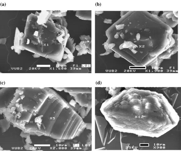
Free crystals extracted from the EPS layer show platy habits (Figs. 11a, b) and coffin shaped habits that are ei-ther distorted pyramidal or typical monoclinic 2/m symme-try forms (Figs. 11c, d). The distorted pyramidal crystals bear transversal lines resembling striations. These lines indi-cate crystal growth. The platy (pinacoidal) and coffin shaped crystals (Figs. 11a, d) are new crystal habits for glushinskite forming in fungal culture.
Prismatic-bipyramidal crystals produced a Raman spec-trum of weddellite. Whewellite was not clearly identified in the analysis, probably due to its very low level of con-centration. The spectrum (Fig. 12) shows complexity espe-cially at low and high bands. Intense bands are observed at 1631, 1474, 913, 600, 506, 148 and 121 cm−1, broad band at 250 cm−1and weak band at 198 cm−1. The major intense band at 1474 cm−1is assigned to the C=O symmetric stretch-ing mode. The observation of a sstretch-ingle band at 1474 cm−1 indicates the equivalence of C=O stretching vibrations and a centrosymmetric structure. The band is attributed to weddel-lite. The intense band at 1631 cm−1appears in antisymmetric stretching region. This band is Raman forbidden. The band is observed probably because the structure is a distorted square antiprism (Frost and Weier, 2003a, b). This band is also at-tributed to weddellite. A single intense band is observed at 913 cm−1and is attributed to the C-C stretching vibration in weddellite. The Raman spectrum of the low wavenumber region of the mineral shows three Raman bands in the 400– 600 cm−1region. The doublet bands at 600 and 598 cm−1 and the intense band at 506 cm−1have been assigned to the bending mode of C-C-O and the M-O ring and M-O stretch-ing modes. The broad band centred at 250 cm−1and the band observed at 198 cm−1is attributed to the Ca-O bending and stretching modes. The position of these bands is in good agreement with previous data on weddellite (Frost et al., 2003a). The bands 148 and 121 cm−1are new bands that have not been reported in previous studies on Ca-oxalates (Frost et al., 2003a, b, 2004a). Only the tetragonal prismatic bipyra-midal crystals of weddellite produced these bands. Tetrag-onal bipyramidal crystals of the same mineral did not yield these very low wavenumber bands. This suggests a struc-tural control at molecular level related to crystal habit since the measurements were done on the {100} plane of the pris-matic bipyramidal crystals and on {101} plane for the bipyra-midal crystals. The hydroxyl-stretching region is also com-plex. This region shows two broad bands centered at 3483 and 3281 cm−1. The first band agrees well with published data on whewellite. The second band is reported here for the first time. The low intensity band at 3072 cm−1is attributed to weddellite and the 2935 cm−1could be assigned to organic impurities. The hydroxyl-stretching region is showing a par-tially dehydrated weddellite that also bears a whewellite sig-nature. 50 Figure 10 2 1 1 1 1 2 2 2 3 2 3 2 3 2 2 1 1: Glushinskite 2: Weddelite 3: Whewellite 0 10 20 30 40 2-Theta - Scale 10 20 30 40 50 1 1 2 Lin (Cps)
Fig. 10. XRD analysis of fungal mass and extracted biominerals showing two groups: Ca-oxalates (weddellite and whewellite) and Mg-oxalate (glushinskite) produced through fungal interaction with standard sea water substrate. Glushinskite formed the major mineral phase in the XRD spectra as well as in the extract.
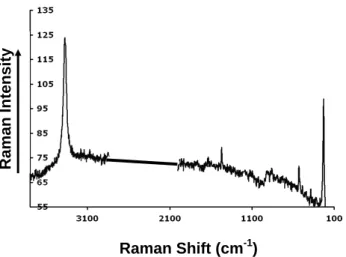
Extracted crystals representing coffin shaped (distorted pyramidal and prismatic) and platy crystals were selected for Raman analysis. The EDX analysis of all these crystal types indicated a Mg peak only. The three crystal forms, although indicating glushinskite, gave slightly different Ra-man spectra (Figs. 13, 14 and 15). The distorted pyrami-dal crystals produced intense bands at 3363, 1470, 586, 532 and 231 cm−1and low intensity bands at 1643, 1659 and 914 and 271 cm−1. The prismatic crystals show intense bands at 3368, 1470, 920, 585, 529, 230, 153, and 117 cm−1 and moderate bands at 3292, 2902, 1643, 1329 and 274 cm−1. The platy crystals produced a simpler Raman spectrum with major intense bands observed at 3377 and 229 cm−1and low intensity bands at 1470, 914 and 529 cm−1. All three spec-tra are in agreement with the previous Raman signature of glushinskite obtained on the spindle crystals. Only the pris-matic crystals show low wavenumbers at 152 and 117 cm−1. The distorted bipyramidal crystal forms of glushinskite have been previously reported (Wilson et al., 1987). The prismatic coffin shaped and platy morphologies are reported here for the first time.
3.3 Ca- and Mg-oxalates formation
All fungal taxa precipitate calcium oxalate on fungal mycelia (Urbanus et al., 1978). This process is considered as detoxi-fication of excess calcium in the fungal growth environment. Fungi and the carbon source they use determine the growth and form of calcium oxalates crystals precipitated on fungal hyphae (Whitney and Arnott, 1988). Oxalic acid, the prin-cipal fungal metabolite reacts with the available Ca2+in the growth environment to precipitate Ca-oxalates:
C2O2−4 +Ca2+→CaC2O4 (R1)
The formation of calcium oxalate monohydrate
[CaC2O4.H2O: whewellite] and calcium oxalate dihydrate [CaC2O4.2H2O: weddellite] depends largely on a variety of factors such as the pH of the growth environment, the
286 K. Kolo and Ph. Claeys: A study on substrate biomineralization and diagenesis by fungi
51
Figure 11 (a) (b)
(c) (d)
Fig. 11. SEM images of free crystals extracted from the EPS layer showing platy habits (a, b) and coffin shaped habits that are either distorted pyramidal or typical monoclinic 2/m symmetry forms (c, d). The distorted pyramidal crystals (c) bear transversal lines resembling striations. These lines indicate crystal growth. The platy (pinacoidal) (a, b) and coffin shaped crystals (d) are new crystal habits of glushinskite that formed in fungal culture. Bar scale=10 µm.
solubility of oxalates (Gadd, 1999) and diagenetic re-crystallization (Verrecchia et al., 1993, 2000), the sequence of formation is driven by the loss of crystallization water: dihydrate oxalates form first, monohydrate second and calcium carbonate CaCO3 third (Verrecchia et al., 1993). Gadd (1999) considered that trihydrate Ca-oxalates are the initial precipitation phase, followed by either the mono- or the dihydrate after losing the water of crystallization. In the present work both types of calcium oxalates (weddellite and whewellite) occur simultaneously in the growth envi-ronment. Diagenetic factors cannot play a major role in the transformation of weddellite into whewellite considering the short experimental time. The co-existence of the two mineral species is attributed to fungal metabolic activities (Horner et al, 1995).
In natural growth environment, subaerial fungi can use wind blown particles, rain or the substrate as sources of Ca for precipitating oxalates (Braissant et al., 2004). In this study, the only source of Ca and Mg is the dolomite (CaMg CO3)2)substrate and the seawater. Oxalic and carbonic acids cause dissolution of carbonate substrates leading to their
sub-sequent weathering and deterioration, which adds an envi-ronmental impact to fungal interaction with buildings and works of art made of this material.
Both Ca and Mg existed in an unbound form in the sea-water growth medium. Thus both metals were available for uptake by fungi. Sayer and Gadd (1997) have demonstrated the fungal ability to form different forms of oxalates, e.g. Ca, Cd, Co, Cu, Mn, Sr, and Zn. Howell et al. (2003) have shown that while Ca-oxalates (weddellite and whewellite) were formed by fungal mycobionts of lichens, no magne-sium oxalates were detected on the fungal thalli despite the Mg-rich substrates. The fungal culture in the present experi-ment provided similar results, which could indicate a mech-anism of Mg-oxalates formation that differs from that of Ca-oxalates.
K. Kolo and Ph. Claeys: A study on substrate biomineralization and diagenesis by fungi 287 52 Figure 12 Raman Shift (cm-1) 0 20 40 60 80 100 120 0 500 1000 1500 2000 2500 3000 3500 Raman Intensity
Fig. 12. Raman analysis of single tetragonal prismatic bipyramidal crystals extracted from fungal mass and EPS layer. The spectrum indicates the mineral weddellite. Intense and low bands are ob-served in the spectrum. The very intense band at 1474 cm−1is a signature of weddellite.
3.3.1 Thin section substrates
A. Ca-oxalates
The oxalic acid excreted by fungal hyphae reacted with the dolomitic substrate and precipitated Ca and Mg oxalates. This process led to dissolution and deterioration of the dolomitic substrate. Carbonic acid (H2CO3)could also form in this environment due to the production of CO2by fungal bioactivity in the aqueous growth environment (Rosling et al., 2004). In this latter case, high levels of CO2 accu-mulating in “microenvironments” would produce sufficient concentrations of carbonic acid to induce dissolution of the substrate. This carbonic acid dissolution process also increases the concentration in Mg2+ and Ca2+ cations available to react with oxalic acid and frees the carbonate ions into the growth environment, which contributes to calcium carbonate precipitation. The extensive formation of the biomineralized minerals weddellite and whewellite on the analysed thin sections indicates the high availability of Ca2+ to react with oxalic acid in the fungal growth environment.
Based on SEM observations, the Ca-oxalates within the fungal mass and on the attacked thin sections displayed three precipitation modes:
(i) Direct deposition of tetragonal prismatic dipyramidal crystals (1–10 µm) on the attacked substrates of the thin sec-tions (Figs. 2a, b, c and d). Here, the precipitation produced dense new layers of single crystals that either replaced the dissolved original substrate or were deposited directly on it. In the latter case, they filled voids and spaces (created mostly by fungal dissolution of the substrate) between and within the original grains. Fungi also extended the precipitation of biominerals to the rims of the glass slide, outside the pri-marily attacked area. This suggests fungal capability of mo-bilising and transporting Ca2+ outside the source area. In
53 Figure 13 Raman Shift (cm-1) Raman Intensity 0 5 0 1 0 0 1 5 0 2 0 0 2 5 0 3 0 0 1 0 0 6 0 0 1 1 0 0 1 6 0 0 2 1 0 0 2 6 0 0 3 1 0 0
Fig. 13. Raman analysis of single distorted pyramidal crystals (cof-fin shaped) extracted from fungal mass and EPS layer produced by fungal interaction with SSW substrate. The spectrum indicates the mineral glushinskite with very intense band at 1470 cm−1.
54 Figure 14 Raman Intensity Raman Shift (cm-1) 0 20 40 60 80 100 120 140 100 600 1100 1600 2100 2600 3100 3600 0 20 40 60 80 100 120 140 100 600 1100 1600 2100 2600 3100 3600
Fig. 14. Raman analysis of single prismatic crystals (coffin shaped) extracted from fungal mass and EPS layer produced by fungal in-teraction with SSW substrate. The spectrum indicates the mineral glushinskite with very intense band at 3368, 1470 and 153 cm−1.
all these cases, the process of forming new substrates or fill-ing the original or created voids can be defined as “biolog-ical precipitation” that produces a “micro-biolog“biolog-ical stratifi-cation” similar to the larger scale sedimentary environment.
The abundant fungal spores (size ≈2 µm) released into the growth environment come in contact with oxalic acid-Ca2+rich medium where they became nucleation sites for Ca-oxalates. Unlike other free or attached ca-oxalates, these oxalates possess a multi-layer and concentric zonation struc-tures that reflect crystal growth mechanism around a nu-cleus (Fig. 16). This structure shows a homogenous and equally spaced smooth internal and external growth surfaces. Most of the crystals show a four layer zonation. Unlike the abundant crystals encrusting the fungal hyphae, the Ca-oxalates deposited around the fungal spores represent pris-matic bipyramidal single crystal growth (4.5×4 µm). The
288 K. Kolo and Ph. Claeys: A study on substrate biomineralization and diagenesis by fungi
55
Figure 15
Raman Intensity
Raman Shift (cm-1)
Fig. 15. Raman analysis of single platy (pinacoidal) crystals ex-tracted from fungal mass and EPS layer produced by fungal in-teraction with SSW substrate. The spectrum indicates the mineral glushinskite with very intense bands at 3377 and 229 cm−1. The typical glushinskite band (1470 cm−1) is weak for this crystal habit.
crystals interior and nuclei are square shaped with a de-pression that represents the spores folding. The nuclei size (2 µm) is equal to the size of the spores (2 µm). The EDX analysis of such fungal crystals revealed a spectrum simi-lar to normal Ca-oxalates. However, the formation of these zoned crystals reflects chemical changes in growth environ-ment. Despite the fact that these concentric crystals differ in size and shape from sedimentary ooids, their formation in a totally biological environment draws attention to the pos-sibility of formation of similar concentric particles outside sedimentary environments. True fungal ooids have been re-ported by Krumbein et al. (2003).
(ii) Precipitation on fungal hyphae. Here, a highly ordered envelope of densely packed Ca-oxalate crystals completely engulfs the fungal sheaths at regular spaces (Figs. 2a, b). The highly mineralized fungal hyphae are mostly seen un-der the microscope as broken fragments even in fresh spec-imens. The biomineralized fungal hyphae lose their flexi-bility and become brittle, probably leading to the death of the affected hyphae. Arocena et al. (2001) suggested the de-position of Ca-oxalates on the fungal hyphae surfaces as a protection against dehydration and grazing. The crystals ad-here strongly to fungal hyphae, resisting even the mechanical force during extraction. This further indicates that the crystal grows within fungal sheath.
The first and the second modes of precipitation have been described in present day caliches layers, weathering of car-bonate buildings or in plants litter (Arnott, 1995; Sterflinger, 2000; Verrecchia, 2000; Garvie, 2003). However, the for-mation of Ca-oxalates as highly packed and ordered crystal envelopes around the fungal sheaths indicates a direct expul-sion of Ca-oxalate through the fungal cellular walls rather
56
Figure 16
Figure 17
Fig. 16. SEM image of Prismatic bipyramidal Ca-oxalates (weddel-lite) crystals showing a multi-layer and concentric zonation struc-tures reflecting crystal growth mechanism around a nucleus. Most crystals show a four layer zonation. The crystals nucleated on fun-gal spores. The size and shape of the nuclei are identical to funfun-gal spores in the growth environment. Black bar scale=1 µm.
56
Figure 16
Figure 17
Fig. 17. SEM images showing a double layer of ordered ca-oxalates engulfing fungal sheath reflecting two growth generations. The crystals of the inner and outer layer differ in size, crystal morphol-ogy and position. Black bar scale=10 µm.
than an exogenous oxalic acid/Ca2+reaction and precipita-tion. The regularity of Ca-oxalates crystals deposition on fungi from plant litter is an indication of the endogenous formation of oxalates followed by expulsion and deposition on the hyphal wall (Arnott, 1995). The occurrence of Ca-oxalates as ordered and packed deposits on fungal hyphal sheaths together with free Ca-oxalate deposits on attacked substrates indicate that both exogenous and endogenous for-mation of Ca-oxalates have occurred in the course of this ex-periment. The intensive biomineralization of fungal hyphae is not a general process. Extensive sampling of the fungal mass demonstrates that biomineralization on fungal hyphae is restricted to the lower parts of fungal hyphae embedded in the EPS layer. Above the EPS layer, hyphae did not show biomineralization.
K. Kolo and Ph. Claeys: A study on substrate biomineralization and diagenesis by fungi 289 SEM images of some fungal hyphae (Fig. 17) show a
dou-ble layer of ordered ca-oxalates engulfing fungal sheath. The crystals of the inner layer are much smaller (2–3 µm) than the second external layer (5–10 µm). The growth of two crystal layers suggests two generations separated by a time sequence: a faster inner one and a slower outer one. These two generations are possibly related to the availability of Ca in the growth environment. With increased fungal attack on the substrate more Ca is released in the growth environment, which is then immobilized as Ca-oxalates either by direct extracellular expulsion or by reaction of excreted oxalic acid with Ca+2. The crystals of the outer envelope seem to be attached to the fungal hypha by the {001} plane. The devel-opment of the {001} pinacoids seems to be unstable result-ing in small {001} faces and truncated {101} faces. While the crystal morphology of the inner layer type and relation to fungal hyphae have been previously reported (e.g. Arnott, 1995), the double crystal layer and the outer crystal morphol-ogy on the hypha are reported here for the first time. The inner crystal layer is densely packed with no apparent ori-entation to the fungal hypha. The outer layer crystals are orientated perpendicularly to the long hyphal axis resting on the basal pinacoid. Many of these crystals are deeply em-bedded in the inner layer. The diameter of the inner layer (≈12 µm) is doubled by the precipitation of the outer crystal layer (≈25 µm). The EDX analysis shows a Ca, O and C composition. Though not a conclusive analysis, the crystals of the inner envelope seems to show a lower calcium content (26 wt %) than the crystals of the outer envelope (34 wt %). The mechanism of formation of the double crystal layers on the hypha is not clear. The general homogeneity of the size of outer layer crystals and their embedding in the fungal hy-phal wall passing through the inner layer suggest an inner secretion of oxalates.
iii) Precipitation within fungal hyphae. In this case, the Ca-oxalates are lining the inner wall of the fungal sheath (Figs. 2d, e, f), indicating an inner formation of weddel-lite or whewelweddel-lite. This third mode of Ca-oxalate formation has been subject of discussion. In higher plants the intra-cellular formation of Ca-oxalates is well established (Horner and Wagner, 1995). In fungi, the issue of intra-cellular or extra-cellular formation is not fully understood (Vincent et al., 1995). Arnott (1995) demonstrated a sequential expul-sion of Ca-oxalates from the hyphal wall. Gadd (1999) re-viewed the different arguments pointing towards an intra-hyphal formation of oxalates as a cellular detoxification pro-cess, where the excess Ca is neutralized in the form of in-soluble oxalates. In the SEM photomicrographs (Figs. 2d, e, f), ordered crystal lining is clearly visible inside the bro-ken fungal hyphae. The crystals form a uniform lining that covers the whole inner curvatures of the broken hollow hy-phal sheath. This uniform distribution of the inner lining can occur through endogenous precipitation of the Ca-oxalates.
B. Glushinskite
Glushinskite as observed under optical microscopy and SEM, showed peculiar crystal morphology. The crystals have a general “spindle” or pear shapes with layered lamellae and locally with transition towards more rhombic or pyrami-dal morphologies (Figs. 5a, b). The “lamellae”, “striations” and “spindle shapes” are common features in the examined glushinskite crystals. Under the optical microscope, the lamellas appear as striations. Wilson (1980) observed at the interface between the lichen Lecanora atra and serpentinite substrates similar striated crystals of naturally formed glushinskite. But the striations on these latter crystals could belong to the other type of glushinskite crystals, namely the distorted pyramidal forms, where they would indicate a growth process involving repetitive twinning. Figures 5a, b also show the genetic relationship between glushinskite and the fungal hyphae. Here glushinskite crystals do not cover or envelope fungal hyphae, but rather appear attached to hyphal tips and sheaths, similar to a fruit-bearing tree, indicating an external reaction of oxalic acid with an Mg+2-rich solution. Glushinskite crystals lie on a layer of finer and more extensively precipitated Ca-oxalates. Garvie (2003) observed the common occurrence of fungal hyphae on the mineral glushinskite and considered that it resulted from the reaction of oxalic acid released by fungi with the Mg-rich solutions supplied by a rotting saguaro cactus.
Glushinskite generally superpose Ca-oxalates in thin sec-tions, indicating a post-Ca-oxalates formation. This se-quence of biomineralization could indicate the availability and relative importance of Ca and Mg cations in the fungal growth environment. In the case of a dolomitic substrate, it may also illustrate the sequence of possible uptake-expulsion processes for Ca2+and Mg2+by fungi. Fungi first neutralize the toxicity of excess Ca through the Ca up-take and subse-quent precipitation of Ca-oxalates (Gadd, 1999; Sterflinger, 2000). This produces solutions rich in Mg within the fun-gal growth environment that could precipitate glushinskite. Since the Mg oxalates have a much higher solubility than Ca-oxalates (Gadd, 1999), the restricted spatial distribution of glushinskite suggests that the high Mg concentrations neces-sary for its precipitation only prevailed in localized microen-vironments.
The peculiar spindle and lamellar crystal morphology of the biomineralized glushinskite could suggest its nucleation on bodies with similar shapes. In Fig. 5c unidentified or-namented round bodies (fungal spores?) with “ribbed” and “ridged” walls, seemingly hollow and with star-shaped orna-mentation locally litter some of the spindle shaped glushin-skite crystals. Numerous EDX spot analyses of these bod-ies revealed a high Mg content. It is possible to speculate that these ribbed and ridged round bodies formed nucleation sites on which the biomineralized glushinskite grew. The for-mation of zoned structures around spores has been already demonstrated. Lamella growing from the “ribs” formed
290 K. Kolo and Ph. Claeys: A study on substrate biomineralization and diagenesis by fungi complete spindle shapes in full mineral growth. This offers
an additional argument for the exogenous formation of the mineral glushinskite.
3.3.2 The seawater substrate
A. Ca-oxalates
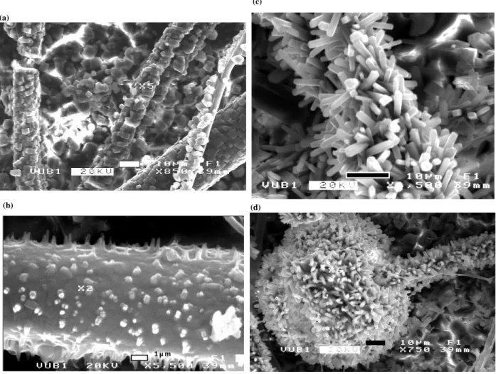
Fungal interaction with the seawater substrate did not produce a substrate of layered Ca-oxalates. Under the optical and SEM microscopy, the Ca-oxalates were either encrusted on the fungal sheaths or embedded in the fungal mass, the former being predominant. The mineral extract produced high abundance of free glushinskite crystals and fewer free Ca-oxalate crystals. Encrustation on fungal hyphae was the main process of Ca-oxalates formation during this interaction. Tetragonal prismatic, bipyramidal and needle prismatic crystals are the observed crystal habits (Figs. 18a, b, c and d). The size and composition are similar to the Ca-oxalates retrieved from the thin section substrates. Here no double encrustations are observed on the fungal sheaths. Small crystals (1 µm) protrude perpendicularly from some fungal sheaths (Fig. 18b). These crystals have a wide base on the fungal sheath and a pointed end, making a triangular shape. They appear growing from the hyphal sheath. These growth shapes are interpreted as incipient Ca-oxalates expulsion from fungal sheath. Arnott (1995) observed a similar case of fungi from plant litter. On other hyphae (Figs. 18a, c) rich Ca-oxalates encrustation are observed with tetragonal prismatic and pyramidal crystals with free crystals on the background. The average crystal size is 5 µm. In Figs. 18c and d the oxalates encrusting the hyphae are classified as needle prisms. The crystals have high length/width ratio. Some of them show penetration twining. The fungal background is littered with prismatic bipyramidal crystals.
Very interesting is the extent of biomineralization. Figure 18d shows total biomineralization of fungal spo-rangium and hypha with prismatic-needle-type Ca-oxalates. The EDX analysis produced a composition of Ca only. Ca-oxalates are known to be related to fungal hyphae in various forms, but sporangia are not known to take part in biomineralization as nucleation sites. The crystals’ form (needle prisms) and sizes (1×2×5 µm) are identical on both fungal hypha and sporangium. The crystals protrude per-pendicularly from the hypha or from the sporangium sphere. This suggests a common biomineralization process different from the background, where much larger (10×10×5 µm), isolated and tetragonal prismatic and bipyramidal crystals are dominant (Fig. 9a). The formation of similar Ca-crystal forms has been reported as an internal hyphal secretion (Arnott, 1987, 1995; Gadd, 1999). If the hyphal wall is a site for Ca-oxalate formation, then apparently the fungal sporangium plays a critical role. Biomineralization on hyphae and sporangium is induced by indigenous processes
through Ca uptake by fungi from the growth medium and subsequent expulsion of Ca-oxalates, while the background Ca-oxalates, imbedded in fungal mycelium are formed through oxalic acid-Ca reaction in the growth medium.
B. Glushinskite
Glushinskite, as demonstrated earlier, is represented here by three crystal morphologies, i.e. coffin shaped (distorted-pyramidal and prismatic) and platy (pinacoids). The first two types formed the bulk of the extracted crystals. Similar to the case of the thin section substrate, glushinskite formed within fungal mass embedded in the EPS layer but never encrusted fungal filaments. All of the EDX analyses on crystals enveloping hyphal sheaths produced Ca-bearing mineral only. Glushinskite in its different forms occurred as single, clusters or beads of crystals embedded in the fungal mucilage. The crystals sometimes adhered to fungal hyphae, but never formed ordered encrustations as is the case for Ca-oxalates. This repeated observation confirms the suggestion that during the fungal growth neither Mg uptake nor Mg expulsion occurred, contrary to calcium, where both could occur (Jackson and Heath, 1993). The main process of glushinskite formation was exogenous. Oxalic acid secreted by fungi reacted with the free Mg in the growth medium and precipitated glushinskite in the three different forms. Since the solubility constant of glushinskite is much higher than that of Ca-oxalate monohydrate (Gadd, 1999), fungi respond to Ca concentration in the growth medium and reaches an equilibrium between Ca needed for hyphal tip growth process and toxicity of the metal. Excess Ca is excreted outside the cellular walls (Jackson and Heath, 1993) and removed from the growth environment in the form of insoluble Ca-oxalates. This fungal metabolic need for intracellular calcium regulation and the lower solubility constant of Ca-oxalates leads to the formation of a one phase or several phases (e.g. the double encrustations on fungal sheaths) fungal of Ca-oxalates precipitation before Mg2+ attains the concentration necessary to trigger Mg-oxalate precipitation in the growth environment The Ca was with-drawn from the growth environment before the initiation of glushinskite formation.
4 Conclusions
The EDX, Raman and XRD analyses of biomineralized crys-tals on thin sections and on fungal hyphae (crystal encrusta-tions) have shown that the only Mg-bearing neoformations are those of glushinskite and are crystallized in spindles, fo-liated, platy and prismatic shapes. The ordered crystals en-crustations engulfing fungal hyphae or forming the new sub-strates are always Ca-oxalates with their specific prismatic, bipyramidal and sometimes rhombic shapes. In this study, glushinskite did not encrust fungal sheaths. This rules out
K. Kolo and Ph. Claeys: A study on substrate biomineralization and diagenesis by fungi 291 57 Figures 18a, b, c, d (a) (b) 1 µ m 58 (c) (d)
Fig. 18. SEM images of various Ca-oxalates encrustation on fungal hyphae produced by fungal interaction with SSW. Tetragonal pris-matic bipyramidal, tetragonal bipyramidal and needle prispris-matic crystals are the observed crystal habits (a, b, c, d). (a) SEM image of intense biomineralization and encrustation of oxalates on fungal hyphae produced by fungal interaction with SSW substrate. Free Ca-oxalate crystals litter the background. The encrustations and free Ca-oxalates indicate endogenous and exogenous processes of formation. Bar scale=10 µm. (b) SEM image showing incipient biomineralization of Ca-oxalates on fungal sheath produced by fungal interaction with SSW substrate. Small crystals (1 µm) are protruding perpendicularly from the fungal sheath. The crystals have wide bases and pointed ends, making a triangular shape. They appear to grow from within the hyphal sheath. Bar scale=1 µm. (c) SEM image showing rich Ca-oxalates encrustation of fungal hyphae with needle prism crystals produced by fungal interaction with SSW substrate. The average crystal size is 5 µm. The crystals have high length/width ratio. Some of them show penetration twining. Bar scale=10 µm. (d) SEM image showing total biomineralization of fungal sporangium and hypha with prismatic-needle-type Ca-oxalates produced by fungal interaction with SSW substrate. Sporangia are not known to take part in biomineralization as nucleation sites for Ca-oxalates. The crystal forms (needle prisms) and size (1×2×5 µm) are identical on both fungal hypha and sporangium. The crystals protrude vertically from the hypha and from the sporangium sphere. This suggests a common endogenous biomineralization process. bar scale=10 µm.
the possibility of Mg-uptake and expulsion as Mg-oxalates. Actually, the much larger glushinskite crystals (40–100 µm) compared to Ca-oxalates (usually <10 µm) suggests slower and longer crystallization processes in localized microenvi-ronments containing high concentrations of Mg2+. The for-mation of glushinskite is due to a direct reaction between ox-alic acid excreted by fungi in the growth environment and the Mg2+produced by the dissolution of the dolomitic substrate of the thin section. The notions of “biological mineral
depo-sition” and the formation of layered stratum (biological strat-ification) are invoked here again. The Ca-oxalates formed the first deposited ”stratum” that was subsequently overlain by a second layer formed of glushinskite. Under natural condi-tions, where extensive fungal-rock surface interaction is en-visaged, this type of microbial deposition could have real ge-ological implications. Lichens cover large areas of exposed rock surfaces almost in any type of environment. Fungi, the mycobiont of lichens interact strongly with rock surfaces
292 K. Kolo and Ph. Claeys: A study on substrate biomineralization and diagenesis by fungi modifying their chemistry and petrography and causing rock
weathering and deposition of biominerals. Fungi growing in vitro from natural airborne spores interact in Petri dishes with dolomitic thin sections and seawater substrates to pro-duce both mineralogical and diagenetic changes on the sub-strates. Mineralogically, the presence of two cations Ca and Mg mobilized by fungi from the substrates and released in the growth environment led to sequential precipitation of Ca-oxalates (weddellite and whewellite) followed by the mag-nesium dihydrate oxalate: glushinskite. The spatial dis-tribution of this paragenetic sequence produced a “micro-biological stratification”. The external Ca-oxalates envelope that engulfed whole fungal hyphae indicates an exogenous process of formation. The SEM images of Ca-oxalates lin-ing the inner hyphal walls clearly prove an endogenous for-mation. Glushinskite compared to Ca-oxalates, only formed exogenously and did not form envelopes on fungal hyphae. Fungal interaction with the dolomitic substrates also pro-duced double-layered encrustations of Ca-oxalates on fungal hyphae. Nucleation on fungal spores produced zoned Ca-oxalates crystals. Nucleation is also the possible cause of spindle shaped glushinskite. On the seawater substrate, the fungal interaction produced Ca-oxalates and glushinskite by mobilizing Ca and Mg from the substrate. Glushinskite could also form in prismatic and platy crystal habits in addition to the known distorted pyramidal habit. Diagenetically, the pe-culiar forms of substrate dissolution and replacement, bios-tratification, grain-grain bridging, pore space filling mark the potential imprint left on hard substrates by fungi that together with the fungal organic material within the substrate matrix and the biominerals can form a set of measurable biomark-ers for the search of life forms on earth as well as on other planets.
Acknowledgements. We thank J. Vereecken, O. Verstaeten and K.
Baerts, Department of Metallurgy, Vrije Universiteit Brussels, for the SEM-EDX and Raman facilities. We also thank A. Bernard, Department of Geology, Universit´e Libre de Bruxelles, for the XRD facility. This work is supported by the Onderzoeksraad van de Vrije Universiteit Brussel.
Edited by: T. W. Lyons
References
Arnott, H. J.: Calcium oxalates (weddelite) crystals in forest litter, Scan. Electron. Microsc., P3, 1141–114, 1982.
Arnott, H. J.: Calcium oxalates in fungi, in: Calcium Oxalates in Biological Systems, edited by: Khan, S. R., pp. 73–111, CRC press, Boca Raton, 1995.
Arocena, J. M. and Glowa, K. R.: Mineral weathering in ectomyc-orrhizosphere of subalpine fir (Abies lasiocarpa (Hook.) Nutt.) as revealed by soil solution composition, Forest Ecol. Manag., 133, 61–70, 2000.
Arocena, J. M., Glowa, K. R., and Massicotte, H. B.: Calcium-rich hypha encrustations on Piloderma, Mycorrhiza, 10, 209– 215, 2001.
Arocena, J. M., Zhu, L. P., and Hall, K.: Mineral accumulations in-duced by biological activity on granitic rocks in Qindhai Plateau, China, Earth Surf. Process. Landf., 28, 1429–1437, 2003. Ascaso, C., Wierzchos, J., and Castello, R.: Study of the biogenic
weathering of calcareous litharenite stones caused by lichens and endolithic microorganisms, Int. Biodeterior. Biodegrad., 42, 29– 38, 1998.
Bishop, J. L. and Murad, E.: Characterization of minerals and bio-geochemical markers on Mars: A Raman and IR spectroscopic study of montmorillonite, J. Raman Spectrosc., 35, 480–486, 2004.
Braissant, O., Cailleau, G., Aragno, M., and Verrecchia, E.: Bi-ologically induced mineralization in the tree Milicia excelsa (Moraceae): its causes and consequences to the environment, Geobiology, 2, 59–66, 2004.
Burford, E. P., Hilier, S., and Gadd, G. M.: Rock and mould: trans-formation of carbonate minerals by fungi, Abstracts, 7th Internat. Mycolog. Congr., Oslo, abstract no. 1083, August 2002. Diaz-Espineira, M., Escolar, E., Bellanato, J., and Medina, J. A.:
Crystalline composition of equine urinary sabulous deposits, Scan. Microsc., 9, 1071–1079, 1995.
Edwards, H. G. M., Seaward, M. R. D., Attwood, S. J., Little, S. J., De Oliveira, L. F. C., and Tretiach, M.: FT-Raman spectroscopy of lichens on dolomitic rocks: an assessment of metal oxalate formation, Analyst, 128, 1218–1221, 2003.
Edwards, H. G. M, Villar, J. S. E., Bishop, J. L., and Bloomfield, M.: Raman spectroscopy of sediments from the Antartic Dry Val-leys; an analogue study for exploration of potential paleolakes on Mars, J. Raman Spectrosc., 35, 458–462, 2004.
Franceschi, V. R. and Loewus, F. A.: Oxalate biosynthesis and func-tion in plants and fungi, in: Calcium Oxalates in Biological Sys-tems, edited by: Khan, S. R., 113–130, CRC Press, Boca Raton, 1995.
Frost, L. R. and Weier, L. M.: Thermal treatment of weddellite – a Raman and infrared emission spectroscopic study, Thermochm. Acta, 406, 221–232, 2003a.
Frost, L. R. and Weier, L. M.: Raman spectroscopy of natural ox-alates at 298 and 77 K, J. Raman Spectrosc., 34, 776–785, 2003b. Frost, L. R. and Weier, L. M.: Thermal treatment of whewellite – a thermal analysis and Raman spectroscopic study, Thermochm. Acta, 409, 79–85, 2004a.
Frost, L. R., Adebajo, M., and Weir, M. L.: A Raman spectroscopic study of thermally treated glushinskite-the natural magnesium oxalate dehydrate, Spectrochimica Acta, part A, 60, 643–651, 2004b.
Gadd, G. M.: Fungal production of citric and oxalic acid: impor-tance in metal speciation, physiology and biogeochemical pro-cesses, Adv. Microb. Physiol., 41, 47–92, 1999.
Gadd, G. M.: Fungal influence on mineral dissolution and metal mobility: mechanisms and biogeochemical relevance, 12th an-nual Goldschmidt Conference Abstracts, A257, 2002.
Gadd, G. M., Burford, E. P., Fomina, M., Harper, F. A., and Jacobs, H.: Fungal influence on metal mobility: Mechanisms and rele-vance to environment and biotechnology, Abstracts, 7th Internat. Mycolog. Cong., Oslo, abstract no. 328, August, 2002.
K. Kolo and Ph. Claeys: A study on substrate biomineralization and diagenesis by fungi 293
Garieb, M. M, Sayer, J. A., and Gadd, G. M.: Solubilization of natural gypsum (CaSO4.2H2O) and the formation of calcium
oxalates by Aspergillus niger and Serpula himantiodes, Mycol. Res., 102, 825–830, 1998.
Garvie, L. A. J.: Decay-induced biomineralization of the saguaro cactus (Carnegiea gigantean), Am. Mineral., 88, 1879–1888, 2003.
Garvie, L. A. J., Bungartz, F., Nash, T. H., and Knauth, L. P.: Caliche dissolution and calcite biomineralization by the en-dolithic lichen Verrucaria rubrocincta Breuss in the Sonoran desert, J. Conf. Absts., 5, 430, 2000.
Hochleitner, R., Tarcea, N., Simon, G., Kiefer, W., and Popp, J.: Micro-Raman spectroscopy: a valuable tool for the investiga-tion of extraterrestrial material, J. Raman Spectro., 35, 515–518, 2004.
Horner, H. T. and Wagner, B. L.: Calcium oxalate formation in higher plants, in: Calcium oxalates in biological systems, edited by: Khan S. R., Boca Raton, FL, USA, CRC Press, 53–75, 1995. Horner, H. T., Tiffany, L. H., and Knaphus, G.: Oak leaf litter rhi-zomorphs from Iowa and Texas: calcium oxalate producers, My-cologia, 87, 34–40, 1995.
Jackson, S. L. and Heath, I. B.: Roles of calcium ions in hyphal tip growth, Microbiol. Rev., 57, 367–382, 1993.
Kolo, K., Mamet, B., and Preat, A.: Dichotomous filamentous dolomite crystal growth in the Lower Carboniferous from North-ern France: A possible direct production of fungal activity? in: Proc. 1st Geologica Belgica Intern. Meeting, edited by: Degryse, P. and Sintubin, M, 117–120, Leuven, 2002.
Mamet, B. and Pr´eat, A.: S´edimentologie de la S´erie Vis´eenne d’Avesnes-Sur-Helpe (Avesnois, Nord de la France), Geologica Belgica, 8, 17–26, 2005.
Preat, A., Kolo, K., Mamet, B., Gorbushina, A., and Gillan, D: Fossil and sub-recent fungal communities in three calcrete series from the Devonian of Canadian Rocky Mountains, Carbonifer-ous of northern France and CretaceCarbonifer-ous of central Italy, in: Fossil and Recent Biofilms, edited by: Krumbein, E. W. and Paterson, D. M, Kluwer Academic Publishers, 291–306, 2003.
Rodr´ıguez-Navarro, C., Sebasti´an, E., and Rodr´ıguez-Gallego, M.: An urban model for dolomite precipitation: Authigenic dolomite on weathered building stones, Sed. Geol., 109, 1–11, 1997. Rosling, A., Lindhal, B. D., Taylor, A. F. S., and Finlay, R. D.:
Mycelial growth and substrate acidification of ectomycorrhizal fungi in response to different minerals, FEMS, Microb. Ecol., 47, 31–37, 2004.
Sayer, J. A. and Gadd, G. M.: Solubilization and transformation of insoluble inorganic metal compounds to insoluble metal oxalates by Aspergillus niger, Mycol. Res., 101, 653–661, 1997. Silverman, M. P. and Munoz, E. F.: Fungal attack on rock:
solu-bilisation and altered infra red spectra, Science, 169, 985–987, 1970.
Sterflinger, K.: Fungi as geological agents, Geomicrob. J., 17, 97– 124, 2000.
Verrecchia, E. P.: Fungi and Sediments, in: Microbial Sediments, edited by: Riding, R. E. and Awramik, S. M., Springer-Verlag Berlin Heidelberg, 68–75, 2000.
Verrecchia, E. P, Dumont, J. L., and Verrecchia, K. E.: Role of calcium oxalates biomineralization by fungi in the formation of calcretes: A case study from Nazareth, Israel, J. Sed. Geol., 63, 1000–1006, 1993.
Urbanus, J. F. L. M, van den Ende, H., and Koch, B.: Calcium oxalates crystals in the wall of Mucor mucedo, Mycologia, 70, 829–842, 1978.
Wilson, M. J. and Bayliss, P.: Mineral nomenclature: glushinskite, Mineral. Mag., 51, 327, 1987.
Wilson, M. J., Jones, D., and Russel, J. D.: Glushinskite a natu-rally occurring magnesium oxalate, Mineral. Mag., 43, 837–840, 1980.
Whitney, K. D and Arnott, H. J.: The effect of calcium on mycelial growth and calcium oxalate crystal formation in Gilbertella


