HAL Id: hal-02881194
https://hal.archives-ouvertes.fr/hal-02881194
Submitted on 26 Jun 2020
HAL is a multi-disciplinary open access
archive for the deposit and dissemination of
sci-entific research documents, whether they are
pub-lished or not. The documents may come from
teaching and research institutions in France or
abroad, or from public or private research centers.
L’archive ouverte pluridisciplinaire HAL, est
destinée au dépôt et à la diffusion de documents
scientifiques de niveau recherche, publiés ou non,
émanant des établissements d’enseignement et de
recherche français ou étrangers, des laboratoires
publics ou privés.
Distributed under a Creative Commons Attribution - NonCommercial - NoDerivatives| 4.0
International License
K. Relizani, E. Mouisel, B. Giannesini, C. Hourde, K. Patel, S. Morales
Gonzalez, K. Julich, A. Vignaud, F. Pietri-Rouxel, D. Fortin, et al.
To cite this version:
K. Relizani, E. Mouisel, B. Giannesini, C. Hourde, K. Patel, et al.. Blockade of ActRIIB signaling
triggers muscle fatigability and metabolic myopathy. Molecular Therapy, Nature Publishing Group,
2014, 22 (8), pp.1423-1433. �10.1038/mt.2014.90�. �hal-02881194�
Myostatin regulates skeletal muscle size via the activin receptor IIB (ActRIIB). However, its effect on muscle energy metabolism and energy-dependent muscle function remains largely unexplored. This question needs to be solved urgently since various therapies for neuromuscular diseases based on blockade of ActRIIB signaling are being developed. Here, we show in mice, that 4-month pharmacological abrogation of ActRIIB signaling by treatment with soluble ActRIIB-Fc trig-gers extreme muscle fatigability. This is associated with elevated serum lactate levels and a severe meta-bolic myopathy in the mdx mouse, an animal model of Duchenne muscular dystrophy. Blockade of ActRIIB signaling downregulates porin, a crucial ADP/ATP shut-tle between cytosol and mitochondrial matrix leading to a consecutive deficiency of oxidative phosphory-lation as measured by in vivo Phophorus Magnetic Resonance Spectroscopy (31P-MRS). Further, ActRIIB blockade reduces muscle capillarization, which further compounds the metabolic stress. We show that ActRIIB regulates key determinants of muscle metabolism, such as Pparβ, Pgc1α, and Pdk4 thereby optimizing different components of muscle energy metabolism. In conclusion, ActRIIB signaling endows skeletal muscle with high oxidative capacity and low fatigability. The severe metabolic side effects following ActRIIB block-ade caution against deploying this strategy, at least in isolation, for treatment of neuromuscular disorders. Received 23 May 2013; accepted 18 May 2014; advance online publication 1 July 2014. doi:10.1038/mt.2014.90
INTRODUCTION
Skeletal muscle has inbuilt control mechanisms to prevent over-growth. This function is executed, at least in part, by secreted mol-ecules including members of the transforming growth factor-β (TGF-β) family, especially myostatin.1 Myostatin signals via its
transmembrane activin receptor IIB (ActRIIB) and suppression of this pathway stimulates muscle growth.2,3 In the past few years,
strategies have been developed to treat muscular dystrophies, muscle wasting, and cachexia by blocking the myostatin/ActRIIB pathway with first of many clinical trials already being concluded (ClinicalTrials.gov NCT01099761, NCT01519349, NCT01423110, NCT01669174, NCT01601600, and NCT01433263). However, it remains a matter of controversy whether the hypertrophic muscles that form as a result of blocking myostatin/ActRIIB signaling confer any functional benefit, because a number of groups have reported loss of specific force of larger muscles in myostatin knockout mice and a faster fatigability (Mstn−/−).4–6 In addition, myostatin
knock-out leads to a change of muscle contractile and metabolic character-istics towards a “glycolytic” phenotype,5,7,8 commonly attributed to
a change in muscle specification during development. In contrast to the constitutive myostatin deficiency of Mstn−/− mice, postnatal treatment with soluble activin IIB receptor (sActRIIB-Fc) in adult mice blocks myostatin/ActRIIB signaling and increases muscle force without altering the fiber-type composition.9,10 Similar results
have been obtained in the mdx mouse model of Duchenne muscu-lar dystrophy (DMD).11,12 However, a recent transcriptome
profil-ing demonstrated a downregulation of genes involved in oxidative phosphorylation and mitochondrial function following treatment with sActRIIB-Fc.13 Another study revealed a faster decline of
muscle force following repetitive stimulation.14 Whether those
changes reflect solely a change towards a faster muscle phenotype
Correspondence: Helge Amthor, Laboratoire «Biothérapies des Maladies du Système Neuromusculaire», UFR des Sciences de la Santé «Simone Veil», Université de Versailles Saint-Quentin-en-Yvelines, 2, avenue de la Source de la Bièvre, 78180 Montigny-le-Bretonneux, France. E-mail:
helge.amthor@uvsq.fr or Markus Schuelke, NeuroCure Clinical Research Center and Department of Neuropediatrics, Charité Universitätsmedizin Berlin,
Augustenburger Platz 1, D-13353 Berlin, Germany. E-mail: markus.schuelke@charite.de
Blockade of ActRIIB Signaling Triggers Muscle
Fatigability and Metabolic Myopathy
Karima Relizani
1–3, Etienne Mouisel
1,4, Benoit Giannesini
5, Christophe Hourdé
1, Ketan Patel
6,
Susanne Morales Gonzalez
2, Kristina Jülich
2, Alban Vignaud
1,7, France Piétri-Rouxel
1, Dominique Fortin
8,
Luis Garcia
3, Stéphane Blot
9, Olli Ritvos
10, David Bendahan
5, Arnaud Ferry
1,11, Renée Ventura-Clapier
8,
Markus Schuelke
2and Helge Amthor
1,3,121Université Pierre et Marie Curie, Institut de Myologie, Unité mixte de recherche UPMC-AIM UM 76, INSERM U 974, CNRS UMR 7215, Paris, France; 2Department of Neuropediatrics and NeuroCure Clinical Research Center, Charité Universitätsmedizin Berlin, Berlin, Germany; 3UFR des Sciences de
la Santé, Université de Versailles Saint-Quentin-en-Yvelines, Montigny-le-Bretonneux, France; 4Current address: Inserm UMR 1048, Université Paul
Sabatier, Toulouse, France; 5Aix-Marseille Université, Centre National de la Recherche Scientifique, Centre de Resonance Magnetique Biologique et
Medicale UMR 7339, Marseille, France; 6School of Biological Sciences, University of Reading, Reading, UK; 7Généthon, 1 bis rue de l’Internationale,
Evry, France; 8INSERM U 769, Université Paris-Sud, Châtenay-Malabry, France; 9Unité de Neurologie, Ecole Nationale Vétérinaire d’Alfort, Université
Paris Est, Créteil, France; 10Department of Bacteriology and Immunology, Haartman Institute, University of Helsinki, Helsinki, Finland; 11Université Paris
or a relevant mitochondrial dysfunction is presently unknown. In the view of ongoing clinical trials, we need to address the ques-tion of how myostatin blockade affects the metabolism of dystro-phic muscle in mdx mice, which already has a preexisting deficit of mitochondrial function.15–17 Here we explored the hypothesis that
ActRIIB signaling is a key regulator of oxidative metabolism in the adult muscle. We thus set out to systematically investigate in adult wild-type and mdx mice, how postnatal blockade of ActRIIB signal-ing ussignal-ing sActRIIB-Fc might affect muscle energy metabolism and energy-dependent muscle function. Our data conclusively show the importance of ActRIIB signaling as a pivotal link that acts to
balance muscle size and strength against endurance capacity via optimization of energy metabolism.
RESULTS
ActRIIB blockade in adult wild-type mice increases fatigability
Muscles of Mstn−/− mice, a model of constitutive inhibition of
signaling via ActRIIB, exhibit a congenital fiber-type profile that is characterized by an increase in the number of fast “glycolytic” myosin heavy chain (MHCIIB) fibers and concomitant loss of “oxidative” (MHCI, MHCIIA) fibers, which entails changes in
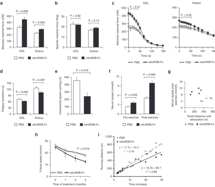
Figure 1 Treatment of adult wild-type mice with soluble activin IIB receptor (sActRIIB-Fc). All tests were done after a 4-month treatment of the wild-type mice with either sActRIIB-Fc or PBS (controls). (a) Absolute maximal force (n = 10 for each condition), and (b) specific maximal force of EDL and soleus muscles (n = 10 for each condition). (c) Force recordings during the fatigue protocol over 180 seconds, and (d) fatigue resistance of EDL (n = 9 for PBS-treated mice and n = 8 for Fc-treated mice) and soleus muscles (n = 10 for PBS-treated mice and n = 9 for sActRIIB-Fc-treated mice). (e) Running distances during incremental speed running until exhaustion (n = 5 for each condition). (f) Serum lactate levels at rest and 5 minutes after incremental speed running until exhaustion (n = 5 for each condition). (g) A plot depicting the relationship between travel distance until exhaustion during incremental speed running and serum lactate, which was measured 5 minutes after exhaustion, for individual mice (n = 5 for each condition). (h) Critical speed before and after 1, 2, and 4 months of treatment with sActRIIB-Fc in comparison to PBS-treated control mice (n = 5 for each condition). (i) A plot depicts the proportional relationship between distance run (y-axis) and time to exhaustion (x-axis) at dif-ferent velocities. The slope of the regression line indicates the Critical Speed. Values are shown as means ± SEM. P values were calculated using the nonparametric U-test. 0 100 200 EDL PBS sActRIIB-Fc Soleus 0 10 20 30 Specific maximal f orce (N/g) EDL PBS 0 100 200 300 400 500 Absolute maximal f orce (mN) 0 60 120 180 PBS Time (s) EDL Soleus sActRIIB-Fc sActRIIB-Fc 100 0 200 300 400 0 60 120 180 PBS Time (s) sActRIIB-Fc Soleus 300 Absolute maximal f orce (mN) 0 30 60 90 120 150 EDL PBS sActRIIB-Fc PBS sActRIIB-Fc PBS sActRIIB-Fc PBS Pre-exercise Post-exercise sActRIIB-Fc PBS sActRIIB-Fc PBS 0 0 4 8 Ser um lactate post-e x ercise (mmol/l) 12 16 200 Travel distance until
exhaustion (m) 400 600 sActRIIB-Fc Soleus F atigue resistance (s) 400 500 P = 0.008 0 100 200 300 Incremental speed r unning (m) 400 500 P = 0.016 P = 0.016 0 0 0 1
Time of treatment (months)
0 200 400 600 Running distance (m) 800 1,000 0 15 30 y = 12.3x + 60.1 r2 = 0.89 y = 17.3x + 39.2 r2 = 0.96 45 Time (minutes) 2 10 20 Cr
itical speed (m/min)
30 3 6 9 12 Ser um lactate (mmol/l) P = 0.028 P = 0.009 P = 0.004 P = 0.026 P = 0.097 P = 0.82 P = 0.13 P < 0.01 P < 0.05 4 a d h i e f g b c
muscle function, exercise capacity, and muscle metabolism.4,7,8,18,19
To circumvent the effects of congenital fiber-type switching, we here inhibited ActRIIB signaling in adult wild-type mice with a soluble form of the activin receptor fused with the Fc-fragment of mouse IgG (sActRIIB-Fc). Four months of treatment pro-moted robust skeletal muscle growth together with a significant increase in total body weight, confirming previously published data (Supplementary Figure S1).20 Importantly, the increase
in muscle mass was not accompanied by fiber-type conver-sion (Supplementary Figure S2). The absolute maximal force of EDL and soleus muscle increased in parallel with muscle size (Figure 1a,c). Specific maximal force was conserved, implicating
a proportional increase of force and muscle mass (Figure 1b). However, sActRIIB-Fc treatment also increased muscle fatigue (Figure 1c,d) and mice exhausted precociously during incremen-tal speed running tests (Figure 1e,g). Serum lactate, being already significantly increased at resting state, rose to pathological levels following incremental speed running (Figure 1f,g). The concept of “Critical Speed” accurately reflects the capacity for aerobic exercise and is based on the proportional relationship between “covered distance” and “time to exhaustion” at different veloci-ties.21 During the 4-month treatment period, we found a steady
decline in Critical Speed in the treatment and control group, how-ever, the decline over time was by far larger in sActRIIB-Fc-treated
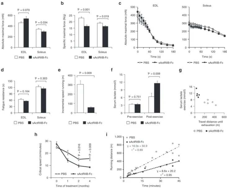
Figure 2 Treatment of adult mdx mice with soluble activin IIB receptor (sActRIIB-Fc). All tests were performed after a 4-month treatment of the mdx mice with either sActRIIB-Fc or PBS (controls). (a) Absolute maximal force (n = 9 for each condition for EDL muscles and n = 10 for each condi-tion for soleus muscles), and (b) specific maximal force of EDL (n = 9 for each condicondi-tion) and soleus muscles (n = 10 for each condicondi-tion). (c) Force recordings during the fatigue protocol over 180 seconds of EDL and soleus muscles, and (d) fatigue resistance for EDL (n = 8 for each condition) and soleus (n = 10 for PBS-treated mice and n = 8 for sActRIIB-Fc-treated mice). (e) Running distance during incremental speed running until exhaustion (n = 5 for each condition). (f) Serum lactate levels at rest and 5 minutes after incremental speed running until exhaustion (n = 5 for each condition). (g) A plot depicts the relationship between travel distance until exhaustion during incremental speed running and serum lactate, which was measured 5 minutes after exhaustion, for individual mice (n = 5 for each condition). (h) Critical speed before and after 1, 2, and 4 months of treatment with sActRIIB-Fc in comparison to PBS (n = 5 for each condition). (i) A plot depicts the proportional relationship between distance run (y-axis) and time to exhaustion (x-axis) at different velocities. The slope of the regression line indicates the critical speed. Values are shown as means ± SEM. P values were calculated using the nonparametric U-test.
0 200 400 600 EDL PBS sActRIIB-Fc Soleus 0 5 10 15 20 25 EDL PBS sActRIIB-Fc Soleus Specific maximal f orce (N/g) Incremental speed r unning (m) Ser um lactate (mmol/l) Ser um lactate e x ercise (mmol/l) Absolute maximal f orce (mN) 0 30 60 90 120 150 EDL PBS sActRIIB-Fc PBS sActRIIB-Fc 0 0 0 0 200 400 Travel distance until
exhaustion (m) 600 4 8 12 16 3 6 9 12 15 100 200 300 400 PBS sActRIIB-Fc PBS Pre-exercise Post-exercise sActRIIB-Fc PBS sActRIIB-Fc y = 16.0x + 50.9 r2 = 0.89 y = 8.6x + 20.2 r2 = 0.95 PBS sActRIIB-Fc Soleus F atigue resistance (s) 0 0 1 2
Time of treatment (months) 4 10
20 30
Cr
itical speed (m/minutes)
0 200 400 600 800 1,000 0 15 30 45 Time (minutes) Running distance (m) 0 0 60 120 180 100 200 300 400 500 EDL Time (s) PBS sActRIIB-Fc 0 0 60 120 180 100 200 300 400 500 Soleus Time (s) PBS sActRIIB-Fc Absolute maximal f orce (mN) P = 0.070 P = 0.001 P = 0.019 P = 0.034 P = 0.164 P = 0.016 P = 0.009 P = 0.303 P = 0.009 P = 0.009 P = 0.751 a b c d h i e f g
animals as compared to phosphate-buffered saline (PBS)-treated mice (Figure 1h,i).
Severe exercise intolerance in dystrophic mdx mice following treatment with sActRIIB-Fc
In Duchenne muscular dystrophy and its mdx mouse model, oxi-dative metabolism is compromised due to membrane damage and the resulting intracellular calcium overload.15–17 Having shown that
ActRIIB blockade decreased aerobic exercise capacity in wild-type mice, we now investigated what effect administration of sActRIIB-Fc would have on the metabolic phenotype of mdx mice. This infor-mation would have important clinical implications for the strategies to use ActRIIB blockade for the treatment of muscular dystrophies. Despite a massive increase in skeletal muscle mass after sActRIIB-Fc treatment (Supplementary Figure S1), absolute maximal force decreased in soleus muscle and most notably specific force in both EDL and soleus muscles (Figure 2a,b), serving as functional evidence
for increased myopathic changes of sActRIIB-treated dystrophic mdx muscle. Interestingly, sActRIIB-Fc treatment did not cause any greater force decline during repetitive stimulation (Figure 2c,d). Electromyography excluded problems in neuromuscular transmis-sion but revealed abnormal spontaneous potentials and the presence of complex repetitive discharges in both PBS and sActRIIB-Fc treat-ment groups of the mdx mice (Suppletreat-mentary Figure S3). Such polyphasic potentials are characteristic of dystrophic mdx muscle.22
As voluntary motor activity seemed reduced when observing sAc-tRIIB-Fc-treated mdx mice, we proceeded to analyze their exercise behavior. Remarkably, at the end of the treatment period, sActRIIB-Fc-treated mdx mice suffered from severe exercise intolerance associated with a pathological serum lactate increase (Figure 2e–g;
Supplementary Video S1). It is important to note, that exercise
capacity of mdx mice declined throughout the 4-month treatment period, however, to a far larger extent in sActRIIB-Fc-treated mdx mice than in the PBS-treated control group (Figure 2h,i).
Figure 3 The effect of myostatin/ActRIIB signaling on vascularization. All investigations were done after a 4-month treatment of wild-type and mdx mice with either soluble activin IIB receptor (sActRIIB-Fc) or PBS (controls). (a–d) Capillarization of wild-type soleus (n = 430 fibers from PBS-treated muscles (n = 3) and n = 300 fibers from sActRIIB-Fc-PBS-treated muscles (n = 3)) and mdx soleus muscle (n = 605 fibers from PBS-PBS-treated muscles (n = 3) and n = 836 fibers from sActRIIB-Fc-treated muscles (n = 4)). Histograms in (a) and (c) depict the distribution of capillaries per muscle fiber (in (%)), whereas diagrams in (b) and (d) depict the capillary domain, the fiber area per capillary (in (μm2)). Values are depicted as means ± SEM.
(e) Vegf-A relative mRNA copy numbers as expressed per 106 × 18S rRNA copies in wild-type TA muscle (n = 5 for each condition). (f) Vegf-A relative
mRNA-copy numbers in C2C12 myotubes following 24 hours treatment with sActRIIB-Fc in comparison to control cultures (n = 3 for each condi-tion). (g) Relative mRNA-copy numbers of MSTN and myostatin receptors ActRIIA/B and ALK4/5 in cultures of human umbilical vein endothelial cells (HUVEC) in comparison to muscle samples from a healthy control and a patient with Duchenne muscular dystrophy. Values are shown as means ± SEM. P values were calculated using the nonparametric U-test.
PBS sActRIIB-Fc PBS 0 250 500 Capillar y domain ( μ m 2) 750 1,000 WT soleus muscle WT soleus muscle sActRIIB-Fc PBS sActRIIB-Fc 0 0 5 10
Number of muscle fibers (%
) 15 20 25 30 1 2 3 4 5 6 7 8 Number of capillaries per muscle fiber
9 10 11 P < 0.0001 12 PBS sActRIIB-Fc mdx soleus muscle 0 0 5 10
Number of muscle fibers (%
)
15 20 25
1 2 3 4 5 6 7 8 Number of capillaries per muscle fiber
9 10 11 12 a b PBS 0 0 30 60 mRNA cop y number s (per 10 6 18S rRNA) mRNA cop y number s (per 10 6 18S rRNA) 0 20 40 60 80 100 120 Control ActRIIB-Fc mRNA cop y number s (per 10 6 18S rRNA) 90 120 150 250 500 Capillar y domain ( μ m 2) 750 1,000 sActRIIB-Fc ActRIIB
Control muscle DMD muscle Endothelial (HUVEC) cells 0 1,000 2,000 0 0 10 20 30 40 50 6,000 12,000 18,000 24,000 3,000 ALK4 ActRIIA WT mdx P < 0.0001 P < 0.001 P = 0.793 P = 0.001 Vegf-A in skeletal muscle Vegf-A in C2C12 myotubes mdx soleus muscle d g e f c ALK5 MSTN
sActRIIB-Fc treatment affects muscle capillarization
The ability of mitochondria to produce ATP critically depends on the oxygen supply via tissue blood perfusion, thus a combination of hypoperfusion plus exercise-induced hypoxemia might explain the severe exercise intolerance. Treatment with sActRIIB-Fc caused a drop in capillary density especially in the oxidative soleus muscle from mdx mice with a subsequent increase in the capillary domain (Figure 3a–d). The increase of the capillary domain was also found in glycolytic EDL muscles of mdx mice, albeit to a lesser degree (Supplementary Figure S4a–d). Treatment of mice as well as of C2C12 myotubes with sActRIIB-Fc downregulated expression of Vegf-A suggesting an indirect negative effect of myostatin block-ade on capillary formation (Figure 3e,f). Interestingly, Vegf-A expression in mdx mice was much lower than in wild-type mice, and treatment with sActRIIB-Fc did not decrease Vegf-A mRNA-abundance any further (Figure 3e), suggesting the presence of additional mechanisms for regulating capillary density. In this
regard, the findings of Hayot et al.23 are of special interest, who
reported an induction of myostatin expression in muscles of rats exposed to chronic hypoxia and in patients with chronic obstruc-tive pulmonary disease. The authors interpreted these findings as a potential cause for the muscle wasting that is often seen in chronic obstructive pulmonary disease patients. These findings, however, could also be interpreted as a compensatory upregula-tion of myostatin to improve the metabolic funcupregula-tioning and to increase capillary density in a state of chronic hypoxia. It is of spe-cial interest that endothelial cells strongly express the mRNAs of transmembrane receptors (ActRIIA/B and ALK4/5) for myostatin or its homologs, whereas myostatin mRNA was only expressed at low levels (Figure 3g). Treatment of endothelial cells (Human umbilical vein endothelial cell line) with increasing dosages of recombinant myostatin in vitro increased the cell doubling time in culture, verifying a direct effect of myostatin or its homologs on endothelial cell proliferation (Supplementary Figure S4e),
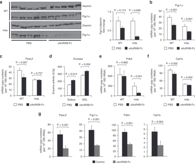
Figure 4 Effect of myostatin/ActRIIB signaling on muscle metabolic phenotype. All investigations were done after a 4-month treatment of wild-type (n = 5 for each condition) and mdx mice (n = 5 for each condition) with either sActRIIB-Fc or PBS (controls). (a) Western blots (left side) depict bands for Pgc1α referenced to Desmin. The bar chart (right side) depicts the quotients of Pgc1α/Desmin band densities. (b) Pgc1α and (c) Pparβ relative copy numbers in the TA muscle from wild-type and mdx mice as expressed per 106 × 18S rRNA copies. (d) Enolase enzymatic activity in
wild-type EDL (n = 5 for each condition) and wild-wild-type soleus muscles (n = 5 for each condition). (e,f) Relative mRNA copy numbers of genes involved in the regulation of the oxidative metabolism in TA muscle from wild-type and mdx mice and (g) from C2C12 myotubes following 24 hours treatment with sActRIIB-Fc in comparison to control cultures (n = 3 for each condition). Values are shown as means ± SEM. P values were calculated using the nonparametric U-test. sActRIIB-Fc, soluble activin IIB receptor.
PBS mdx WT sActRIIB-Fc PBS sActRIIB-Fc PBS sActRIIB-Fc PBS sActRIIB-Fc
PBS sActRIIB-Fc PBS sActRIIB-Fc PBS sActRIIB-Fc
Desmin 0 0.5 Pgc1 α /desmin fold change 1 1.5 WT mdx WT mdx WT mdx P = 0.174 0 10 20 30 40 50 WT mdx P < 0.001 Pgc1α P < 0.001 0 10 20 30 40 50 0 0 Control sActRIIB-Fc 0 1 2 3 4 5 6 7 20 40 60 80 100 120 10 20 30 40 50 mRNA cop y number s (per 10 6 18S rRNA) WT 0 100 200 300 400 Enolase Pdk4
Enzyme activity (IU/g)
0 300 600 900 mRNA cop y number s (per 10 6 18S rRNA) 0 20 40 60 80 mRNA cop y number s (per 10 6 18S rRNA) 0 30 60 90 120 mRNA cop y number s (per 10 6 18S rRNA) mRNA cop y number s (per 10 6 18S rRNA) Soleus EDL mdx Pparβ Pparβ Pgc1α Pdk4 Cpt1b P = 0.727 P = 0.050 P = 0.983 P = 0.001 P = 0.002 P = 0.052 P = 0.014 P < 0.001 P < 0.001 P = 0.001 P = 0.004 P = 0.007 P = 0.009 Pgc1α Desmin Pgc1α Cpt1b a c g d e f b
however, the exact ligands of muscle endothelial cell regulation in vivo remain to be determined.
ActRIIB signaling regulates Pgc1α and Ppar transcription factors
The exercise intolerance and lactic acidosis following ActRIIB blockade suggests underlying changes in muscle metabolism, a hypothesis supported by previous transcriptome profiling.13 In
agreement, we show that the copy numbers of Pgc1α and Pparβ, which are key transcription factors promoting oxidative metabo-lism in skeletal muscle, are downregulated after treatment with sActRIIB-Fc (Figure 4b,c) and following treatment of C2C12 myo-tubes with sActRIIB-Fc (Figure 4g). On the protein level, down-regulation of Pgc1α was more pronounced in the mdx muscle if referred to desmin abundance (Figure 4a). Such loss of oxidative properties was accompanied by a compensatory activity increase of enolase, a key glycolytic enzyme (Figure 4d). Furthermore, mRNA levels of Pdk4, an inhibitor of pyruvate dehydrogenase (Pdh) and a regulatory switch of substrate utilization from glucose towards fatty acids,24 was strongly decreased following
sActRIIB-Fc treatment in mdx mice (Figure 4e). We thus expected an inhib-itory effect of sActRIIB-Fc treatment on β-oxidation and found a downregulation of Cpt1b mRNA levels (Figure 4f). Likewise, sActRIIB-Fc treatment of C2C12 myotubes reduced expression of genes controlling oxidative metabolism and β-oxidation within 24 hours of treatment (Figure 4g), implying a direct effect of myo-statin signaling in the regulation of these genes.
We further focused our attention on the neuronal nitric oxide synthase (Nos1, nNos), because it is well known that the sarco-lemmal presence of the Nos1 enzyme is strongly reduced in the absence of its binding partner dystrophin in patients with DMD and in mdx mice.25,26 The resulting dysregulation of NO synthesis
entails a failure of contraction-induced vasodilatation as well as changes in the cellular calcium homeostasis associated with exac-erbated postexercise fatigability, exercise-induced muscle edema, and cell necrosis.27,28 We thus wondered, whether sActRIIB-Fc
treatment would influence sarcolemmal Nos1 expression, hence further compromising the pathophysiological effect of sActRIIB-Fc on vasculature and oxidative metabolism. As expected, we found a strong decrease of Nos1 mRNA copy numbers in mdx mus-cles of both treatment groups in comparison to wild-type muscle (Supplementary Figure S11a), which was paralleled by a strong decrease of sarcolemmal expression of Nos1 protein in mdx mus-cle (Supplementary Figure S11b). Furthermore, treatment with sActRIIB-Fc diminished Nos1 transcription in wild-type and mdx muscle (Supplementary Figure S11a). However, subsarcolem-mal Nos1 protein content remained unchanged (Supplementary
Figure S11b), and western blot did not reveal any changes in Nos1
protein levels in wild-type mice (Supplementary Figure S11c), whereas Nos1 protein levels in mdx mice were below detection levels (data not shown). This suggests that sActRIIB-Fc treatment unlikely aggravates NO dysregulation of dystrophin deficient muscle, although further experiments are required to ascertain or to exclude a role of the ActRIIB-receptor on NO signaling.
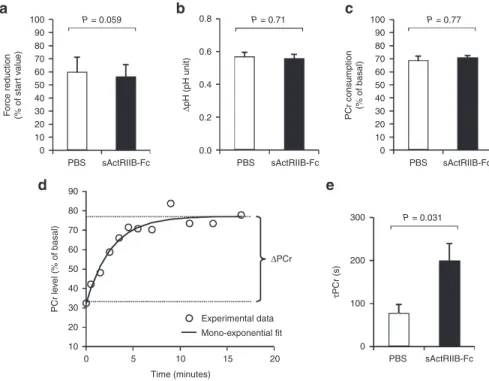
Figure 5 In vivo investigation of muscle function and oxidative metabolism in mdx calf muscles by noninvasive 31P-MRS. Investigations were
done in mdx mice that were treated either with sActRIIB-Fc (n = 5) or with PBS (controls; n = 4). (a) The extent of force reduction was measured at the end of the 6 minutes period of electrostimulation and is represented as percent of the starting value. (b) The drop of intracellular pH and (c) phosphocreatine (PCr) concentration was determined at the end of the 6-minute in vivo electrostimulation period. (d) For each animal, the post-stimulation time course of PCr was fitted to a mono-exponential function with a least mean-squared algorithm in order to calculate the PCr recovery time constant (τPCr): τPCr = −t/ln(PCrt/ΔPCr). (e) τPCr was significantly larger in sActRIIB-Fc-treated mdx mice (200 ± 39 versus 78 ± 20 seconds in the
control group). Values are shown as means ± SEM. P values were calculated using the nonparametric U-test.
0 0.0 0.2 0.4 Δ pH (pH unit) 0.6 0.8
PBS sActRIIB-Fc PBS sActRIIB-Fc PBS sActRIIB-Fc
10 20 30 40 F orce reduction (% of star t v alue) 50 60 70 80 90 10 0 5 10 Time (minutes) 15 Experimental data ΔPCr Mono-exponential fit 20 20 30 40 PCr le v el (% of basal) 50 60 70 80 90 100 0 10 20 30 40 PCr consumption (% of basal) PBS sActRIIB-Fc 0 100 200 300 τPCr (s) 50 60 70 80 90 100 P = 0.059 P = 0.71 P = 0.031 P = 0.77 a d e b c
Reduced oxidative metabolism in mdx muscle following treatment with sActRIIB-Fc
It should be noted that the mRNA and protein levels of key regu-latory genes (Pparβ, Pgc1α) important for oxidative metabolism (Figure 4a–c) were significantly lower in mdx than in wild-type mice, supporting previous findings that oxidative muscle metabo-lism is depressed in dystrophic muscle to some extent.15,29
We therefore studied in real-time the response of the oxidative metabolism to a standardized bout of exercise in anesthetized mdx mice either treated with PBS (controls) or sActRIIB-Fc. Muscle function and energy metabolism were assessed strictly nonin-vasively in calf muscle with an innovative experimental setup using phosphorus (31P) nuclear magnetic resonance spectroscopy
(MRS).30 An exercise bout of 6 minutes consisting of repeated
maximal isometric contractions was induced in vivo by transcu-taneous electrostimulation. After induced repeated contractions fatigue levels (Figure 5a), intracellular acidosis (Figure 5b) as well as phosophocreatine (PCr) consumption (Figure 5c) were similar in both groups. However, the time constant of postexercise phos-phocreatine resynthesis (τPCr) was significantly prolonged in sActRIIB-Fc-treated mdx mice (Figure 5d,e). Given that PCr syn-thesis during the postexercise recovery period relies exclusively on oxidative ATP synthesis, τPCr has largely been acknowledged as an important in vivo index of oxidative mitochondrial capacity. Hence, the prolonged τPCr demonstrates that sActRIIB-Fc treat-ment reduces oxidative metabolism in vivo.
The MRS results pointed to an underlying functional deficit of the skeletal muscle respiratory chain complexes or β-oxidation in response to sActRIIB-Fc treatment. However, on contrary to our hypothesis, we found (i) largely unaffected ex vivo activities of isolated key mitochondrial enzymes (Krebs cycle: citrate syn-thase; respiratory chain: cytochrome C oxidase; and β-oxidation: hydroxyacyl-CoA-dehydrogenase) and (ii) similar succinate dehy-drogenase and cytochrome C oxidase fiber profiles (Supplementary
Figures S5–S7). In fact, cytochrome C oxidase and succinate
dehy-drogenase enzyme activities even appeared somewhat increased in EDL muscles (Supplementary Figures S5c,d, S6a, S7a), likely reflecting a compensatory increase in response to decreased aero-bic energy production. Furthermore, mitochondrial DNA copy numbers remained largely unchanged following treatment with sActRIIB-Fc (Supplementary Figure S8a). The normal mitochon-drial DNA copy numbers together with unaltered citrate synthase enzyme activities (Supplementary Figure S5a,b) let us conclude that ActRIIB blockade did not affect mitochondrial mass.
Treatment with sActRIIB-Fc downregulates porin expression in wild-type and mdx muscle
Given the abnormal postexercise τPCr along with normal respi-ratory chain activities, we wondered whether the ATP transport from the mitochondrial matrix into the cytosol of skeletal muscle cells could be affected, which might explain the diminished rate of aerobic energy production. Keeping with such hypothesis, decreased protein levels of Vdac3 had already been reported for mdx muscle, hinting towards a derangement of the ADP/ATP-shuttling system through the outer mitochondrial membrane via the voltage-dependent anion channels (Vdac, syn. porin).31–33
In line with these findings, a proteomic survey of differentially
expressed proteins from wild-type and mdx mouse hearts had discovered a substantial loss of Vdac1 protein.34 Indeed, here
we show that wild-type and to an even larger extent mdx mus-cles exhibited a considerable reduction of porin mRNA tran-scripts (Supplementary Figure S8a) and porin protein levels (Supplementary Figure S9) after sActRIIB-Fc treatment. This pushes the muscle even further into global mitochondrial dys-function than dystrophin deficiency alone. Such secondary mito-chondriopathy might explain the high lactic acidosis and rapid fatigability of sActRIIB-Fc-treated mdx mice.
Myopathic changes in mdx mice following treatment with sActRIIB-Fc
We next investigated the consequences of sActRIIB-Fc treatment on the extent of muscle dystrophy in mdx mice. Muscles from both sActRIIB-Fc and PBS-treatment groups revealed typical dystrophic changes comprising muscle fiber necrosis, regenerat-ing fibers, fibers with central nuclei, inflammatory infiltrates, and increased fibrosis, which are difficult to quantify (Supplementary
Figure S10a). Muscle degeneration is accompanied by a leak of
cytoplasmic enzymes such as creatine kinase. We measured serum creatine kinase levels, which were largely elevated in mdx mice from both treatment groups; however, we did not detect any significant differences since interindividual variation was large (Supplementary Figure S10b). In mdx mice, muscle degenera-tion is followed by excessive regeneradegenera-tion with abundant split-ting of regenerated fibers, which appear as small fiber profiles on transverse sections. Such excessive regeneration leads to an increase of muscle mass (see comparison between wild-type and mdx mice: Supplementary Figure S1). Following treatment with sActRIIB-Fc, muscles enlarged on average, if compared to PBS treatment, by ≈1.6-fold in mdx and by ≈1.3-fold in wild-type mice (Supplementary Figure S1). However, analysis of morphomet-ric features of EDL muscles from sActRIIB-Fc-treated mdx mice, revealed a further increase in the number of small fiber profiles if compared to PBS-treated mdx mice (Supplementary Figure
S10c). This finding suggestes that the excessive increase of muscle
weight was triggered by abnormal regeneration and not by fiber hypertrophy. The soleus muscle of mdx mice, while not increasing its mass after sActRIIB-Fc treatment, exhibited an increased fiber size variation (Supplementary Figure S10d). In conclusion, the dystrophic phenotype of dystrophin deficient muscle persisted or even increased following treatment with sActRIIB-Fc.
DISCUSSION
Myostatin/ActRIIB signaling exerts three major functions on skeletal muscle. (i) It acts to limit its size, (ii) promotes oxidative properties, and (iii) balances glucose versus fat utilization. The changes in muscle physiology in hypermuscular mice follow-ing treatment of adult mice with sActRIIB-Fc highlights the fact that myostatin/ActRIIB blockade confers little functional advan-tage over wild-type muscle due to its rapid fatigability. Improved muscle strength, however short lived, comes at the cost of increased fatigability and exercise intolerance, which is often seen in patients with mitochondrial disorders such as mitochondrial encephalopathy, lactic acidosis, stroke-like episodes (MELAS) or myoclonic epilepsy with ragged-red fibers (MERFF) syndrome.35
Interestingly, muscle cramps are frequently observed in whippet dogs with Mstn mutations.36 Moreover, “double muscle cattle”,
several breeds of which have been identified to carry Mstn muta-tions,37,38 are prone to exercise induced lactic acidosis and severe
rhabdomyolysis.39,40 In myostatin deficient animals, such exercise
failure could be attributed to congenital fiber-type disproportion with a shift toward the expression of the fast IIB MHC isoform,4,18
which is well known to be associated with loss of oxidative prop-erties of skeletal muscle and increased fatigability.5,8 In contrast
to animals born with mutations in the Mstn gene, we show that blockade of ActRIIB signaling in adult wild-type mice beyond the period of muscle development does not have any impact on fiber-type composition, thus confirming previous reports.10
After sActRIIB-Fc treatment, the mice exhibit clinical signs of early muscle fatigue, exercise intolerance, and lactic acidosis— characteristic signs for a depression of β-oxidation, a shift toward anaerobic glycolysis, and ATP deficiency. Interestingly, whereas ex vivo mitochondrial respiratory chain enzyme activities and mitochondrial DNA copy numbers were within the normal range, in vivo 31P-MRS clearly demonstrated a downregulation of oxidative
energy metabolism in sActRIIB-Fc-treated mdx mice. We further demonstrate a significant loss of the porin complex in sActRIIB-Fc-treated mice, pointing towards an underlying defect of ATP handling and ATP transport as one causative mechanism for the metabolic phenotype. A second aggravating factor, which further compromised exercise tolerance, is the decrease of capillary density and the increase of the capillary domain. Previous reports attrib-uted such reduced capillary density to the increase of muscle fiber size.41 However, here we demonstrate a net numerical loss of
capil-laries per fiber following treatment with sActRIIB-Fc. This finding was most pronounced in mdx mice with a profound rarefication of the capillary bed in dystrophic muscle. In dystrophinopathies diminished sarcolemmal Nos1 results in dysregulation of the cap-illary adaptive response to exercise leading to functional muscle ischemia.42 Our protein analysis argues against a further
aggrava-tion of the capillary adaptive response by addiaggrava-tional loss of Nos1 in sActRIIB-Fc-treated animals, despite the fact that Nos1 mRNA lev-els were clearly reduced. sActRIIB-Fc treatment of dystrophic mdx mice dramatically worsened the myopathic phenotype as shown by the large deficit in specific force. As lack of dystrophin per se alters mitochondrial function in DMD patients and mdx mice,15,16,29 the
blockade of myostatin signaling initiates a vicious cycle resulting in severe secondary metabolic myopathy. We also found an increased fiber size variation after sActRIIB-Fc treatment of mdx mice point-ing towards increased myopathic changes even at the tissue level.
In the past, several investigators have used different strategies of interfering with the ActRIIB receptor mediated signaling path-way in order to treat mdx mice and Golden Retriever muscular dystrophy (GRMD) dogs. This was done either through injection of sActRIIB-Fc,12,43 AAV-mediated gene transfer11 or
antibod-ies directed against the ActRIIB receptor.44 Overall, the
conclu-sions were optimistic about the usefulness of such strategy to treat dystrophinopathies. However, it should be noted that our results and conclusions differ from previously published work in vari-ous aspects. We think the reason for that mainly lies in the choice of endpoints to define success or failure of such a treatment. For DMD patients, clinically relevant and quantifiable improvements
would comprise better performance in the 6-minute walk45 and
improvement of respiratory function. This implies the ability of the patient’s body to maintain a certain workload for a pro-longed time period and not just to be able to produce single bouts of maximum short duration muscle activity as tested by tetanic muscle contractions11,12 or by the whole body tension method.43
In most studies, the increase of muscle size was taken as an end-point,11,44 automatically assuming that big muscles are healthier
muscles. This basic assumption is put into question by our results. None of the studies investigated endurance capacity, which evalu-ates the effect of a treatment on the physiology of the entire body over a longer time period and would thus be a relevant parameter that could translate into improvement of life quality in patients. Several studies, one using the identical sActRIIB-Fc compound,12
reported a small, but significant decline of creatine kinase values after ActRIIB blockade.11,12 We were unable to reproduce this
find-ing, the reason for these differences remaining unresolved. We show that myostatin controls the metabolic profile of skel-etal muscle, and its blockade depresses the main molecular deter-minants of oxidative metabolism and β-oxidation. Interestingly, genetic inactivation of Pparβ, similar to myostatin/ActRIIB blockade, reduced oxidative properties of skeletal muscle.46 This
adds further evidence that myostatin controls the muscle oxida-tive phenotype via peroxisome proliferator-activated receptors (PPAR) and Pgc1α, the downstream target of Pparβ, while the down-regulation of Pdk4 indicates a shift away from β-oxidation towards glucose metabolism. These quantitative polymerase chain reaction data are strongly corroborated by a recent transcriptome study following treatment with sActRIIB-Fc of wild-type mice.13
However, we lack direct evidence to ascertain a shift towards higher glucose metabolism, although the downregulation of Pdk4 and upregulation of Enolase can be counted as indirect indica-tors for such a change. The unfavorable combination of decreased vascularization and metabolic changes after ActRIIB blockade is likely to cause a rapid imbalance between increased cytosolic ATP hydrolysis and insufficient mitochondrial ATP synthesis during exhaustive exercise. The subsequent shift toward anaerobic glyco-lytic ATP synthesis explains the rapid fatigability and the patho-logically increased lactate production.47 However, the metabolic
adaption in response to myostatin may differ in diverse physi-ological and pathophysiphysi-ological contexts, e.g., myostatin inhibi-tion was reported to improve motor performance in aged mice.48
Further work is required to elucidate the metabolic function of myostatin in different disease situations and during ageing.
In conclusion, our results suggest that myostatin/ActRIIB sig-naling optimizes oxidative metabolism of skeletal muscle leading to lower muscle fatigability and amelioration of endurance capac-ity. Such fundamental functions of myostatin should be taken into account in the development of therapies based on myostatin/ ActRIIB blockade. However, it should be kept in mind that our experimental design does not allow to determine which effects can be ascribed to myostatin blockade alone and which to the inactiva-tion of other TGF-β family members such as bone morphogenetic proteins, growth and differentiation factors, and activins that may also be sequestered by the soluble ActRIIB. Further investigations are required to answer the question, such as whether emerging therapies based on PPAR agonists might be able to prevent such
adverse effects of ActRIIB blockade on the oxidative metabo-lism and on exercise tolerance. Furthermore, dose regime stud-ies could answer the question, whether short-term treatment or pulse treatment may circumvent secondary effects of myostatin/ ActRIIB blockade on muscle metabolism.
MATERIALS AND METHODS
Animals. Male mdx mice (on a C57BL/10ScSn background) were bred in the animal facility of the Medical Faculty of Paris VI and kept according to institutional guidelines. Wild-type male C57BL/6J control mice were pur-chased from Charles River (France). Two-month-old wild-type and mdx mice were injected twice weekly subcutaneously with 10 mg/kg with the rodent form of the soluble activin receptor IIB (sActRIIB-Fc; Acceleron Pharma, Cambridge, MA) for a total of 4 months before killing. The meth-ods of sActRIIB-Fc synthesis have previously been described.49 All animal
studies have been approved and were carried out under the laboratory and animal facility licenses A75-13-11 and A91-228-107.
Evaluation of the critical speed. Mice were subjected to three or four sep-arate bouts of runs until exhaustion at various treadmill speeds (between 20 and 80 cm/second according to individual motor capacity, one run per day) according to previously published protocols.21 Critical speed, an index
of the aerobic exercise capacity, was calculated from the slope (a) of the regression line, plotting the distance (y) against the time to exhaustion (x) from the different runs.
Blood lactate assessment during exhaustive exercise. Lactate concentra-tions were determined in blood samples collected from the tip of the tail using a Lactate pro LT device (Arkray, Kyoto, Japan) at rest before exercise (0 minute) and 5 minutes after treadmill running-induced exhaustion. Exhaustion was defined as the time point at which the mice were unable to run anymore and stayed on the grid despite repeated electric stimula-tion. The running test started at the lowest speed of 5 cm/second to allow a warm-up and then increased by 1 cm/second every 30 seconds until exhaustion. This protocol is illustrated by the Supplementary Video S1, which demonstrates the running test of sActRIIB-Fc-treated wild-type (left) and mdx mice (right) side by side. The starting speed of 5 cm/second was increased by 1 cm/second every minute until exhaustion of the mdx mouse at 19 cm/second after 14 minutes, while the wild-type mouse was still able to run at a speed of 40 cm/second.
Electromyographic examination. Electromyographic examination of the
triceps brachialis, tibialis, gastrocnemius, and quadriceps femoris muscles
was performed in mice anesthetized with isoflurane. Standard noninvasive needle electromyography was conducted on a Viking Quest EMG appa-ratus (Viasys, Nicolet Biomedical, Madison, WI) using concentric bipolar needle electrodes. Insertional activity and pathological spontaneous activ-ity were recorded.
Measurement of contractile properties. Absolute maximal isometric tetanic force (P0) was measured during tetanic contractions (frequency of 50–100 Hz, train of stimulation of 1,500 ms for soleus and 750 ms for
EDL). Specific maximal isometric force (sP0) was given as the quotient
between force and muscle weight. For analysis of fatigue resistance, mus-cles were stimulated at 75 Hz for 500 ms, every 2 seconds over 3 minutes. See Supplementary Methods.
Western blot. Protein was extracted from frozen tibialis anterior muscle of wild-type and mdx mice and processed as described.50 Briefly, after
homog-enization of the muscle in radioimmunoprecipitation assay buffer with a proteinase inhibitor cocktail (Complete; Roche-Diagnostics, Mannheim, Germany) proteins were separated through denaturating sodium dodecyl sulphate polyacrylamide gel electrophoresis with the Laemmli system and blotted onto nitrocellulose membranes by the semidry method (Biometra, Göttingen, Germany). The blots were probed with anti-porin as primary
antibody (VDAC 31HL, AB-2; Calbiochem, Darmstadt, Germany), anti-PGC1α (Santa-Cruz, Heidelberg, Germany), anti-Nos1 (Abcam, Paris, France), and corresponding peroxidase-labeled secondary antibodies. The desmin and GAPDH bands were used as loading control for muscle. Bands were visualized by chemiluminescence. The protein bands were quantified by measuring their integrated density within a rectangle that covered the entire individual band and subtracting the integrated density of an empty rectangle of exactly the same size in the vicinity using the ImageJ software (NIH, Bethesda, MD).
Enzyme measurements. Enolase, citrate synthase, cytochrome C oxi-dase, and hydroxyacyl-CoA-dehydrogenase activities were determined in extracts from frozen cryostat sections using a coupled enzyme assay as detailed in Supplementary Methods.
Histology. H&E, succinate dehydrogenase and cytochrome C oxidase staining were performed using routine histological protocols. The fol-lowing primary antibodies were used for immunohistochemistry: anti-CD31 (Pharmingen, Ranges, France), anti-MHCIIA (SC-71, DSMZ, Braunschweig, Germany), anti-MHCI (BAD5, DSMZ), anti-Nos1 (Abcam), and antilaminin (Dako, Les Ulis, France) followed by second-ary antibodies with various fluorophores (Alexa Fluor; Invitrogen, Saint Aubin, France) (see Supplementary Methods).
Morphometric analysis of capillary number and capillary domains.
Cryosections of 12 μm of the EDL and soleus muscles of PBS- and sAc-tRIIB-Fc-treated animals were stained with antilaminin to delineate the muscle fibers. Muscle capillaries were stained with anti-CD31. Fluorescent photographs were taken with a ×20 objective on a Microscope (Zeiss, AxioImager Z1, Marly-le-Roi, France) and saved as TIFF files. These images were projected on a flatscreen coupled with a graphic tablet, which enabled the manual retracing of the muscle fiber outlines and the counting of capil-laries that were found around it. For the EDL, the fibers of the entire muscle cross section were analyzed, and for the soleus muscle, the fibers from 10 representative nonoverlapping visual fields. For each muscle fiber, we deter-mined the cross-sectional plane (μm2) and counted the number of
border-ing capillaries. The capillary domain (μm2) for each fiber was calculated by
dividing its cross-sectional plane by the number of bordering capillaries.
Cell culture. C2C12 cells were grown in Dulbecco's Modified Eagle's Medium (DMEM) (Gibco 41966-029, Darmstadt, Germany) supple-mented with 1% Pen/Strep and 20% fetal bovine serum (Gibco 10500-064) to semiconfluency at 37 °C and 5% CO2 for 2 days. Thereafter, the medium was replaced by DMEM plus 10% horse serum (Gibco 26050-088) to induce fusion into multinucleated myotubes. sActRIIB-Fc was added to a final concentration of 200 ng/ml to the culture medium of the myotubes. After 24 hours, the myotubes were harvested by trypsinization, washed and pelleted for RNA extraction. Human umbilical vein endothelial cells were grown to confluency, trypsinized and pelleted for RNA extraction. The doubling time of the cells was determined using the AlamarBlue reagents from Invitrogen. Briefly, cells were plated at 20% confluency and allowed to settle for 12 hours before introducing recombinant myostatin (R&D Systems, Wiesbaden, Germany). The cells were grown for 24 hours before addition of 0.1 V of AlamarBlue reagent and incubation at 37 °C for 20 minutes before photometric analysis. The cells were then washed and cultured in fresh medium containing myostatin. Cell proliferation was monitored every 24 hours for 4 days after initial introduction of myostatin. Cell number was determined by comparison of absorbance against a stan-dard curve. All experiments were performed in triplicate.
Reverse transcription quantitative polymerase chain reaction. Real-time polymerase chain reaction was performed according the SYBR Green protocol (Applied Biosystems, Darmstadt, Germany) on the Eco real-time polymerase chain reaction System (Illumina, Eindhoven, The Netherlands) with a HotStart Taq polymerase (Applied Biosystems). For primer sequences, See Supplementary Methods. Fold changes were
calculated according to the efficiency corrected −ΔΔCt method. The use of normal and of DMD muscle from patients was covered by the approval of the ethical review board of the Charité (#216/2001). All patients or their legal guardians provided written informed consent according to the Declaration of Helsinki.
In vivo MRS investigation of muscle function and oxidative
metabo-lism. Mice were anesthetized with 4% isoflurane in 100% air at a flow of 3 l/minute and were placed into a home-built cradle specifically designed for the strictly noninvasive MRS investigation of muscle function and ener-getics.30 Throughout the experiment, anesthesia was maintained using a
facemask continuously supplying 1.75% isoflurane in 33% O2 (0.2 l/minute) and 66% N2O (0.4 l/minute). Animal body temperature was controlled by a rectal probe and maintained at physiological values by a feedback loop that regulated an electrical heating blanket. MR spectra were recorded in the 4.7 T horizontal magnet of a 47/30 Biospec Avance MR system (Bruker, Karlsruhe, Germany) equipped with a Bruker 120-mm BGA12SL (200 mT/m) gradient insert. Calf muscle were electrostimulated transcuta-neously to produce maximal repeated isometric contractions at a frequency of 1.7 Hz. Mechanical performance was measured using a foot pedal cou-pled to a force transducer. Concentrations of phosphorylated compounds and intracellular pH of the calf muscle were continuously measured with an elliptic (8 × 12 mm2) 31P-MRS surface coil during 6 minutes of rest,
6 minutes of electrostimulation and 16 minutes of recovery. MRS data were processed using a custom-written analysis program developed on the IDL software (Research System, Boulder, CO). In order to determine the time constant of postexercise phosphocreatine resynthesis (τPCr, an in vivo index of oxidative mitochondrial capacity), the time course of phosphocreatine concentrations during the poststimulation period was fitted to a monoex-ponential function with a least mean-squared algorithm (Figure 5d): τPCr = −t/ln(PCrt/ΔPCr), where ΔPCr is the extent of PCr depletion measured at the start of recovery period.
Statistical analysis. Data were analyzed and significance levels calculated using the nonparametric Wilcoxon–Mann–Whitney U-test, as stated in the legends and detailed in Supplementary Methods. Values are presented as means ± SEM. Significance levels were set at P < 0.05.
SUPPLEMENTARY MATERIAL
Figure S1. Effect of sActRIIB-Fc on body weight and muscle weight
in wild-type and mdx mice.
Figure S2. Effect of sActRIIB-Fc on fiber-type distribution of EDL and
soleus muscles from wild-type and mdx mice.
Figure S3. Effect of sActRIIB-Fc on EMG recordings of mdx mice. Figure S4. Effect of myostatin on capillaries and endothelial cell
proliferation.
Figure S5. Effect of sActRIIB-Fc on enzymes activities of
mitochon-drial respiratory chain in EDL and soleus muscles from wild-type and mdx mice.
Figure S6. Effect of sActRIIB-Fc on SDH and COX enzyme activity on
muscle sections of EDL and soleus muscles from wild-type mice.
Figure S7. Effect of sActRIIB-Fc on SDH and COX enzyme activity on
muscle sections of EDL and soleus muscles from mdx mice.
Figure S8. Effect of sActRIIB-Fc on mitochondrial DNA copy
num-ber and transcription of the gene encoding the mitochondrial protein porin in muscles from wild-type and mdx mice.
Figure S9. Effect of sActRIIB-Fc on the expression of the
mitochon-drial protein porin in muscles from wild-type and mdx mice.
Figure S10. Effect of myostatin/ActRIIB signaling on muscle phenotype.
Figure S11. Effect of sActRIIB-Fc on the expression of Nos1 in
mus-cles from wild-type and mdx mice.
Video S1. Effect of sActRIIB-Fc on incremental speed running in mdx
mice.
Methods
ACKNOWLEDGMENTS
We acknowledge Acceleron Pharma for the gift of sActRIIB-Fc. This work was supported by the Association Française contre les Myopathies towards H.A., A.F., A.V., L.A., L.G., and E.M., Association Monegasque contre les Myopathies and the Parents Project France toward H.A. and C.H., Aktion Benni & Co toward H.A., the Deutsche Forschungsgemeinschaft and the Université Franco-Allemand towards K.R., H.A., and M.S. (as part of the MyoGrad International Graduate School for Myology DRK 1631/1 and CDFA-06-11), and NeuroCure (Exc 257) to M.S. M.S. is a member of mitoNET (01GM1113D) sup-ported by the BMBF (Germany). The authors do not declare any con-flict of interest. K.R., E.M., C.H., O.A., K.P., S.L., K.J., B.G., F.P.-R., L.A., A.V., D.F., S.B., A.F., M.S., and H.A. performed the experiments; M.S. contributed patient material; O.R. contributed new reagents; K.R., E.M., D.F., S.B., B.G., D.B. A.F., R.V.C., M.S., and H.A. analyzed the data; M.S. did the statistical analysis; K.R., E.M., B.G., D.B., M.S., and H.A. wrote the manuscript, K.P. revised the article critically for impor-tant intellectual content. All authors read the final version of the manu-script and gave final approval of the manumanu-script to be published.
REFERENCES
1. McPherron, AC, Lawler, AM and Lee, SJ (1997). Regulation of skeletal muscle mass in mice by a new TGF-beta superfamily member. Nature 387: 83–90.
2. Lee, SJ and McPherron, AC (2001) Regulation of myostatin activity and muscle growth. Proc Natl Acad Sci USA 98:9306–9311.
3. Schuelke, M, Wagner, KR, Stolz, LE, Hübner, C, Riebel, T, Kömen, W et al. (2004). Myostatin mutation associated with gross muscle hypertrophy in a child. N Engl J Med
350: 2682–2688.
4. Amthor, H, Macharia, R, Navarrete, R, Schuelke, M, Brown, SC, Otto, A et al. (2007). Lack of myostatin results in excessive muscle growth but impaired force generation.
Proc Natl Acad Sci USA 104: 1835–1840.
5. Matsakas, A, Mouisel, E, Amthor, H and Patel, K (2010). Myostatin knockout mice increase oxidative muscle phenotype as an adaptive response to exercise. J Muscle Res
Cell Motil 31: 111–125.
6. Qaisar, R, Renaud, G, Morine, K, Barton, ER, Sweeney, HL and Larsson, L (2012). Is functional hypertrophy and specific force coupled with the addition of myonuclei at the single muscle fiber level? FASEB J 26: 1077–1085.
7. Baligand, C, Gilson, H, Ménard, JC, Schakman, O, Wary, C, Thissen, JP et al. (2010). Functional assessment of skeletal muscle in intact mice lacking myostatin by concurrent NMR imaging and spectroscopy. Gene Ther 17: 328–337.
8. Savage, KJ and McPherron, AC (2010) Endurance exercise training in myostatin null mice. Muscle Nerve 42:355–362.
9. Akpan, I, Goncalves, MD, Dhir, R, Yin, X, Pistilli, EE, Bogdanovich, S et al. (2009). The effects of a soluble activin type IIB receptor on obesity and insulin sensitivity. Int J Obes
(Lond) 33: 1265–1273.
10. Cadena, SM, Tomkinson, KN, Monnell, TE, Spaits, MS, Kumar, R, Underwood, KW
et al. (2010). Administration of a soluble activin type IIB receptor promotes skeletal
muscle growth independent of fiber type. J Appl Physiol (1985) 109: 635–642. 11. Morine, KJ, Bish, LT, Selsby, JT, Gazzara, JA, Pendrak, K, Sleeper, MM et al. (2010).
Activin IIB receptor blockade attenuates dystrophic pathology in a mouse model of Duchenne muscular dystrophy. Muscle Nerve 42: 722–730.
12. Pistilli, EE, Bogdanovich, S, Goncalves, MD, Ahima, RS, Lachey, J, Seehra, J et al. (2011). Targeting the activin type IIB receptor to improve muscle mass and function in the mdx mouse model of Duchenne muscular dystrophy. Am J Pathol 178: 1287–1297.
13. Rahimov, F, King, OD, Warsing, LC, Powell, RE, Emerson, CP Jr, Kunkel, LM et al. (2011). Gene expression profiling of skeletal muscles treated with a soluble activin type IIB receptor. Physiol Genomics 43: 398–407.
14. Chiu, CS, Peekhaus, N, Weber, H, Adamski, S, Murray, EM, Zhang, HZ et al. (2013). Increased muscle force production and bone mineral density in ActRIIB-Fc-treated mature rodents. J Gerontol A Biol Sci Med Sci 68: 1181–1192.
15. Jongpiputvanich, S, Sueblinvong, T and Norapucsunton, T (2005). Mitochondrial respiratory chain dysfunction in various neuromuscular diseases. J Clin Neurosci 12: 426–428.
16. Kuznetsov, AV, Winkler, K, Wiedemann, FR, von Bossanyi, P, Dietzmann, K and Kunz, WS (1998). Impaired mitochondrial oxidative phosphorylation in skeletal muscle of the dystrophin-deficient mdx mouse. Mol Cell Biochem 183: 87–96.
17. Millay, DP, Sargent, MA, Osinska, H, Baines, CP, Barton, ER, Vuagniaux, G et al. (2008). Genetic and pharmacologic inhibition of mitochondrial-dependent necrosis attenuates muscular dystrophy. Nat Med 14: 442–447.
18. Girgenrath, S, Song, K and Whittemore, LA (2005). Loss of myostatin expression alters fiber-type distribution and expression of myosin heavy chain isoforms in slow- and fast-type skeletal muscle. Muscle Nerve 31: 34–40.
19. Matsakas, A, Macharia, R, Otto, A, Elashry, MI, Mouisel, E, Romanello, V et al. (2012). Exercise training attenuates the hypermuscular phenotype and restores skeletal muscle function in the myostatin null mouse. Exp Physiol 97: 125–140.
20. Lee, SJ, Reed, LA, Davies, MV, Girgenrath, S, Goad, ME, Tomkinson, KN et al. (2005). Regulation of muscle growth by multiple ligands signaling through activin type II receptors. Proc Natl Acad Sci U S A 102: 18117–18122.
21. Billat, VL, Mouisel, E, Roblot, N and Melki, J (2005). Inter- and intrastrain variation in mouse critical running speed. J Appl Physiol (1985) 98: 1258–1263.
22. Han, JJ, Carter, GT, Ra, JJ, Abresch, RT, Chamberlain, JS and Robinson, LR (2006). Electromyographic studies in mdx and wild-type C57 mice. Muscle Nerve 33: 208–214.
23. Hayot, M, Rodriguez, J, Vernus, B, Carnac, G, Jean, E, Allen, D et al. (2011). Myostatin up-regulation is associated with the skeletal muscle response to hypoxic stimuli. Mol
Cell Endocrinol 332: 38–47.
24. Zhao, G, Jeoung, NH, Burgess, SC, Rosaaen-Stowe, KA, Inagaki, T, Latif, S et al. (2008). Overexpression of pyruvate dehydrogenase kinase 4 in heart perturbs metabolism and exacerbates calcineurin-induced cardiomyopathy. Am J Physiol Heart
Circ Physiol 294: H936–H943.
25. Brenman, JE, Chao, DS, Xia, H, Aldape, K and Bredt, DS (1995). Nitric oxide synthase complexed with dystrophin and absent from skeletal muscle sarcolemma in Duchenne muscular dystrophy. Cell 82: 743–752.
26. Chang, WJ, Iannaccone, ST, Lau, KS, Masters, BS, McCabe, TJ, McMillan, K et al. (1996). Neuronal nitric oxide synthase and dystrophin-deficient muscular dystrophy.
Proc Natl Acad Sci USA 93: 9142–9147.
27. Heydemann, A and McNally, E (2009). NO more muscle fatigue. J Clin Invest 119: 448–450.
28. Kobayashi, YM, Rader, EP, Crawford, RW, Iyengar, NK, Thedens, DR, Faulkner, JA et
al. (2008). Sarcolemma-localized nNOS is required to maintain activity after mild
exercise. Nature 456: 511–515.
29. Percival, JM, Siegel, MP, Knowels, G and Marcinek, DJ (2013). Defects in mitochondrial localization and ATP synthesis in the mdx mouse model of Duchenne muscular dystrophy are not alleviated by PDE5 inhibition. Hum Mol Genet 22: 153–167.
30. Giannesini, B, Vilmen, C, Le Fur, Y, Dalmasso, C, Cozzone, PJ and Bendahan, D (2010). A strictly noninvasive MR setup dedicated to longitudinal studies of mechanical performance, bioenergetics, anatomy, and muscle recruitment in contracting mouse skeletal muscle. Magn Reson Med 64: 262–270.
31. Guzun, R, Gonzalez-Granillo, M, Karu-Varikmaa, M, Grichine, A, Usson, Y, Kaambre, T
et al. (2012). Regulation of respiration in muscle cells in vivo by VDAC through
interaction with the cytoskeleton and MtCK within Mitochondrial Interactosome.
Biochim Biophys Acta 1818: 1545–1554.
32. Braun, U, Paju, K, Eimre, M, Seppet, E, Orlova, E, Kadaja, L et al. (2001). Lack of dystrophin is associated with altered integration of the mitochondria and ATPases in slow-twitch muscle cells of MDX mice. Biochim Biophys Acta 1505: 258–270. 33. Massa, R, Marliera, LN, Martorana, A, Cicconi, S, Pierucci, D, Giacomini, P et al.
(2000). Intracellular localization and isoform expression of the voltage-dependent anion channel (VDAC) in normal and dystrophic skeletal muscle. J Muscle Res Cell Motil
21: 433–442.
34. Lewis, C, Jockusch, H and Ohlendieck, K (2010). Proteomic Profiling of the Dystrophin-Deficient MDX Heart Reveals Drastically Altered Levels of Key Metabolic and Contractile Proteins. J Biomed Biotechnol 2010: 648501.
35. Janssen, AJ, Schuelke, M, Smeitink, JA, Trijbels, FJ, Sengers, RC, Lucke, B et al. (2008). Muscle 3243A–>G mutation load and capacity of the mitochondrial energy-generating system. Ann Neurol 63: 473–481.
36. Mosher, DS, Quignon, P, Bustamante, CD, Sutter, NB, Mellersh, CS, Parker, HG et al. (2007). A mutation in the myostatin gene increases muscle mass and enhances racing performance in heterozygote dogs. PLoS Genet 3: e79.
37. Grobet, L, Martin, LJ, Poncelet, D, Pirottin, D, Brouwers, B, Riquet, J et al. (1997). A deletion in the bovine myostatin gene causes the double-muscled phenotype in cattle. Nat Genet 17: 71–74.
38. McPherron, AC and Lee, SJ (1997) Double muscling in cattle due to mutations in the myostatin gene. Proc Natl Acad Sci USA 94:12457–12461.
39. Holmes, JH, Robinson, DW and Ashmore, CR (1972). Blood lactic acid and behaviour in cattle with hereditary muscular hypertrophy. J Anim Sci 35: 1011–1013. 40. Holmes, JH, Ashmore, CR and Robinson, DW (1973). Effects of stress on cattle with
hereditary muscular hypertrophy. J Anim Sci 36: 684–694.
41. Personius, KE, Jayaram, A, Krull, D, Brown, R, Xu, T, Han, B et al. (2010). Grip force, EDL contractile properties, and voluntary wheel running after postdevelopmental myostatin depletion in mice. J Appl Physiol (1985) 109: 886–894.
42. Sander, M, Chavoshan, B, Harris, SA, Iannaccone, ST, Stull, JT, Thomas, GD et al. (2000). Functional muscle ischemia in neuronal nitric oxide synthase-deficient skeletal muscle of children with Duchenne muscular dystrophy. Proc Natl Acad Sci USA 97: 13818–13823.
43. George Carlson, C, Bruemmer, K, Sesti, J, Stefanski, C, Curtis, H, Ucran, J et al. (2011). Soluble activin receptor type IIB increases forward pulling tension in the mdx mouse.
Muscle Nerve 43: 694–699.
44. Lach-Trifilieff, E, Minetti, GC, Sheppard, K, Ibebunjo, C, Feige, JN, Hartmann, S
et al. (2014). An antibody blocking activin type II receptors induces strong
skeletal muscle hypertrophy and protects from atrophy. Mol Cell Biol 34: 606–618.
45. Goemans, N, Klingels, K, van den Hauwe, M, Boons, S, Verstraete, L, Peeters, C et al. (2013). Six-minute walk test: reference values and prediction equation in healthy boys aged 5 to 12 years. PLoS One 8: e84120.
46. Schuler, M, Ali, F, Chambon, C, Duteil, D, Bornert, JM, Tardivel, A et al. (2006). PGC1alpha expression is controlled in skeletal muscles by PPARbeta, whose ablation results in fiber-type switching, obesity, and type 2 diabetes. Cell Metab 4: 407–414.
47. Robergs, RA, Ghiasvand, F and Parker, D (2004). Biochemistry of exercise-induced metabolic acidosis. Am J Physiol Regul Integr Comp Physiol 287: R502–R516. 48. LeBrasseur, NK, Schelhorn, TM, Bernardo, BL, Cosgrove, PG, Loria, PM and
Brown, TA (2009). Myostatin inhibition enhances the effects of exercise on performance and metabolic outcomes in aged mice. J Gerontol A Biol Sci Med Sci 64: 940–948.
49. Morrison, BM, Lachey, JL, Warsing, LC, Ting, BL, Pullen, AE, Underwood, KW et al. (2009). A soluble activin type IIB receptor improves function in a mouse model of amyotrophic lateral sclerosis. Exp Neurol 217: 258–268.
50. Rajab, A, Straub, V, McCann, LJ, Seelow, D, Varon, R, Barresi, R et al. (2010). Fatal cardiac arrhythmia and long-QT syndrome in a new form of congenital generalized lipodystrophy with muscle rippling (CGL4) due to PTRF-CAVIN mutations. PLoS Genet