Publisher’s version / Version de l'éditeur:
Vous avez des questions? Nous pouvons vous aider. Pour communiquer directement avec un auteur, consultez la première page de la revue dans laquelle son article a été publié afin de trouver ses coordonnées. Si vous n’arrivez pas à les repérer, communiquez avec nous à PublicationsArchive-ArchivesPublications@nrc-cnrc.gc.ca.
Questions? Contact the NRC Publications Archive team at
PublicationsArchive-ArchivesPublications@nrc-cnrc.gc.ca. If you wish to email the authors directly, please see the first page of the publication for their contact information.
https://publications-cnrc.canada.ca/fra/droits
L’accès à ce site Web et l’utilisation de son contenu sont assujettis aux conditions présentées dans le site LISEZ CES CONDITIONS ATTENTIVEMENT AVANT D’UTILISER CE SITE WEB.
Scientific Reports, 8, 1, 2018-12-05
READ THESE TERMS AND CONDITIONS CAREFULLY BEFORE USING THIS WEBSITE. https://nrc-publications.canada.ca/eng/copyright
NRC Publications Archive Record / Notice des Archives des publications du CNRC :
https://nrc-publications.canada.ca/eng/view/object/?id=8fd28cea-a0f9-4a33-9762-569f3c0f038a
https://publications-cnrc.canada.ca/fra/voir/objet/?id=8fd28cea-a0f9-4a33-9762-569f3c0f038a
Archives des publications du CNRC
This publication could be one of several versions: author’s original, accepted manuscript or the publisher’s version. / La version de cette publication peut être l’une des suivantes : la version prépublication de l’auteur, la version acceptée du manuscrit ou la version de l’éditeur.
For the publisher’s version, please access the DOI link below./ Pour consulter la version de l’éditeur, utilisez le lien DOI ci-dessous.
https://doi.org/10.1038/s41598-018-35924-0
Access and use of this website and the material on it are subject to the Terms and Conditions set forth at
An accurate TMT-based approach to quantify and model lysine
susceptibility to conjugation via N-hydroxysuccinimide esters in a
monoclonal antibody
Hill, Jennifer J.; Tremblay, Tammy-Lynn; Corbeil, Christopher R.; Purisima,
Enrico O.; Sulea, Traian
An accurate TMT-based approach
to quantify and model lysine
susceptibility to conjugation via
N-hydroxysuccinimide esters in a
monoclonal antibody
Jennifer J. Hill
1, Tammy-Lynn Tremblay
1, Christopher R. Corbeil
2, Enrico O. Purisima
2&
Traian Sulea
2Conjugation of small molecules to proteins through N-hydroxysuccinimide (NHS) esters results in a random distribution of small molecules on lysine residues and the protein N-terminus. While mass spectrometry methods have improved characterization of these protein conjugates, it remains a challenge to quantify the occupancy at individual sites of conjugation. Here, we present a method using Tandem Mass Tags (TMT) that enabled the accurate and sensitive quantiication of occupancy at individual conjugation sites in the NIST monoclonal antibody. At conjugation levels relevant to antibody drug conjugates in the clinic, site occupancy data was obtained for 37 individual sites, with average site occupancy data across 2 adjacent lysines obtained for an additional 12 sites. Thus, altogether, a measure of site occupancy was obtained for 98% of the available primary amines. We further showed that removal of the Fc-glycan on the NIST mAb increased conjugation at two speciic sites in the heavy chain, demonstrating the utility of this method to identify changes in the susceptibility of individual sites to conjugation. This improved site occupancy data allowed calibration of a bi-parametric linear model for predicting the susceptibility of individual lysines to conjugation from 3D-structure based on their solvent exposures and ionization constants. Trained against the experimental data for lysines from the Fab fragment, the model provided accurate predictions of occupancies at lysine sites from the Fc region and the protein N-terminus (R2 = 0.76). This predictive model
will enable improved engineering of antibodies for optimal labeling with luorophores, toxins, or crosslinkers.
Conjugation of small molecules to antibodies and other proteins is widely used to produce both therapeutics and assay reagents1. Recently, antibody drug conjugates (ADCs) have been shown to be a promising approach for
can-cer therapy, combining the speciicity of an antibody with the potency of small molecule toxins2. Several diferent
conjugation chemistries have been utilized to attach molecules to reactive groups on amino acids, such as sulhy-dryl groups on cysteine residues, or primary amines on lysine residues and the protein N-terminus. Alternative conjugation chemistries have also been described and are an area of intense development aimed to produce more stable and homogenous ADC products3.
Despite the high level of research on alternative conjugation schemes, many of the ADCs that are currently progressing to the clinic continue to be based on lysine conjugation of MCC-DM14. MCC-DM1 consists of the
cytotoxic agent emtansine (DM1) linked to the maleimide of the heterobifunctional crosslinker succinimidyl trans-4-(maleimidylmethyl)cyclohexane-1-carboxylate (SMCC)4. Conjugation of antibodies to MCC-DM1
proceeds through the reaction of the SMCC-derived N-hydroxysuccinimide (NHS) ester reactive group with primary amines on the protein N-termini and on lysine residues. Although standardization of the conjugation reaction conditions can produce ADCs with consistent average drug-antibody ratios (DARs) and drug distribu-tions (percentage of IgG with a speciic DAR), a broad population of species occurs since the NHS ester reacts
Human Health Therapeutics Research Centre, National Research Council Canada, Sussex Dr, Ottawa, ON, K A R , Canada. Human Health Therapeutics Research Centre, National Research Council Canada, Royalmount Ave, Montreal, QC, H P R , Canada. Correspondence and requests for materials should be addressed to J.J.H. (email: jennifer.hill@nrc-cnrc.gc.ca) or T.S. (email: traian.sulea@nrc-cnrc.gc.ca)
Received: 29 June 2018 Accepted: 9 November 2018 Published: xx xx xxxx
with any accessible primary amine5. For example, mapping studies have found evidence of MCC-DM1
conjuga-tion at more than 40 unique sites, on lysines or at the N-termini, in monoclonal antibodies5,6.
Generally, protein small-molecule conjugates are characterized on the basis of their DAR and drug distribu-tion proile. An improved understanding of the susceptibility of individual lysine residues to conjugadistribu-tion may enable engineering eforts to minimize conjugation to sites that are required for biological activity. his knowl-edge would also improve our understanding of how changes to the antibody sequence, glycosylation proile, or other post-translational modiications may afect the inal conjugated product. Site occupancy measurements would allow for a careful assessment of batch-to-batch variability under the controlled conditions chosen for conjugation. Finally, accurate proiling of conjugation susceptibilities would allow development of predictive structure-based molecular models that can potentially be used to bias conjugation towards a preferred subset of lysine residues in a controllable manner.
To date, however, it has proven diicult to accurately determine the relative level of conjugation at individual lysine residues, especially at the lower levels of conjugation typical for an ADC. For batch-to-batch comparisons, the MS signal intensity for each conjugated peptide was shown to efectively identify diferences in site occupancy between samples6. However, this method did not provide information on the actual conjugation level at each site.
Recently, several mass spectrometry (MS) approaches have been used to provide some information on site occu-pancy at individual lysines in ADCs by comparing the ion signal for conjugated peptides relative to unconjugated ones7,8. his can be done with or without correction for diferences in ionization eiciency between these two
species. However, these studies provided site occupancy data for a limited number of lysine sites, and the accuracy of their measurements was diicult to assess due to the inherent diiculties in comparing two diferent peptide species. hese ‘apples-to-oranges’ comparisons are complicated by the fact that conjugation can afect not only the ionization and chromatographic properties of a peptide, but also the local susceptibility to protease digestion, even when non-speciic or non-lysine dependent proteases are used.
One way to overcome some of the challenges associated with these ‘apples-to-oranges’ site occupancy meas-urements, would be to develop a worklow where both peptides (conjugated and unconjugated) are modiied in a similar way. For example, in the context of protein crosslinking for structural elucidation, Jhan et al. have conjugated Cytochrome C to NHS-acetate to form a protein conjugate9. hey then labeled all remaining lysines
with a deuterium-containing acetate using anhydride chemistry. his method determined site occupancy values for most lysines in Cytochrome C. However, since the purpose of their work was to improve the results in protein crosslinking studies, they provided site occupancy measurements at relatively high levels of conjugation with minimal statistical analysis.
In this study, we have utilized tandem mass tags (TMTs) as a “toxin” surrogate in order to accurately quantify the susceptibility of individual lysine residues to conjugation, using a worklow that compares peptides of identi-cal molecular structure. TMTs are conjugated to lysine and the protein N-terminus through the same NHS-based chemistry that is used to produce MCC-DM1 ADCs. TMTs have several advantages over toxins for this type of analysis, including their smaller size and most importantly, their well-deined fragmentation characteristics under MS-MS conditions which provide a unique mass tag for accurate quantiication. Previous work by Gautier et al.7 has demonstrated the use of TMTs as surrogate ADCs to demonstrate the use of native mass spectrometry
for intact mass analysis. In addition, they were able to approximate the relative reactivity of 43% of the available lysines, using a bottom-up approach that relied on quantiication of the MS signal intensity of the conjugated pep-tide relative to the unconjugated one. However, this coverage dropped to 27% of the available lysines when labe-ling was done at levels commonly used in the production of ADCs, where only ~5 labels per antibody were added. Here, we describe a novel worklow using TMTs as a surrogate ADC molecule that enables the accurate quan-tiication and increased sensitivity of site occupancy at individual lysine residues with high coverage. We take advantage of the unique nature of TMTs by producing a surrogate ADC through the addition of TMT126 and then completely labeling all remaining primary amines in the intact, denatured protein with TMT127. In contrast to the TMT approach used by Gautier et al.7, this process enables direct determination of site occupancy for the
TMT126 label by relying only on the ratio of the TMT126 label to the TMT127 label at any given site. By compar-ing peptides that are essentially identical in molecular composition and only difer in the position of a 13C isotope,
this method prevents inaccuracies due to conjugation efects on protease susceptibility or ionization properties. Using this approach, we demonstrate that the site occupancy at individual lysine residues can be determined with a greatly improved level of accuracy, even at low conjugation ratios. his accurate proiling allowed us to develop a structure-based computational model of lysine susceptibility to conjugation, a predictive tool that can be useful for molecular engineering and optimization of future ADCs.
Methods
Production of TMT-conjugated antibody using TMT126.
400 µg of NIST monoclonal antibody stand-ard was diluted to 5 µg/µL and bufer exchanged into 1x conjugation bufer (0.1 M potassium phosphate, 20 mM NaCl, 2 mM EDTA, pH 7.2) using 2 subsequent Zeba Spin Desalting Columns (7 K MWCO, hermo Scientiic), as suggested by the manufacturer. he bufer exchanged NIST antibody was aliquoted into 3 tubes of 25 µl, contain-ing 125 µg each. A vial of TMT126 reagent containcontain-ing 0.8 mg, as provided by the manufacturer, was resuspended in 235 µl of acetonitrile (ACN). For the 30X molar excess condition, 2.5 uL (containing 8.5 ug TMT126) was added to the NIST mAb. Subsequently, the TMT solution was further diluted with ACN such that 2.5 ul contained the required amount of TMT126 for each remaining labeling condition (2.1 ug for 8X, 4.25 ug for 15X). All samples were incubated overnight at 25 °C in the dark. Following labeling, each antibody was diluted with 100 µL of PBS pH 7.4 prior to bufer exchange into PBS pH 7.4, as described above. For the deglycosylation experiment, 400 µg of NIST mAb was diluted to 5 mg/mL through the addition of 45 µL of PBS (control glycosylated sample) or PNGaseF (Sigma, deglycosylated sample) and incubated at 37 °C for 5 h, prior to the initial bufer exchange step.Intact mass analysis of TMT126-conjugated antibody.
To remove sample heterogeneity due to gly-cans and C-terminal lysine residues prior to intact mass analysis, a 20 µg aliquot of each TMT-labeled sample was treated with 4 µL PNGaseF overnight at 37 °C followed by a 2 h incubation with 4 µL of Carboxypeptidase B (Sigma, 0.05 mg/mL). Samples were desalted using an Agilent HP1100 system with a 2.1 × 30 mm Poros-R2 reverse phase column (Applied Biosystems) by running a 3 min linear gradient from 20% to 90% Solvent B (Solvent A: 0.1% formic acid; Solvent B: 100% acetonitrile). Mass spectra were collected using an in line LTQ-Orbitrap XL (FTMS analyzer at 7500 resolution from 400–4000 Da). Spectra were summed over the protein elution range and the protein signal in the m/z range from 1800 to 3800 Da was deconvoluted using MaxEnt1. DAR values (in this work, the average number of attached TMT molecules per antibody molecule) were calcu-lated using the weighted average of the MS signal intensity of each assigned TMT-conjugate species in the decon-voluted intact mass spectrum.Complete TMT-labeling of lysines with TMT127.
Complete labeling was achieved following a modi-ied version of the TAILS protocol10. Briely, 100 µg of TMT126 labeled proteins were precipitated with 8 samplevolumes of freezer cold (−20 °C) ACN and 1 sample volume of freezer cold methanol, incubated at −80 °C for at least 2 h and centrifuged. he protein pellet was washed twice with freezer cold methanol and briely air dried. Each pellet was solubilized in 15 µL 6 M guanidine-HCl prior to addition of 25 µL dd-H2O and 10 µL 1 M HEPES,
pH 8.0. Disulide bonds were reduced and alkylated with 10 mM TCEP (30 min at RT) and 25 mM iodoaceta-mide (25 °C in the dark for 30 min). Each supplied vial of TMT-127, containing 0.8 mg of reagent, was resus-pended in 110 µL of DMSO. For each sample, 55 µL of this TMT-127 solution and added to the reduced and alkylated protein, resulting in 50% v/v DMSO during labeling. Samples were incubated at 25 °C in the dark for 1 h, quenched with 100 µM ethanolamine, and incubated for an additional 30 min at RT to quench unreacted TMT labels. Unreacted reagents were removed by acetone/methanol precipitation as described above. Pellets were resuspended in 25 µL of 0.5% Rapigest in 50 mM ammonium bicarbonate (AMBIC) and diluted with 100 µL 50 mM AMBIC prior to bufer exchange into 50 mM AMBIC using a 7 K Zeba spin-column.
Protease digestion.
Each sample was diluted to 0.1 µg/uL with 50 mM AMBIC and split into three 25 µg aliquots for digestion with either (1) Trypsin (Promega, 1:20, 2–5 h at 37 °C)/GluC (Promega, 1:20, O/N at 37 °C), (2) Chymotrypsin (SIGMA C-3142, 1:40, O/N at 25 °C), or (3) Elastase (SIGMA E-0258, 1:20, O/N at 37 °C). Samples were stored at 4 °C or −80 °C prior to MS analysis.SDS-PAGE analysis.
An equivalent of 1 µg of TMT-labeled NIST mAb ater low labeling and high labeling were run on an SDS-PAGE gel (Bio-Rad). Proteins were visualized by Sypro ruby stain (Bio-Rad) as recom-mended by the manufacturer and imaged on a Biorad ChemiDoc MP Imaging system, using manufacturer sug-gested settings for Sypro ruby stained gels.Peptide-level LC-MS analysis.
For the peptide-level analysis, 0.3 µg of digested protein was analyzed by automated nanoLC-MS(/MS) on an LTQ-Orbitrap XL (hermo Scientiic) coupled to a NanoAcquity UPLC system (Waters). Peptides were trapped using an inline C8 precolumn (LC Packings, 161194) and C18 trap col-umn (Waters, 186003514) and separated on a 10 cm × 100 µm I.D. C18 colcol-umn (Waters, 1.7 µm BEH130C18, 186003546) at ~250 nL/min using a 60 min gradient from 1% to 40% Solvent B (Solvent A: 0.1% formic acid, Solvent B: 100% ACN/0.1% formic acid), followed by a 3 min ramp to 85% Solvent B and a 9 min equilibration at 1% Solvent B. Blanks with a 25 min gradient were run between samples to minimize carryover. MS spectra were acquired in the Orbitrap between 400 and 2000 Da m/z in proile mode at 60k resolution, while data-dependent CID (ion trap, centroid mode) and HCD (Orbitrap, proile, 7500 resolution) scans of the top 2 ions were acquired with dynamic exclusion (20 s) using the following settings: isolation width = 2.0 HCD and 3.0 CID, activation Q = 0.250, activation time = 30 ms, and normalized collision energy = 45 HCD and 35 CID. All samples were injected a minimum of 2 times as technical repeats (TRs). In some runs, an inclusion list was used to provide quantiication data on speciic lysine containing peptides of interest. All samples were analyzed a minimum of 2 times for each digest enzyme.Database searches for peptide identiications.
CID data was converted to mzXML using Msconvert from the ProteoWizard package11 with the following parameters: –mzXML -32 –ilter ‘peakPicking true [2,3]’.MGF files (*.mgf) were generated from the mzXML file using MzXML2Search from the Trans Proteomics Pipeline project and searched with Mascot against a database containing the NIST mAb sequence with the following parameters: enzyme = none; modiications = carbamidomethyl (C, ixed), oxidation (M, variable), TMT-duplex (K and protein N-term, variable), Pyroglutamic acid (protein N-term Q); peptide tolerance = 1.2 Da; fragment tolerance = 1.2 Da. Peptides were subsequently iltered to remove peptides with a Mascot score <10 and a delta mass >5 ppm following correction for systemic mass error based the median ppm value of high scoring peptides (score >40). hese iltering parameters lead to a false discovery rate (FDR) of <1%, based on a decoy search strategy against a scrambled NIST mAb sequence. As previously described12, we found the wide peptide
mass tolerance followed by a tight mass ilter led to a lower false identiication rate than setting a tight mass tol-erance during the search.
TMT data analysis and statistics.
he MS intensity of peaks with 20 ppm of the expected mass for the 126 and 127 reporter ions was extracted from the HCD MS/MS spectra and assigned to a Mascot search result based on the corresponding CID data. Site occupancy was calculated based on the intensity of the 126 reporter ion divided by the sum of the 126 and 127 reporter ion intensities. All spectra with a summed reporter ion intensity less than 120000 were removed from the analysis (see Results). Peptides that contained the same contingent oflysine residues were grouped and the average and standard deviation of the site occupancy values were calculated. Occupancy values were normalized based on experimental DAR values to allow direct comparison between tech-nical replicates. All analyses were performed using in-house designed Perl scripts. Data from 2 to 3 individual experiments were combined in the inal analysis.
he site occupancy of single lysine values calculated from peptides containing 2 or 3 lysines (‘doubles’ or ‘triples’ respectively) were determined using the following equations, where C is a correction value for the com-pleteness of lysine labeling (C = 0.95 for data presented here):
= · −
Occupancy Occupancy Occupancy
Doubles: Y C (2 XY X) (1)
= · − ·
Occupancy Occupancy Occupancy
Triples: Y C (3 XYZ 2 XZ) (2)
Calculation of molecular descriptors from 3D structures.
he crystal structure of the NIST Fab (PDB code 5K8A) was used for the Fab fragment, with neutral capping (acetyl, ACE) of the heavy chain N-terminus and neutral capping (methylamino, NME) of the C-termini of both heavy chain (Cys223) and light chain (Cys213). he four independently reined Fab molecules present in the asymmetric unit were each prepared separately. he glycosylated Fc fragment including the hinge region (heavy chain D224-K450) was extracted from the crystal structure of a human IgG1 mAb (PDB code 1HZH), and neutral ACE groups were added at the N-termini of the dimeric Fc structure. he missing fragments T226-T228 in the hinge region of one heavy chain and P448-K450 from the C-terminus of the other heavy chain were reconstructed using the conformations observed in the com-plementary chain in Sybyl v8.1.1 (Tripos, Inc., St-Louis, MO) and then relaxed by constrained energy minimiza-tion with the AMBER force ield13,14. he carbohydrate structures present in the crystal structure were retainedand linked to N300 in the Fc H and K chains.
Molecular dynamics (MD) simulations were carried out with the AMBER16 CUDA sotware (University of California, San Francisco, CA) using the prepared crystal structures as input. The AMBER14SB15 and
GLYCAM06j-116 force ields were used to parameterize the protein and carbohydrates, respectively. A 12-Å
trun-cated octahedron was used to solvate the system. Na and Cl counterions were added to neutralize the system to a inal salt concentration of 0.1 M. TIP3P water parameters17 were used for the solvent. Each system irst underwent
an energy minimization where all heavy atoms were restrained using a harmonic potential with a force constant of 4 kcal mol−1 Å−2 followed by 30-ps NVT simulation to heat the system to 150 K, then 30-ps NPT simulation
to heat to 300 K. his was followed by slowly removing the restraints on the side chain atoms of the protein over a 1-ns NPT simulation. A 10-ns production NPT run was obtained for each of the four Fab copies and for the Fc homodimer. Restraints having a harmonic potential with a force constant of 4 kcal mol−1 Å−2 were applied to
the protein backbone and carbohydrate heavy atoms during the production run. To constrain the bond length of hydrogen atoms, we used SHAKE18. he time step was set to 2 fs and an 8-Å non-bonded cutof was used. For
long-range electrostatic treatment, particle mesh Ewald (PME)19 was used. Snapshots were taken every 100 ps
over the last 8 ns of each simulation to generate conformational ensembles.
Prepared crystal structures (four Fab copies and two Fc chains) or generated MD conformational ensembles were then used to calculate average values for molecular properties. Acidity constants (pKa = −logKa) of
ioniz-able groups were calculated using the H++ 20,21, DelPhiPKa22,23 and PropKa24,25 programs. For pKa calculations
with H++, recommended values for protein interior dielectric constant of 10, water dielectric constant of 80, and salt concentration of 0.15 M were used, whereas DelPhiPKa was run with default values of these parameters including a protein dielectric constant of 8. For both H++ and DelPhiPKa, the carbohydrate structures present in the glycosylated Fc fragment were parametrized with partial charges and atomic radii from the GLYCAM force ield16. he PropKa empirical method was used with default parameters. Solvent-accessible surface area (SASA)
calculations were carried out using a marching tetrahedral algorithm26,27 and scaled AMBER van der Waals radii28
increased by a water probe radius of 1.4 Å.
Multiple linear regressions (MLRs) were carried out with the glm function in R29. DAR-independent
sus-ceptibility values over experimental data at all three antibody:TMT126 ratios (1:8, 1:15 and 1:30) were used as the independent variable, and the calculated average molecular properties (pKa, SASA) as dependent variables.
he assumptions of this model were that lysine reactivity would depend linearly on the surface exposure and the ionization potential of the side chain N atom, and that both properties would depend on the 3D structural envi-ronment. MLR models were trained on single lysine residues from the Fab fragment. Models were then externally tested on single lysine residues from the Fc fragment and the N-terminus of the Fab light chain, as well as on single lysine residues whose data was estimated from ‘doubles’ and ‘triples’.
Results
A two-step TMT labeling approach to enable accurate quantification of lysine site
occu-pancy.
To determine the site occupancy of individual lysines ater conjugation to an NHS-ester based mol-ecule, we developed a method that relies on the relative quantitative accuracy enabled by the commercially available TMT reagents, which contain an NHS ester for conjugation. As depicted in Fig. 1, our proposed approach irst uses the TMT126 reagent to produce pseudo-ADC conjugates of a monoclonal antibody (mAb). In these studies, we used the NIST mAb standard, a highly characterized member of the therapeutically-rele-vant IgG1k class of antibodies. Next, all remaining lysines in the antibody were conjugated to a complementary TMT, TMT127. Importantly, TMT127 has the same structure and molecular weight as the TMT126 used in the original conjugate and difers only in the location of a 13C isotope. his second-step labeling is performedunder denaturing conditions with the aim of fully labeling all unconjugated lysines in the antibody. Finally, the fully labeled NIST mAb is digested with a variety of proteolytic enzymes to maximize the number of peptides
available for analysis. he average site occupancy for the lysines present in a given peptide can be calculated by determining the ratio of the MS signal intensity of the TMT126 reporter ion to the total reporter ion intensity (TMT126 + TMT127).
As proof-of-concept for this worklow, we conjugated NIST mAb to TMT126 under identical conjugation conditions using 8x, 15x, and 30x molar excess of TMT126 reagent to antibody. To characterize the resulting TMT126 conjugates, prior to the TMT127 labeling step, the intact conjugated antibodies were analyzed by mass spectrometry ater addition of PNGaseF and carboxypeptidase B to reduce heterogeneity due to glycosylation or C-terminal lysine cleavage, respectively. Figure 2 shows the deconvoluted mass spectra from each of these con-jugates. Each conjugate contains a distribution of molecules with diferent numbers of TMT moieties added. he average number of TMT126 molecules added to each antibody, commonly referred to as the drug-to-antibody ratio (“DAR”) in the ADC ield, ranged from 3 to 11 for the diferent conjugation conditions that were used. As expected, increasing the amount of TMT126 in the conjugation reaction led to a linear increase in the number TMT labels, with an average of 3.0, 5.7, and 11.0 TMT molecules conjugated to each antibody for the 8X, 15X, and 30X conditions, respectively. Repeats of these conjugations under identical conditions proved to be quite consistent, with only minor variations in DAR (±0.2) and TMT-distribution proiles, even when the conjugations were performed months apart.
Accurate quantiication with our proposed worklow requires high eiciency in the second step TMT127 labeling reaction such that nearly all primary amines in the monoclonal antibody (lysine residues and protein N-termini) are labeled. To evaluate this, we subjected the NIST mAb TMT126 conjugates to our full TMT127 labeling protocol and then estimated the eiciency of complete lysine labeling in two ways. First, we visualized the proteins on an SDS-PAGE gel ater the irst TMT126 labeling and ater the second TMT127 labeling. As can be seen in Fig. 3a, there is a clear upward shit in the apparent molecular weight of both the heavy chain and light chain following the TMT127 labeling and the post-TMT127 sample appears as a single tight band in this analysis. As a second approach, we used Mascot to search the MS/MS data collected from LC-MS(/MS) analysis of peptide digests of the post-TMT127 labeled samples. In the search parameters, we included the TMT-label mass as a var-iable modiication on lysine and then compiled all lysine-containing peptides with a unique m/z. his approach led to the identiication of 638 peptides where all lysines were modiied with a TMT label and only 21 peptides that contained an unlabeled lysine (3%).
Most ADCs are conjugated to an average of three or four toxins per molecule, suggesting that the site occu-pancy at any given lysine is likely to be quite low, especially considering that antibodies typically have more than 80 lysine residues available for conjugation. In order to determine if our method would have the necessary dynamic range to analyze antibody conjugates at these low levels, we examined the 126 and 127 reporter ions pro-duced by individual peptides from the 8x NIST mAb conjugate. Evaluation of this data revealed that many spectra with a quantiied TMT127 reporter ion did not have a detectable signal for the TMT126 reporter ion. While it is possible that this relects a low level of TMT126 conjugation at this lysine, it is more likely to relect a sensi-tivity limit in our mass spectrometry analysis. Based on this observation, we decided to empirically determine the minimum intensity value of the TMT127 reporter ion required to reliably detect the TMT126 reporter ion signal, if present. To evaluate this, we used the TMT data collected from this 8x NIST mAb conjugate to calculate the positive likelihood ratio of TMT126 detection using diferent cutof values for the minimum intensity of the TMT127 reporter ion (Supplementary Figure S1). In this context, the positive likelihood ratio acts as a measure of
Figure 1. Concept and worklow. NIST mAb conjugates are produced by NHS ester-based conjugation of TMT126 under conditions which add an average of 3 to 11 TMT126 molecules per IgG molecule. he remaining available conjugation sites are then completely labeled with TMT127 under denaturing conditions. Following proteolytic digestion, the resulting peptides are subjected to LC-MS1 and MS2 analysis. An accurate quantiication of the TMT126 site occupancy is obtained from the MS2 spectra derived from peptides containing a lysine or protein N-terminus, based on the ratio of the TMT126 reporter ion signal relative to the TMT127 reporter ion. TMT ratio data derived from all peptides that contain the same contingent of conjugated sites is combined to enable statistical analysis.
the “detectability” of the TMT126 reporter ion signal at each TMT127 intensity value, while also considering the loss of data points due to overly stringent iltering. As seen in Supplementary Figure S1, the maximum positive likelihood ratio is reached at a reporter ion intensity of 120000. hus, in all future experiments, we only include data from peptides with a reporter ion intensity above this threshold.
Lysine susceptibility to NHS-based conjugation in NIST mAb.
To determine the site occupancy at individual lysine residues, the peptide digests from NIST mAb samples conjugated at the three diferent ratios (8x, 15x, 30x) were analyzed by MS and the TMT ratios, representing site occupancy of the TMT126 label, were calcu-lated for all spectra. Subsequently, all peptides containing the same cohort of lysines were grouped and their aver-age TMT ratio was calculated. As can be seen in the individual peptide data shown in Supplementary Dataset S1, all peptides that contained the same cohort of lysines had very similar site occupancy values to each other, even when the parent peptides had difering amino acid compositions overall. Site occupancy data was obtained for most of the lysines present in the NIST mAb, as depicted in Fig. 3b, using site occupancy values measured for 29 single lysines and the light chain N-terminus, in addition to 16 peptides that contained 2 diferent lysines and 8 peptides that contained 3 lysines. he TMT ratios of these ‘double’ and ‘triple’ peptides represent an average occu-pancy of all sites that are present in the peptide. In some cases, it was possible to mathematically determine indi-vidual site occupancy values based on diferent combination of peptides (see Methods). his analysis provided data for an additional 7 single lysines, marked with an asterisk in Fig. 3b. In this way, site occupancy data was collected for 37 individual conjugation sites, representing 74% of the sites available for amine-based conjugation in the NIST mAb. An additional 12 sites are found exclusively in ‘double’ or ‘triple’ peptides without suicient data available to determine site occupancy at each individual site; however, the average occupancy across the two or three sites was determined. When including these average occupancy values, our analysis was able to provide an indication of site occupancy for 49 of the possible 50 sites, representing coverage of 98% of available primary amines. Altogether, this dataset provided information on lysine susceptibility to conjugation with NHS-esters for all except one lysine (K216 in the HC) in the NIST mAb sequence.he inal site occupancy data from this complete analysis are depicted in Fig. 4 with detailed numerical data and statistics provided in Supplementary Dataset S2. As expected, the site occupancy for the conjugates with a higher DAR, as determined by our intact mass analysis in Fig. 2, is relected in a higher site occupancy at each individual lysine. he average site occupancy per lysine was 2.9%, 5.9%, and 10.5% for the 1:8, 1:15, and 1:30 con-jugates respectively. Individual lysines, however, displayed a range of site occupancy values. For example, some amines, such as K58/K59 in the heavy chain (HC), the K187 in the light chain (LC), and the N-terminus of the LC, are very amenable to conjugation, as relected in their relatively high site occupancies. In contrast, other lysines,
Figure 2. Mass spectrometry analysis of intact TMT126 conjugates to determine average number of TMT126 molecules added per antibody. Conjugates were produced by labeling under 3 diferent conjugation conditions difering only in the molar ratio of TMT label to antibody (8X, 15X, and 30X). To reduce heterogeneity, NIST-mAb was deglycosylated and the C-terminal lysine was removed with carboxypeptidase B prior to intact mass analysis. A peak corresponding to the expected mass of unconjugated intact NIST mAb (predicted: 145151 Da, observed “+0”: 145149 Da) was seen only in the 8X conjugation sample. For each conjugation condition, a series of peaks corresponding to the addition of ~225 Da are present, representing the addition of increasing numbers of TMT labels (+1,+2, etc). he average number of TMT molecules added to each NIST mAb molecule under each condition (DAR) was calculated based on the weighted average of the peak intensity for each population.
Figure 3. Accurate quantiication of site occupancies at high sequence coverage. (a) NIST mAb was visualized by SDS-PAGE using a total protein stain, prior to conjugation with either 0.5 or 1 µg loaded on gel, ater the TMT126 conjugation, and ater the subsequent TMT127 conjugation steps. Although only minor mobility shits are seen ater the low-level TMT126 labeling, the TMT127 labeling results in a strong upward shit in the apparent molecular weight, indicative of successful labeling. he entire SDS-PAGE separating gel is shown. (b) Proteolytic digestion with Trypsin/GluC, Chymotrypsin, and Elastase enabled a high coverage of lysines and protein N-termini for site occupancy determinations, as shown on the amino acid sequence of the heavy chain and light chain of the NIST mAb. Individual site occupancy data was found for all conjugation sites numbered in red. Most of these were obtained from peptides which contained only a single site of conjugation. However, sites marked with an asterisk (*) were calculated using occupancy values obtained from peptides containing two or three lysine residues. In addition, we were able to determine average site occupancy data for 2 adjacent lysines for an additional 12 sites, labeled in blue, providing a measure of site susceptibility for 49 of the possible 50 sites. Only a single site (HC-216, labeled in black) did not have any site occupancy data in this analysis.
including K150, K341, K373 in the HC and K38 in the LC, show a very low level of conjugation, suggesting NHS attachment at these sites is unfavorable. As relected by the error bars representing the 95% conidence interval, the quantiication provided by this method is quite accurate. he accuracy of this method is improved with higher site occupancy values which better absorb minor inaccuracies in the quantiication of the TMT reporter ion intensities, as relected by the average relative standard deviation (RSD) for all measured site occupancy values, which ranges from 60% in the low level conjugate (1:8 sample) to 17% in the higher level conjugate (1:30 ratio) (Supplementary Figure S2).
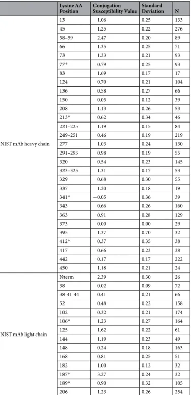
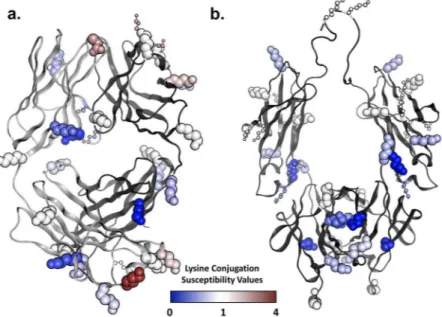
In general, it appears that most if not all sites respond in a linear manner to an increase in TMT label present during conjugation, suggesting that each lysine has a particular conjugation probability that does not depend on DAR. We have taken advantage of this observation to calculate a conjugation susceptibility factor that is inde-pendent of the DAR, by normalizing the observed site occupancy values to the expected average occupancy value if all available conjugation sites were conjugated equally. By doing this, a conjugation susceptibility factor of 1 represents a site that is conjugated with a site occupancy equal to the expected site occupancy value based on the DAR alone. he conjugation susceptibility factors for the NIST mAb is shown in Table 1 and mapped onto the crystal structures of the NIST Fab and Fc fragments in Fig. 5.
Efect of Fc-glycosylation on lysine susceptibility to conjugation with NHS esters.
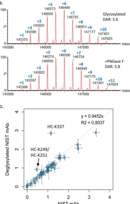
Next, we uti-lized our method to determine whether the presence of a glycan at the N-linked site in the Fc region of the NIST antibody afects the susceptibility of individual lysines to conjugation with NHS esters. To do this, we produced 15x conjugates of deglycosylated NIST mAb. Prior to conjugation, NIST mAb was deglycosylated by treatment with PNGaseF, an enzyme that removes the N-linked glycan normally present at position 300 in the Fc region of the heavy chain, replacing it with an aspartic acid and the site susceptibility values were compared to those found in the glycosylated NIST mAb. As shown in Fig. 6, the susceptibility of most lysines to conjugation was not afected by deglycosylation of the NIST antibody. However, signiicant increases in conjugation were noted at NIST mAb peptides containing K249/K251 and K337 in the heavy chain in the deglycosylated sample. he 3D crystal structure suggests that these glycosylation efects on the site occupancy at heavy chain positions K249 andFigure 4. Site occupancy data for the 8X, 15X, and 30X TMT126 conjugates. he average site occupancy ratio is plotted for each lysine and for the protein N-termini of the NIST mAb standard, under each of the three conjugation conditions tested. As demonstrated by the error bars, representing the 95% conidence interval, the data showed a high-level of consistency between experiments.
K337 labeling can be explained by interactions of carbohydrates with these lysines, which limit their accessibility in the glycosylated state (Supplementary Figure S3).
A model to predict lysine susceptibility based on 3D structure.
It is expected that conjugation sus-ceptibilities of various residues relate to the reactivity of their primary amine, which in turn should depend on solvent exposure and nucleophilicity. Both parameters are 3D-structure dependent and are afected by the local geometry and electrostatics around each lysine residue. Hence, a bi-parametric linear model of lysine conjugation susceptibility was derived by describing solvent exposure by the solvent-accessible surface area (SASAN) andnucleophilicity by the acidity constant (pKa) at aliphatic N atoms of the NIST mAb. Model calibration was done
on the set of 18 Lys residues from the Fab fragment with measurements based on peptides containing a single Lysine AA Position Conjugation Susceptibility Value Standard Deviation N
NIST mAb heavy chain
13 1.06 0.25 133 45 1.25 0.22 276 58–59 2.47 0.20 89 66 1.35 0.25 71 73 1.33 0.21 93 77* 0.79 0.25 93 83 1.69 0.17 17 124 0.70 0.21 104 136 0.58 0.27 66 150 0.05 0.12 39 208 1.13 0.26 53 213* 0.62 0.34 46 221–225 1.19 0.15 84 249–251 0.46 0.19 219 277 1.03 0.24 130 291–293 0.98 0.19 55 320 0.54 0.23 145 323–325 1.31 0.17 53 329 0.68 0.30 55 337 1.20 0.18 19 341* −0.05 0.36 39 343 0.66 0.26 160 363 0.91 0.28 129 373 0.00 0.00 29 395 1.37 0.70 32 412* 0.37 0.35 38 417 0.66 0.23 38 442 0.17 0.17 222 450 1.18 0.21 24
NIST mAb light chain
Nterm 2.39 0.30 26 38 0.02 0.09 72 38-41-44 0.41 0.21 66 52 0.48 0.22 158 102 0.32 0.21 174 106* 1.23 0.27 164 125 1.62 0.22 61 144 1.19 0.23 49 148 0.24 0.18 163 168 0.81 0.25 51 182 1.00 0.12 32 187* 3.27 0.24 32 189* 0.90 0.32 105 206 1.23 0.26 254
Table 1. Susceptibility of lysines in the NIST mAb to NHS-ester conjugation. Conjugation Susceptibility Values: 1 indicates expected level of conjugation based on DAR if all conjugation sites were equal. Asterisks mark calculated values.
TMT-labeled lysine and averaged parameters calculated from the 4 crystal copies of the NIST mAb. his led to the following linear regression equation:
= . · − . · + .
Susceptibility 0 0091 SASAN 0 6397 pKa 7 5824 (3) his itted linear model achieves a good correlation with R2 = 0.62 (Fig. 7a) and a mean-unsigned-error
(MUE) of 0.24 units on the DAR-independent susceptibility scale. he model indicates increased susceptibility to conjugation with increased surface accessibility (positive SASAN coeicient) and with decreased probability of
protonation (negative pKa coeicient) at the N atom, in line with the expected physics for the conjugation
reac-tion. As shown in Table 2, the pKa descriptor makes a larger contribution to this model (R2 = 0.56; MUE = 0.29)
than the SASAN descriptor (R2 = 0.19; MUE = 0.38).
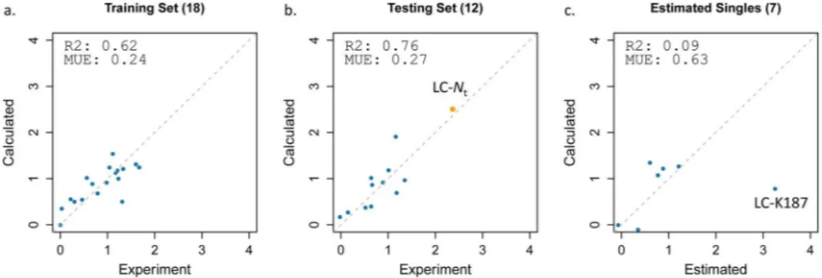
In order to further test its robustness, the bi-parametric model was employed for external predictions of con-jugation susceptibilities at the 11 Lys residues from the Fc fragment, for which data could be obtained from pep-tides with a single TMT-label. Also included in this external prediction was the conjugation susceptibility at the N-terminus of the LC. Parameters were calculated based on a crystal structure of the Fc homodimer and averaged between the two chains for each lysine site. As seen in Fig. 7b showing predicted versus measured susceptibilities, the linear model in Eq. (3) exhibits signiicant predictive power with a high level of correlation to experimental data (R2 = 0.76; MUE = 0.27; see also Table 2). Finally, the model was tested against the set of 7 lysine residues
with susceptibility estimated from measurements based on peptides with 2 and/or 3 TMT-labeled lysines (i.e., ‘doubles’ and ‘triples’). While signiicant statistics cannot be extracted due to the small size of this subset, it is vis-ually apparent from Fig. 7c that the model makes reasonable predictions at 6 lysine sites, with the one noteworthy outlier being LC-K187 for which the SASA/pKa model severely underestimates its susceptibility to conjugation.
he model in Eq. (3) was based on pKa values calculated with the H++ method20,21. Since computational pKa
predictions are considered to be relatively challenging, we have attempted to test other alternative methods. We employed DelPhiPKa22,23, which like H++ includes a relatively high-level treatment of solvation efects, both
using Poisson-Boltzmann electrostatics. DelPhiPka lead to models that compare favorably with those based on H++, albeit with a weaker performance (Table 2). We also tested the empirical method PropKa24,25, which
how-ever led a more marked decrease in performance.
Finally, we tested the impact that structural lexibility has on the quality of the susceptibility model in Eq. (3), which was derived based on only a few crystallographic conformations of the protein. We generated MD confor-mational ensembles starting from each crystal conformation and recalculated the SASA and pKa values as
aver-ages over conformational snapshots along the MD trajectories. As shown in Supplementary Figure S4 for the Fab domain, using MD ensembles seems to reduce the variability in model quality as compared with the correspond-ing individual crystal structures. However, over all MD trajectories the model was not improved relative to the model averaged over the few crystal structures (Supplementary Figure S5). All calculated molecular properties based on crystal structures and MD ensembles are provided in Supplementary Dataset S3.
Figure 5. Mapping of lysine and protein N-terminus susceptibilities on the 3D structure. (a) Fab domain of NIST antibody; (b) Fc domain of NIST antibody. Lysine side chains are shown as CPK models, except for lysines with data estimated from ‘doubles’ and ‘triples’, which are shown as ball-and-stick models. Each lysine side chain is then color coded according to the susceptibility data from Table 1 as per the color scale legend shown.
Discussion
In this study, we describe a novel method using TMTs to directly measure the site occupancy at individual sites on a monoclonal antibody following conjugation via an NHS-ester reactive group. he TMT molecule ofers several advantages in the described approach, most importantly, the very high level of similarity between the TMT126 and the TMT127 molecules, which difer only in the position of a 13 C isotope label. Our method relies on the use of one TMT (TMT126) to make the conjugate and subsequent labeling of the same sample with the other TMT (TMT127) under denaturing conditions such that TMT-127 is added to all remaining lysines, with an eiciency of approximately 95%. Using this approach, the ratio of the TMT126 signal to the TMT127 signal can be used directly to calculate site occupancy. his approach avoids comparing a modiied to an unmodiied peptide, elim-inating inaccuracies due to diferences in ionization properties or protease susceptibility due to the presence of the modiication.
Figure 6. he Fc glycan afects the susceptibility of 2 heavy chain lysines to conjugation by NHS esters. (a) Intact mass analysis shows that conjugation of NIST mAb with TMT126 ater removal of the Fc glycan with PNGaseF (‘+PNGaseF’) results in a similar DAR and distribution pattern to NIST mAb with the Fc glycan remaining (‘Glycosylated’). (b) Comparison of lysine susceptibility values between standard NIST mAb and deglycosylated NIST mAb identiies one single lysine (heavy chain K337) and one double lysine pair (HC- K249/HC-K251) that demonstrate a diference in conjugation susceptibility due to removal of the Fc-glycan with PNGaseF prior to conjugation, suggesting that the Fc glycan plays a role in the susceptibility of these lysines to conjugation via NHS-esters. he dotted line indicates perfect correlation.
TMTs have been previously used to estimate the site occupancy of NHS-conjugation in a monoclonal antibody by Gautier et al.7. However, the approach that they described is fundamentally diferent from the one described
in this work, as depicted in Supplementary Figure S6A. Gautier et al. used TMTs to label a monoclonal antibody under diferent conditions such that each TMT reporter ion represented an individual antibody conjugate sample with various numbers of TMT molecules attached. hus, in their approach, the TMT reporter ions were used to determine the relative level of TMT addition between diferent samples. Gautier et al. then relied on a compari-son between the MS signal intensity of each TMT-conjugated peptide to its equivalent unmodiied peptide, in a separate MS run, to estimate the actual site occupancy values. here are 35 lysines in common between the NIST mAb used in our study and the IgG1 used by Gautier et al. Comparing the 120 µM labeling condition in Gautier et al. with our 30X labeling condition (DAR = 13 vs 11, respectively), Gautier et al. report measurable occupancy values (above 0) for 12 of the 35 sites that are in common between our 2 antibodies. Here, we describe measurable site occupancy values for 26 of these individual sites, a 2-fold improvement in coverage. Furthermore, this work provides average occupancy data between 2 adjacent lysines for an additional 8 of these sites found in ‘double’ peptides. For many sites, the occupancy values that we have obtained show similar trends to those obtained by Gautier et al., with a few exceptions (Supplementary Figure S6B). However, the majority of the occupancy values obtained by Gautier et al. were considerably higher than the ones obtained here, possibly relecting a diference in the MS signal intensity of TMT conjugated peptides over unconjugated ones.
he NIST mAb has 35 unique lysines in the heavy chain and 14 unique lysines in the light chain plus one avail-able N-terminus on the light chain. We did not see conjugation at the heavy-chain N-terminus as the NIST mAb contains a pyroglutamic acid at this site. Since an antibody molecule contains two identical heavy chains and two identical light chains, a total of 100 primary amines are available for conjugation in the intact IgG molecule. In the study described here, we produced a series of conjugates with increasing DAR values, as measured by intact mass analysis. Although our results show that individual lysine residues show variability in their susceptibility to conjugation by NHS esters, the average site occupancy for molecules in this DAR series, as calculated based on our method, averaged 2.9%, 5.9%, and 10.5% for molecules with DAR of 3.0, 5.7, and 11.0 respectively – very
Figure 7. Performance of the bi-parametric linear model of amine susceptibilities to conjugation via NHS esters. Experimental DAR-independent susceptibility values are from Table 1. Only sites corresponding to peptides with a single lysine are used. Calculated molecular descriptors are averages over crystal structure conformations of the Fab and Fc fragments (see Methods). (a) Model training on 18 lysines from the Fab fragment. he model corresponds to Eq. (3). (b) Model testing on 11 lysines from the Fc fragment plus the N-terminus of the light chain (orange symbol). (c) Model testing on 7 lysines with susceptibility values estimated from data for ‘doubles’ and/or ‘triples’.
Model Descriptors R2 MUEb Training set Fab(N = 18) Testing set Fc(N = 12) + Nt Testing set Fab(N = 18) Training set Fc(N = 12) + Nt Nulla 0.00 0.00 0.44 0.45 SASAN 0.19 0.05 0.38 0.42 pKa(H++) 0.56 0.78 0.29 0.29 pKa(DelPhiPKa) 0.49 0.75 0.32 0.32 pKa(PropKa) 0.01 0.48 0.43 0.41 SASAN; pKa(H++) 0.62 0.76 0.24 0.27 SASAN; pKa(DelPhiPKa) 0.50 0.76 0.31 0.30 SASAN; pKa(PropKa) 0.40 0.78 0.32 0.26
Table 2. Performance of various structure-based linear models of conjugation susceptibility. aNull model is
based on average experimental value for the given set. bMean-unsigned-error (MUE) in DAR-independent
close to the predicted values of 3.0%, 5.7%, and 11.0% calculated from the DAR values. hese indings are reassur-ingly close to the expected values. Furthermore, by summing the site occupancy values for all measured sites, the results agree well with the DAR values identiied by intact mass: 2.8, 5.3, and 10.3. he site occupancy values are also quite consistent among replicates conjugated at diferent times, suggesting that the site occupancy at a given lysine is a feature of the protein and not simply the results of a stochastic process. his consistency enabled us to develop a lysine susceptibility value that is independent of the total level of conjugation and DAR.
In this analysis, several peptides contain multiple lysine residues, such that the ratio of TMT126-127 corre-sponds to an average of the site occupancy at each individual lysine. In some instances, it proved possible to cal-culate the individual site occupancies from data collected for diferent overlapping lysine groupings. However, in others, this was not possible as the lysines were not efectively separated by any of the 3 proteolytic enzymes used. It may be possible to separate these lysines using alternative enzymes, such as pepsin, at the expense of adding to the analysis time. Alternatively, it may be possible to use MS3 data to collect TMT ratios for individual peptides through fragmentation of MS2 ions that only contain a single lysine. While we were unable to achieve suicient sensitivity with our mass spectrometer for this analysis, newer instruments may facilitate this approach, improv-ing coverage for individual lysines. Buildimprov-ing an efective MS3-based worklow for this analysis may also lead to signiicant savings in analysis time. heoretically, if MS3 can directly identify individual site occupancy values for each of the 2 or 3 sites present in a ‘double’ or ‘triple’ peptide, a single protease digestion may be able to provide similar coverage levels to the 3 diferent digestion conditions that were used in this work.
he recording of lysine conjugation susceptibilities at high resolution and coverage provided by this study allowed the development for the irst time of a robust structure-based computational model of susceptibility. his simple 3D-structure based linear model highlights the nitrogen atom’s surface exposure (SASA) and nucleop-hilicity (described by pKa) as the main factors to lysine reactivity. Importantly, the present model quantitates
the key contribution of nucleophilicity to predicting the reactivity of surface-exposed lysines. To that end, the computational treatment of solvation efects for pKa calculations was found to be critical to the development of a
high-correlation model. It is also noteworthy that averaging descriptors over multiple conformations, either taken from crystal structures or simulated MD ensembles, was also important to obtaining correlative models. he contributions of nucleophilicity and structural lexibility to conjugation were not evaluated quantitatively in the previous TMT-based study7, which also aforded a more limited coverage unsuitable for statistical model training
and testing as it was carried out in the present study. An intriguing question is whether there are other meaningful structure-based molecular descriptors that can be considered in order to further account for the variance in the conjugation susceptibility data. he case in point is K187 in the light chain of NIST mAb (constant region of the human kappa chain) which is underestimated by the present SASA/pKa model.
he robustness of the calibrated SASA/pKa model in external testing suggests that it can also be employed
in the predictive mode. An immediate useful application will be evaluating the risk that lysines present in the antigen-binding regions (CDR) of antibodies will afect biological activity by estimating their likely susceptibility levels. he model can also be generally applied for predictions of amine susceptibility to conjugation via NHS esters for any protein as long as 3D-structural data are available or a homology model can be built. In this regard, we found that the worst model based on an MD ensemble was superior to the worst model based on a single crys-tal structure (Supplementary Figure S4). his provides a strategy to performing predictions with the proposed SASA/pKa model on antibodies and proteins for which multiple crystal structure will not be available.
he same TMT based approach could also be used to analyze conjugation reactions beyond NHS ester chem-istry. Commercial TMT tags are sold for cysteine conjugation via alkylation with an iodo group. With some efort to optimize the complete labeling step, additional conjugation chemistries could be explored through this method by synthesizing additional molecules with TMT-like properties. It is likely that the structure-based computational model will need to be retrained and tested for the speciic conjugation reagent and reactive residues of the protein. In conclusion, the TMT method described here, combined with molecular modeling, will enable improved engi-neering of antibodies for optimal labeling with luorophores, toxins, or crosslinkers and will provide the basis for the development of proteins with improved conjugation properties.
References
1. deGruyter, J. N., Malins, L. R. & Baran, P. S. Residue-Speciic Peptide Modiication: A Chemist’s Guide. Biochemistry 56, 3863–3873, https://doi.org/10.1021/acs.biochem.7b00536 (2017).
2. Beck, A., Goetsch, L., Dumontet, C. & Corvaia, N. Strategies and challenges for the next generation of antibody-drug conjugates. Nat. Rev. Drug Discov. 16, 315–337, https://doi.org/10.1038/nrd.2016.268 (2017).
3. Gao, W. et al. Recent Advances in Site Speciic Conjugations of Antibody Drug Conjugates (ADCs). Curr. Cancer Drug Targets 16, 469–479 (2016).
4. Polakis, P. Antibody Drug Conjugates for Cancer herapy. Pharmacol. Rev. 68, 3–19, https://doi.org/10.1124/pr.114.009373 (2016). 5. Wang, L., Amphlett, G., Blattler, W. A., Lambert, J. M. & Zhang, W. Structural characterization of the maytansinoid-monoclonal antibody immunoconjugate, huN901-DM1, by mass spectrometry. Protein Sci. 14, 2436–2446, https://doi.org/10.1110/ps.051478705 (2005).
6. Chen, L. et al. In-depth structural characterization of Kadcyla(R) (ado-trastuzumab emtansine) and its biosimilar candidate. MAbs
8, 1210–1223, https://doi.org/10.1080/19420862.2016.1204502 (2016).
7. Gautier, V., Boumeester, A. J., Lossl, P. & Heck, A. J. Lysine conjugation properties in human IgGs studied by integrating high-resolution native mass spectrometry and bottom-up proteomics. Proteomics 15, 2756–2765, https://doi.org/10.1002/pmic.201400462 (2015).
8. Sang, H. et al. Conjugation site analysis of antibody-drug-conjugates (ADCs) by signature ion ingerprinting and normalized area quantitation approach using nano-liquid chromatography coupled to high resolution mass spectrometry. Anal. Chim. Acta 955, 67–78, https://doi.org/10.1016/j.aca.2016.11.073 (2017).
9. Guo, X. et al. Partial acetylation of lysine residues improves intraprotein cross-linking. Anal Chem 80, 951–960, https://doi. org/10.1021/ac701636w (2008).
10. Kleifeld, O. et al. Identifying and quantifying proteolytic events and the natural N terminome by terminal amine isotopic labeling of substrates. Nat Protoc 6, 1578–1611, https://doi.org/10.1038/nprot.2011.382 (2011).
11. Chambers, M. C. et al. A cross-platform toolkit for mass spectrometry and proteomics. Nat. Biotechnol. 30, 918–920, https://doi. org/10.1038/nbt.2377 (2012).
12. Hsieh, E. J., Hoopmann, M. R., MacLean, B. & MacCoss, M. J. Comparison of database search strategies for high precursor mass accuracy MS/MS data. Journal of proteome research 9, 1138–1143, https://doi.org/10.1021/pr900816a (2010).
13. Cornell, W. D. et al. A Second Generation Force Field for the Simulation of Proteins, Nucleic Acids, and Organic Molecules. J. Am. Chem. Soc. 117, 5179–5197 (1995).
14. Hornak, V. et al. Comparison of multiple Amber force ields and development of improved protein backbone parameters. Proteins
65, 712–725 (2006).
15. Maier, J. A. et al. f14SB: Improving the Accuracy of Protein Side Chain and Backbone Parameters from f99SB. J Chem heory Comput 11, 3696–3713, https://doi.org/10.1021/acs.jctc.5b00255 (2015).
16. Kirschner, K. N. et al. GLYCAM06: a generalizable biomolecular force ield. Carbohydrates. J Comput Chem 29, 622–655, https://doi. org/10.1002/jcc.20820 (2008).
17. Jorgensen, W. L., Chandrasekhar, J., Madura, J. D., Impey, R. W. & Klein, M. L. Comparison of simple potential functions for simulating liquid water. he Journal of Chemical Physics 79, 926–935 (1983).
18. Ryckaert, J. P., Ciccotti, G. & Berendsen, H. J. C. Numerical integration of the cartesian equations of motion of a system with constraints: molecular dynamics of n-alkanes. Journal of Computational Physics 23, 327–341 (1977).
19. Darden, T., York, D. & Pedersen, L. Particle mesh Ewald: An N log(N) method for Ewald sums in large systems. he Journal of Chemical Physics 98, 10089–10092 (1993).
20. Gordon, J. C. et al. H++: a server for estimating pKas and adding missing hydrogens to macromolecules. Nucleic Acids Res. 33, W368–W371 (2005).
21. Anandakrishnan, R., Aguilar, B. & Onufriev, A. V. H++ 3.0: automating pK prediction and the preparation of biomolecular structures for atomistic molecular modeling and simulations. Nucleic Acids Res. 40, W537–541, https://doi.org/10.1093/nar/gks375 (2012).
22. Wang, L., Li, L. & Alexov, E. pKa predictions for proteins, RNAs, and DNAs with the Gaussian dielectric function using DelPhi pKa. Proteins 83, 2186–2197, https://doi.org/10.1002/prot.24935 (2015).
23. Wang, L., Zhang, M. & Alexov, E. DelPhiPKa web server: predicting pKa of proteins, RNAs and DNAs. Bioinformatics 32, 614–615, https://doi.org/10.1093/bioinformatics/btv607 (2016).
24. Sondergaard, C. R., Olsson, M. H., Rostkowski, M. & Jensen, J. H. Improved Treatment of Ligands and Coupling Efects in Empirical Calculation and Rationalization of pKa Values. J Chem heory Comput 7, 2284–2295, https://doi.org/10.1021/ct200133y (2011). 25. Olsson, M. H., Sondergaard, C. R., Rostkowski, M. & Jensen, J. H. PROPKA3: Consistent Treatment of Internal and Surface Residues
in Empirical pKa Predictions. J Chem heory Comput 7, 525–537, https://doi.org/10.1021/ct100578z (2011).
26. Chan, S. L. & Purisima, E. O. Molecular surface generation using marching tetrahedra. Journal of Computational Chemistry, 1268–1277 (1998).
27. Chan, S. L. & Purisima, E. O. A new tetrahedral tesselation scheme for isosurface generation. Computers and Graphics, 83–90 (1998). 28. Naim, M. et al. Solvated Interaction Energy (SIE) for Scoring Protein-Ligand Binding Ainities. 1. Exploring the Parameter Space.
J Chem Inf Model 47, 122–133 (2007).
29. R Development Core Team. R: A Language and Environment for Statistical Computing. (he R Foundation for Statistical Computing, 2011).
Acknowledgements
We would like to acknowledge Luc Tessier, Sam Williamson, and other members of the Analytics Team for expert help with the mass spectrometry infrastructure. We would also like to acknowledge Hervé Hogues and Christophe Deprez for assistance with the molecular simulation sotware. he work described here was funded by the Human Health herapeutics portfolio of the National Research Council Canada.
Author Contributions
J.J.H. conceived and developed the described MS method, analyzed the MS data, and wrote the manuscript; T.-L.T. designed and executed the wet lab and MS work; C.R.C. conceived the structure-based methodology, performed molecular simulations, and analyzed the computational data; E.O.P. conceived the structure-based methodology and oversaw the computational studies; and T.S. conceived the structure-based methodology, performed molecular simulations, analyzed the computational data, and wrote the manuscript. All authors edited the manuscript and approved of the inal version.
Additional Information
Supplementary information accompanies this paper at https://doi.org/10.1038/s41598-018-35924-0.
Competing Interests: he authors declare no competing interests.
Publisher’s note: Springer Nature remains neutral with regard to jurisdictional claims in published maps and institutional ailiations.
Open Access This article is licensed under a Creative Commons Attribution 4.0 International License, which permits use, sharing, adaptation, distribution and reproduction in any medium or format, as long as you give appropriate credit to the original author(s) and the source, provide a link to the Cre-ative Commons license, and indicate if changes were made. he images or other third party material in this article are included in the article’s Creative Commons license, unless indicated otherwise in a credit line to the material. If material is not included in the article’s Creative Commons license and your intended use is not per-mitted by statutory regulation or exceeds the perper-mitted use, you will need to obtain permission directly from the copyright holder. To view a copy of this license, visit http://creativecommons.org/licenses/by/4.0/.