HAL Id: hal-02891577
https://hal.archives-ouvertes.fr/hal-02891577
Submitted on 19 Nov 2020
HAL is a multi-disciplinary open access
archive for the deposit and dissemination of
sci-entific research documents, whether they are
pub-lished or not. The documents may come from
teaching and research institutions in France or
abroad, or from public or private research centers.
L’archive ouverte pluridisciplinaire HAL, est
destinée au dépôt et à la diffusion de documents
scientifiques de niveau recherche, publiés ou non,
émanant des établissements d’enseignement et de
recherche français ou étrangers, des laboratoires
publics ou privés.
Physcomitrella patens
Ursina Rathgeb, Min Chen, Flavien Buron, Nadja Feddermann, Martine
Schorderet, Axelle Raisin, Gabrielle-Yasymi Häberli, Sophie Marc-Martin,
Jean Keller, Pierre-Marc Delaux, et al.
To cite this version:
Ursina Rathgeb, Min Chen, Flavien Buron, Nadja Feddermann, Martine Schorderet, et al..
VAPYRIN-like is required for development of the moss Physcomitrella patens. Development (Cambridge,
Eng-land), Company of Biologists, 2020, 147 (11), pp.dev184762. �10.1242/dev.184762�. �hal-02891577�
RESEARCH ARTICLE
VAPYRIN-like is required for development of the moss
Physcomitrella patens
Ursina Rathgeb1,*, Min Chen1,*, Flavien Buron1, Nadja Feddermann1, Martine Schorderet1, Axelle Raisin1,
Gabrielle-Yasymi Hä berli1, Sophie Marc-Martin2, Jean Keller3, Pierre-Marc Delaux3, Didier G. Schaefer2and
Didier Reinhardt1,‡
ABSTRACT
The VAPYRIN (VPY) gene in Medicago truncatula and Petunia hybrida is required for arbuscular mycorrhizal (AM) symbiosis. The moss Physcomitrella patens has a close homolog (VPY-like, VPYL), although it does not form AM. Here, we explore the phylogeny of VPY and VPYL in land plants, and study the expression and developmental function of VPYL in P. patens. We show that VPYL is expressed primarily in the protonema, the early filamentous stage of moss development, and later in rhizoids arising from the leafy gametophores and in adult phyllids. Knockout mutants have specific phenotypes in branching of the protonema and in cell division of the leaves (phyllids) in gametophores. The mutants are responsive to auxin and strigolactone, which are involved in regulation of protonemal branching, indicating that hormonal signaling in the mutants is not affected in hormonal signaling. Taken together, these results suggest that VPYL exerts negative regulation of protonemal branching and cell division in phyllids. We discuss VPY and VPYL phylogeny and function in land plants in the context of AM symbiosis in angiosperms and development in the moss.
KEY WORDS:Physcomitrella patens, VAPYRIN, Vesicle trafficking, Secretion, Symbiosis
INTRODUCTION
Most plants live in symbiotic associations with fungi or bacteria, which improve their nutrient supply and stress resistance (Oldroyd et al., 2011; Smith and Read, 2008). Symbiotic communication and establishment of the functional symbiosis requires a suite of genes, known as the common symbiosis signaling (CSS) genes, including SYMBIOSIS RECEPTOR KINASE (SYMRK) and CALCIUM AND CALMODULIN-DEPENDENT PROTEIN KINASE (CCaMK). SYMRK and CCaMK are involved in a pathway that mediates perception of symbiotic signals from the cell periphery to the nucleus, where the transcription of hundreds of downstream symbiosis-related genes is induced (Gutjahr and Parniske, 2013). Although most downstream genes have not been functionally characterized, they are thought to be required for intracellular
accommodation of the microbial partner and for installation and functioning of the symbiotic interface. This involves containment of the symbiont within a membrane that controls nutrient and signal exchange, and synthesis of the transport proteins that mediate nutrient transfer (Rich et al., 2017; Roth and Paszkowski, 2017; Wang et al., 2017). The transition of nonsymbiotic root cortex cells to symbiotic cells involves major reprogramming of multiple cellular pathways. Central aspects of this transition are repositioning of the nucleus, establishment of an infection structure known as the pre-penetration apparatus (PPA) (Genre et al., 2008) and reorientation of vesicular trafficking towards the symbiotic interface (Pumplin et al., 2012). Although the cellular reprogramming is of central importance for symbiosis, its regulation is poorly understood.
A central element required for intracellular accommodation of arbuscular mycorrhizal (AM) fungi has been identified in forward genetic screens in Medicago truncatula and Petunia hybrida. It was named VAPYRIN (VPY), because of its two domains: a vesicle-associated membrane protein (VAMP)-vesicle-associated protein (VAP) domain and an ANKYRIN domain. Both domains are known as protein:protein interaction domains. While the VAP domain is known primarily from vesicle trafficking (Lev et al., 2008), the ANKYRIN domain is special because it occurs in a large range of sizes, from 1 to 24 ankyrin repeats, and it is one of the most common protein domains in eukaryotes (Michaely et al., 2001). In plants, both domains are very common; however, only in VPY are the two domains combined and only VPY has a large ankyrin domain of 11 predicted repeats (Feddermann and Reinhardt, 2011). Such large ankyrin domains are common in animals, in particular in the ankyrin protein, in which the ankyrin repeat was first described. Ankyrins in animals serve as connectors of resident membrane proteins to the spectrin-actin cytoskeleton (Mohler et al., 2002). In plants, the 11-repeat ankyrin domain of VPY is unique (Feddermann et al., 2010; Feddermann and Reinhardt, 2011).
Most land plants have a closely related VPY-like gene (VPYL) indicative of a duplication. The predicted VPY and VPYL proteins are very similar. They have no transmembrane domain and no peptide signature indicative of a post-translational modification with a membrane anchor; therefore, they might be expected to be localized to the cytoplasm. However, in M. truncatula and P. hybrida, GFP-tagged VPY and VPYL are localized to small mobile subcellular compartments of unknown function (Feddermann et al., 2010; Liu et al., 2019; Pumplin et al., 2010), known as VPY bodies, which have an identity that includes trans-Golgi network (TGN) and endosomal features (Bapaume et al., 2019). Based on the interaction of VAPYRIN with EXO70I (Zhang et al., 2015) and EXO70H4 (Liu et al., 2019), two components of the exocyst complex, VPY bodies are likely to be involved in the transport of an important cargo in symbiotic cells.
Initial phylogenetic inference using transcriptomic data indicated that VPY and VPYL are absent from algae, but can be found in most
Handling Editor: Ykä Helariutta
Received 20 September 2019; Accepted 19 April 2020
1Department of Biology, University of Fribourg, 1700-Fribourg, Switzerland.2Institut
de Biologie, Université de Neuchâtel, 2000-Neuchâtel, Switzerland.3Laboratoire
de Recherche en Sciences Végétales, Université de Toulouse, CNRS, Auzeville, 31326 Castanet Tolosan, France.
*These authors contributed equally to this work
‡Author for correspondence ([email protected])
U.R., 0000-0002-2762-0010; P.-M.D., 0000-0002-6211-157X; D.R., 0000-0003-3495-6783
DEVEL
O
land plants that engage in symbiosis (Delaux et al., 2015; Feddermann et al., 2010; Pumplin et al., 2010). Angiosperm clades that do not form symbiosis, such as the Brassicaceae and the Amaranthaceae, have lost VPY and VPYL (Bravo et al., 2016; Delaux et al., 2014; Favre et al., 2014). This indicates that, in angiosperms, the only selection pressure to maintain VPY and VPYL is symbiosis, indicative of its specific function in symbiosis.
Although the occurrence of VPY and VPYL in the genomes of angiosperms correlates with the ability to engage in AM (Delaux et al., 2014), this relationship seems less obvious in non-angiosperms. For example, a putative VPYL orthologue has been identified in the moss Physcomitrella patens, despite the fact that mosses in general, and P. patens in particular, are not able to engage in mycorrhizal associations under natural conditions (Wang and Qiu, 2006), although in vitro conditions allow for some level of interaction with AM fungi (Hanke and Rensing, 2010). The only moss genus that has been reported to be mycorrhizal, Takakia (Wang and Qiu, 2006), is considered a sister clade of all other mosses (Cox, 2018; Volkmar and Knoop, 2010), indicating that the mosses lost mycorrhizal capacity early during their evolution. The maintenance of VPYL in the absence of symbiosis suggests that VPYL may have nonsymbiotic functions in mosses. Because VPY function is restricted to symbiosis, based on the symbiosis-specific
vpy mutant phenotypes in petunia and M. truncatula, the question arises whether VPYL in angiosperms has retained such developmental functions, after the duplication and involvement of VPY in symbiosis.
To elucidate the origin and function of VPY, we established its phylogeny in land plants and investigated VPYL function in the nonsymbiotic model plant P. patens, which offers excellent molecular genetic tools for exploring gene function, including gene targeting by homologous recombination (Schaefer, 2002). It is therefore an excellent tool for reverse genetic analyses and has become a standard model system for developmental analysis (Prigge and Bezanilla, 2010). P. patens spores germinate to form a juvenile protonema that displays a filamentous growth pattern. The leafy gametophores, nonvascular shoots carrying simple leaves ( phyllids) formed by a single cell layer, then differentiate by caulinary growth (Kofuji and Hasebe, 2014). These features mean that P. patens is an excellent experimental system for the study of cellular aspects of VPYL function in a nonsymbiotic context.
RESULTS
Evolution and phylogeny of the VAPYRIN clade in plants
To explore the origin and phylogeny of VPY and VPYL, we first performed a sequence analysis of the predicted VPY homologs in
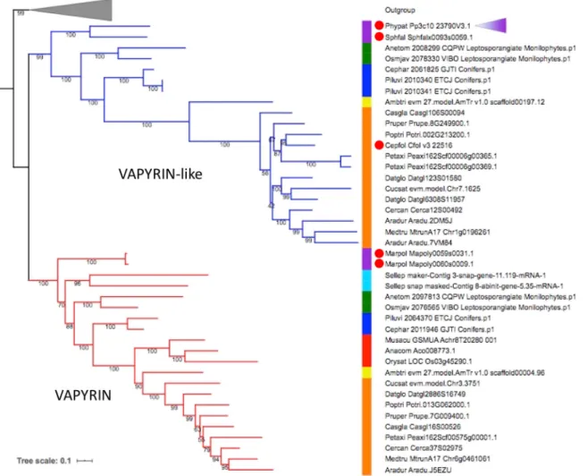
Fig. 1. Phylogenetic tree of the VAPYRIN gene family in selected land plants. Maximum likelihood tree (model JTT+R8) of VAPYRIN (red) and VAPYRIN-like (blue). The tree was rooted using closely related sequences (outgroup, gray triangle). Node supports are indicated by numbers on branches. The colored bar indicates the following clades: purple, bryophytes; cyan, lycophytes; green, monilophytes; blue, gymnosperms; yellow, basal angiosperms; red, monocots; orange, eudicots. The position of the P. patens VAPYRIN-like (VPYL) sequence is indicated by a purple triangle. Red dots indicate non-mycorrhizal species. See Table S1 and protein sequences in the supplementary Materials and Methods for further information.
DEVEL
O
several major land plant taxa, including dicotyledonous and monocotyledonous angiosperms, gymnosperms, ferns, lycophytes and nonvascular representatives of the mosses and liverworts (bryophytes) (Table S1; see protein sequences in the supplementary Materials and Methods). We also included five recently published genomes of streptophyte algae, the closest algal relatives to land plants. The best hits identified in streptophyte algae all lacked either the ankyrin or the VAP/MSP domain, indicating that the VPY/ VPYL clade evolved in land plants, as previously suggested on the basis of transcriptomic data (Delaux et al., 2015). Most land plant taxa have at least one VPY homolog, and many have a similar VPYL gene (Fig. 1) that encodes a protein with a similar predicted domain structure. Based on the overall structure of the phylogenetic tree, VPY is likely to have emerged in an early common ancestor of land plants, followed by a duplication event resulting in two paralogous clades (VPY and VPYL; Fig. 1; Fig. S1). Both clades show strong purifying selection (ωVPY=0.2250;ωVPYL=0.1178), as estimated by
comparison of non-synonymous and synonymous nucleotide substitutions (dN/dS ratio, as assessed using the yn00 method), suggesting a conserved biochemical function. Comparing the dN/ dS ratio of P. patens VPYL with VPYL from all other species revealed the same trends (mean, 0.3167733; standard error, 0.02458039). Most taxa retained both paralogues (e.g. dicots, gymnosperms, ferns), but others maintained only VPY (monocots,
lycophytes, liverworts) or VPYL (mosses). On the other hand, many dicots displayed additional gene copies of one or the other paralogue, indicative of additional rounds of duplication, as for instance in the Papilionoideae, the largest subfamily of the Fabaceae, members of which have each at least two VPY copies, with the exception of M. truncatula that lost one of these recent paralogues (Fig. S2). This duplication probably resulted from an ancestral whole genome duplication that occurred before the radiation of this subfamily (Koenen et al., 2019 preprint).
As observed previously (Delaux et al., 2014; Feddermann et al., 2010; Pumplin et al., 2010), nonsymbiotic angiosperms such as the Brassicaeae (with Arabidopsis thaliana) and the Amaranthaceae, as well as the monocot Zostera marina lost both VPY and VPYL, presumably as a consequence of the relaxed selection pressure following the loss of symbiosis in these taxa. This phylogenomic pattern, and the fact that vpy mutants in P. hybrida and M. truncatula have no developmental phenotypes (Feddermann et al., 2010; Murray et al., 2011; Pumplin et al., 2010), suggests that VPY (and perhaps VPYL) in seed plants is dispensable for plant development and that its function is specific to symbiosis.
Interestingly, the correlation between the presence of VPY/VPYL and the competence to engage in AM does not hold true in nonvascular plant lineages. The liverwort Marchantia polymorpha has lost VPYL, but has retained two copies of VPY (Fig. 1).
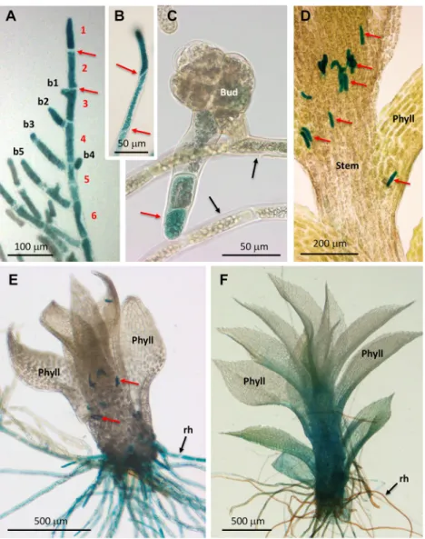
Fig. 2. GUS expression pattern under control of the P. patens VPYL promoter. (A-F) Samples of the pVPYL:: GUS line stained forβ-glucuronidase activity.
(A) Chloronema with perpendicular cell walls (red arrows). Cells of the main branch are numbered in red (1-6), lateral branches are numbered in black (b1-b5). Older protonemal cells exhibit decreased VPYL expression relative to the tip. (B) Caulonema with oblique cell walls (red arrows). (C) Protonema and bud with a young rhizoid (red arrow). Note decreased VPYL expression in protonema (black arrows). (D) Gametophore with stem-borne rhizoid initials (red arrows). (E) Young gametophore with five phyllids, developing basal rhizoids and stem-borne rhizoid initials (red arrows). (F) Mature gametophore with >12 phyllids and mature brownish rhizoids. Note increasing expression levels in phyllids. Phyll, phyllids; rh, rhizoids.
DEVEL
O
Conversely, the moss P. patens has lost VPY, but has retained VPYL (Phypat Pp3c10 23790V3.1). Interestingly, both these nonvascular plants are nonsymbiotic. This indicates that these VPY homologs in nonvascular species have a nonsymbiotic function, which may be related to the ancestral function of the first VPY in early embryophytes.
Expression pattern of theVPYL gene in P. patens
P. patens allows straightforward gene replacement by homologous recombination (Schaefer, 2002); hence, we decided to assess the function of P. patens VPYL using this reverse genetic tool. As a first step, we assessed its expression pattern across the different cell types and developmental stages. To assess gene expression with cellular resolution, we chose a promoter-reporter strategy by inserting the bacterial β-glucuronidase (GUS) gene UidA downstream of the VPYL promoter, thereby leaving about 800 nt of duplicated promoter (5′-TS) sequence of the original VPYL promoter (downstream of the GUS insertion cassette) to control the VPYL ORF, resulting in the reporter line pVPYL::GUS (Fig. S3). This line exhibited no developmental phenotype (compared with the knockout phenotype described below), indicative of normal VPYL function, thereby showing that the 800 bp 5′-UTR can be regarded as a fully functional VPYL promoter.
At early stages of development, P. patens produces filamentous protonemata that grow by tip growth in combination with lateral branching from the apical end of subapical cells (Menand et al., 2007) (Fig. 2A). The protonema consists of two distinct cell types, the photosynthetic chloronemata and the adventitious caulonemata, which mediate rapid radial expansion of the protonema (Menand et al., 2007). VPYL expression was consistently high, both in chloronema and caulonema, in particular in the youngest cells at the tip and in young branches (Fig. 2A,B). At later stages of development, VPYL expression in the protonema decreased (Fig. 2A), but resumed in the lateral buds that give rise to the gametophores. Remarkably, the main body of the buds showed low expression, whereas VPYL was strongly induced in cells at the
bottom that develop into rhizoids (Fig. 2C). In developing gametophores, VPYL expression was highest in aerial rhizoid initials (Sakakibara et al., 2003) (Fig. 2D) and in growing basal rhizoids (Fig. 2E), whereas it decreased in fully grown rhizoids (Fig. 2F). The stem and the leaf-like structures ( phyllids) exhibited low expression at early stages (Fig. 2E); however, at later stages of gametophore development, VPYL expression increased from the base of the stem into the phyllids (Fig. 2F). Taken together, these results show that P. patens VPYL expression is highest in cell types with a filamentous growth mode ( protonema and rhizoids), with lower expression levels in gametophores, in particular in mature phyllids. This global expression pattern is consistent with the pattern revealed in a large scale transcriptomic analysis accessible at the Physcomitrella eFP Browser (http://bar.utoronto.ca/efp_ physcomitrella/cgi-bin/efpWeb.cgi) (Ortiz-Ramírez et al., 2016) (Fig. S4).
Phenotypic analysis ofP. patens vpyl knockout mutants
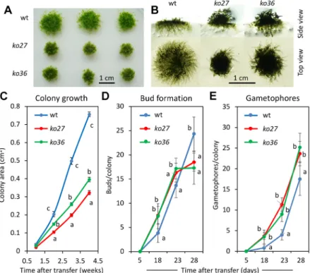
To explore the function of VPYL in P. patens, the VPYL ORF was deleted by homologous recombination (HR) to generate loss-of-function vpyl mutants (Figs S3 and S5). For that purpose, the entire VPYL ORF was replaced by a selectable resistance cassette for the antibiotic G418 (Fig. S3). Two independent mutant lines (ko27 and ko36) were identified by PCR genotyping based on the presence of the expected recombined junctions generated by HR, and on the absence of the VPYL coding region (Fig. S5B) and the VPYL transcript (Fig. S5C). These two knockout lines were used for detailed phenotypic characterization. First, we assessed overall growth of the plants under standard growth conditions on solid BCD agar medium. Overall, mutant plants grew more slowly in diameter and initiated gametophores earlier than the wild type (Fig. 3A,B), whereas development of the protonema was strongly reduced (Fig. 3B). Quantification of colony growth over a time course confirmed the slower expansion growth and the earlier initiation of gametophores in vpyl mutants (Fig. 3C-E). Whereas overall colony surface increased more slowly
Fig. 3. Global phenotype ofP. patens vpyl knockout mutants ko27 and ko36. (A) Overall colony growth of wild type (wt) and the two vpyl mutants (ko27, ko36) represented by three replicate plants each after 4 weeks of culture on agar plates with BCD medium. (B) Side view and top view of representative colonies as in A at higher magnification. (C) Plant area growth over a time course of 4 weeks. (D) Bud formation over a time course of 4 weeks. (E) Emergence of gametophores over a time course of 4 weeks. Values represent the mean±s.d. of 15 (C) or 6 (D,E) biological replicates. Different letters indicate significant differences (two-way ANOVA, P<0.05). See P-values in the supplementary Materials and Methods.
DEVEL
O
in the mutants (Fig. 3C), they produced more buds and gametophores (Fig. 3D,E). At the 4 week time point, however, bud formation in the wild type exceeded that in the mutants, presumably as a result of the larger total area covered by the protonema (Fig. 3B) and a concomitant increase in the number of protonemal cells competent for bud formation.
Slower colony expansion in mutants could potentially be a result of retarded transition of protonemata from the slow-growing chloronema cell type to the fast-growing caulonema cell type. Hence, we carried out growth measurements under conditions that favor caulonema development. Initially, the three genotypes were grown on BCD plates with 0.5% glucose for 18 days under low light conditions, then they were transferred to very low unilateral light. Under these conditions, protonemata acquire caulonema identity and grow phototropically towards the dim light source. The directional growth pattern and the synchronized transition from chloronema to caulonema identity allows quantification of caulonemal growth dynamics. At 6 days after initiation of the dark treatment, all three genotypes had initiated caulonemata, which had grown to approximately 2 mm in length (Fig. S6). The mutants had grown slightly longer (difference significant only for ko27). These results show that the vpyl mutants are not affected in the initiation of caulonemata, nor in their elongation rate.
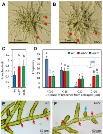
Closer inspection revealed that the protonemata in mutants produced more branches than in the wild type (Fig. 4A,B). Quantification confirmed that the mutants exhibited a significantly increased average branch number per cell (Fig. 4C), whereas cell length was not affected in the mutants (Fig. S7A,B). Interestingly, branching sometimes occurred in abnormal positions in the mutants (Fig. 4D,F). In the wild type, branches were initiated almost exclusively at the apical end of subapical cells (Fig. 4D,E, arrows), whereas in the mutants lateral branches were often formed in a more proximal position, including the middle of the cells (Fig. 4D,F, blue arrow).
Apart from the site of branch initiation, the length and size distribution of branches in P. patens vpyl mutants were also affected (Fig. 5). Normally, lateral branches grow progressively longer after their initiation, resulting in a‘pine-tree’ organization of P. patens protonemata (Fig. 5A). This results in a clear correlation of branch length with distance from the tip (coefficient of determination R2=0.78; Fig. 5B). In the vpyl mutants ko27 and
ko36, this relationship was much less obvious (R2=0.046 and 0.103,
respectively; Fig. 5C,D). This was mainly caused by a generally reduced extension rate of protonemal branches (Fig. 5C-E), but also to a lack of coordination between the position and length of the branches (Fig. 5C,D). Overall branch extension was significantly lower in mutants than in the wild type (Fig. 5E). Log10 transformation of the data set revealed that branch length was log-normally distributed (Fig. S8); hence, statistical analysis was performed on log10-transformed data (Fig. 5E; Fig. S8). Taken together, these data show that the mutants branch more frequently and at illegitimate positions, relative to the wild type, and that the branches develop more slowly after initiation. This can explain the global growth phenotype of the mutants (Fig. 3A,B).
Gametophore phenotype ofP. patens vpyl mutants
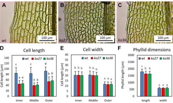
Although gametophores were formed earlier in the mutants, they were macroscopically indistinguishable in their overall morphology. However, closer examination at higher magnification revealed that the cells of the fully mature leaves ( phyllids) were smaller in the mutants relative to the wild type (Fig. 6A-C). On average, mutant cells had approximately half the length of wild-type cells (Fig. 6D).
This was true for cells close to the midrib (inner), cells in the middle of the lamina (middle), and cells at the leaf edge (outer) (Fig. 6D). Interestingly, cell width was indistinguishable between mutants and wild type (Fig. 6E). Quantification of overall phyllid dimensions showed that vpyl mutants and the wild type had very similar phyllid sizes (Fig. 6F). Although mutants exhibited a small but significant reduction in phyllid length, the small difference of 8.7% does not compare with the approximately 50% reduction in cell length. Hence, P. patens vpyl mutant phyllids have more, but shorter, cells than the wild type, suggesting that the mutants undergo additional rounds of cell division compared with the wild type.
Hormones rescue the protonemal phenotype ofP. patens vpyl mutants
Branching of protonema in P. patens is known to be under hormonal control (Knight et al., 2009). Strigolactone inhibits branching (Proust et al., 2011), whereas auxin causes a combination of changed identity (from chloronema to caulonema) and reduced branching (Thelander et al., 2018). To test whether the mechanism Fig. 4. Branching phenotype ofP. patens vpyl knockout mutants ko27 andko36. (A) Protonemal appearance of a young wild-type plant. Arrows indicate lateral branches that grow into the air. (B) As in A with a plant of ko27. (C) Average branching index for the first five cells of protonemal files in wild type, ko27 and ko36 (n=100 per genotype). (D) Distance of branching site from the apical end of cells in protonemal files in wild type, ko27 and ko36 (compare with E,F). For each branch, its distance from the upper end of the protonemal cell (see schematic inset) was measured. Only the first five cells in a protonemal file were considered (n=125 per genotype). (E) Branching pattern in the wild type. Red arrows indicate the apical end of the cells. (F) Branching pattern in ko27. Red arrows indicate the apical end of the cells. Blue arrow indicates an illegitimate branch site in the middle of a cell. Values represent the mean+s.d. Different letters indicate significant differences (two-way ANOVA, P<0.05). See P-values in the supplementary Materials and Methods.
DEVEL
O
involving VPYL interacts with hormonal regulation, we exposed mutants and wild-type protonemata to the synthetic strigolactone GR24 and to the auxin 1-naphthylacetic acid (NAA) at concentrations of 1-10 µM. These concentrations did not significantly alter overall colony growth of P. patens, indicating that such concentrations are within the physiological range (Fig. S9A). A partial loss of chlorophyll in response to NAA (Fig. S9A) is
consistent with the onset of the transition from chloronema to caulonema identity in response to auxin treatment. Quantification of protonemal branching revealed a strong inhibitory effect of both hormones on branch numbers (Fig. 7). The strigolactone GR24, as well as the auxins NAA and indole-3-acetic acid (IAA) all significantly inhibited protonemal branching, both in the wild type and in the two vpyl knockout lines (Fig. 7). These results
Fig. 5. Branch length distribution in wild type and knockout mutantsko27 and ko36. (A) Branch length distribution is defined as the length of branches in dependence of their distance from the tip of the protonemal file. (B) Branch length distribution in the wild type (wt). (C) Branch length distribution in ko27. (D) Branch length distribution in ko36. (E) Branch length in wild type, ko27 and ko36. Values represent the mean+s.d. (n=105). Branch length distribution was log10-normally distributed (see Fig. S8). Therefore, values were log10 transformed followed by two-way ANOVA and Tukey post hoc tests.
Fig. 6. Phyllid phenotype ofP. patens vpyl knockout mutants ko27 and ko36. (A) Representative phyllid of a wild-type gametophore. (B) Representative phyllid of a ko27 gametophore. (C) Representative phyllid of a ko36 gametophore. (D) Average phyllid cell length in the wild type, ko27 and ko36 (n=100). (E) Average phyllid cell width in the wild type, ko27 and ko36 (n=100). (F) Overall phyllid length and width in the wild type, ko27 and ko36 (n=20). Bars represent the mean+s.d. Different letters indicate significant differences (two-way ANOVA, P<0.05). See P-values in the supplementary Materials and Methods.
DEVEL
O
indicate that the vpyl phenotype is not caused by insensitivity to the negative regulators of protonemal branching, SL and auxin. Quantification of chloronemal and caulonemal cells by measuring cell wall angles of the first four cells in protonemal files showed that the material shown in Fig. 7 consisted mainly of chloronemal cells (angle of the wall between successive cells 80-90°; compare with Fig. 2A). However, a gradual shift to smaller angles towards the protonemal tip (first cell) was observed after treatment with IAA (Fig. S9B, arrows), consistent with a gradual transition from chloronemal to caulonemal identity. This auxin-induced developmental response was similar in the wild type and in vpyl mutants (Fig. S9B), indicating that auxin-inducible initiation of the transition to caulonemal identity is not compromised in vpyl mutants. Taken together, these results suggest that P. patens vpyl mutants are not affected in their responses to SL and auxin.
Subcellular localization ofP. patens VPYL protein
To assess the subcellular localization in P. patens, the VPYL gene was fused in frame with the fluorescent protein Citrine under the control of the constitutive promoter of the gene encoding elongation factor 1-α (EF1a), resulting in pEF1a::Citrine-VPYL. This construct was inserted by homologous recombination at the P. patens intergenic1 (PIG1) locus, which has been shown to be a suitable site for transformation because it does not cause mutant phenotypes upon disruption (Okano et al., 2009). Thus, the PIG1 locus can serve as a recipient site for gene knock-ins without interfering with development of the moss.
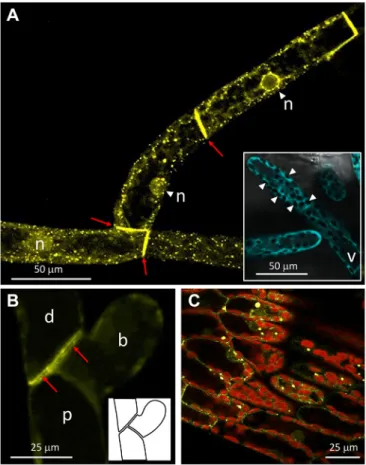
In protonemal cells, Citrine-VPYL exhibited pronounced localization to small subcellular compartments (Fig. 8), apart from a weaker signal in the cytoplasm and nuclei. The subcellular localization to small compartments is reminiscent of the VPY bodies observed in P. hybrida, M. truncatula, and Nicotiana benthamiana (Bapaume et al., 2019; Feddermann et al., 2010; Liu et al., 2019; Pumplin et al., 2010). In analogy to the structures characterized in tobacco (Bapaume et al., 2019), we refer to these structures as VPYL bodies. As in the case of M. truncatula (Liu et al., 2019), these compartments were most prominent in areas that are rich in cytoplasm, such as around the nuclei (Fig. 8A). In contrast, free fluorescent protein expressed from a heat-inducible Cerulean construct inserted at the PIG1 site, showed a general cytoplasmic signal that was just excluded from the chloroplasts and the vacuole (Fig. 8A, inset). Strong Citrine-VPYL signal was also observed along the plasma membrane of adjacent protonemal cells
next to their shared cell wall (Fig. 8B), at sides where actin also accumulates (Vidali et al., 2009). As in protonemal cells, a subcellular localization to punctate objects was found in phyllids, although in this case the fluorescent compartments were slightly larger and more heterogenous in size than in the protonema (Fig. 8C).
DISCUSSION
Origin, evolution and expression pattern of the VAPYRIN gene family
The small VAPYRIN gene family (including VPY, VPYL and additional copies thereof ) occurs in most land plants. Based on our phylogenetic analysis, the ancestral VPY gene emerged at the base of the embryophytes, probably via fusion of the ankyrin domain with the MSP domain, and underwent early duplication. Both copies, VPY and VPYL, were retained in most angiosperm clades, except in those that lost the capability to engage in AM symbiosis. This phylogenetic signature, and the fact that vpy mutants in petunia and M. truncatula have no developmental phenotypes, suggests that VPY has only symbiosis-related functions. However, the finding that some nonsymbiotic mosses and liverworts have retained VPY or Fig. 7. Hormone-dependent reversal of the branching phenotype inko27,
andko36. Average branch number per cell was measured as in Fig. 4C in the wild type, ko27 and ko36 in the presence of 10 µM of the auxins NAA and IAA, or of the strigolactone GR24 (SL). Error bars represent the mean+s.d. (n=100). Different letters indicate significant differences (two-way ANOVA, P<0.05). See P-values in the supplementary Materials and Methods.
Fig. 8. Subcellular localization of Citrine-tagged VPYL inP. patens. (A) Protonemal cell file with nuclei in the focal plane expressing N-terminally tagged Citrine-VPYL under control of the P. patens elongation factor 1-α promoter. Note many Citrine-labeled VPYL bodies and accumulation of signal at cell boundaries (arrows). Inset: Free Cerulean is localized throughout the cytoplasm, but excluded from the vacuole and chloroplasts (arrowheads). (B) Localization of Citrine-VPYL in three adjacent protonemal cells, including a distal cell, proximal cell and an early branch. Signal was deliberately kept low to reveal details of the cell boundaries (arrows). Inset shows cellular context. (C) Citrine-labeled VPYL bodies in phyllid cells. Note chlorophyll
autofluorescence of chloroplasts (red). b, early branch; d, distal cell; n, nucleus; p, proximal cell; v, vacuole.
DEVEL
O
VPYL (Fig. 1; Fig. S1) indicates that it has nonsymbiotic functions in nonvascular plants. The developmental phenotype of P. patens vpyl mutants is consistent with this assumption.
The fact that vpy mutants in petunia and M. truncatula have a strong symbiosis-defective phenotype indicates that there can be only limited redundancy between VPY and VPYL, if any, and that the function of the latter in angiosperms remains to be found. In this context, it is interesting that, according to the M. truncatula Gene Expression Atlas (https://mtgea.noble.org/v3/), the M. truncatula VPY gene is expressed almost exclusively in roots (Fig. S10A) and is highly responsive to nodulation (Fig. S10B), mycorrhizal infection (Fig. S10C), nod factor and rhizobia in root hairs (Fig. S10C). In contrast, M. truncatula VPYL has a broader expression domain, including shoot tissues (Fig. S10A), and is not influenced by symbiotic status or symbiotic signals in the roots (Fig. S10B,C). This indicates that VPYL has nonsymbiotic functions in M. truncatula (and presumably in other angiosperms) that remain to be discovered.
Expression pattern and developmental function ofVPYL inP. patens
Our analysis using a pVPYL::GUS construct revealed that the VPYL promoter is most active in protonema (caulonema and chloronema) and in young rhizoids (Fig. 2). The common feature of these cell types is their growth mode, which is restricted to tip growth as in the root hairs and pollen tubes of angiosperms. The high expression of VPYL at early stages of development in these cell types ( protonemal tip cells; rhizoid initials) could indicate that VPYL is involved in tip growth. However, the fact that P. patens vpyl knockout mutants had no phenotype in tip growth or protonemal cell size (Fig. S7) indicates that the role of VPYL in the protonema of P. patens is not in cell growth or cell division. Because P. patens vpyl mutants show excessive and ectopic branching (Fig. 4), VPYL may be involved in the determination of branch sites, thereby influencing protonemal patterning. The initial acceleration of gametophore formation in vpyl knockout mutants (Fig. 3) seems at first to concern a different aspect of moss development. However, the buds that give rise to gametophores differentiate from protonemal side branch initials, which subsequently undergo a specific cell division pattern to generate a three-dimensional leafy shoot (Moody, 2019). Hence, the common aspect of all these phenotypic traits in vpyl mutants is increased protonemal branching. Hence, P. patens VPYL is a determinant of branch site selection and limits excessive branching, thereby controlling protonemal patterning.
Uncoupling of phyllid size and cell size invpyl mutants
A striking aspect of the P. patens vpyl knockout phenotype was decreased cell length in phyllids, whereas cell width and total phyllid size was almost normal (Fig. 6). This suggests that phyllid size and cell size are controlled independently. Final organ size is defined by a combination of expansion growth and the cell cycle (Hepworth and Lenhard, 2014). Mechanistically, the two processes can proceed independently; however, cell division is coordinated with expansion in such a way that cell size remains nearly constant at early stages of organ growth, whereas cell division ceases at later stages and the organ reaches its final size and shape by cell expansion. Experiments on tobacco mutants with a dominant negative version of a cell cycle regulator showed that normal organ shape can be attained with fewer (but larger) cells (Hemerly et al., 1995); thus, organ growth and cell division are separable to a certain degree. The opposite phenotype in P. patens vpyl mutants (normal leaf size with smaller cells) reflects either increased cell cycle activity
or decreased cell diffuse growth in the absence of VPYL, whereas the determinants of phyllid size control organ growth independently of cell size and cell number. Strikingly, mutation of VPYL only affects one cell dimension (length), indicating that P. patens VPYL function in cell division control has a polar component.
Cellular function of VPYL inP. patens
An important element in all tip-growing cells is the cytoskeleton (Bascom et al., 2018). In pollen tubes and root hairs, as well as in moss protonemata (Wu et al., 2018), the cytoskeleton is thought to mediate the vesicle transport required for cell wall extension at the tip (Bascom et al., 2018). Although the role of the cytoskeleton in tip growth is well established, its role in protonemal branching is less clear. Mutants in actin-related genes in P. patens show a more severe and pleiotropic phenotype than vpyl mutants (Finka et al., 2008; Harries et al., 2005; Perroud and Quatrano, 2008). More informative are mutants in myosin VIII genes, which encode motor proteins that move along actin strands. A mutant defective in all five myosin VIII genes (ΔmyoABCDE) of P. patens shares several striking similarities in its developmental phenotype with P. patens vpyl mutants (Wu et al., 2018, 2011). In particular,ΔmyoABCDE exhibits increased protonemal branching, illegitimate branching sites, uneven distribution of branch length and accelerated gametophore formation. It is interesting to see such similarities to the vpyl phenotype because the motility and function of VPYL bodies is probably related to motor proteins such as myosins.
Functional parallels of VPY and VPYL in symbiosis and cell branching
In the angiosperms P. hybrida, M. truncatula and N. benthamiana, GFP-tagged VPY localizes to small subcellular compartments (VPY bodies) with endosomal identity (Bapaume et al., 2019; Feddermann et al., 2010; Pumplin et al., 2010). A similar subcellular localization was found in P. patens (Fig. 8). Could this indicate commonalities in the cellular events associated with endosymbiosis and with protonemal branching? One common aspect of both phenomena is the accumulation of large amounts of cytoplasmic constituents and organelles, both in the accommodation of an intracellular microbe and in the establishment of a new tip-growing branch. Cellular events in the host associated with microbial infection and accommodation have been compared with cytokinesis because of overlapping gene expression patterns (Breakspear et al., 2014) and prominent similarities in assembly of the phragmoplast and infection structures such as the PPA (Genre et al., 2012). In addition, symbiosis requires assembly of the large amounts of membrane material that surrounds the growing tips of arbuscules (in AM) and the invading or accommodated rhizobia (in RNS). With their trans-Golgi and endosomal identities (Bapaume et al., 2019), and given the interaction with exocyst components (Liu et al., 2019; Zhang et al., 2015), VPY bodies may well play an important role in these processes.
An additional striking parallel is the involvement of nuclear repositioning in all three processes: protonemal branching is associated with migration of the nucleus to an apical position next to the branch site (Doonan et al., 1986; Jensen, 1981; Schmiedel and Schnepf, 1979) (Fig. S11A). On the other hand, fungal and bacterial accommodation in AM and RNS is guided by the nucleus, which dictates the trajectories of the PPA (Genre et al., 2008, 2005) and infection thread, respectively (Fournier et al., 2008) (Fig. S11B,C). A unifying interpretation of these phenomena could be a scenario in which new foci of tip growth are initiated by nuclear positioning and assembly of a tip-growing
DEVEL
O
machinery, after which the newly established tip grows outward (in branching moss protonema) or inward (in PPA and infection thread). It remains to be explored how VPY and VPYL impinge on these processes to promote accommodation of endosymbionts and to limit protonemal branching.
MATERIALS AND METHODS
Phylogenetic analysis
VAPYRIN homologs were searched against a custom genome database of 77 plant species from the SymDB database (Radhakrishnan et al., 2020) covering the main orders of plant lineages from bryophytes to angiosperms. Additionally, data from the 1KP project (Matasci et al., 2014) were added to improve coverage of the gymnosperm and monilophyte lineages (Table S1; see protein sequences in the supplementary Materials and Methods). Searches were performed with the tBLASTn algorithm v2.9.0+ (Camacho et al., 2009) with an e-value threshold of 1e-10 and the protein sequence of VAPYRIN from the model legume Medicago truncatula as query. Obtained coding sequences were aligned with MAFFT v7.407 (Katoh and Standley, 2013) with default parameters. The resulting alignment was cleaned by removing all positions with more than 20% of gaps using trimAl v1.2 (Capella-Gutierrez et al., 2009). The clean alignment was used as matrix for subsequent phylogenetic analysis by maximum likelihood using IQ-TREE v1.6.7 (Nguyen et al., 2015). First, the best-fitted evolutionary model was tested using ModelFinder (Kalyaanamoorthy et al., 2017) according to the Bayesian Information Criteria. Maximum likelihood analysis was then performed with 10,000 replicates of UltraFast Bootstraps (Hoang et al., 2018) to test the branch support. The resulting phylogeny revealed three main clades, with two paralogous groups corresponding to VAPYRIN and VAPYRIN-like. Sequences from the third clade were used as an outgroup. From this first analysis, three subsets were extracted and subjected to detailed phylogenetic analysis: the clade VAPYRIN/VAPYRIN-like with all sequences (Fig. S1), the VAPYRIN/VAPYRIN-like clade with only representative species of plants orders (for readability reasons) (Fig. 1) and the Fabaceae VAPYRIN clade (Fig. S2). In all cases, the obtained topologies were similar, with slight differences in the positions of some terminal branches. Each of the subsequent phylogenetic analyses was performed on both protein and protein-coding sequences (with a gap trimming threshold set as 50% for the protein-coding sequences). Similar results were obtained and the tree with the best resolution was selected (the only difference between trees based on proteins and coding sequences was the resolution of some terminal branches). Proteins were aligned using MUSCLE v3.8.31 (Edgar, 2004) with the default parameters. The alignment was then subjected to maximum likelihood analysis following the procedure described above.
Trees were visualized and annotated throughout the Interactive Tree Of Life (iTOL) platform v4.3.3 (Letunic and Bork, 2019).
Analysis of selection signature
Analysis of the selective constraint acting on the VPY and VPYL genes was conducted using the PAML4.9j package (10.1093/molbev/msm088). Protein alignments from phylogenic analysis were used, and sequences with more than 50% of gaps removed before realignment of sequences using MUSCLE. Obtained alignments were used as templates to generate the codon alignment using the PAL2NAL Perl script (10.1093/nar/gkl315) with the option to remove all gapped position for subsequent analysis. The resulting alignment of 166 sites was subjected to a dN/dS analysis using the YN00 method from the PAML package (10.1093/molbev/msm088; 10.1093/oxfordjournals.molbev.a026236).
Plant material, culture conditions and protoplast transformation
P. patens (standard lab strain Gransden) was grown under sterile conditions on 0.8% agar containing BCD medium in 9 cm Petri dishes as described (Ashton and Cove, 1977; Ashton et al., 1979; Trouiller et al., 2006). Growth chamber conditions were adjusted to a day:night photoperiod of 16:8 h (25/22°C) at a photon fluence rate of approximately 50 µmol/m2/s. P. patens
was routinely grown on cellophane discs except for analysis of total colony growth (Fig. 3).
For detailed microscopic analysis, the moss was grown between two cellophane sheets (sandwich method). In the case of hormonal treatments, P. patens was first grown on BCD medium for one week before transferring the cellophane sandwich to a BCD plate with IAA (Sigma-Aldrich), NAA (Fluka), or GR24 (Strigolab) at the indicated concentrations for one week. Hormones were diluted in 40μl of solvent (36 μl ethanol and 4 μl hormonal stock in DMSO) in Petri plates before adding 40 ml BCD medium. Protoplast isolation and polyethylene glycol-mediated transformation of P. patens protoplasts were performed as previously described (Schaefer and Zrÿd, 1997; Trouiller et al., 2006).
Cloning of transformation vectors
The knockout vector pVPYL-KO was constructed in the vector pBNRF that carries an nptII expression cassette conferring G-418 resistance (Schaefer et al., 2010). Two 800 bp targeting sequences (5′-TS and 3′-TS) covering the sequences located immediately up- and downstream of the P. patens VPYL ORF were amplified by PCR using primers ko-5′F and ko-5′R, and ko-3′F and ko-3′R, respectively (see primer list in the supplementary Materials and Methods). The amplified fragments were cloned in pBNRF to generate the replacement vector pVPYL-KO (see vector constructs in the supplementary Materials and Methods). For transformation, 15μg of vector DNA was linearized with Sal1 and Mlu1, precipitated with ethanol and resuspended in sterile water. Homologous recombination on both TSs resulted in the replacement of the P. patens VPYL ORF by the nptII resistance gene (Fig. S3).
A GUS reporter line was generated by insertion of the UidA gene, which encodes bacterial β-glucuronidase (GUS), under the control of the full-length P. patens VPYL promoter. First, a 1.6 kb fragment containing the coding sequence ofβ-glucuronidase and the NOS-terminator, including an XhoI restriction site on each side, was amplified by PCR from the plasmid pMDC163 (Curtis and Grossniklaus, 2003) using primers #19fw and #20rev (see the primer list in the supplementary Materials and Methods). This fragment was inserted between the 5′-TS and the resistance marker in the knockout vector VPYL-KO. For transformation, 15μg vector DNA was linearized only with Sal1, precipitated with ethanol, and resuspended in sterile water. This construct was introduced into the P. patens genome at the VPYL locus by homologous recombination only at the 5′-TS, resulting in insertion of the linearized vector, instead of replacement of the VPYL ORF (Fig. S3).
For analysis of subcellular localization, an N-terminal Citrine-VPYL fusion was built in the gateway-compatible destination vector pOYG1 (http://moss.nibb.ac.jp/protocol.html) designed for insertion at the PIG1 locus. The PIG1 locus is considered silent and therefore suited for insertion (Okano et al., 2009). The VPYL ORF including the stop codon was amplified by PCR and integrated into the pENTRTM/D-TOPO vector, followed by an LR recombination reaction with the preformed Gateway-compatible transformation vector pOYG1. The VPYL ORF was thereby fused in frame to Citrine at its N-terminal end. Expression of the fusion protein is under control of the strong constitutive EF1a promoter. For transformation, 15μg vector DNA was linearized with PmeI, precipitated with ethanol, and resuspended in sterile water. As a cytoplasmic control, heat shock-inducible free Cerulean ( pPIG1HCG) (Aoyama et al., 2012) was used 24 h after heat shock at 38°C for 1 h.
β-Glucuronidase assay
GUS activity was determined by staining in the following staining solution: 0.5 mg/ml 5-bromo-4-chloro-3-indolylβ-D-glucuronidecyclohexylammonium salt (X-Gluc) in 125 mM sodium phosphate buffer ( pH 7.5), 0.2 mM EDTA, 0.02% NaN3, 0.1% Triton-X-100, 5 mM ferricyanide and 5 mM ferrocyanide. Staining was performed for 3 h (Fig. 2B,C,F) or overnight (Fig. 2A,D,E) at 37°C. Chlorophyll was removed by consecutive immersion in 50, 75 and 100% ethanol.
Microscopy
For detailed phenotypic characterization, moss grown in a cellophane sandwich was excised from Petri plates with care to avoid shifts of the protonema between the cellophane layers. These samples were then
DEVEL
O
mounted with water on object slides and images acquired with an Axiocam color CCD camera (Zeiss Axiophot I) operated by AxioVision 2.05 software (Zeiss) and mounted on a Leica DMR microscope. Confocal analysis was performed on a Leica SP5 with an argon laser (458, 476, 488 and 514 nm) and a helium-neon laser (543 nm) using the respective laser for Citrine and chlorophyll autofluorescence. Pictures were edited with the Leica LAS AF Lite software and ImageJ.
Phenotypic analysis ofP. patens vpyl mutants
For quantitative analysis of colony growth, normalized tissue suspensions of P. patens wild type, ko27 and ko36 were pre-grown for 1 week on BCD medium overlaid with cellophane. At the start of the experiment, protonemal fragments of about 1 mm in diameter were transferred to fresh BCD plates (without cellophane) as triplets and grown for an additional 4 weeks under standard conditions. Colony expansion was determined by analyzing scans of the bottom of each Petri dish. The area of expansion was measured using the Particle Analysis function of ImageJ software on 8-bit Image type pictures with an auto-adjusted threshold. Numbers of buds and gametophores present in each moss colony were counted by eye through a binocular (Leica MZ-FLIII) after dissection.
For quantification of caulonemal growth, pre-grown protonemata were aligned in cellophane sandwiches on BCD supplemented with 5 mM ammonium tartrate and 0.5% glucose. About three-quarters of each plate surface was wrapped with aluminum foil to initiate directional phototropic growth perpendicular to the aligned protonemata for 18 days. Subsequently, plates were wrapped with black plastic and aluminum foil to leave only a narrow light window of ca. 1 cm diameter, towards which the directionally growing protonemata were oriented. After 6 days of further culture, all protonemata had undergone transition to caulonemal identity. The length of caulonemata was determined from pictures acquired with a Nikon Digital Sight DS-U1 camera mounted an a binocular dissection microscope (Leica MZ-FLIII). For detailed analysis of branch number, branch distribution, average branch length and branch position, P. patens wild type, ko27 and ko36 were grown under standard conditions (see above) in cellophane sandwiches. Approximately 100 randomly chosen cells or branches were analyzed per treatment, as indicated in the figure legends. Phyllid analysis was performed with fully grown leaves of gametophores after 4 weeks of culture under standard conditions without cellophane. At least five different gametophores were chosen per genotype to measure the dimensions of 20 phyllids or 100 cells per genotype. For cell length measurements, the protonema was stained with Calcofluor White as described (Kofuji et al., 2018) to reveal cell walls and the samples analyzed by confocal analysis. All experiments were repeated at least twice with similar results.
Genotyping
Genomic DNA was isolated by extraction with 20 mM Tris HCl ( pH 7.5), 250 mM NaCl, 25 mM EDTA, 0.5% (w/v) SDS and 10 mM β-mercaptoethanol. Extracts were cleared by centrifugation (10 min, 15,000 g) and precipitated with isopropanol. DNA was recovered by centrifugation (10 min, 15,000 g and washed with 75% ethanol. Genotyping was performed by PCR with the primers listed in the supplementary Materials and Methods (see also Fig. S3).
Gene expression analysis
RNA from protonemal cultures was extracted employing the NucleoSpin RNA Plant Kit (Macherey-Nagel). RNA was reverse transcribed with iScript cDNA Synthesis Kit (BioRad). Quantitative real-time RT-PCR (qRT-PCR) was carried out with primers #51f and #52r (see primer list in the supplementary Materials and Methods) using the qPCR SYBRGreen master mix (ThermoScientific, http://www.thermo.com) in a Rotorgene thermocycler (Corbett Life Science). The PCR conditions included an initial denaturation cycle for 15 min at 95°C, followed by 40 cycles of denaturation for 15 s at 95°C, annealing for 20 s at 60°C and extension for 20 s at 72°C, followed by a final extension for 5 min at 72°C. qPCR analysis was performed with P. patens TUA1 (GenBank Accession Number AB096718.1) as reference gene (see primer list in the supplementary
Materials and Methods). Relative gene expression values were calculated using theΔΔCt method (Pfaffl, 2001).
Acknowledgements
We thank Guillaume Gouzerh and Noémie Fahr for confocal microscopy, Mitsuyasu Hasebe (NIBB Okasaki, Japan) for providing us with the cloning vectors pOYG1 and pPIG1HCG, and Chiara Pollheimer for assistance in phenotypic analysis. Special thanks go to Rudolf Rohr for help with statistical analysis.
Competing interests
The authors declare no competing or financial interests.
Author contributions
Conceptualization: D.R., D.G.S., P.-M.D.; Methodology: U.R., M.C., N.F., M.S., J.K., P.-M.D., D.G.S., D.R.; Software: J.K.; Validation: U.R., D.G.S.; Formal analysis: U.R., M.C., F.B., N.F., D.G.S.; Investigation: D.R., U.R., N.F., M.S., A.R., S.M., G.-Y.H., J.K., P.-M.D., D.G.S.; Resources: P.-M.D., D.G.S.; Data curation: U.R., F.B., J.K., P.-M.D.; Writing - original draft: D.R., D.G.S.; Writing - review & editing: D.R., U.R., M.C., J.K., D.G.S.; Visualization: D.R., U.R., M.C., F.B., M.S.; Supervision: D.R., D.G.S.; Project administration: D.R.; Funding acquisition: D.R.
Funding
This work was supported by the Schweizerischer Nationalfonds zur Fö rderung der Wissenschaftlichen Forschung to D.R. (31003A-169732).
Data availability
The P. patens VPYL gene sequence can be accessed at GenBank: MN971578TS.
Supplementary information
Supplementary information available online at
http://dev.biologists.org/lookup/doi/10.1242/dev.184762.supplemental
Peer review history
The peer review history is available online at
https://dev.biologists.org/lookup/doi/10.1242/dev.184762.reviewer-comments.pdf
References
Aoyama, T., Hiwatashi, Y., Shigyo, M., Kofuji, R., Kubo, M., Ito, M. and Hasebe, M. (2012). AP2-type transcription factors determine stem cell identity in the moss Physcomitrella patens. Development 139, 3120-3129. doi:10.1242/dev.076091 Ashton, N. W. and Cove, D. J. (1977). The Isolation and preliminary
characterisation of auxotrophic and analogue-resistant mutants of the moss, Physcomitrella patens. Mol. Gen. Genet. 154, 87-95. doi:10.1007/BF00265581 Ashton, N. W., Grimsley, N. H. and Cove, D. J. (1979). Analysis of gametophytic
development in the moss, Physcomitrella patens, using auxin and cytokinin resistant mutants. Planta 144, 427-435. doi:10.1007/BF00380118
Bapaume, L., Laukamm, S., Darbon, G., Monney, C., Meyenhofer, F., Feddermann, N., Chen, M. and Reinhardt, D. (2019). VAPYRIN marks an endosomal trafficking compartment involved in arbuscular mycorrhizal symbiosis. Front. Plant Sci. 10, 666. doi:10.3389/fpls.2019.00666
Bascom, C. S., Hepler, P. K. and Bezanilla, M. (2018). Interplay between ions, the cytoskeleton, and cell wall properties during tip growth. Plant Physiol. 176, 28-40. doi:10.1104/pp.17.01466
Bravo, A., York, T., Pumplin, N., Mueller, L. A. and Harrison, M. J. (2016). Genes conserved for arbuscular mycorrhizal symbiosis identified through phylogenomics. Nat. Plants 2, 15208. doi:10.1038/nplants.2015.208
Breakspear, A., Liu, C. W., Roy, S., Stacey, N., Rogers, C., Trick, M., Morieri, G., Mysore, K. S., Wen, J. Q., Oldroyd, G. E. D. et al. (2014). The root hair “infectome” of Medicago truncatula uncovers changes in cell cycle genes and reveals a requirement for auxin signaling in rhizobial infection. Plant Cell 26, 4680-4701. doi:10.1105/tpc.114.133496
Camacho, C., Coulouris, G., Avagyan, V., Ma, N., Papadopoulos, J., Bealer, K. and Madden, T. L. (2009). BLAST plus : architecture and applications. BMC Bioinformatics 10. doi:10.1186/1471-2105-10-421
Capella-Gutierrez, S., Silla-Martinez, J. M. and Gabaldon, T. (2009). trimAl: a tool for automated alignment trimming in large-scale phylogenetic analyses. Bioinformatics 25, 1972-1973. doi:10.1093/bioinformatics/btp348
Cox, C. J. (2018). Land plant molecular phylogenetics: a review with comments on evaluating incongruence among phylogenies. Crit. Rev. Plant Sci. 37, 113-127. doi:10.1080/07352689.2018.1482443
Curtis, M.D. and Grossniklaus, U. (2003). A gateway cloning vector set for high-throughput functional analysis of genes in planta. Plant Physiol. 133, 462-469. doi:10.1104/pp.103.027979
Delaux, P.-M., Varala, K., Edger, P. P., Coruzzi, G. M., Pires, J. C. and Ané, J.-M. (2014). Comparative phylogenomics uncovers the impact of symbiotic
DEVEL
O
associations on host genome evolution. PLoS Genet. 10, e1004487. doi:10.1371/ journal.pgen.1004487
Delaux, P.-M., Radhakrishnan, G. V., Jayaraman, D., Cheem, J., Malbreil, M., Volkening, J. D., Sekimoto, H., Nishiyama, T., Melkonian, M., Pokorny, L. et al. (2015). Algal ancestor of land plants was preadapted for symbiosis. Proc. Natl. Acad. Sci. USA 112, 13390-13395. doi:10.1073/pnas.1515426112 Doonan, J. H., Jenkins, G. I., Cove, D. J. and Lloyd, C. W. (1986). Microtubules
connect the migrating nucleus to the prospective division site during side branch formation in the moss, Physcomitrella patens. Eur. J. Cell Biol. 41, 157-164. Edgar, R. C. (2004). MUSCLE: multiple sequence alignment with high accuracy and
high throughput. Nucleic Acids Res. 32, 1792-1797. doi:10.1093/nar/gkh340 Favre, P., Bapaume, L., Bossolini, E., Delorenzi, L., Falquet, L. and Reinhardt,
D. (2014). A novel bioinformatics pipeline to discover genes related to arbuscular mycorrhizal symbiosis based on their evolutionary conservation pattern among higher plants. BMC Plant Biol. 14, 333. doi:10.1186/s12870-014-0333-0 Feddermann, N. and Reinhardt, D. (2011). Conserved residues in the ankyrin
domain of VAPYRIN indicate potential protein-protein interaction surfaces. Plant Signal. Behav. 6, 680-684. doi:10.4161/psb.6.5.14972
Feddermann, N., Duvvuru Muni, R. R., Zeier, T., Stuurman, J., Ercolin, F., Schorderet, M. and Reinhardt, D. (2010). The PAM1 gene of petunia, required for intracellular accommodation and morphogenesis of arbuscular mycorrhizal fungi, encodes a homologue of VAPYRIN. Plant J. 64, 470-481. doi:10.1111/j. 1365-313X.2010.04341.x
Finka, A., Saidi, Y., Goloubinoff, P., Neuhaus, J.-M., Zrÿd, J.-P. and Schaefer, D. G. (2008). The knock-out of ARP3a gene affects F-actin cytoskeleton organization altering cellular tip growth, morphology and development in moss Physcomitrella patens. Cell Motil. Cytoskelet. 65, 769-784. doi:10.1002/cm.20298 Fournier, J., Timmers, A. C. J., Sieberer, B. J., Jauneau, A., Chabaud, M. and Barker, D. G. (2008). Mechanism of infection thread elongation in root hairs of Medicago truncatula and dynamic interplay with associated rhizobial colonization. Plant Physiol. 148, 1985-1995. doi:10.1104/pp.108.125674
Genre, A., Chabaud, M., Timmers, T., Bonfante, P. and Barker, D. G. (2005). Arbuscular mycorrhizal fungi elicit a novel intracellular apparatus in Medicago truncatula root epidermal cells before infection. Plant Cell 17, 3489-3499. doi:10. 1105/tpc.105.035410
Genre, A., Chabaud, M., Faccio, A., Barker, D. G. and Bonfante, P. (2008). Prepenetration apparatus assembly precedes and predicts the colonization patterns of arbuscular mycorrhizal fungi within the root cortex of both Medicago truncatula and Daucus carota. Plant Cell 20, 1407-1420. doi:10.1105/tpc.108. 059014
Genre, A., Ivanov, S., Fendrych, M., Faccio, A.,Žárský, V., Bisseling, T. and Bonfante, P. (2012). Multiple exocytotic markers accumulate at the sites of perifungal membrane biogenesis in arbuscular mycorrhizas. Plant Cell Physiol. 53, 244-255. doi:10.1093/pcp/pcr170
Gutjahr, C. and Parniske, M. (2013). Cell and developmental biology of arbuscular mycorrhiza symbiosis. Annu. Rev. Cell Dev. Biol. 29, 593-617. doi:10.1146/ annurev-cellbio-101512-122413
Hanke, S. T. and Rensing, S. A. (2010). In vitro association of non-seed plant gametophytes with arbuscular mycorrhiza fungi. J. Endocytobiosis Cell Res. 20, 95-101.
Harries, P. A., Pan, A. H. and Quatrano, R. S. (2005). Actin-related protein2/3 complex component ARPC1 is required for proper cell morphogenesis and polarized cell growth in Physcomitrella patens. Plant Cell 17, 2327-2339. doi:10. 1105/tpc.105.033266
Hemerly, A., Engler, J. D., Bergounioux, C., Vanmontagu, M., Engler, G., Inzé, D. and Ferreira, P. (1995). Dominant-negative mutants of the Cdc2 kinase uncouple cell division from iterative plant development. EMBO J. 14, 3925-3936. doi:10.1002/j.1460-2075.1995.tb00064.x
Hepworth, J. and Lenhard, M. (2014). Regulation of plant lateral-organ growth by modulating cell number and size. Curr. Opin. Plant Biol. 17, 36-42. doi:10.1016/j. pbi.2013.11.005
Hoang, D. T., Chernomor, O., von Haeseler, A., Minh, B. Q. and Vinh, L. S. (2018). UFBoot2: Improving the ultrafast bootstrap approximation. Mol. Biol. Evol. 35, 518-522. doi:10.1093/molbev/msx281
Jensen, L. C. W. (1981). Division, growth, and branch formation in protonema of the moss Physcomitrium turbinatum: studies of sequential cytological changes in living cells. Protoplasma 107, 301-317. doi:10.1007/BF01276832
Kalyaanamoorthy, S., Minh, B. Q., Wong, T. K. F., von Haeseler, A. and Jermiin, L. S. (2017). ModelFinder: fast model selection for accurate phylogenetic estimates. Nat. Methods 14, 587. doi:10.1038/nmeth.4285
Katoh, K. and Standley, D. M. (2013). MAFFT multiple sequence alignment software version 7: Improvements in performance and usability. Mol. Biol. Evol. 30, 772-780. doi:10.1093/molbev/mst010
Knight, C., Perroud, P.-F. and Cove, D. (2009). The Moss Physcomitrella Patens. Oxford, UK: Wiley-Blackwell.
Koenen, E. J. M., Ojeda, D. I., Steeves, R., Migliore, J., Bakker, F. T., Wieringa, J. J., Kidner, C., Hardy, O., Pennington, R. T., Herendeen, P. S. et al. (2019). The origin and early evolution of the legumes are a complex paleopolyploid phylogenomic tangle closely associated with the Cretaceous-Paleogene (K-Pg) boundary. bioRxiv 577957. doi:10.1101/577957
Kofuji, R. and Hasebe, M. (2014). Eight types of stem cells in the life cycle of the moss Physcomitrella patens. Curr. Opin. Plant Biol. 17, 13-21. doi:10.1016/j.pbi. 2013.10.007
Kofuji, R., Yagita, Y., Murata, T. and Hasebe, M. (2018). Antheridial development in the moss Physcomitrella patens: implications for understanding stem cells in mosses. Philos. Trans. R. Soc. B Biol. Sci. 373, 20160494. doi:10.1098/rstb.2016. 0494
Letunic, I. and Bork, P. (2019). Interactive Tree Of Life (iTOL) v4: recent updates and new developments. Nucleic Acids Res. 47, W256-W259. doi:10.1093/nar/ gkz239
Lev, S., Ben Halevy, D., Peretti, D. and Dahan, N. (2008). The VAP protein family: from cellular functions to motor neuron disease. Trends Cell Biol. 18, 282-290. doi:10.1016/j.tcb.2008.03.006
Liu, C.-W., Breakspear, A., Stacey, N., Findlay, K., Nakashima, J., Ramakrishnan, K., Liu, M. X., Xie, F., Endre, G., de Carvalho-Niebel, F. et al. (2019). A protein complex required for polar growth of rhizobial infection threads. Nat. Commun. 10, 2848. doi:10.1038/s41467-019-10029-y
Matasci, N., Hung, L.-H., Yan, Z. X., Carpenter, E. J., Wickett, N. J., Mirarab, S., Nguyen, N., Warnow, T., Ayyampalayam, S., Barker, M. et al. (2014). Data access for the 1,000 Plants (1KP) project. Gigascience 3, 17. doi:10.1186/2047-217X-3-17 Menand, B., Calder, G. and Dolan, L. (2007). Both chloronemal and caulonemal cells expand by tip growth in the moss Physcomitrella patens. J. Exp. Bot. 58, 1843-1849. doi:10.1093/jxb/erm047
Michaely, P., Machius, M., Tomchick, D. and Anderson, R. G. W. (2001). Structure of the ANK repeats of ankyrin. Mol. Biol. Cell 12, 2477.
Mohler, P. J., Gramolini, A. O. and Bennett, V. (2002). Ankyrins. J. Cell Sci. 115, 1565-1566.
Moody, L. A. (2019). The 2D to 3D growth transition in the moss Physcomitrella patens. Curr. Opin. Plant Biol. 47, 88-95. doi:10.1016/j.pbi.2018.10.001 Murray, J. D., Duvvuru Muni, R. R., Torres-Jerez, I., Tang, Y., Allen, S.,
Andriankaja, M., Li, G., Laxmi, A., Cheng, X., Wen, J. et al. (2011). Vapyrin, a gene essential for intracellular progression of arbuscular mycorrhizal symbiosis, is also essential for infection by rhizobia in the nodule symbiosis of Medicago truncatula. Plant J. 65, 244-252. doi:10.1111/j.1365-313X.2010.04415.x Nguyen, L.-T., Schmidt, H. A., von Haeseler, A. and Minh, B. Q. (2015). IQ-TREE:
a fast and effective stochastic algorithm for estimating maximum-likelihood phylogenies. Mol. Biol. Evol. 32, 268-274. doi:10.1093/molbev/msu300 Okano, Y., Aono, N., Hiwatashi, Y., Murata, T., Nishiyama, T., Ishikawa, T., Kubo,
M. and Hasebe, M. (2009). A polycomb repressive complex 2 gene regulates apogamy and gives evolutionary insights into early land plant evolution. Proc. Natl. Acad. Sci. USA 106, 16321-16326. doi:10.1073/pnas.0906997106 Oldroyd, G. E. D., Murray, J. D., Poole, P. S. and Downie, J. A. (2011). The rules of
engagement in the legume-rhizobial symbiosis. Annu. Rev. Genet. 45, 119-144. doi:10.1146/annurev-genet-110410-132549
Ortiz-Ramı́rez, C., Hernandez-Coronado, M., Thamm, A., Catarino, B., Wang, M. Y., Dolan, L., Feijó, J. A. and Becker, J. D. (2016). A transcriptome atlas of Physcomitrella patens provides insights into the evolution and development of land plants. Mol. Plant 9, 205-220. doi:10.1016/j.molp.2015.12.002
Perroud, P.-F. and Quatrano, R. S. (2008). BRICK1 is required for apical cell growth in filaments of the moss Physcomitrella patens but not for gametophore morphology. Plant Cell 20, 411-422. doi:10.1105/tpc.107.053256
Pfaffl, M. W. (2001). A new mathematical model for relative quantification in real-time RT-PCR. Nucleic Acids Res. 29, e45. doi:10.1093/nar/29.9.e45
Prigge, M. J. and Bezanilla, M. (2010). Evolutionary crossroads in developmental biology: Physcomitrella patens. Development 137, 3535-3543. doi:10.1242/dev. 049023
Proust, H., Hoffmann, B., Xie, X. N., Yoneyama, K., Schaefer, D. G., Yoneyama, K., Nogue, F. and Rameau, C. (2011). Strigolactones regulate protonema branching and act as a quorum sensing-like signal in the moss Physcomitrella patens. Development 138, 1531-1539. doi:10.1242/dev.058495
Pumplin, N., Mondo, S. J., Topp, S., Starker, C. G., Gantt, J. S. and Harrison, M. J. (2010). Medicago truncatula Vapyrin is a novel protein required for arbuscular mycorrhizal symbiosis. Plant J. 61, 482-494. doi:10.1111/j.1365-313X.2009.04072.x Pumplin, N., Zhang, X., Noar, R. D. and Harrison, M. J. (2012). Polar localization of a symbiosis-specific phosphate transporter is mediated by a transient reorientation of secretion. Proc. Natl. Acad. Sci. USA 109, E665-E672. doi:10. 1073/pnas.1110215109
Radhakrishnan, G. V., Keller, J., Rich, M. K., Vernié, T., Mbadinga Mbaginda, D. L., Vigneron, N., Cottret, L., San Clemente, H., Libourel, C., Cheema, J. et al. (2020). An ancestral signalling pathway is conserved in plant lineages forming intracellular symbioses. Nat. Plants 6, 280-289. doi:10.1038/s41477-020-0613-7
Rich, M. K., Nouri, E., Courty, P.-E. and Reinhardt, D. (2017). Diet of arbuscular mycorrhizal fungi: bread and butter? Trends Plant Sci. 22, 652-660. doi:10.1016/j. tplants.2017.05.008
Roth, R. and Paszkowski, U. (2017). Plant carbon nourishment of arbuscular mycorrhizal fungi. Curr. Opin. Plant Biol. 39, 50-56. doi:10.1016/j.pbi.2017.05.008 Sakakibara, K., Nishiyama, T., Sumikawa, N., Kofuji, R., Murata, T. and Hasebe,
M. (2003). Involvement of auxin and a homeodomain-leucine zipper I gene in
DEVEL
O
rhizoid development of the moss Physcomitrella patens. Development 130, 4835-4846. doi:10.1242/dev.00644
Schaefer, D. G. (2002). A new moss genetics: Targeted mutagenesis in Physcomitrella patens. Annu. Rev. Plant Biol. 53, 477-501. doi:10.1146/ annurev.arplant.53.100301.135202
Schaefer, D. G. and Zrÿd, J.-P. (1997). Efficient gene targeting in the moss Physcomitrella patens. Plant J. 11, 1195-1206. doi:10.1046/j.1365-313X.1997. 11061195.x
Schaefer, D. G., Delacote, F., Charlot, F., Vrielynck, N., Guyon-Debast, A., Le Guin, S., Neuhaus, J. M., Doutriaux, M. P. and Nogué, F. (2010). RAD51 loss of function abolishes gene targeting and de-represses illegitimate integration in the moss Physcomitrella patens. DNA Repair 9, 526-533. doi:10.1016/j.dnarep.2010.02.001 Schmiedel, G. and Schnepf, E. (1979). Side branch formation and orientation in the
caulonema of the moss, Funaria hygrometrica: normal development and fine structure. Protoplasma 100, 367-383. doi:10.1007/BF01279323
Smith, S. E. and Read, D. J. (2008). Mycorrhizal Symbiosis, 3rd edn. New York: Academic Press.
Thelander, M., Landberg, K. and Sundberg, E. (2018). Auxin-mediated developmental control in the moss Physcomitrella patens. J. Exp. Bot. 69, 277-290. doi:10.1093/jxb/erx255
Trouiller, B., Schaefer, D. G., Charlot, F. and Nogue, F. (2006). MSH2 is essential for the preservation of genome integrity and prevents homeologous recombination in the moss Physcomitrella patens. Nucleic Acids Res. 34, 232-242. doi:10.1093/nar/gkj423
Vidali, L., Rounds, C. M., Hepler, P. K. and Bezanilla, M. (2009). Lifeact-mEGFP reveals a dynamic apical F-actin network in tip growing plant cells. PLoS ONE 4, e5744. doi:10.1371/journal.pone.0005744
Volkmar, U. and Knoop, V. (2010). Introducing intron locus cox1i624 for phylogenetic analyses in bryophytes: On the issue of Takakia as sister genus to all other extant mosses. J. Mol. Evol. 70, 506-518. doi:10.1007/s00239-010-9348-9 Wang, B. and Qiu, Y.-L. (2006). Phylogenetic distribution and evolution of mycorrhizas
in land plants. Mycorrhiza 16, 299-363. doi:10.1007/s00572-005-0033-6 Waese, J., Fan, J., Pasha, A., Yu, H., Fucile, G., Shi, R., Cumming, M.,
Kelley, L.A., Sternberg, M. J., Krishnakumar, V. et al. (2017). ePlant: Visualizing and exploring multiple levels of data for hypothesis generation in plant biology. Plant Cell 29, 1806-1821. doi:10.1105/tpc.17.00073
Wang, W., Shi, J., Xie, Q., Jiang, Y., Yu, N. and Wang, E. (2017). Nutrient exchange and regulation in arbuscular mycorrhizal symbiosis. Mol. Plant 10, 1147-1158. doi:10.1016/j.molp.2017.07.012
Wu, S.-Z., Ritchie, J. A., Pan, A.-H., Quatrano, R. S. and Bezanilla, M. (2011). Myosin VIII regulates protonemal patterning and developmental timing in the moss Physcomitrella patens. Mol. Plant 4, 909-921. doi:10.1093/ mp/ssr068
Wu, S.-Z., Yamada, M., Mallett, D. R. and Bezanilla, M. (2018). Cytoskeletal discoveries in the plant lineage using the moss Physcomitrella patens. Biophys. Rev. 10, 1683-1693. doi:10.1007/s12551-018-0470-z
Zhang, X. C., Pumplin, N., Ivanov, S. and Harrison, M. J. (2015). EXO70I is required for development of a sub-domain of the periarbuscular membrane during arbuscular mycorrhizal symbiosis. Curr. Biol. 25, 2189-2195. doi:10.1016/j.cub. 2015.06.075