Publisher’s version / Version de l'éditeur:
Journal of Sedimentary Petrology, 39, 1, pp. 90-105, 1969-03
READ THESE TERMS AND CONDITIONS CAREFULLY BEFORE USING THIS WEBSITE. https://nrc-publications.canada.ca/eng/copyright
Vous avez des questions? Nous pouvons vous aider. Pour communiquer directement avec un auteur, consultez la
première page de la revue dans laquelle son article a été publié afin de trouver ses coordonnées. Si vous n’arrivez pas à les repérer, communiquez avec nous à [email protected].
Questions? Contact the NRC Publications Archive team at
[email protected]. If you wish to email the authors directly, please see the first page of the publication for their contact information.
NRC Publications Archive
Archives des publications du CNRC
This publication could be one of several versions: author’s original, accepted manuscript or the publisher’s version. / La version de cette publication peut être l’une des suivantes : la version prépublication de l’auteur, la version acceptée du manuscrit ou la version de l’éditeur.
Access and use of this website and the material on it are subject to the Terms and Conditions set forth at
Study of the fabric of fine-grained sediments with the scanning electron
microscope
Gillott, J. E.
https://publications-cnrc.canada.ca/fra/droits
L’accès à ce site Web et l’utilisation de son contenu sont assujettis aux conditions présentées dans le site LISEZ CES CONDITIONS ATTENTIVEMENT AVANT D’UTILISER CE SITE WEB.
NRC Publications Record / Notice d'Archives des publications de CNRC:
https://nrc-publications.canada.ca/eng/view/object/?id=6a28b5e4-9d4e-413a-91e1-b79c1284ffad
https://publications-cnrc.canada.ca/fra/voir/objet/?id=6a28b5e4-9d4e-413a-91e1-b79c1284ffad
S e r
TH1
N21r2
no.
389
ANALYZED
c.
2
NATIONAL RESEARCH COUNCIL O F CANADA
BLDG
CONSEIL NATIONAL DE RECHERCHES DU CANADA
STUDY OF T H E FABRIC OF FINE-GRAINED SEDIMENTS
W I T H T H E SCANNING ELECTRON MICROSCOPE
BY
J.
E. GILLOTT
Reprinted f r o ~ n
JOURNAL O F SEDIMENTARY PETROLOGY Vol. 39, No. 1, March 1969
pp. 90-105
liesearch Paper
No. 359
of the
- - -
.&;:
4--At
Division of Building Research
OTTAWA
March
1969
Price
25
cents
E T U D E D E L A T E X T U R E D E S SEDIMENTS D E F I N E
GRANULOMETRIE A L'AIDE D'UN MICROSCOPE
ET,ECTRONTQUE A BA1,AYAGE
La r6partition des particules minkrales et des interstices de tailles infCrieures au micron d6fie
I'observation par microscopic optique. Les limitations dues
A
I'instrument peuvent etre surmoiltCes
par l'einploi d'un appareil klectroniclue. Le microscope Olectronique
A
balayage constitue un
outil iclkal pour ce genre cle travail, car il doilile une excellente r6solution aux forts grossisse-
inents avec unc profoncleur de foyer relativement Cnonlle. L'auteur utilise cles microphotog-
raphies obteilues avec ce microscope pour illustrer la texture de quelclues sols calcaires et
argileux
A grains fins qui possi.dent des propriPtCs spPciales au point de vue g@nie civil. La
structure fine apparait avcc uile clarti: inhabituelle. Les points pasticuli~rement int6ressants soilt
les suivants: la nature tles contacts des fills cristaus entre eux el avec les cristaux plus gros
et pasticulii.1-eme~~t
ceux de clolo~llie
;la rkpartition tles mat6i-iaux autres que les carbo~latcs et
cles mntkriaux de cimentage entre les grains, et l'osientation tles minCraux du genre mica clans les
schistes argileux, les schistes pklitiques et les sols asgileux. 1,'auteur compare les m6rites cles
diucsses m6thodes de pr6paration des Cchantillons.
NATIONAL IiESEARCH COUNClI, O F CANADA
CONSEIL NATIONAL D E RECHERCHES DU CANADA
STUDY O F T H E FABRIC O F FINE-GRAINED SEDIMENTS
W I T H T H E SCANNING E L E C T R O N MICROSCOPE
B Y
J. E. GILLOTT
Reprinted from
J O U R S A L O F SEDIMENTARY PETliOLOGY Vol. 39, No. 1, March 1969
pp. 90-105
Research Paper No.
359
of the
Division of Building Research
I'ricc
25 cents
NRC
10603
S T U D Y O F T H E FABRIC O F FINE-GRAINED SEDIMENTS W I T H
T H E SCANNING ELECTRON MICROSCOPE1
J. E. GILT,OTT
National ICese;ircl~ Council o l Canada, Ottawa 7, Canatln
ABSTRACT
Tlle mutual tlisposition of mineral particles and voids in the niicron and sub-micron size ranges is gc~lerally inaccessil~le to observation by light ~nicroscopy. This instrun~ctital limitation can hc overcome 11y tllc usc oT electron optical systcm. The scanni~ig clcctron microscope is ;un idcal tool ior tllis l i i i i i l of
work, for it produces good resolution at high magnifications with relatively cnormous tlepth of focus. Micro- graphs obtained \\lit11 a scanning electron microscope arc uscd to illustrate the fabrics of some finc-g-raincd limestones and clay soils which have properties of special enginecring intcrest. Fine structure is revealctl with unus~~al clarity. Of particular intcrest are: tile nature of contacts of fine-grainetl crystals with one anotlicr ant1 with coarser crystals, pa~.ticnlarly dolomites; the disposition of non-carbonates ant1 (or) cemct~titious matcrial along grain hou~idarics; and tlle orientation of mica-type ~ilinerals in shales, mudstoncs, and clay soils. The relative nierit of se~reral methotls of sample preparation is discussed.
T h e fabric of the fine-grained setliments often holds the key to understanding t h e pro- cesses involved in their formation ant1 their properties. I n the studics cond~ictccl by the Divi- sion involving the mecl~anical behavior of cer- tain clays, the mechanical properties of concrctc and other structural materials, and the rcaction of dolomitic limestone aggregate in concrete, it has been found useful to relate the bullc propcr- ties and Ijchavior of thc material to its micro- structul-c. Many rocks contain ~ n i n e r a l s which a r e in the micro11 ant1 sull-micron size ranges. F o r csamplc, clays a r e by definition composed of p;lrticlc\ \\hicli a r e lc5s than 2 microns ecluivalent spherical tliameter ( e s u ) . Clay min- erals a r e almost ul~iquitous in setlimcntary cle- posits. 111 many limestones, calcite and some- times dolomite a r e micron-sized; there are a150 often many other minerals of small particle size. These constituents a r e too small t o be con- veniently observetl 11y mealis of the optical mi- croscope, as thc resolution is limited to a b o l ~ t 0.2 microns, even with a n oil immersion o l ~ j c c - tive. Furthermore, at high magnifications the liglit microscope has only a very small clcpth of focus \\rhich also severely restricts its use in f a l ~ r i c studies. These instrumental limitations can be largely overcome by resorting to an elec- tron optical system. And in this rcgarcl the scan- ning electroll microscope has been found partic- ularly useful.
I n petrology, a s in other fields, the scanning electroll microscope is lilcely to be of greatest
'
Manuscript received March 27, 1968.use for investigations in which the very high resolution of the transmission electron micro- scope is not required, but in which t h e resolving power or tleptli of focus of t h e optical micro- scope a r e insufficient. T h e instrument has 01,- 1-ious applications in elucidating the fabric of fine-grained rocks ancl gives a clear imagc of t h e m l ~ t ~ ~ a l tlisposition of mineral particles and voids. I t sl~ould also be of use in the stucly of diagenesis, the investigation of changes in size, shape, ancl orientation of micron ancl s u l m i - cron-sized crystals during c o ~ ~ s o l i c l : ~ t i o ~ ~ alicl litli- ification, and research into reel-ystallization ancl replacement reactions of all kinds. T h e n a t ~ i r e o f fracture slirfaces and the orientatio~i of fine crystallites close to fracture and shear planes a r e also of interest. I\4icrofossils may be stuclietl with unusual clarity.
T h e scanning electron microscope is designed to produce a magnified image of a surface. Some o f t h e main aclvantages of this instr~lment a r e : the large depth of focus a t high magnifica- tions, which gives considerable perspective to the image; the possibility of examining rela- tively large samples ( - 1 cm cliam) a t low power ant1 a t high magnification when o l ~ j e c t s of interest have been located; and the relative sin~plicity of sample preparation. I t has been used t o study the gro\vth of crystals by T h o r n - ton, James, Lewis, and Bradford (1966), G;~rtl- ner and Cahn (1966), ant1 Minkoff ancl Nixon
( 1966) ; crystal decomposition reactions 11y Bo\\rden and McAuslan, (1956) ; oxidation and corrosion reactions by Pease and Ploc (1965) and Castle and Masterson (1966) ; ill electron- ics by Mackintosh (1965) and Thornton
(1965) ; and in investigating fracturc and failurc by s c c o n c l a r ~ ~ clcctrons, ant1 thc most important mccl~anisms 11y hiIcGrat11, 13~1ch:tnan, and Thurs- cause of contl-ast is topography in thc samplc ton (1962) and Tipper, D;tgg, and Wclls (1959). surfacc. Composition and variation of potential
Advantage was takcn of the opportunity to i l l the surface also havc solnc cffcct.
csplorc the u s c f u l ~ ~ c s s of this i ~ ~ s t r u m c n t in thc 1)cpth of focus is anothcr inlportant factor study of certain lilncstoncs and clay soils, thc a f c c t i n g tllc quality of thc imagc produccd by a I~ulli propcr-tics of which a r c of engineering microscopc. T h i s is nowhcre morc clcarly tlcm- concern.
-.
onstratcd than by the scanning elcctron micro- l l i c prcscnt papcr is intcr~dcrl to dcmonst~-atc scopc. I n t h c light n~icroscopc, resolution ant1 by illustrations that thc s c a n n i ~ ~ g clcctron mi- dcpth of focus a r c small a t high magnification. croscopc rcvcals ncw data on t h e fabrics of Outsidc thc clcpth of focus light is scattered by fine-grained scdi~ncnts.-
thc sample and contributes to thc clirtusencss ofIIISSCRCPTION 01: T I I E I X S T I < U h L E N T
r .
l h c scanning clcctron micl-oscope has been dcscribcd by I'case ancl Nison (1965), Smith ( 1961), and Oatley, Nison, and Pcasc (1965). T h c clcctron I ~ c a m is focuscd t o a point a l ~ o u t
l0OK in cliamctcr I.)y clcctromag.nctic or clcc- trostatic lenses. T o scan the surface of thc sam- pic, thc focusctl spot is movctl 11y dcflccti~lg coils. T h c bcam stimulates the cmission of scc- ontlary clcctrons, ;und high cncrgy clcctrons a r e also "1-cflcctcd" fro111 tllc SIIS~;LCC. A coIIcctor, which is usually a scintillation coumtcr, intcr- ccpts a portion of thc clcctron currcnt. T h c out- put is amplified and uscd to modulate tlic signal of a cathotlc ray t u l ~ c , t h c spot of which is de- flcctcd in synchronis~n with the movemcnt of thc focuscd clcctron 11cam ovcr t l ~ c sample sur- face. An image of thc surface is built up on the scrccn of the catl~oclc ray tu11c. I n practice, two cathode ray tubes a r e cmployctl: onc for dircct viewing and one for photographit~g the imagc. T h e visu;tl display t u l ~ c has ;I lonx-persistcncc
scrccn, whcrcas the cathodc-ray tubc usetl for photographic recording has a high resolution scrcen with short afterglow.
T h c magnificatiorl depends upon t h e ratio of tllc Icngths of the final and initial scanning n~ovcmcnts. T h c length of thc initial movement is rcatlily changcd, and thc sanlplc rnay bc c s - amincd ovcr a wide range of magnifications. T h e n i a s i ~ n u m uscful magnification is limited by thc i ~ ~ s t r u m c n t ' s resolving powcr o r al~ility to scparatc finc dctail in the imagc of t h c object. T h c thcorctical limit a t thc present tiinc is ap- pl-osimatcly 10011, t h o ~ t g h for many s p c c i m c n ~ it is 1~01)al)Iy closcr t o 200~4.
T h c qt~ality of an image is also affected by contrast, o r clificrcnces in light intcnsity o r bright~lcss bctwccn its different parts. Tn t h e op- tical rnicroscopc, contrast rcsults mai11ly from spcctral absorption of thc light by the samplc, \vhcreas scattering of the clcctrons is the most important cause of contrast in the image ob- tainecl by t h e electron rnicroscopc. I n t h e s c a m ning microscope t h e image is ilormally formed
thc image. T o o l ~ s c r v c dctail closc to thc rc- solvable limit the sample must bc thin ( n o t morc than 1 or 2 microns). T h i s is onc rcason why vcry finc dctail can gcncrally not 11c ob- scrvctl in standard petrographic microscopc scc- tions, \vIiich a r c 30 microns thick.
Ilcpth of focus in an clcctron microscopc is ;~pproxin~atcly
J4
to 1 micron (5000 to 10,000ii). When a transmission clcctron rnicroscopc i s ~111- ployed, howcvcr, it may not l ~ c possiblc to c s - ploit t h c full dcpth of focus, a s thc samplc has to I)c thin cnough to allo\\~ thc clcctron I ~ c a ~ n to pcnctratc. This thicl<ncss dcpcncls upon thc atomic numl)cr of thc atoms in thc sample and thc accclcrating voltage, bt1t \\hcn thc micro- scope is operated a t 50 t o 100 k V it is gcncrally I)ct\vccn 1000 ancl 2000:i. I-Icncc, in dircct vicw- ing by thc transmission arrangement, limitations imposcd by samplc thiclincss rcquircnlents may prcvcnt the utilization of t h c full clcpth of focus potentially available. T h c r e a r c proccdurcs f o r making ultra-thin scctions of rock-lilcc matcrials (1)olicrty and T,combruno, 1964) and soils ( S m a r t , 1967), but a s clamage to t h c sample is a possil)ility, t h e scctiori may not be truly reprc- scntative of t h e ~ n a t c r i a l in bulk. Surface rcpli- cas may bc cn~ploycd, but thcse clo not nccessar- ily givc coniplctc i ~ ~ f o r m a t i o n if rc-cntrant an- glcs csist o r i f the surfacc is r o ~ r g h or irrcgu- lar. Ultra-thin sectioning and surface replica- tion tccl~niclucs require considcral~le sliill ancl a r c also t i m e - c o n n i g and oftcn tcdious.T h c scanning clcctron microscopc has thc ad- vantage, bccai~sc of the reflection gcomctry in- volved, that thc surface is esamirlcd directly. T h e very 1;lrgc depth of focus of the clcctro~l optical systcnl may therefore bc complctcly c s - ploited, anel 11cithcr replication nor ulti-a-thin sectioning is ncccssary. F o r non-conducting matcrials like roclis and soils, a thin conducting lapcr such a s cvaporatcd golcl has to be clcpos- itcd on thc surfacc of t h e sample t o prcvcnt the builelup of an clcctric charge ~ v h i c h \\roulcI cause a serious loss in resolution.
I n t h c scanning electron microscopc t h e sam- ple is i~lclined a t a n angle of 20' t o 45' to thc
clcctron bcam, a s this incrcascs the fraction of t h c sccondal-y c l c c t r o ~ ~ s n.hich cscapc from the samplc antl reach tlic collector. As the clcctron 11cam stri1;cs the sui-face ol)liclucly, t h c image is forcshortenctl antl the m;lgiiification varies from a maximum to a n ~ i n i m l ~ m in orthogo~i:~l dircc- tions. A s a result the sample appears as if it xvcrc vim\-ctl a t a n angle, but i n t c r p r c t a t i o ~ ~ is not atlx-crscljr aSicctcd by this llccausc thc maxi- munl magnification is only tn-o o r three tiines the minimum.
Pi-eparatio~i of the s a ~ n p l e involvcs the drying of materials xvhich contain water in porcs and voitl spaces, ancl thc exposure of the surface to be csatnined.
T h c sc;~nniilg clcction microscope, likc other clcctron microscopes, function5 untlcr contlitions of high vacuum. Untlcr normal operating contli- lions t h c specimen ch;lml)cr is at the same low pi-cssurc thc rcst of t h c colum~l. Bccausc of this, substances with a motlci-ate vapor prcssurc such as Ivatcr 11;lvc to l ~ c rcmovcd from the samplc. Othcl-wise gas evolution antl cvapol-a- tion may causc enough prcssurc in tlic instru- mcnt to maltc possil~lc clcctl-ical tliscl~argc from thc high voltage terminal. T h c molcc~ilcs of gas antl vapor coming off tlic samplc may also scat- tcr thc electrons and calisc a serious loss in rcs- o l ~ ~ t i o n . 'Thus tlic s:unl~lc must IIC tlrictl unlcss special tcchniclucs a r c usctl.
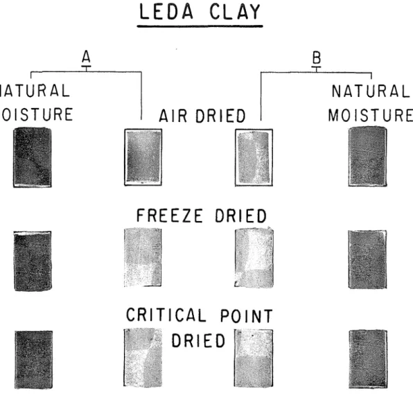
T h e shape ; ~ n t l composition of most minerals and the fabric of wcll intluratctl roclts a r c prob- aldy not scriously :lfTcctctl 11y (11-ying. Koncthc- Icss, a s m o i s t ~ i r c is rcmovctl, there is a consider- able rise in the force of surf;lcc tcnsio~l. ant1 this may seriously ~notlify thc arr:ungcmcnt of tl1c particlcs in \vc:lk matci-ials such as clays ant1 soils. 'Thcrc is also a possibility that thin platy particlcs may roll 1111 ant1 11ccomc t u l ~ u l a r . P r c - c;lutions havc to bc taken, thcreforc, to cnsurc that the m o r l ) l ~ o l o g ~ of the m ~ n c r a l s mid their spatial tlisposition in t h c sample c s a ~ n i n e d a r e the same as in thc ~ l a t r ~ r a l state of the material. Clay soils wcrc tlr~ctl by thrcc principal n ~ c t h - otls. I n the first mcthotl thc sample \\?as tlrictl u~itlcr ambient coiltlitions. hfany clay soils shoxv consitlerablc volume dccrcasc when a ~ r - t l r i e d
(fig. 1 ) aiid thcl-c is a strong lilielihood that fabric is affcctctl by this shrinkage.
I n the sccolitl mclhotl \\ a t c r was rcmovetl hy
f r c c ~ c - t l r j i ~ l j i o r lyol)hil~zation. A small sainple was frozen nut1 thc I ~ C rcmovcd 11y s ~ t l j l ~ m a t i o n
undcr v ; ~ c u u n ~ TVh~lc th e ice is I-rcing rc~novetl no meniscus shoultl fol-m in thc porcs and capil-
lary sl);~ccs l)et\\-ccn tlic pzlrticlcs, so surf:lcc tcnsion forccs shoultl I)c allsent. It is tlesiral~lc to avoitl the form;ltion of cryst:lls of ice, w l ~ i c h a r c 1)clicvcd to be oftcll rcsponsil~lc lor tlic i l l -
troduction of artifacts 1)y c a u s i ~ i g d;lmagc to the original fabric. Icc may IIC formctl in the vitrc- ous 01- glassy state most cflicic~itly by ultra-
I-apitl freezing to a tcml)cr;lturc of less than -130°C. I t is thought I~cst to frcczc the s;lrnplc l ~ y immcrsio~i in ;L vcsscl containil~g isopcnt:~nc
surrountlctl 11y licluitl
N,
in an outcr coiitxincr. If immcrscd tlircctly in liquidN,
thc s;lml)lc 11c- .:omcs s ~ i r r o ~ ~ n t l c t l 11y a layer of gas n.hicl1 has a n insu1;lting cficct, so thc tlrop in tcmpcraturc is less ral~itl ant1 ice crystals havc nlorc chnncc to form. Crystals xvon't grow in ice glass I~clon; -130°C (h'lcryman ant1 Icafig, 1955, 13. .542)I ~ u t , 111-cdictal)ly, sul~limation is also very slow a t this tcmpcraturc. I-Icncc frcczc-tlrying is nor- mally cal-rictl o ~ i t at liighcr t c m p c r ; ~ t u r c s : it is hopcd that the fabric of the clay iinpcdcs rc- crystallization sufficicntly to p~.c\.c~it scrious tlamagc from thc gro\vth of icc cryst;~ls.
T h c third mcthotl of drying may 11c rcfcrrcd to as thc critical poilit method. A t a t c m p c r ; l t ~ ~ r c ant1 prcss111-c allovc a ccrtain critical point the physical propcrtics of a liquid antl its vapor bc- come intlistinguisi~;il~lc. T11crcfo1-c the t \ r o phascs a r c no longer scjsaratcd 11); a l~ountlai-y
layer ant1 surfacc tension forccs \.anish. By this tcchniclue the water m;ly eitl~ci- l)c I-cmovctl cli- rcctly o r first I)e rel11;lcrtl 1)y a \\~atc~--lniscil)lc licliiid silch as alcohol antl t h c 11cw licl~iitl pllasc s~~l~scc[iicntly rcmovcd a t a tcnipcrat~u-c ancl jxcssurc ;!llovc its critical j)oi~ii. T h e \vatcr is rcl)l;~cctl l~ c c a u s c its critical point is somcwh:~t high (374°C. 3266 psi). 11~11crcas t h c critic;~l point of cth!.I alcohol is lo\vcr (213"C, 946.5 nsi). T h c r c a r c othcr l i t ~ l ~ i d s for \\.Ilicli tllc
a ,
critical point occurs ; ~ t still lo\\.cr t c m p c r a t ~ ~ r c s . Antlcl-son (1950), f o r c r ; u m ~ ~ l c , I-cl)l;lcctl t h c \vatel- in 1)iologic;ll 1i1ntcri;lls 11y licluitl C'O.!, t h c critical point of which occurs at 31°C :untl 73 atmosphcrcs prcssurc. As liq~iitl CO, is not miscible n ~ i t h water, the scrics rcplaccmcnt water
+
alcohol - - 3 atnyl acctate+
CO,(liquitl) was usctl. S o ~ n c samples of clay soil \Yere prcparcd in this xvay tlui-inx this I\-ork, 1)ut tllc f;ll)ric ;lppcarctl itlcntical to that of samples prcnarctl 1)y thc alcohol ~ncthotl.
By resorting t o spccial artifices it may IIC pos- sible to examine s a ~ n p l c s \vhich still contain w a t e r using t h e scanning clcctl-on microscope. Oatlcy, Nixon, ant1 Pcasc (1965) tlcscril~c a n arrangcmcnt usctl I)y Smith and 1)y Thol-~ilcy ill which t h c s a ~ n p l c cnclosctl in a spcci:~l ccll w a s surroundctl by water vapor a t thc satr1r;~tion prcssurc. T h c electrons gainctl access to t h e
TI-IE S C A N N I N G EL-ECTRON 11IICROSCOPE
L E D A
C L A Y
N A T U R A L
M O I S T U R E
1
A I R D R I E D
1
N A T U R A L
M O I S T U R E
F R E E Z E D R I E D
C R I T I C A L P O I N T
D R I E D
FIG. 1.-Volumc change o i scnsitivc clay soils di-ictl by tliffercnt mctl~ods.
A. Sample ovcrconsoli:ialcd; platy niincrals show prcferrctl orientation.
I?.
Salnplc allnost normally consolidated ; platy mincrals randomly oriented. samplc through a 50-micron wintloxv covcrc(lwith a thin film of collodion able to \vitI~stantl the pressure difference between the sample cell and the botly of the microscope. Alternativcly, i f thc samplc holder is conncctcd t o a supply of liquid K,;, outside t h e microscope, t h e specimen may bc csamincd in the frozen state i v i t l ~ o ~ ~ t re- moving the ice, since the vapor pressure of II,O is very low a t those temperatures.
A s previously noted thc scanning clcctroli mi- croscope givcs information about a surface. I t is thcrefore vitally importalit that the s ~ u f a c e examined be representative of the bulk mate- rial.
A s11rface may I)c csposcd 11y f r a c t m e o r cut- ting. T11 metallurgy it is a standard procedure to
csposc surfaces of polycrystallinc materials by fract~u-e. Cutting is a shcaring action (Kay, 1965, p. 216, p. 2 4 s ) which intluccs damage, thc depth ant1 estcllt of \vhich dcpcntl t ~ p o n thc mcthocl of cutting and thc nature of the mate- rial. It has I~ccn shoxvn to orient wet clay ( M a r - tin, 1966, p. 2 S I ) , f o r csample, and thc arrange- mcnt of the crystals on a cut surface is there- fore unlikely to rcprcsent the truc fabric of the l~ulli material. ~ 2 l t h o ~ 1 g h thc distul-bed layer can- not be cornplctcly rclnoved by mechanical grind- ing and polishing, its thickness can be consider- ably reduced. T h e extent of the damaged layer which remains is dependent upon the type and
similar to those in the Solcnlnofcn li~ncstonc. other non-carbonates. I t s concentratio11 at grain Figlire
2C
shows another view of the m a t r i sof the same rock. T h c contact area I~ctwcen many of the crystals is s u r p r i s i ~ ~ g l y small, ant1 the rock has a sintcrcd appearance. Some of the crystals have p r o j c c t i o ~ ~ s whicli 111-itlgc gaps from one crystal to the nest. 11s many present- day lime mutls a r c composcd of aragonite nccdlcs, the c l o ~ ~ g a t c f o w l of some of thcsc crjstals may illustrate a sl;lgc in the transfor- mation of ~iccdlc-like crystals into thc morc common, almost ccluiasial gi-ains of typical 111ic1-itc. T h e mccl~anisms Ily which this pro- cess may take p1;~cc havc Ilccn tliscusscd 11y 1;ollc
(1965, p. 3 5 ) , Frictlman (1961), Dodd (1966), Eathurst (1959, 1961), ant1 othcrs.
Pressure s o l ~ ~ t i o n ant1 clicn~ical d c p ~ s i i i o n of c:llcitc on free sul-faces of ~ i i l g l c ci-ystals and polycrystallinc aggregations al-c follo~vcd Ily gro\vtln into voids and porc spaces. Tlicsc, to- gcthcr ~ v i t h other processes, ccmcnt c a r l ~ o n a t c mud into rock. Fignrcs
2C,
D, ant1E
ant1 figurc 311 show samples of Ortlovician limcstonc col- lcctcd near Ottawa, Ontario. h cast shown i l lfigurc 2, D ant1 E, appcars to 11c a coating of ccrncntitious material n~liich cncloscd a grain that pullcd out when the roclc was fracturctl. T h e cast has a moltlctl appcaralicc and tlisplays a lobe-likc cstcnsion along the I ~ o u ~ l t l a r y IIC- tween t ~ v o grains. A somc\vl~at similar view of impul-c limcsto~ic is shown in fig~lrc
2F
along with the ~ ~ 1 s t of a rhoml~ic crystal m11icl1 mas p111lcd from the largcr anlictlral crystal when the 1-oc1~ mas fractnrctl.Tn nxuny of thc pl~otographs (figs. 217; 3A, P,, C, D,
E,
1;; 411) there a r c I ~ r a n c l ~ i n g aggrcga- tions which appcar somc~vhat lilic sma1lc1- vcl-- sions of the astrosclcrcids fo~lntl in ccrtain upper soil horizons in Vancouver (Rrytlon, Dorc, and C:lark, 1963). T h c aggrcgations may 11e ccmcntitous ~ n a t c r i a l tlcpositcd along gl-ail1 I~ountlarics or pl;itc-likc crystals of clay miner- als si~nilarly tlisposctl. Tf this matel-i;ll is a cc- menting agent, it is cvitlcntly younger in a g c than tlic crystals of which thc 111:ltris is c o n - posctl and diffcrcnt from then1 in composition. A s t l ~ c s c roclis a r c impure limcstoncs and con- tain 10 pcrccnt o r morc :~citl insolulrle r c s i d ~ ~ c , itsccms most lilicly that the grain bountlary matc- rial is composctl 1 : ~ r ~ c l y of clay minerals ant1
I~ountlarics nay 11e the r c s ~ ~ l t of espulsion from the carbonates tluring tliagcnesis, I ~ u t there is ;!lso ;uiothcr possil~ility. Clay mincl-als f l o c c ~ ~ l a t e in solutions containing a motlcratc c o ~ ~ c c ~ ~ t r a - tion of clcctl-olytes, ant1 clays dcpositctl under marine contlitio~ls gcncrally havc an open fab- ric. T h i s f ; ~ l ~ r i c may have I ~ c c n prcscrvcd 11y the early tlcposition of calcite ccmcnt upon the clay minerals, particulal-ly at the interparticle con- tacts; then carl~onatc diagcncsis may have talccn place later within a patchily tlistl-il~utctl clay mineral fran~c\\rork. T i so, thc sizc t o which t h e calcitc crystals coultl grtnv was limitcd by thc space available 11ct\vccn thc platy clay ~ilincrals, thc d i s p o s i t i o ~ ~ of n~hicli also prcdctcrmincd t h c location of tlic calcite grain-l~o~~~ltlzlries. T h u s tlic clay may liavc playctl a n ;~ctivc rather t h a n a passive role i n thc tliagcncsis of t l ~ c c a r l ~ o n - atcs.
Tt is also possil~lc that clay may affect the pcrmcal~ility of a sctlimcnt, causing a tlccrcasc in the ratc of migr;ition of porc solutions. In figurc 3 h tlicrc is a tl-ansition in tlic sizc of t h e calcitc cryslals from ;~l)ont
2
microns up t o ahout 10 microns. T h c grain bountlary m;~tcrial is conccntratctl in thc region whcrc the smallest crystals a r c locatctl, \vhcrcas contan~i~i:ltion is laclcing where the calcitc crystals a r c I:lrgc~-. TTcncc for thii \:lmplc, a t Icast, tlccrcasctl pcr- mcal~ility is prol)al~ly not tlic reason for thc smnllcr siyc of the c:~lcitc crystals in thc clay- co~ltaining arcas, 11cc;111sc a s l o ~ v ratc of supply of c;lrl~onatc wo111d 11c cspcclctl to Icatl to the g r o ~ v t h of largcr crystals (Gathurst, 1964, p. 3 3 ) .~ ' ~ O C C S P C P \ v l ~ i ~ I i affcct thc sizes of the con-
stitncnt CI-ystals in a rock have rccci\:ctl niucln attention in r e c c ~ ~ t litcraturc. Tf final crystal sizc \\;as tlctcrminctl I I ~ rccrystallization, irnp~ll-- itics I)ct\vccn the cl.ystals may havc h a ~ n p c r c d grain I~onntlary migration ant1 so have c:u~sctl tlccrcasctl coarscncss of c;~lcitc in clay-rich RVCaS.
!>olomitc crystals in a fine m a t r i s in 0 1 - d o v - cian roclis from near Icingston, Ontario, a r c shown in Iig111-cs 313 to 112; polishctl and ctchctl surfaces a r c slio\vn in f i g ~ ~ l - c 3P,,
C,
ant1 D ant1 fractured ant1 ctcllcd s ~ l r f a c c s in figures 3E, F , ant1 l.A. Polishing protl~lccs a t1isturl)cd s ~ ~ r f a c cFIG. 3.-Fine-grained limestones ant1 dolon~itic litnestones. ( 4 ) Ordovician limestoi~e, Ottawa, Ontario, C;ui~ada.
(B, C, D ) Ordovician tlolomitic limestone, I<ingstoti, Ontario, Ca~iatl:~. Polishcd, etchcd.
J .
E.
GILLOT?'
which h a s to he rcmovcd bcforc tlctail c a n 11c tlisccrtictl. P a r t of this d i s t ~ l r b c d l a ~ c r ~vliicli tlie etch failecl to rclnovc is visible in tlic lowcr p a r t of figurc 3B.
Dolomite-to-tlolo~~iitc contacts niav 11c sccn in figure 3 C ;~ncl clolomitc-to-matrix contacts in ligurcs 3 D , E ,
F,
ant1 4A. Tlic contact 1,ctwccn clolo~nitc r l i o m l ~ s and m a t r i x lias a n appcaralicc which I-cscml~lrs a lionrycoml~. Tlic apparclitly discontinuous ~ i a t u r c of tlic contact is surpris- ing. Tlic holcs m a y I)c casts of crystals prlllctl 01-torn out cl~iring samplc prcpar:ltion, t l i o ~ ~ g l i tlic
appeal-ancc is siliiilar on l~otli polislictl antl f r a c - tllrctl s111-fzlccs. If tliis is tlic c o r l ~ c t c s p l a n a t i o n tlicl-e shoultl IIC a s 111;lny p r o j c c t i o ~ i s a s clcprcs- sions on a f r a c t ~ i r e s~ ~ l - f a c c , alitl tliis sccms not t o bc tlic c a s c : frirtlicr~norc, tlie crposccl s ~ l r - faccs of somc tlolomitc crystals a r c quite ror~gli
(figs. 31;, 1 A ) . I n m a n y instances f r a c t ~ ~ r c lias not follo\vctl clcavngc plallcs in thc tlolomitc crystals 11rit lias t a l i c ~ l placc in tlic atlj;lccnt nia- tl-is. I t thris a p p c a r s t h a t p a r t of t h c ccmcnt which formcl-ly attachctl tlic tlolomitc CI-ystals t o t h c rcst of tlic m a t r i x h a s :~tlhcrctl to tlic crystal faccs \vlicn tlic rock nras fractured.
A n -
otlicr lilicly csplanation is tliat t h c ctcli rlsctl dllring samplc prcparation tlissol\lctl o111y tlic calcitc crvstals. T l i c lioncvcomb t c s t l l r c m a v I)? a I-csicll~c ( I F impurities originally concrntratctl a l o n g grai11 11o11nda1-ics. T h i s tcxtllrc is oftcn most a p p a r c n t n c a r tlc~lomitc r1ioml)s (fig. 3B, C, D ) a n d pcrhaps tlic distri1)ution of clay mincrals \vithin tlic sctlimcnt hat1 somc inflrlcncr on t h c sitcs of tlolomitization. I" 1)arti;ll c l c a r i n g o f non-car1)onatc.s sricli a s clay minerals f r o m within tlic g r o w i n g tlolomitc crystals m a y liavc taken placc ant1 so carlsctl a conccntl-ation to I~llild u p near tlic margins. I t is also possil~lc t h a t tlic h o ~ ~ c y c o r n l ~ t c s t l l r c rcprcscnts a tli+ tortcd prcscrvation of tlic original cdgc-to-facc o r randomly 5taclictl arraiigcmcnt i o r m c d I)y tlic dcpo\ltion of floccrllatctl a g g r c g a t i o n s of clay platclcts. T h i 5 clay m a y liavc 1)ccn partly assini- ilatccl (Gillott, 1963, fig. 1 ) and partly prlslied asiclc 11y tlic g r o w i n g clolomitc CI-ystals. Dcposi- tion of calcitc c r m c n t m a y liavc strcngthcncd tlic original flocculatctl clay fabric antl Icd to its prcscrvation. A tlolomitic linirstonc f r o m Ala- bama in \ \ ~ l i ~ c l i tllc a c i t l - i ~ ~ \ o l u l ~ l c contcnt w a s
only 0.57 pcrccnt did not show t h e l i o n c y c o ~ n l ~ tcstul-c a t m2ltrix-to-tlololiiitc collt;lcts (fig. 4 B ancl C ) . T h e tlolomitic limcstoncs f r o m n e a r Icingston a1-c typical r x ; ~ n i p l c s of I-oclis which m a y causc cspansion ant1 tlisl-rlption of concrctc \vhcn usctl a s coarsc aggi-rgatc with high alliali ccmcnt. Tnvcstigations suggest tliat t h e fallric of tlic rocli m a y 11c significant to a propcr untlcr- stantling of tlic mccllanism of tlic rcaction (Gil- lott, 1961; FTatllcy, 1964; S\\:cnson ant1 Gillott, 1967).
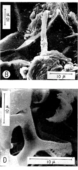
'I'lic l a r g c dcptli of focus of tlic s c a n n i ~ l g clcctron microscopc somctimcs givcs a ncur pcr- spccti\rc t o tllr al-I-angcmcilt of coarsc crystals. T h c cfrcct on Georgia wliitc m a r l ~ l c of a 6-mi1111tc ctcli in 1 : 1 acetic a c i d : w a t e r is illus- tratctl in figlll-c 41). l'hc gr;lin 1rountl;rry h a s 11ccn p r c f c r c ~ ~ t i a l l y attaclictl, ant1 t h c appc:lr;lncc of tlic calcitr c1cav;lgc is q11itc striliing. Dolo- mitc-to-tlolomitc ctlgr- ant1 poi~lt-contacts a r c sccn in figurc 4 E ancl 1; alltl clctails of ccmcntctl jrlnctions in figurc
5,
A alltlR.
ilr-gilltrcco~ts Rocks a ~ l d S o i l s
S:lmplcs of Ortlovician sli;llc antl muclsto~ic f r o m O ~ i t a r i o \vcrc csaminctl o n tlic s c : l n ~ ~ i ~ i g clrctl-on m i c r o s c o ~ ) ~ , a ~ i t l tlic appcarancc of t h r s c ind~lratctl roclis \\.as comparctl with t h a t of ~ n a r i n c tlcl~ositctl clay soils of post-Plcisto- ccnc a g c f r o m E;istc~-n C;a~iatla. Tlic oriclitation of tlic m i c a - t y l ~ c minrl-als ol)scr\~c(l in t h i ~ i scc- tions 011 tlic pct~-ogl-al>hic miel-oscopc was com-
parrtl \vitli tlir fine tlc'ail of tlic crystal a r r a n g c - m c n t s c r n 11y mr:lns of tlic s c a ~ i n i l i g c l c c t r o ~ i microscopc.
7 .
L
lic s1i:~lc samplc displayrtl wcll marIi?tl fissil- ity in liantl sprcimcn. i\,Iic~-oscopic thin s c c t i o ~ i s \\lcrc c ~ i t iiorm;~l antl parallel t o tlic lamination. E x a m i n a t i o n o n tlic p c t r o ~ r ; ~ p l i i c rnicroscopc under crossctl polarizcrs int1ic;ltctl a strong, tliougli not p r r f c c t , tlrgl-cc of 111-cfcl-I-ctl oricnta- tion of tlic niic;lccous minct-als. Tn tlic nintl- stoncs tlierc w c r c f c w casy plancs of parting, a n d micl-oscopic cz;uiiination confirmctl tliat tlicrc xvas littlc, if ally, prcfrrrctl orientation a m o n g tlic p1;lty mincrals.Practul-c sul-f:lccs of tlicsc s:l~nplcs w c r c e x - aminctl o n tlic scanning clcctron rnicroscopc. A t l o w - p o ~ v c r magnificatio~l tlic finely laycrcd
FIG. 4.-Contacts I~etwecn carl~onate mincrals.
(.A) Ortlovician tlolomitic limestone, ICingstoll, Ontario, Canada. Fractr~red, ctcl~ccl.
(B,
C) Dolomitic limestone, Alabama, u.S.11. ( D ) Georxia \VIiite Marhle. Polished, etcl~ed.FIG.
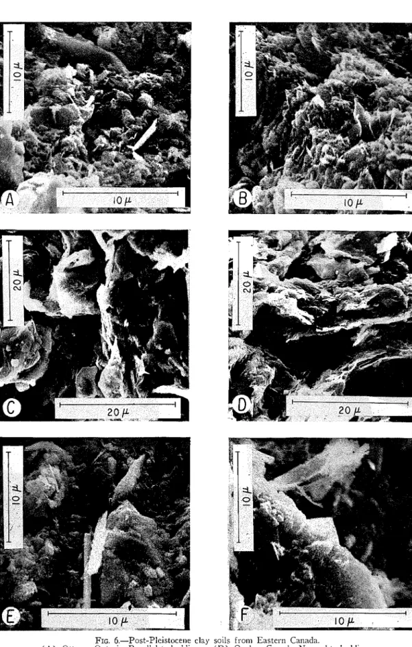
6.-Post-Pleistoce~le clay soils fro111 Eastern Canada.( A ) Ottawa, Ontario. Parallel to 1)etltling. ( D ) Quebcc, Canada. Nor111aI to 11cdtli11~.
(13) Ottawa, Ontario. Norm:~l to betltling. ( E ) Quebec, Canatla. Normal to bedtli~ig.
FIG. 7.-Siliceous rocks.
( A , B ) Agate, Brazil.
104
J .
E. GILLOTT
( ? ) crystals, togcthcr with riiorc ilcarly cq~li-di- mensional silt particles a r c t o bc scen in figure 6E. I n t h c upper portion of figure 6F t h e r e is w h a t a p p e a r s t o be a partially unrollcd crystal of a t t ~ b u l a r mineral. T h e remaintlcr of t h e mi- c r o g r a p h is taken up by platy mica-typc crystals together nlith somc laths anel a t lcast o n e o t h c r crystal which m a y bc tubular.
Silica llditzerals
A limitcd investigation h a s bccn m a d c of tlic appearance of f r a c t u r e s u r f a c e s of a g a t c a n d diatomaceous cartli. A t t h e magnifications em- ployed, a g a t e s u r f a c c s a r e generally not tlis- tinctivc, bnt q u a r t z crystal f o r m is occasionally devclopcd. T h i s is illustratccl in figure 7, A a n d B, in which a n ~ o r p l ~ o ~ ~ s - l o o l c i ~ ~ g silica, w i t h a f o r m rcminisccnt of a waterfall, is dcposited on crystals with wcll devclopcd faccs.
Diatornaccous e a r t h is composcd of t h e sili- ccotis tests of diatoms. T h c Inaterial h a s a strik- ing a p p e a r a n c e whcn vie\vcd o n t h c s c a n n i n g electron microscopc (fig. 7 , C , D , E , F ) . T h i s mcthod of investigation slio~lltl facilitate s t ~ ~ t l p of tlic micro-fossils ant1 m a y help in tlnder- stantling t h c h i g h sorptive capacity a n d impor- t a n t intlustrial propcrtics of t h e material. S o m e diatom slteletons a r e finely striated, a n d obser- vations under t h e transmission elcctron micro- scope h a v e shown t h a t t h e striations a r e m a d e
up o f ro\vs of small pores. A s the spacing is regular, tliatoms h a v c been ~isccl fo r sizc indica- tion a s a rcatly m a d c scalc.
CONCLUSIONS
T l l c s c a n n i n g clcctron rnicroscopc h a s consitl- cral)lc potential a s a tool in t h c petrographic in- vcstigation of fine-grained I-oclcs. T h c l a r g c depth of f o c ~ ~ s a t 11ig11 magnifications inakcs it itlcally suitcd t o observation of rough, i r r c g ~ l l a r ant1 finely p o r o ~ l s sl ~ r f a c c s . S a m p l c prcparation is simpler t h a n f o r t h e t r a n s n ~ i s s i o n clcctron mi- croscope, s o a l a r g e r numbcr of samplcs c a n bc cxamincd f o r a comparable e x p c n t l i t t ~ r c of t i ~ n c a n d effort.
I t is a p l c a s ~ ~ l - c t o aclaiowleclgc t h c courtcsy anel hclp of tlic staff a t t h e Canatlian P u l p and P a p c r Rcscarcli Association w h o o w n tllc scan- n i n g electron rnicroscopc o n which t h c rnicro- g r a p h s shown in this papcr w e r c talcen. T h a n k s a r e also exprcsscd t o 1'. J. L c f e b v r e f o r con- scientious t e c l ~ n i c a l assistance in prcparation o f samplcs. 'Iclpful colnments o n t h c m ; ~ ~ l u s c r i p t \vcre m a d c 1 ~ y P.
J.
Serctla. T h i s papcr is a con- tr11111tion from t h e Division of B ~ ~ i l d i n g R c - search, Kational R c s c a r c h Council of Canacla, a n d is p~tblislied \\-it11 tlic approval of t h e Direc- t o r of t h e Division.R E F E R E X C E S
ANDRRSON, T . F., 1950, A 111ctl10d for elimi~latitig gross artifacts in drying spccimcns: 211d International Conference Electron h,Iicroscopy Proc., p. 567-576.
ASIIHEE, M.
R.
A,, 1961, Thc crfects oE step strain pheliornena on scttlcmcnt calcl~latio~ls: 5th Inter~lational Conference on Soil Mcclia~i~cs and Foundation Eng. 3, p. 119.AYLMORE, L. A. G., A N D QUIRK, J. P., 1960, Domain o r turbostl.atic structure of clays: Nature, v. 187, p.
1016-1018.
I~.V~IIURS.T, R. G. C., 1959, Diagencsis in Mississil~pian calcih~titcs and ~~seutlol~rcccias: Jour. Seditiicnt:u.y Petrology, v. 29, p. 365-376.
--- 1964. The replaccnicnt of aragoliite by calcite in t11e ~nolluscan sl~cll wall, p. 3.77-376, .in Embric, J., Approaches to I'alcoccology. J. \\?ilcy. Ncw Yorlc.
BO\\JDEN, F . B., A X D MCAUSLAN, J., 1956, Slow dccotiiposition of cxplosivc crystals: Nature, v. 178, p.
408-410.
I ~ R Y D O N , J. E., DORE, 141. G., A N D CLARK, J. S., 1963, Silicified plant asterosclcreids prescrvecl in soil: Soil
Sei. Soc. America Proc., v. 27, p. 476177.
CASAGRANDE, ARTI-TUR, 1940, T h e Structure of clay and its importa~lce in fou~itlation e n g i n e e r i i ~ ~ , ,irz Con- tril~utions to soil mechanics, 1924 to 1940: Boston Soc. Civil Engrs., p. 72-125.
CASTLE, J. E., A N D MASTERSON, H. G., 1966, T h e role of difillsion in tlic oxitlati011 of mild stccl in high
tc~liperature aqueous solutions : Corrosion Scicnce, v. 6, p. 93-!04.
CI-I.AVE, I<. E., 1954a, Aspects of the biogeochcinistry of magnesium: 1, Calcareous r n a r i ~ ~ c organisms: Jour.
Geology, v. 62, p. 266-283.
1954b, Aspects of the b i ~ ~ e o c h e m i s t r y o l iiiagnesillm : 2, caleareor~s scdiiiicnts aiid roc1.r~ : Jour. Geol- ogy, v. 62, p. 587-599.
CLOUD, P . E., JR., 1962, Environment of calcium carbonate tleposition wcst of Andros Island, Bahamas: U.S. Geol. Sur. Prof. Paper 350, 138 p.
CRAWFORD, C. B., 1968, Quick clays oE Eastern Cailada: gngiileering Gcology, in press.
DODD, J. R., 1966, Processes of conversioii of aragon~tc to calcite with examples from the Cretaceous oE Texas: Jour. Sedimentary Petrology, v. 36, p. 733-711.
DOIIRRTY, P .
E.,
A N D LEOMBRUNO, R. R., 1961, Transmission electron microscopy of glass-ceramics: Jour. Am. Ccratn. Soc., v. 47, p. 368-370.EMERSON, \V. W., 1962, The swelling of Ca-rnontmorillonite due to water absorption; 2. Water uptalte in
T H E SCAlVNIATG E L E C T R O N AlICXOSCOPE
105
FOLK, R L., 1939, Practical petrograpl~ic classification of limcstoncs : Am. Assoc. I'ctrol. Geologists Cull., v. 13, p. 1-38.
-
1963, Some aspects of recrystallization in ancient limestor~cs, p. 1-8 irz I'ray, L. C., and Murray, I?. C., eds. L)olomitization ant1 limestone tliagciicsis, a synl~osium. Soc. Econ. Paleo~~tologists & Mineral- ogists, Spcc. Piibl. No. 13, Tulsa, 180 p.I;I<ILDBIAN, C;. U., 1964, Early tliagcncsis ant1 iitliificatioi~ in carbonate sctlimcnts: Jour. Scdimcntary Pct- rology, v. 31, p. 777-813.
G.\HI)NER, G. A., A N D CAIIN, R.
Mr.,
1966, Tlic use of a scaniii~ig clcctron microscopc to csarninc whislicr g r o ~ t h 011 an iron-aluminum alloy: .Tour. Materials Sci., v. 1, p. 211-212.GILLOTT, J. E . , 1963, Petrology of dolomitic limestones, IGllgston, Ontario, Canatla: Gcol. Soc. Amcrica Bull., v. 74, p. 759-778.
-- 1964, Meclia~ilsm and 1.rinctics o i es1)ansion in tllc alliali-carbo~~atc rock reaction: Canadian Jour. E:lrtli Sci., v. 1, p. 121-145.
:L-[..\I)LEY, . D. .~ \V., 1961, Alkali reactivity of dolomitic carbonate rocl;s: Hig11w;iy Rcscarcl~ Rccortl, No. 45,
p. 1-17.
IIOPMANN, E., 1912, Ncucs ;ui dcr Chemic tlcs Tones: Ange\vantltc-Clic~iiic, 1.. 55, p. 283-289.
ICAY, D. H., etl., 1965, Techniques for clcctron microscopy. Blacliwcll, O x f o r d , 560 p.
I ~ a r ~ e , T . \V., 1953, T h e structure of inorganic soil: Am. Soc. Civil Engrs. Proc., Jour. Soil Mcci~. Fountl. Div., 79, Scparate No. 315, p. 1 1 9 .
I > o w r : ~ s , r ~ a r , I-I. A,, 1963, Biologic probleins rclating to the composition and diagcnesis of setliments, itt Don-
nclly, T . \\'., etl., T h c earth sciences. Rice Univ. Semi-centennial Publ., p. 137-195.
MACICINTOSII, I.
M.,
1965, Applications of the scanning clcctron microscope to solitl-state dcviccs: Inst. Electrical and Electronics Engrs. Proc., v. 53, p. 370-377.MCGR~\TII, J.
T.,
BUCHANAN, J. G., A N D TIIURSTON, R . C. A., 1962, h stildy 01 fatigue and impact lr;ic- turcs with tlie scanning electron niicroscope: Jour. Inst. Metals, v. 91, p. 3 4 3 9 .L ~ : \ I ~ T I N , R. T., 1966, Quantitative fabric of \vet kaolinite: Clays alltl Clay h~linerals, Proc. 11th National Conf., Berl;cley, Calif., p. 271-287,-
>II:RYNAN, 1-1. T., . ~ N D KAFIG, E., 1933, T h e study of froze^^ specimens, ice crystals ant1 ice crystal ~ r o w t h
I)y-electron microscopy: S a v a l klctlical 1Ces. Inst., Bctllesda, Maryl;uid, Res. Report N31 000 018.01.09, p 329-544.
L<~INI<OFF, I., ANn NIXON, \\I. C., 1966, Scanning electron microscopy of graphite growth in iron ant1 nicltcl
alloys: Jour. Appl. Phys., 1.. 37, p. 181818.53.
MUSC..AN, NI:CBIE.~I'IN, A N D JESSEN, 'F. \I[., 1963, Stutlics in iractionatetl mo~~tmorillonitc suspensions: Clays
ant1 Clay Minerals, Jour. Proc. 11th Conf., Pergamon Prcss, Oxford, p. 282-291.
OATLEY, C. \'I[., NIXON, \V. C., A N D PEASE, R. F. W., 1965, Scanning clcctron niicroscopy : Atlvan. Elec- tronic and Electron Pllysics, v. 21, 1). 181-247.
PE.~SE,
R.
F. \<I., A X D NIXOX,\Y.
C., 1963, High resolution scanning electron microscopy: Jour. Sci. Instr., v. 42, p. 81-83.PI:ASE, I?. F. W., A N D PI.OC, R . A., 1965, Scanning electron microscopy a s applietl to tlie oxidation o i iron: hlctallurgical Soc. of A.I.M.E. Trans., v. 233, p. 1949-1954.
PURDY,
E..
G., 1963, Recent calcium carbonatc facics of tlie Great Bal~:uila Ea111.r: 2. Scdimcntary facies: Tour. Geology, v. 71, p. 472497.SCI-IOPIELD, R . I<., A N D SAMSON, H . R., 1953, T h e dcflocculation o l l<aoli~~itc suspensio~is ant1 tile accompally-
ing cl~ange-over from positive to negative cl~loritle atlsorption : Clay Minerals Bull., v. 2, p. 4.5-51. SLIART, PETER, 1967, Particle arrangen~ents in Iiaolin: Clays allti Clay Mincr:~ls, Proc. 15th Conference;
Pittsburgh, Pennsylvania, p. 211-254.
Sarr.1.1~1, K. C. .4., 1961, Scanning, 1). 211-2.51, i i r Encyclopctlia o i microscopy. Kcinl~oltl, S e w Yorl;, 1). 693. S \ \ r ~ ~ ~ ~ ~ , 1:. G., AYI) GILI.O.IT, .I. E., 1967, Alkali rcacti\iit>, of dolomitic limestone aggregate: Mag. COII-
cretc Research. v. 19, p. 95-101.
TI:I:ZAGHI, I<:\RL, 1925, Ertll~aumcchanil~ auf I~otlcnphysiknlisclic~- G r u ~ ~ t l l a g c , Leipzig/Vielllia, 399 p. ' r r r o n ~ r o x , P. R., 1965, T h e scanning electroll microscope: Science, v. 1, p. 66-71.
TI[OI:NTON, P .
R.,
JAMES, D . \<I. F., LE\\;IS, C., )\XU RR.~I)IIOKD, A , , 1966, Silicoli whisker growth hy thevapour-liquitl-solitl process : Pliil. Max., v. 14, p. 165-177.
TII'FER, C. F . , DAM, D. I., A N D WI:I.I.S, 0. C., 19.59, S ~ ~ r f a c e fracture 111arLings 011 alpha iron crystals : Jour.
Iron and Stcel Illst., v. 193, p. 132-111.
Tnor.r.or~, D . H., A N D CFIAN. C. I<., 1960, Soil structure ant1 tlie step strain gl~cnomcna: Jour. Soil Mecli.