HAL Id: hal-02428142
https://hal.archives-ouvertes.fr/hal-02428142
Submitted on 5 Jan 2020
HAL is a multi-disciplinary open access
archive for the deposit and dissemination of
sci-entific research documents, whether they are
pub-lished or not. The documents may come from
teaching and research institutions in France or
abroad, or from public or private research centers.
L’archive ouverte pluridisciplinaire HAL, est
destinée au dépôt et à la diffusion de documents
scientifiques de niveau recherche, publiés ou non,
émanant des établissements d’enseignement et de
recherche français ou étrangers, des laboratoires
publics ou privés.
Space rescaling in the MFS method improves the ECGI
reconstruction
Pauline Migerditichan, Mark Potse, Nejib Zemzemi
To cite this version:
Pauline Migerditichan, Mark Potse, Nejib Zemzemi. Space rescaling in the MFS method improves the
ECGI reconstruction. CinC 2019 - Computing in Cardiology 2019, Sep 2019, Singapour, Singapore.
�hal-02428142�
Space rescaling in the MFS method improves the ECGI reconstruction
Pauline Migerditichan
1 2 3, Mark Potse
1 2 3, Nejib Zemzemi
1 2 31
Institute of Mathematics, University of Bordeaux, Talence, France
2
INRIA Bordeaux Sud-ouest, CARMEN Team, Talence, France
3
Electrophysiology and Heart Modeling Institute (IHU-Lyric), Pessac, France
Abstract
The method of fundamental solutions (MFS) has been extensively used for the electrocardiographic imaging (ECGI) inverse problem. One of its advantages is that it is a meshless method. We remarked that the using cm in-stead of mm as a space unit has a high impact on the re-constructed inverse solution. Our purpose is to refine this observation, by introducing a rescaling coefficient in space and study its effect on the MFS inverse solution. Results are provided using simulated test data prepared using a reaction-diffusion model. We then computed the ECGI in-verse solution for rescaling coefficient values varying from 1 to 100, and computed the relative error (RE) and cor-relation coefficient (CC). This approach improved the RE and CC by at least 10 % but can go up to 40 % indepen-dently of the pacing site. We concluded that the optimal coefficient depends on the heterogeneity and anisotropy of the torso and does not depend on the stimulation site. This suggests that it is related to an optimal equivalent conduc-tivity estimation in the torso domain.
1.
Introduction
Electrocardiographic imaging (ECGI) is a noninvasive technique to assess the electrical potential on the epicardial surface from measures realized on the thoracic surface. A meshless approach that employs the method of fundamen-tal solutions (MFS) has been adopted [1] to solve the in-verse problem of electrocardiography. Its performance is similar to other, more elaborate techniques such as finite-element models [2]. However, the MFS assumes a homo-geneous isotropic torso conductivity. In reality several or-gans have highly deviating conductivities and some, such as the skeletal muscle, are strongly anisotropic. These heterogeneities have an important effect on forward solu-tions of the surface potentials [3, 4], and therefore might affect the quality of the MFS solution as well. To as-sess the severity of this problem we tested the MFS on data generated with homogeneous, inhomogeneous, and inhomogeneous-anisotropic forward models. In addition
we investigated whether a scaling factor added to the MFS kernel can compensate for it.
2.
Methods
2.1.
Standard MFS
The solution given by the MFS approach is represented in the form of a linear superposition of source functions (Laplace fundamental solutions) located on a set of virtual source points yj, j = 1 . . . M over an auxiliary surface [1].
The method uses a set of measured body surface potentials u(xi) at electrode positions xi, (xi|i = 1 . . . N ) and
at-tempts to express these in terms of the source functions with weight factors aj. The source functions and aj can
then be used to predict the potential at any point on the torso or cardiac surface.
When using Dirichlet and Neumann conditions we ob-tain the linear system
A~a = ~b (1) where ~a = (a0, a1, . . . , aM)T, ~b = (u(x1), . . . u(xN), 0, . . . , 0)T, and A = 1 f (kx1− y1k) · · · f (kx1− yMk) .. . ... · · · ... 1 f (kxN − y1k) · · · f (kxN− yMk) 0 ∂f (kx1− y1k) ∂n · · · ∂f (kx1− yMk) ∂n .. . ... · · · ... 0 ∂f (kxN− y1k) ∂n · · · ∂f (kxN − yMk) ∂n (2)
in which f (r) = 1/(4πr) is the fundamental solution of Laplace’s equation in 3D, and kx − yk is the 3D euclidean distance between points x and y.
2.2.
MFS with scaling factor
We added a rescaling coefficient α to the kernel of the MFS matrix. The approximate solution of the MFS relies upon the Laplace fundamental solution which hinges upon the euclidean distance r = kx − yk . We made the as-sumption that reducing the distance will help to make our problem better conditioned. To do this let R be the new reduced distance expressed by:
R = r α , with α ≥ 1 f (R) = f (r α) = αf (r). (3) Similarly, ∂f (R) ∂n = ∂f (αr) ∂n = α 2∂f (r) ∂n (4)
Taking this new distance R in the MFS matrix (2) amounts to use the old distance r in the matrix Aα given by the
expression 1 αf (kx1− y1k) · · · αf (kx1− yMk) .. . ... · · · ... 1 αf (kxN − y1k) · · · αf (kxN − yMk) 0 α 2∂f (kx 1− y1k) ∂n · · · α2∂f (kx 1− yMk) ∂n .. . ... · · · ... 0 α 2∂f (kx N − y1k) ∂n · · · α2∂f (kx N− yMk) ∂n (5) In both cases the Tikhonov regularization method [5] with a fixed parameter λ = 10−2was used to stabilize the solu-tion and obtain ~a.
2.3.
Simulated data
Test data were prepared with a reaction-diffusion model of the heart on a finite-difference mesh with 0.2 mm res-olution. Computed transmembrane currents were trans-ferred to a torso model with 1-mm resolution to com-pute torso potentials [6]. Three sets of parameters were used for the torso conductivities. The first was fully ho-mogeneous and isotropic (HOM), the second piecewise heterogeneous (lungs, liver, blood, skeletal muscles, and
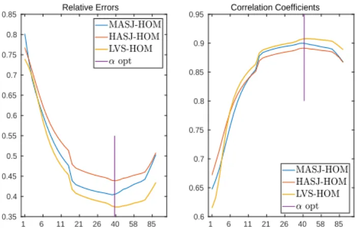
1 6 11 21 26 40 58 85 0.35 0.4 0.45 0.5 0.55 0.6 0.65 0.7 0.75 0.8 0.85 Relative Errors 1 6 11 21 26 40 58 85 0.6 0.65 0.7 0.75 0.8 0.85 0.9 0.95 Correlation Coefficients
Figure 1. Relative errors and Correlation coefficients of homogeneous model (HOM) with pacing site in mid ante-rior septal junction (MASJ), high anteante-rior septal junction (HASJ) and left ventricular septum (LVS).
the remaining tissue) (HET) and isotropic, and the third model was the same as the second but the skeletal mus-cles were anisotropic (ANISO). We simulated three cases with different stimulation sites: mid anterior septal junc-tion (MASJ), high anterior septal juncjunc-tion (HASJ) and left ventricular septum (LVS). All simulations were performed with the Propag-5 software [7] on a Bullx cluster machine.
2.4.
Evaluation of reconstructed potentials
To compare the reconstructed potentials on the epi-cardium with the simulated ones correlation coefficients (CC) and relatives errors (RE) were computed through time and then averaged to get only two values for each scaling factor. RE quantifies the amplitude difference and CC the pattern similarity between the simulated and the computed potentials.
3.
Results
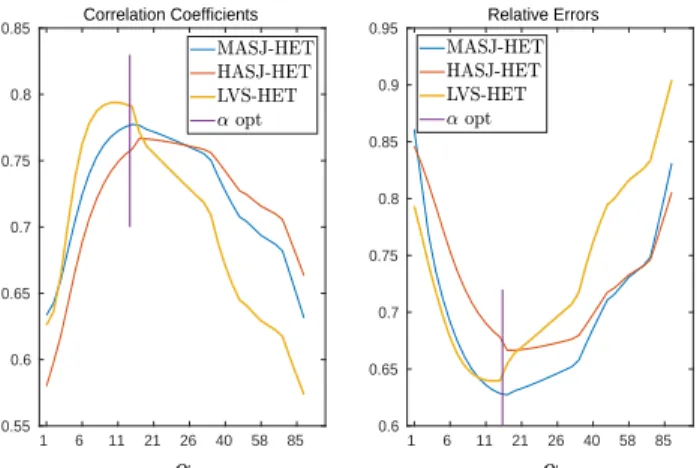
We studied the influence of the α factor inside the MFS matrix for α ∈ [1, . . . , 95] with three different pacing sites in each of the three models. Results for a com-pletely homogeneous and isotropic torso model are shown in Figure 1, those for a heterogeneous and isotropic one in Figure 2, and those for a heterogeneous model with anisotropic skeletal muscle in Figure 3.
The results show that RE and CC vary widely with α and that the shape of the error function was different for each model. For the HOM model, small values of α resulted in the largest errors (Figure 1), whereas for the ANISO model they resulted in the smallest errors (Figure 3). For the HET model, both small and large α values resulted in
1 6 11 21 26 40 58 85 0.55 0.6 0.65 0.7 0.75 0.8 0.85 Correlation Coefficients 1 6 11 21 26 40 58 85 0.6 0.65 0.7 0.75 0.8 0.85 0.9 0.95 Relative Errors
Figure 2. Relative errors and Correlation coefficients of heterogeneous and isotropic model (HET) with pacing site in mid anterior septal junction (MASJ), high anterior septal junction (HASJ) and left ventricular septum (LVS).
1 6 11 21 26 40 58 85 0.6 0.7 0.8 0.9 1 1.1 1.2 1.3 Relative Errors 1 6 11 21 26 40 58 85 0.5 0.55 0.6 0.65 0.7 0.75 0.8 Correlation Coefficients
Figure 3. Relative errors and Correlation coefficients of heterogeneous and isotropic with anisotropic skeletal mus-cles model (ANISO) with pacing site in mid anterior septal junction (MASJ), high anterior septal junction (HASJ) and left ventricular septum(LVS).
relatively large errors and the optimum solution was found for intermediate values.
For all six cases an “optimal” scale factor could be found. The effect of this factor on the reconstruction of the signal is summarized in tables 1 and 2.
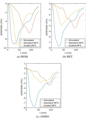
To illustrate the impact of α-optimization on recon-structed electrograms we reconrecon-structed the signal on the heart for the standard MFS approach and the one with op-timal scaling and compared them to the simulated one for a location near the stimulation site (LVS in this case) as shown in Figure 4.
Table 3 shows the estimated activation time at the pac-ing site position. It is defined as the instant of steepest
Pacing Site Model MFS Scaled MFS MASJ HOM 0.80 0.40 HET 0.86 0.63 ANISO 0.88 0.72 HASJ HOM 0.77 0.45 HET 0.85 0.67 ANISO 0.87 0.77 LVS HOM 0.74 0.37 HET 0.83 0.69 ANISO 0.83 0.64
Table 1. Mean RE of the reconstructed potentials along the time for the standard MFS Method and the “optimal” scaled method.
Pacing Site Model MFS Scaled MFS MASJ HOM 0.65 0.90 HET 0.63 0.78 ANISO 0.62 0.72 HASJ HOM 0.67 0.89 HET 0.58 0.77 ANISO 0.57 0.68 LVS HOM 0.61 0.91 HET 0.63 0.79 ANISO 0.63 0.78
Table 2. Mean CC of the reconstructed potentials along the time for the standard MFS Method and the “optimal” scaled method.
Model Simulated MFS Scaled MFS HASJ-HOM 9 ms 54 ms 25 ms
HASJ-HET 9 ms 55 ms 23 ms HASJ-ANISO 9 ms 52 ms 22 ms Table 3. Estimated activation time near the pacing site for the standard MFS Method and the “optimal” scaled method.
downstroke in the electrogram. The error approximates 45 ms for the original MFS method in each case, and ap-proximately 15 ms error for the scaled MFS approach. The modified method was also better at reconstructing the deep S wave seen in the simulated signal.
4.
Discussion and conclusions
By applying the MFS to torso potentials with homoge-neous and inhomogehomoge-neous torso models we have shown that torso heterogeneity reduces the quality of the MFS in-verse solution, both in terms of RE and CC. In addition we have shown that the use of a scaling factor in the MFS can reduce the RE in estimated cardiac potential by upto 50 % and improve the CC by upto 30 percent-points. We
0 50 100 t (ms) -10 -8 -6 -4 -2 0 potentials (mv) Simulated Standard MFS Scaled MFS (a) HOM 0 50 100 t (ms) -8 -7 -6 -5 -4 -3 -2 -1 0 potentials (mv) Simulated Standard MFS Scaled MFS (b) HET 0 50 100 t (ms) -8 -7 -6 -5 -4 -3 -2 -1 0 1 potentials (mv) Simulated Standard MFS Scaled MFS (c) ANISO
Figure 4. Comparison of reconstructed potentials for HASJ - standard MFS (red line) and scaled MFS (orange line) - against simulated one (blue line) in a point near the pacing site for three different torso models (a), (b) and (c).
conclude that the optimal coefficient depends on the het-erogeneity of the torso and does not depend on the stimu-lation site. In fact for the three stimustimu-lation cases the value of the optimal coefficient was 38 (respectively, 11 and 13) for the HOM (respectively HET and ANISO) case. We can suppose that the obtained optimal coefficients are cor-related with an optimal equivalent conductivity in the torso domain.
In terms of RE and CC our correction worked best in case of a homogeneous torso model. Thus, even with this correction, the torso heterogeneity remains a prob-lem for the MFS. Yet, if we focus on activation time this method shows some promising results by reducing errors by around 30 ms.
In this study we fixed the Tikhonov factor to understand the results more easily. Using a method to find the
opti-mal regularization factor as described by Barnes and John-ston [8] could improve them. Moreover, to determine the optimal value of α we need to know the true solution. For practical applications it would be of interest to develop a method looking for both the optimal regularization factor and the optimal scaling factor at the same time.
Acknowledgments
This work was granted access to HPC resources of CINES under GENCI allocation 2019-A0050307379. This work was also supported by the French National Research Agency, grant references ANR-10-IAHU04-LIRYC.
References
[1] Wang Y, Rudy Y. Application of the method of fundamen-tal solutions to potential-based inverse electrocardiography. Annals of biomedical engineering 2006;34(8):1272–1288. [2] Karoui A, Bear L, Migerditichan P, Zemzemi N. Evaluation
of fifteen algorithms for the resolution of the electrocardio-graphy imaging inverse problem using ex-vivo and in-silico data. Front Physiol 2018;9:1708.
[3] Gulrajani RM, Mailloux GE. A simulation study of the ef-fects of torso inhomogeneities on electrocardiographic po-tentials, using realistic heart and torso models. Circ Res 1983;52:45–56.
[4] Keller DUJ, Weber FM, Seemann G, D¨ossel O. Ranking the influence of tissue conductivities on forward-calculated ECGs. IEEE Trans Biomed Eng 2010;57:1568–1576. [5] Tikhonov AN, Arsenin VI. Solutions of ill-posed problems,
volume 14. Washington DC: Winston & Sons, 1977. [6] Potse M. Scalable and accurate ECG simulation for
reaction-diffusion models of the human heart. Front Physiol 2018; 9:370.
[7] Krause D, Potse M, Dickopf T, Krause R, Auricchio A, Prinzen FW. Hybrid parallelization of a large-scale heart model. In Keller R, Kramer D, Weiss JP (eds.), Facing the Multicore-Challenge II, volume 7174 of Lecture Notes in Computer Science. Berlin: Springer, 2012; 120–132. [8] Barnes JP, Johnston PR. Application of robust generalised
cross-validation to the inverse problem of electrocardiology. Computers in biology and medicine 2016;69:213–225.
Address for correspondence: Pauline Migerditichan Inria Bordeaux Sud-Ouest,
200 avenue de la vieille tour, 33405 Talence, France. [email protected]