HAL Id: inserm-00922212
https://www.hal.inserm.fr/inserm-00922212
Submitted on 24 Dec 2013
HAL is a multi-disciplinary open access
archive for the deposit and dissemination of
sci-entific research documents, whether they are
pub-lished or not. The documents may come from
teaching and research institutions in France or
abroad, or from public or private research centers.
L’archive ouverte pluridisciplinaire HAL, est
destinée au dépôt et à la diffusion de documents
scientifiques de niveau recherche, publiés ou non,
émanant des établissements d’enseignement et de
recherche français ou étrangers, des laboratoires
publics ou privés.
Delayed Hemolytic Transfusion Reaction in Sickle Cell
Disease
Narcisse Elenga
To cite this version:
Narcisse Elenga. Delayed Hemolytic Transfusion Reaction in Sickle Cell Disease. American Journal
of Clinical Medicine Research, 2013, 1 (3), pp.40-44. �10.12691/ajcmr-1-3-2�. �inserm-00922212�
Available online at http://pubs.sciepub.com/ajcmr/1/3/2 © Science and Education Publishing
DOI:10.12691/ajcmr-1-3-2
Delayed Hemolytic Transfusion Reaction in Sickle Cell
Disease
Narcisse Eleng a*
Service de Pédiatrie, Centre hospitalier de Cayenne,Cayenne cedex, Guyane française
*Corresponding author: [email protected]
Received December 31, 2012; Revised May 16, 2013; Accepted May 17, 2013
Abstract
Patients with sickle ce ll disease frequently require red blood cell transfusions . However, transfusions can cause delayed hemolytic transfusion reaction (DHTR) , a serious and potentially life-threatening complication of alloimmunization that results in hemolysis of transfused as well as patients’ own red cells . Although we are beginning to understand some of the pathophysiology and risk factors associated with alloimmunization, optimal management of DHTR in this patient population is still under debate. Here, I will review the clinical features, pathophysiology, laboratory evaluation, current strategies for management and prevention of DHTRs. Given that DHTRs are associated with massive hemolysis , it is reco mmended that all patients with sickle cell disease receiving transfusions are carefully and systematically monitored after each transfusion.Keywords
: delayed hemolytic transfusion reaction, sickle cell disease1. Background
Red blood cell (RBC) transfusion is often used for treatment and management of sickle ce ll disease (SCD). The purpose of RBC transfusion is to increase oxygen distribution to the tissues and/or to replace the rig id sickle-shaped RBCs with healthy deformable RBCs. However, because of the frequency of trans fusions in their life-t ime, patients with SCD beco me e xposed to RBCs alloantigens of donor units, ma king the m more like ly to produce alloantibodies, which in turn puts them at risk for delayed hemolytic transfusion reactions (DHTRs). In addition, e xchange transfusions, which a re often used for these patients, may increase the risk o f DHTR. Th is is because there is increased red cell utilizat ion with e xchange transfusions, with more e xposure to foreign erythrocyte antigens and increased volume of transfused erythrocytes susceptible to he molysis during DHTR. A llo immun izat ion in SCD has a reported inc idence of 20% to 50% [1,2]. Red cell alloimmunizat ion is frequent because of the antigen disparities between patients of African descent and donors of European ancestry [3,4,5].
Table 1. Pathophysiological mechanisms of DHTR 1- Differences in erythrocyte antigens between blood donors of European descent and patients of African descent (Yazdanbakhsh et al. 2012)
2- Interaction and destruction of the patient’s own cells (HbSS/reticulocytes) and transfused cells by activated macrophages (Platt et al. 2000)
3- Destruction of antigen negative RBCs during immune hemolysis of antigen positive RBCs « bystander hemolysis » (King et al, 1997) 4- Increased RBC exposure of phosphatidylserine (Lang et al, 2005) 5- Acceleration of eryptosis( Petz et al, 1997)
6- Association of alloimmunization with particular HLA subtypes (Hoppe et al. 2009)
The DHTR syndrome is a serious and potentially life-threatening complication of a llo immun ization, wh ich has
a reported incidence of 4% to 11% [1,3]. It is characterized by recurrence of d isease complications such as recipient RBCs destruction. In some cases, no detectable antibody is present. DHTR is associated with a significant morb idity or mortality. The pathophysiology of this syndrome re mains unclear; especially when there is no detectable antibody. Accelerated apoptosis of donor RBCs has been recently suggested as a potential mechanis m of rec ipient RBC destruction during DHTR
[6,7,8].
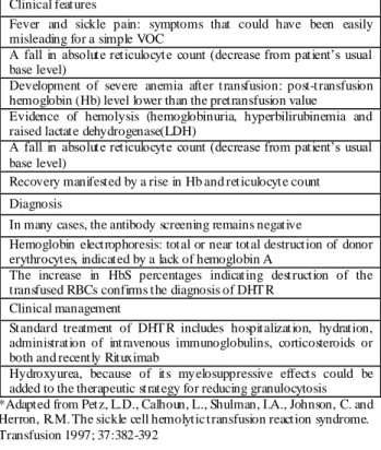
Table 2. Components of delaye d haemolytic transfusion reaction in sickle cell disease*
Clinical features
Fever and sickle pain: symptoms that could have been easily misleading for a simple VOC
A fall in absolute reticulocyte count (decrease from patient’s usual
base level)
Development of severe anemia after transfusion: post-transfusion hemoglobin (Hb) level lower than the pretransfusion value
Evidence of hemolysis (hemoglobinuria, hyperbilirubinemia and raised lactate dehydrogenase(LDH)
A fall in absolute reticulocyte count (decrease from patient’s usual
base level)
Recovery manifested by a rise in Hb and reticulocyte count Diagnosis
In many cases, the antibody screening remains negative
Hemoglobin electrophoresis: total or near total destruction of donor erythrocytes, indicated by a lack of hemoglobin A
The increase in HbS percentages indicating destruction of the transfused RBCs confirms the diagnosis of DHT R
Clinical management
Standard treatment of DHT R includes hospitalization, hydration, administration of intravenous immunoglobulins, corticosteroids or both and recently Rituximab
Hydroxyurea, because of its myelosuppressive effects could be added to the therapeutic strategy for reducing granulocytosis *Adapted from Petz, L.D., Calhoun, L., Shulman, I.A., Johnson, C. and Herron, R.M. The sickle cell hemolytic transfusion reaction syndrome. Transfusion 1997; 37:382-392
American Journal of Clinical Medicine Research 41
Signs and symptoms of DHTR are quite simila r to those of vaso-occlusive crisis (VOC) in patients with sickle cell disease [9,10]. Th is makes the diagnosis of DHTR and appropriate therapeutic decisions still a cha llenging issue to clinic ians in charge of patients affected by DHTR.
In this review, we p resent a synthesis of the current understanding of the pathophysiology of allo immun izat ion, as well as the clinica l features, diagnosis and manage ment strategies of DHTR in SCD (Tab le 1 and Table 2).
2. Pathophysiology
The pathophysiology of DHTR in SCD patients has not been completely e lucidated. Classically, DHTR is ascribed to a reaction between anti-RBC antibodies produced by the recipient and antigens expressed by the donor RBCs
[6,7,8,9,10]. It re ma ins unclear why some patients develop strong alloantibody responses following initia l RBC transfusions, while others do not despite mult iple transfusions. However, anti-RBC antibodies are often undetectable in SCD patients with DHTR [1,4,7]. The high proportion of DHTR cases without identifiable antibodies suggests that multip le factors are involved in the pathophysiology of this life-threatening comp lication. 1- Differences in e rythrocyte antigens between blood donors of European descent and patients of African descent.
It is generally accepted that the high rate of allo immun izat ion in SCD patients is main ly due to the polymorphic diffe rences in immunologic RBC antigens between the predominantly wh ite general blood donors and patients of predominantly African descent [11,12]. In the countries, where donors and patients are racially more homogeneous, low rates of alloimmunization have been reported [13,14]. These reports support the idea that rac ial antigenic differences account for inc reased allo immun izat ion rates.
Antigenic diffe rences between donors and SCD patients could be represented at three levels of co mple xity:
- First, there is a substantial difference in the prevalence of some co mmon but highly immunogenic RBC antigens between donors and transfusion recipients. The following blood groups: C and E in the Rhesus (RH), K in the Kell (KEL), Fya in the Duffy (FY), Jkb in the Kidd (JK), and S in the MNS a re more frequently encountered in whites than in black African descents.
- Second, other antigens are more immunogenic in blacks: RH32 encoded by the RN haplotype, DAK encoded by DIIIa, DOL, and RN [15,16], Jsa and Cob antigens [17,18].
- Third, a lloimmun ization in SCD patients is also possible when the recip ient lac ks an antigen that is e xpressed in almost all donor RBCs. These main concerned blood groups are RH (the absence of Hs, HrB, or RH46), KEL (the absence of Jsb) and of MNS (absence of U) [19].
Partia l antigens are frequently encountered in Afro-Carribean patients. These patients make a lloantibodies when e xposed to the complete antigen through transfusion or pregnancy.
2- The theory of apoptosis
According to one hypothesis, stored donor RBCs may experience accelerated eryptosis in the bloodstream of SCD patients, with externalization of the phosphatidylserine (PS)
me mb rane at the outer surface of the cell [6]. The e xposure of PS in RBC has recently been reported to be significantly increased following the incubation of donor RBC with pre -transfusion plasma samples fro m SCD patients who develop DHTR co mpared to other SCD patients who do not develop this complication [8]. Furthermore, the e xposure of PS in RBC progressively increased in patients with DHTR, particu larly when donor erythrocytes were completely destroyed. As PS e xposure is a signal for apoptosis, this increases the suicidal death of RBC. PS e xposure may contribute to the increased hemolysis observed during DHTR. The ro le of PS in RBC adhesion to endothelium may also, at least in part, e xp lain the severe pain episodes observed during DHTR. RBCs may be rapid ly destroyed by macrophages, which are probably activated. By performing a hemoglobin electrophoresis, it is easy to show that donor RBCs are often completely destroyed [20]. The reason for this is obvious when new antibodies directed against the transfused units are observed. However, a lloantibodies may not be detected at the time of the DHTR, may become detected later, or may never be detected [21]. The worsening anemia probably represents a co mbination of hemolysis of transfused cells, hyperhemolysis related to the immunologic response, and suppression of erythropoesis.
The so-called “bystander hemolysis” may be a major mechanis m in DHTR in SCD. In addition to a lloimmune and autoimmune RBC lysis, a third category of immune destruction of blood cells should be recognized [22]. This additional immunologic response occurs when RDC are injured by immunologic reactions in which the RBC act as
“innocent bystanders.” Bystander hemolysis may be
defined as the destruction of antigen-negative RBCs during immune he molysis of antigen-positive RBCs. King et al. suggested the bystander haemolysis mechanism [23]. One mechanism by wh ich an immune response to an e xogenous antigen leads to the destruction of autologous RBC is the te mporary development of autoantibodies. This is actually an alloimmune reaction which results in a
temporary state of “pseudo”-autoimmunity. Although
originally described as a type of he mo lysis of autologous cells, the concept of bystander immune cytolysis has been e xtended to include other instances in which immune destruction of cells is caused by antibody that is not developed in response to intrinsic antigens on the cell being lysed. Bystander hemolysis during DHTR may occur following activation of co mple ment as a result of the reaction of alloantibodies with transfused RBCs or other antibody reactions with transfused foreign antigens, leading to the attachment o f act ivated comp le ment components to autologous RBCs. The de layed form of DHTR represents a typical e xa mple of th e bystander hemolytic mechanism [24]. The suppression of erythropoeisis that accompanies transfusion may also contribute to the increased anemia observed follo wing a DHTR. Marked ret iculocytopenia is not always a feature of DHTR. As patients with SCD have a shortened RBC survival, suppression of erythropoiesis has a profound effect on hemoglobin concentration compared to patients with norma l red cell lifespan.
3- The theory of infla mmation
SCD has been postulated to be a chronic infla mmatory disease. One possibility is that a non-clinically significant
antibody may become significant when the donor RBCs undergo accelerated senescence in the bloodstream. This mechanis m wh ich worsens anaemia fo llo wing RBC transfusion still re ma ins controversial. Based on the finding of erythroid hyperplasia on a bone marrow aspirate during DHTR, a more recent case report suggests that the observed reticulocytopenia is not due to suppression of erythropoiesis, but rather is like ly related to peripheral RBC consumption. While no experimental data were provided, a fe w authors postulated that this could be related to enhanced macrophages activation [25,26,27].
4- The murine models of DHTR
The murine mode ls of DHTR have strongly demonstrated that SCD is characterized by chronic infla mmat ion. It is possible that infla mmation may play a role in the high rate of a lloimmunization observed in these patients. However, there are , to date, no published data in SCD patients [28].
5- The DHTR and HLA types.
Associations of HLA types with alloimmunization in SCD patients with nu merous transfusions have been described [29,30]. More recently, a case-controlled study demonstrated associations of allo immun ization with particular HLA subtypes [31]. The HLA -DRB1*1503 alle le was associated with an increased risk of allo immun izat ion [26,28,32], while the HLA-DRB1*0901 alle le appeared to confer protection from developing alloantibodies.
Specific risk factors for DHTR in patients without detectable antibodies are unknown. Consequently, patients at risk fo r a first or recurrent episode of DHTR cannot be identified.
3. Clinical Features
The clinica l features of DHTR include [1,20,23,25,33-35]:
Fever and sic kle pain : sy mptoms that could have been easily mistaken for a simp le VOC in the SCD population.
Develop ment of severe anemia after t ransfusion: post-transfusion hemoglobin (Hb) level lower than the pretransfusion value.
Evidence of hemolysis (hemoglobinuria, hyperbilirubinemia and raised lactate dehydrogenase (LDH).
A fall in absolute reticulocyte count (decrease from
patient’s usual base level).
Recovery manifested by a rise in Hb and reticulocyte count.
Additional transfusion may e xacerbate ongoing hemolysis (may lead to protracted course or even death).
Further c lassified into -acute(in the acute form of DHTR, both autologous cells and transfused cells are destroyed in the absence of red cell a lloantibodies).
-and delayed form (in the delayed form of DHTR, new RBC a lloantibodies are often identified in post-transfusion
patients’ samples).
DHTR may recur following subsequent transfusion (although this is extre me ly ra re).
4. Diagnosis
The diagnosis of a DHTR is often difficult and de layed. Clin icians in charge of SCD patients should sufficiently be sensitized and informed on this co mplication. The
initia l sympto ms of DHTR a re often co mp le x and mimic other complications of SCD such as severe VOC. He molysis is often present in these patients. Reticu locytopenia may mimic ap lastic crisis. DHTR may be more co mmon than is generally recognized and should be considered when a patient has a sic kle ce ll pa in crisis approximately 5-15 days after rece iving a transfusion. Because of difficu lties in getting compatible blood for future transfusions, it is important to identify the responsible red cell antigen at the time of the DHTR.
It is important that DHTR be included in the diffe rential diagnosis of severe acute pain episodes and decreasing Hb (often lowe r than it was at the time of o rig inal t ransfusion), following a RBC t ransfusion in a patient with SCD.
Laboratory testing should include a co mp lete blood count with reticulocyte count, an antibody screening (indirect antiglobulin test and direct antiglobulin test) to detect new RBC a lloantibodies and/or autoantibodies and a He moglobin e lectrophoresis. Blood che mistry testing should include a serum bilirubin (total and indirect) and LDH to estimate for inc reased hemolysis, and a urine sample to measure he moglobinuria [18].
Inability to identify a new antibody does not exc lude the diagnosis of a DHTR, and in many cases, the antibody screening rema ins negative. A he moglobin e lectrophoresis is also helpful in establishing the diagnosis of a DHTR. He moglobin electrophoresis often demonstrates total or near total destruction of donor erythrocytes, indicated by a lack of he moglobin A in the specimen [25-38]. The increase in HbS percentages indicating destruction of the transfused RBCs confirms the diagnosis of DHTR
[25,34,36,37,38].
5. Treatement
Although DHTR is a serious and potentially life-threatening complication in ch ildren with SCD, a standard evidence-based approach to therapy has not yet been established. Standard treatment of DHTR inc ludes hospitalization, hydration, administration of intravenous immunoglobulins, corticosteroids or both and recently Ritu ximab [3,5,9,39-57]. Ho wever, no randomized controlled trials have been ca rried out to assess treatments for SCD patients with DHTR. Continuation of blood transfusion may be lethal, as this can further e xacerbate hemolysis. Fo r thos e who develop severe acute hemolysis, Corticosteroids (CS) are be lieved to be acceptable therapeutic options, because of their potent immunosuppressive properties [12,51]. In SCD patients however, there is controversy concerning the effects of steroids. The high frequency of rebound attacks and readmission in SCD children treated with de xa methasone for painful c rises or moderate acute chest syndromes argues against the use of CS in these patients [51,52].
In addition, severe pain episodes and stroke were reported to occur in four SCD patients after ad ministration of steroids for rheumatoid arthritis and autoimmune hepatitis
[55,56,57]. More recently, long-term administration of steroids for the treatment of auto-immune diseases in SCD children was reported to be poorly tolerated with worsening of the course of SCD [56]. The basis for the association between CS and the neurological complications in children is speculative
American Journal of Clinical Medicine Research 43
hemolysis, when corticosteroids become necessary, transfusion therapy designed to maintain the hemoglobin S percentage below 30%, should be considered. Such a protective effect has been recently reported for SCD patients treated with both corticosteroids and transfusion therapy for severe acute chest syndrome [56]. A recent study suggested that hydroxyurea, because of its mye losuppressive effects , could be added to the therapeutic strategy for reducing granulocytosis [3,58]. Few authors have also used high doses of erythropoietin, although baseline levels are like ly to be elevated in patients with norma l renal function. Pa in episodes and other complications associated with DHTR should be treated as required. Immune-modulating medicat ions provide an exc iting possibility for new moda lit ies of DHTR prevention. Although clinica l tria ls are lac king, a case report presented a patient with three prior ep isodes of DHTR for who m ritu ximab, a monoclonal antibody that targets B cells by binding with CD20, allowed successful transfusion [49]. Th is will like ly be an active area of research in upcoming years .
Finally, patients should be educated about DHTR, providing them with a letter (or card) that e xp lains this complication and listing any known alloantibodies, so that med ical providers who are otherwise not familiar with the
patient’s medical history avoid unnecessary transfusions.
6. Conclusion
Since DHTR frequently mimics other manifestations of SCD and since post-transfusion screening tests are usually negative, its diagnosis may be underestimated in SCD patients. Patients in whom the diagnosis of DHTR is missed may receive repeated transfusions, which may contribute to the mortality associated with SCD. The high proportion of DHTR cases without identifiable antibodies suggests that different factors are involved in the physiopathology of this potentially life-threatening complication. A mong these factors, infla mmation may play a substantial role. Further research on genetic associations with DHTR will likely provide considerable help in eluc idating risk factors, and perhaps the pathogenesis, of this syndrome. M inimizing RBC transfusion and the use of more e xtensive phenotypic matching of b lood, particula rly the Rh and Kell blood group systems, when transfusion is required are essential to decrease the risk for allo immunizat ion.
The optima l t reatment of DHTR is still not defined. However, many patients appear to respond to treatment with high-dose steroids, with or without intravenous immunoglobulin. More studies are needed to define the optima l treat ment of this life threatening complication in SCD.
Acknowledgements
The author thanks Dr Ka rina Ya zdanbakhsh (New York Blood Center, USA) for her co mments and corrections on the manuscript.
References
[1] Talano, J.A., Hillery, C.A., Gottschall, J.L., Baylerian, D.M. and Scott, J.P., Delayed hemolytic transfusion reaction/hyperhemolysis syndrome in
children with sickle cell disease. Pediatrics. 2003 Jun; 111(6 Pt 1): e661-5.
[2] Vichinsky EP . Current issues with blood transfusions in sickle cell disease. Semin Hematol 2001; 38(1):14-22.
[3] Elenga, N., Mialou, V., Kebaïli, K., Galambrun, C., Bertrand, Y. and Pondarre, C., Severe neurologic complication after delayed hemolytic transfusion reaction in 2 children with sickle cell anemia: significant diagnosis and therapeutic challenges. J Pediatr
Hematol Oncol. 2008 Dec; 30(12):928-30.
[4] de Montalembert, M., Dumont, M.D., Heilbronner, C., Brousse, V., Charrara, O., Pellegrino, B., Piguet, C., Soussan, V.and Noizat -Pirenne, F., Delayed hemolytic transfusion reaction in children with sickle cell disease. Haematologica. 2011 Jun; 96(6):801-7.
[5] Noizat -Pirenne, F. and T ournamille, C., Relevance of RH variants in transfusion of sickle cell patients.Transfus Clin Biol. 2011 Dec; 18(5-6):527-35. Epub 2011 Oct 22.
[6] Chadebech, P., Habibi, A., Nzouakou, R., Bachir, D., Meunier-Costes, N., Bonin, P., Rodet, M., Chami, B., Galacteros, F., Bierling, P. and Noizat-Pirenne, F., Delayed hemolytic transfusion reaction in sickle cell disease patients: evidence of an emerging syndrome wit h suicidal red blood cell death.Transfusion. 2009 Sep; 49(9):1785-92.
[7] Aygun, B., Padmanabhan, S., Paley, C. and Chandrasekaran, V., Clinical significance of RBC alloantibodies and autoantibodies in sickle cell patients who received transfusions.Transfusion. 2002 Jan;42(1): 37-43.
[8] King, K.E., Shirey, R.S., Lankiewicz, M.W., Young-Ramsaran, J. and Ness, P.M., Delayed hemolytic transfusion reactions in sickle cell disease: Simultaneous destruction of recipients' red cells.
Transfusion 1997; 37(4):376-381.
[9] Diamond, W.J., Brown, F.L. Jr., Bitterman, P., Klein, H.G., Davey, R.J. and Winslow, R.M., Delayed hemolytic transfusion reaction presenting as sickle-cell crisis. Ann Intern Med. 1980 Aug; 93(2): 231-4.
[10] Fabron, A. Jr., Moreira, G. Jr. and Bordin, J.O., Delayed hemolytic transfusion reaction presenting as a painful crisis in a patient with sickle cell anemia. Sao Paulo Med J. 1999 Jan 7; 117(1):38-9.
[11] Yazdanbakhsh K, Ware RE, Noizat-Pirenne F. Red blood cell alloimmunization in sickle cell disease: pathophysiolo gy, risk factors, and transfusion management. Blood. 2012 Jul 19; 120(3):528-37.
[12] Noizat -Pirenne F. Relevance of blood groups in transfusion of sickle cell disease patients.C R Biol. 2013 Mar;336(3):152-8.
[13] Natukunda B., Schonewille H., Ndugwa C, Brand A. Red blood cell alloimmunization in sickle cell disease patients in Uganda. Transfusion 2010; 50(1):20-25.
[14] Olujohungbe A., Hambleton I., Stephens L., Serjeant B., Serjeant G. Red cell antibodies in patients with homozygous sickle cell disease: a comparison of patients in Jamaica and the United Kingdom. Br J Haematol 2001; 113(3):661-665.
[15] Rouillac C., Gane P., Cartron J., Le Pennec P .Y., Cartron J.P., Colin Y. Molecular basis of the altered antigenic expression of RhD in weak D(Du) and RhC/e in RN phenotypes. Blood 1996; 87(11):4853-4861.
[16] Reid M.E., Storry J.R., Sausais L., Tossas E., Rios M., Hue-Roye K., Gloster E.S., Miller S.T ., Wolf C., Lomas-Francis C. DAK, a new low-incidence antigen in the Rh blood group system. Transfusion 2003; 43(10):1394-1397.
[17] Anderson R.R., Sosler S.D., Kovach J., DeChristopher P.J. Delayed hemolytic transfusion reaction due to anti-Js(a) in an alloimmunized patient with a sickle cell syndrome. Am J Clin Pathol 1997; 108(6):658-661.
[18] Scheunemann LP , Ataga KI. Delayed hemolytic transfusion reaction in sickle cell disease. Am J Med Sci. 2010 Mar;339(3):266-9.
[19] Noizat -Pirenne F. [Immunohematologic characteristics in the Afro-Caribbean population: consequences for transfusion safety]. Transfus Clin Biol 2003; 10(3):185-191.
[20] Petz, L.D., Calhoun, L., Shulman, I.A., Johnson, C., Herron, R.M. The sickle cell hemolytic transfusion reaction syndrome.
Transfusion 1997; 37:382-392
[21] Lang, K.S., Lang, P.A., Bauer, C., Duranton, C., Wieder, T., Huber, S.M. and Lang, F., Mechanisms of suicidal erythrocyte death. Cell Physiol Biochem 2005; 15:195-202.
[22] Petz LD. Bystander immune cytolysis Transfus Med Rev. 2006 Apr;20(2):110-40.
[23] King K.E., Shirey R.S., Lankiewicz M.W., Young-Ramsaran J., Ness P.M. Delayed hemolytic transfusion reactions in sickle cell disease: simultaneous destruction of recipients' red cells.Transfusion. 1997 Apr;37(4): 376-81.
[24] Darabi K., Dzik S. Hyperhaemolysis syndrome in anaemia of chronic disease. Transfusion 45, 1930-1933 (2005).
[25] Win, N., Doughty, H., Telfer, P., Wild, B.J. and Pearson, T .C., Hyperhemolytic transfusion reaction in sickle cell disease.
Transfusion. 2001; 41(3): 323-8.
[26] Platt O.S., Sickle cell anemia as an inflammatory disease. J Clin
Invest 2000; 106:337-338.
[27] Ni Choileain N. and Redmond H.P., Cell response to surgery.
Arch Surg. 2006; 141(11):1132-40.
[28] Zimring J.C. and Hendrickson J.E., The role of inflammation in alloimmunization to antigens on transfusedred blood cells. Curr
Opin Hematol 2008; 15:631-635.
[29] 29-Alarif L., Castro O., Ofosu M., Dunston G., Scott R.B. HLA-B35 is associated with red cell alloimmunization in sickle cell disease. Clin Immunol Immunopathol 1986; 8: 178-183.
[30] Reisner E.G., Kostyu D.D., Phillip G., Walker C., Dawson D.V. Alloantibody responses in multiply transfused sickle cell patients.
Tissue Antigens 1987; 30:161–166.
[31] Hoppe C., Klitz W., Vichinsky E., Styles L. HLA type and risk of alloimmunization in sickle cell disease. Am J Hematol 2009; 84: 462-464.
[32] Hebbel R.P., Osarogiagbon R., Kaul D. The endothelial biology of sickle cell disease: Inflammation and a chronic vasculopathy.
Microcirculation 2004; 11: 129-151.
[33] Cullis J.O., Win N., Dudley J.M., Kaye T. Post transfusion hyperhaemolysis in a patient with sickle cell disease: use of steroids and intravenous immunoglobulin to prevent further red cell destruction. Vox Sang.4,355-357 (1995).
[34] Win N., T ullie Y., Needs M., Chen P.E., Okpala I. Use of intravenous immunoglobulin and intravenous methylprednisolone in hyperhaemolysis syndrome in sickle cell disease.
Haematology9, 433-436 (2004).
[35] Win N., New H., Lee E., de la Fuente J. Hyperhemolysis syndrome in sickle cell disease: case report (recurrent episode) and literature review. Transfusion48, 1231-1238 (2008).
[36] Reeves, W.B. and Ballas, S.K., Delayed hemolytic transfusion reaction in sickle cell anemia. Transfusion. 1980 Jul-Aug; 20(4): 477.
[37] Natukunda B., Schonewille H., Ndugwa C., a Brand A. Red blood cell alloimmunization in sickle cell disease patients in Uganda.
Transfusion. 2010 Jan; 50(1): 20-5.
[38] Reyes M.A., Illoh O.C., Hyperhemolytic transfusion reaction attributable to anti-Fy3 in a patient with sickle cell disease.Immunohematology. 2008; 24(2): 45-51.
[39] Seeyave D., Desai N., Miller S., Rao S.P., Piecuch, S. Fatal delayed transfusion reaction in a sickle cell anemia patient with Serratia marcescens sepsis. J Natl Med Assoc. 2006 Oct;98(10): 1697-9.
[40] Kalyanaraman M., Heidemann S.M., Sarnaik A.P., Meert K.L., Sarnaik S.A. Anti-s antibody-associated delayed hemolytic transfusion reaction in patients with sickle cell anemia. J Pediatr
Hematol Oncol. 1999 Jan-Feb; 21(1):70-3.
[41] Strupp A., Cash K., Uehlinger, J. Difficulties in identifying antibodies in the Dombrock blood group system in multiply alloimmunized patients.Transfusion. 1998 Nov-Dec; 38(11-12): 1022-5.
[42] Larson P.J., Lukas M.B., Friedman D.F., Manno C.S. Delayed hemolytic transfusion reaction due to anti-Go(a), an antibody
against the low-prevalence Gonzales antigen. Am J Hematol. 1996 Dec;53(4): 248-50.
[43] Hillyer C.D., Hall J.M., T iegerman K.O., Berkman E.M. Case report and review: alloimmunization, delayed hemolytic transfusion reaction, and clinically significant anti-Yt (a) in a patient with Beta-thalassemia/sickle cell anemia.
Immunohematology. 1991; 7(4): 102-6.
[44] Bowen D.T., Devenish A., Dalton J., Hewitt P.E. Delayed haemolytic transfusion reaction due to simultaneous appearance of anti-Fya and Anti-Fy5. Vox Sang. 1988; 55(1):35-6.
[45] Ballas S.K., Dignam C., Harris M., Marcolina M.J. A clinically significant anti-N in a patient whose red cells were negative for N and U antigens. Transfusion. 1985 Jul-August; 25(4): 377-80.
[46] Chan-Shu S.A. The second example of anti-Duffy5. Transfusion. 1980 May-Jun; 20(3): 358-60.
[47] Ohene-Frempong K. Indications for red cell transfusion in sickle cell disease. Sem in Hematol. 2001; 38(1 Suppl 1): 5-13.
[48] Hannema S.E., Brand A., van Meurs A., Smiers, F.J. Delayed hemolytic transfusion reaction with hyperhemolysis after first red blood cell transfusion in child with beta-thalassemia: challenges in treatment. Transfusion. 2010 Feb; 50(2):429-32.
[49] Noizat -Pirenne F., Bachir D., Chadebech P., Michel M., Plonquet A., Lecron J.C., Galactéros F., Bierling, P. Rituximab for prevention of delayed hemolytic transfusion reaction in sickle cell disease. Haematologica. 2007 Dec; 92(12):e132-5.
[50] Win N., Yeghen T., Needs M., Chen F.E., Okpala I. Use of intravenous immunoglobulin and intravenous methylprednisolone in hyperhaemolysis syndrome in sickle cell disease. Hematology. 2004 Oct -Dec; 9(5-6):433-6.
[51] Griffin T.C., McIntire D., Buchanan G.R. High-dose intravenous methylprednisolone therapy for pain in children and adolescents with sickle cell disease. N Engl J Med. 1994 Mar17; 330(11):733-7.
[52] Bernini J.C., Rogers Z.R., Sandler E.S., Reisch J.S., Quinn C.T., Buchanan G.R. Beneficial effect of intravenous dexamethasone in children with mild to moderately severe acute chest syndrome complicating sickle cell disease. Blood. 1998 Nov 1; 92(9):3082-9.
[53] Nistala K., Murray K.J. Co-existent sickle cell disease and juvenile rheumatoid arthritis. T wo cases with delayed diagnosis and severe destructive arthropathy. J Rheumatol. 2001 Sep; 28(9):2125-8.
[54] Gladman D.D., Bombardier C. Sickle cell crisis following intraarticular steroid therapy for rheumatoid arthritis. Arthritis
Rheum. 1987 Sep; 30(9): 1065-8.
[55] Lykavieris P ., Benichou J.J., Benkerrou M., Feriot J.P., Bernard O., Debray, D., Autoimmune liver disease in three children with sickle cell disease. J Pediatr Gastroenterol Nutr. 2006 Jan; 42(1): 104-8.
[56] Couillard S., Benkerrou M., Girot R., Brousse V., Ferster A., Bader-Meunier B.Steroid treatment in children with sickle-cell disease. Haematologica. 2007 Mar;92(3): 425-6.
[57] Isakoff M.S., Lillo J.A., Hagstrom J.N. A single-institution experience with treatment of severe acute chest syndrome: lack of rebound pain with dexamethasone plus transfusion therapy. J
Pediatr Hematol Oncol. 2008 Apr; 30(4): 322-5.
[58] Jane A. Little, Vicki R. McGowan, Gregory J. Kato, Kristine S. Partovi, Jordan J. Feld, Irina Maric, Sabrina Martyr, James G. Taylor, VI, Roberto F. Machado, Theo Heller, Oswaldo Castro, Mark T. Gladwin. Combination Erythropoietin-Hydroxyurea Therapy in Sickle Cell Disease: NIH experience and literature review. Haematologica. 2006 August; 91(8): 1076-1083.